R E V I E W
Open Access
Chimeric antigen receptor T cell (CAR-T)
immunotherapy for solid tumors: lessons
learned and strategies for moving forward
Jian Li
1†, Wenwen Li
2†, Kejia Huang
3, Yang Zhang
3, Gary Kupfer
4and Qi Zhao
5*Abstract
Recently, the US Food and Drug Administration (FDA) approved the first chimeric antigen receptor T cell (CAR-T) therapy for the treatment CD19-positive B cell acute lymphoblastic leukemia. While CAR-T has achieved remarkable success in the treatment of hematopoietic malignancies, whether it can benefit solid tumor patients to the same extent is still uncertain. Even though hundreds of clinical trials are undergoing exploring a variety of tumor-associated antigens (TAA), no such antigen with comparable properties like CD19 has yet been identified regarding solid tumors CAR-T immunotherapy. Inefficient T cell trafficking, immunosuppressive tumor microenvironment, suboptimal antigen recognition specificity, and lack of safety control are currently considered as the main obstacles in solid tumor CAR-T therapy. Here, we reviewed the solid tumor CAR-T clinical trials, emphasizing the studies with published results. We further discussed the challenges that CAR-T is facing for solid tumor treatment and proposed potential strategies to improve the efficacy of CAR-T as promising immunotherapy.
Keywords:CAR-T, Solid tumor, Tumor microenvironment, Antigen recognition specificity, Safety control
Background
Recently, the US Food and Drug Administration (FDA) ap-proved the first chimeric antigen receptor T cell (CAR-T) therapy for the treatment of children and young adults with relapsed or refractory B cell acute lymphoblastic leukemia (ALL) positive for CD19 antigen [1, 2]. Chimeric antigen receptors (CARs) are chimeric immunoglobulin T cell re-ceptor (TCR) molecules derived from transgenes encoding for single-chain variable fragments (scFv), which originate from antibodies capable of recognizing tumor-associated antigens (TAA) [3,4]. Mechanistically, the CAR-T cell rec-ognizes and binds to TAA, inducing a conformational change that transmits the binding signal into the CAR-T cell. Activation signal through the CD3ζdomain and costi-mulatory domains activate CAR-T cell, leading to cytokine release and transcription factor expression, which promote T cell survival and function and eventually induce cytotoxic activities against tumor cells [5, 6]. The molecular
mechanisms of CAR-T immunotherapy were summarized in Fig.1.
The designs of CARs are grouped schematically into three generations with increasing costimulatory activity [7]. The first generation CARs are conjugated with TCR-CD3ζ chain alone, which is capable of providing a comparable stimulatory signal to that of the entire CD3 complex [8,9]. However, this CAR configuration is insufficient to prime resting T cells for proliferation and cytokine production, affecting sustained antitumor responses in vivo [10]. With the aim to enhance the stimulation effect, the second-generation CARs include a costimulatory module on the basis of the first generation, which was initially designed in the 1990s [8,9,11,12]. CD28 is one of the most commonly utilized molecules for this purpose to promote interleukin-2 (IL-interleukin-2) secretion and improve T cell activity [13–16]. On top of CD3ζand co-stimulators like CD28, additional cost-imulatory domains, such as OX40 or 4-1BB, were added to the third generation CARs to further enhance the signaling capacity [17,18]. The fourth-generation CARs added IL-12 to the base of the second-generation constructs, which are known as T cell redirected for universal cytokine-mediated killing (TRUCKs). TRUCKs augment T cell activation and * Correspondence:[email protected]
†Equal contributors
5College of Pharmacy and Biological Engineering, Chengdu University, Chengdu 610106, China
Full list of author information is available at the end of the article
activate and attract innate immune cells to eliminate antigen-negative cancer cells in the targeted lesion. Such TRUCK T cells can also treat viral infections, metabolic disorders, and auto-immune diseases [19–21].
Whereas ongoing CAR-T clinical trials for the treat-ment of leukemia and lymphoma have demonstrated durable remission of the disease or even cure, CAR-T therapy targeting solid tumor is still in an infant stage. One of the most frequently asked questions is whether CAR-T can benefit solid tumor patients to the same ex-tent as it does for blood malignancies. Here, we reviewed the published results of clinical studies for solid tumor CAR-T treatment. We further discussed the challenges that CAR-T is facing for solid tumor treatment and pro-posed potential strategies to improve the efficacy of CAR-T as promising immunotherapy.
Clinical trials using engineered CAR-T cells to treat solid tumor
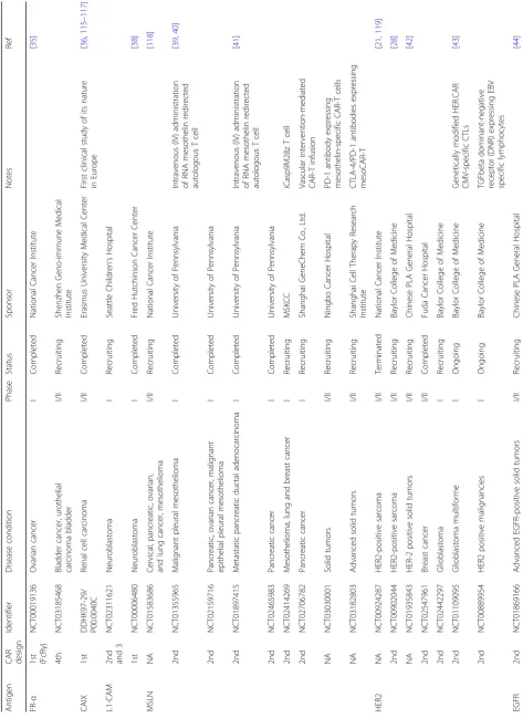
Because of the success achieved in CAR-T therapy tar-geting B cell malignancies and the advancements in CAR-T preclinical studies for solid tumors, more than 100 CAR-T clinical trials targeting solid tumors have been initiated at medical centers all over the world (Table1).
Tumor-associated antigens and CAR design
So far, no such cell surface antigen with comparable properties as CD19 has yet been identified regarding solid tumors. An ideal molecule for CAR targeting should be overexpressed on cancer cell surface of many patients, with zero or very low expression in normal tis-sues. Currently, TAAs, including mesothelin (MSLN),
Fig. 1Molecular mechanism of chimeric antigen receptor T cell-mediated antitumor activity.aThe chimeric T cell receptor coding sequence is
delivered by viral vector. After entering into T cells (beige), virus was uncoated and transgene was preferably integrated at genome transcriptional
start sites using specific vector designs, such as MLV retrovirus and piggyBac transposon.bCAR transgenes were endogenously transcript by host
machinery, translated, and later inserted onto the T cell surface.cAssociation of CARs to TAA (orange) on tumor cell surface activates T
cell for immunological response, for example, signaling network of CAR-T composed of CD8-CD28-CD137-CD3ζdomains was shown in
(d). CAR-T-mediated immune response was reported to be amplified by ZAP70, TRAF1, PI3K, and GRB2 as well as other uncharacterized
factors, giving rise to upregulation of signaling intermediates and subsequent pro-death gene transcriptions.eUpon CAR activation, T cells
secreted cytokines (brown), perforins (bright yellow), and granzymes (blue) as well as activated death receptors, which triggered downstream targets. These
subcellular events directly or subsequently contribute to specific death of tumor cells, including perforin and granzyme release, cytokine production, direct
lysis, apoptosis, necrosis, reprogrammed phenotype, and immuno-memory formation in T cells, tumor cells (gray), macrophages (pink) (via IL-6, IL-10, IL-12,
MCP-1, IP-10, TNF-α, MIP-1α, MIP-1β, IFN-γ), NK cells (cyan) (via IL-12, TNF-α, IFN-γ), Treg cells (navy) (via IL-2, IL-4, IL-7, IL-12, IL-15, IFN-β, IFN-γ, TSLP), and
dendritic cells (yellow) (via IL-6, IL-10, IL-12, TNF-α, MIP-1α, MIP-1β, IFN-γ). Abbreviation:NK cellsnatural killer cells,TAAtumor-associated antigen,Treg cells
Table
1
Selected
CAR-T
clinical
trials
targeting
solid
tumor-associated
antigens
(Co
ntinued)
Antige
n
CAR design
Iden
tifier
D
isease
condit
ion
Phase
Stat
us
Spo
nsor
Notes
Ref
EpCAM
NA
NC
T0272
9493
Live
r
neopl
asms
Recr
uiting
Sin
obiow
ay
Cell
The
rapy
Co.,
Ltd
.
NA
NC
T0272
5125
Sto
mach
neoplasms
Recr
uiting
Sin
obiow
ay
Cell
The
rapy
Co.,
Ltd
.
2nd
NC
T0291
5445
Mal
ignant
neoplasm
of
nasopharynx,
breast
Canc
er
I
Recr
uiting
Sichu
an
Universi
ty
2nd
NC
T0301
3712
Sol
id
tumors
I/II
Recr
uiting
First
Af
filiated
Hospit
al
of
Cheng
du
Medi
cal
Colleg
e
MUC16
ecto
2nd
NC
T0249
8912
Sol
id
tumors
I
Recr
uiting
MSKCC
T
cells
genetic
ally
eng
ineere
d
to
secret
e
IL-12
[
13
0
]
Representative
clinical
trials
using
CAR-T
to
treat
solid
tumors
are
grouped
based
on
the
tumor-associated
antigens
they
are
targeting.
Trial-rela
ted
information
were
mainly
collected
from
published
results
and
ClinicalTrials.gov
Abbreviations:
CAIX
carboxyanhyd
rase-IX,
CEA
carcinoembryon
ic
antigen,
cMET
hepatocyte
growth
factor
receptor,
EGFR
epidermal
growth
factor
receptor,
EpCAM
epithelial
cell
adhesion
molecule,
EphA2
EPH
receptor
A2,
FAP
fibroblast
activation
protein
α
,
GD2
disialoganglioside,
GPC3
glypican-3,
HER2
human
epidermal
growth
factor
receptor-2,
L1-CAM
L1
cell
adhesion
molecule,
MSLN
mesothelin,
MUC1
mucin,
NA
not
applicable,
PSMA
prostate-specific
membrane
antigen,
ROR1
receptor
tyrosine
kinase-like
orphan
receptor
1,
VEGFR
vascular
endothelial
growth
factor
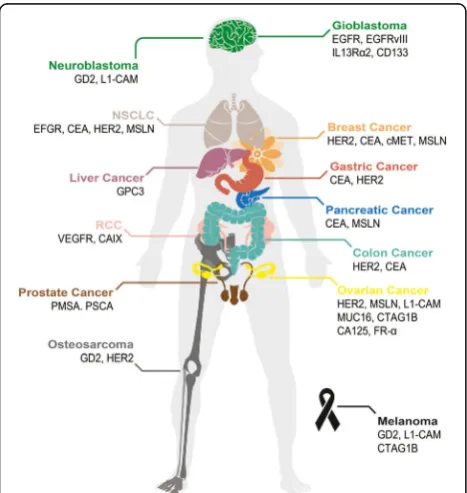
HER2, EGFR/EGFRvIII, GD2, CEA, IL13Rα2, MUC1, FAP, PSMA, and PSCA, are extensively investigated in CAR-T therapy for solid tumors [22,23]. TAAs currently being exploited for CAR-T therapy in solid tumors are summarized (Fig.2). Yu and colleagues comprehensively discussed these antigens regarding their biological func-tions and antitumor activities [22]. As shown in Table1, most of the solid tumor CAR-T clinical trials utilize the second or third generation of CARs, which contain either CD28 alone or CD28-4-BB1/OX40 as the costi-mulatory signal. Notably, a few of these studies, e.g., the trials targeting GD2 (NCT02765243, NCT02992210), PMSA (NCT03185468), FR-α (NCT03185468), investi-gated the efficacy of the fourth-generation CARs, i.e., TRUCK, which includes a transgenic cytokine expres-sion cassette in the CAR constructs [24]. Because of the tremendous phenotypic diversity in solid tumor lesions, a reasonable number of cancer cells are not recognized by a given CAR. The introduction of a transgenic cyto-kine such as interleukin-12 (IL-12) initiates universal cytokine-mediated killing towards those cancer cells that are invisible to CAR-T cells [19].
CAR-T cell therapy must carefully balance efficient T cell activation, to ensure antitumor activity, with the
potential for uncontrolled cytotoxicity. Some recent clinical trials included an inducible caspase 9 (iCasp9) “safety switch” in their CAR construct, which allows for the removal of inappropriately activated CAR-T cells [25,26]. The administration of the small molecule drug AP1903 causes the dimerization and activation of iCasp9, resulting in rapid induction of apoptosis in transduced cells. Two CAR-T clinical trials targeting GD2 performed at Baylor College of Medicine incorpo-rated such iCasp9 switch into their third generation CAR constructs (NCT01822652, NCT02439788). In another recently initiated trial led by Chang, a fourth-generation CAR was fused with the iCasp9 domain (NCT02992210).
PD-L1 expression in solid tumors counteracts the effi-cacy of CAR-T cell [27]. To overcome the inhibitory effect of PD-LI expression on CAR-T therapy, some clin-ical trials (e.g., NCT03030001, NCT03182803-MSLN,
NCT03182816, NCT02873390, NCT02862028-EGFR,
NCT03170141-EGFRvIII, and NCT03179007-MUC1)
add PD-1 dominant-negative receptor expressing gene to CAR-T cells, providing cell-intrinsic checkpoint blockade and increase antitumor efficacy. Based on the currently available data, anti-PD-1 combination therapy approach may be useful to augment CAR-T cell efficacy and persistence in patients [27].
T cell dosage, administration, and persistence
According to the published results and clinical trial de-scription available on the website, the majority of the CAR-T clinical trials use a dose escalation regime, which usually covers two log steps with a starting point ranged from around 1 × 10⁶ and 1 × 10⁹ CAR-T cells [24]. It is important to keep in mind that the percentage of CAR-positive T cells varies significantly not only among differ-ent trials but also among differdiffer-ent batches within single trial [24]. One of the major sources for such variation is the different production procedure of CAR-T cells adopted by different trials, emphasizing the importance of standardizing the CAR-T cells production protocol. Notably, to increase the tolerability of the treatment and to lower the risk of side effects, the given CAR-T cell dose is often split over multiple injections. For example, Ahmed and colleagues reported administration of 1 × 10⁴–1 × 10⁸/m2 HER2 specific CAR-T cells in up to 9 infusions (NCT00902044) [28]. Typically, CAR-T cells are infused intravenously. However, intratumoral (NCT02587689) [29], intracranial (NCT00730613) [30], hepatic artery (NCT01373047) [31], and pleural [32] ad-ministration are being investigated as well. The CAR-T cell administration method could have a dramatic impact on the efficacy of the treatment. Brown et al. compared the effectiveness of two intracranial CAR-T cell delivery route-infusion into the resected tumor
Fig. 2Tumor-associated antigens targeted in CAR-T therapy. Schematic illustration of a human body whose tissues or organs have been investi-gated in preclinical and clinical studies for solid tumor immunotherapy
using CAR-T, including brain (green), lungs (beige), mammary gland
(orange), liver (purple), stomach (red), pancreas (blue), kidneys (pink), colon
(cyan), male reproductive system (brown), ovary (yellow), and bones (gray)
as well as skin (black). Abbreviation:CCAcholangiocarcinoma,MPM
malignant pleural mesothelioma,NSCLCnon-small-cell lung carcinoma,
cavity and infusion into the ventricular system [33]. While intracavitary therapy appeared to control local tumor recurrence, progression of glioblastoma at distant sites was observed. By contrast, intraventricular adminis-tration of CAR-T cells leads to the regression of all cen-tral nervous system tumors, including spinal tumors. After the administration, CAR-T cell persistence is a key factor that determines the efficacy of the therapy. One major problem of current CAR-T cell immunotherapy is that T lymphocytes have limited replicative lifespans [34], which potentially influences the long-term antitu-mor effect of CAR-T cell immunotherapy. The reported T cell persistence in published clinical trials ranged from up to 4 weeks (DDHK97-29/P00.0040C, BB-IND 12084) to up to 192 weeks (NCT00085930).
Clinical outcomes and toxicities
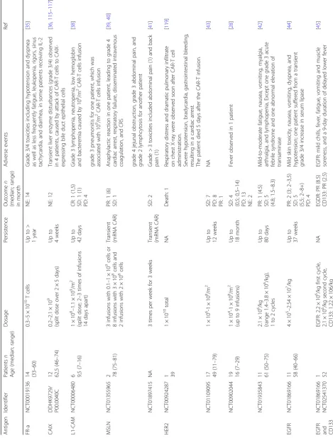
Some early attempts using CAR-T to treat solid tumors were not successful. In 2006, Kershaw and colleagues tested the efficacy of CAR-T cells targeting ovarian cancer cells expressing alpha-folate receptor (FR-α) (NCT00019136) [35]. Even though the patients can tol-erate the administration of FR-αspecific CAR-T cells, no clinical response was observed in all the patients (14 out of 14), likely because of the inefficient localization and short-term persistence of the CAR-T cells [35]. Lamers and colleagues treated 12 patients with CAIX-expressing metastatic renal cell carcinoma with CAIX specific CAR-T cells [36, 37]. Unfortunately, no clinical re-sponses were observed, and some patients developed anti-CAR-T cell antibodies and severe live enzyme disturbance.
Park et al. reported treating metastatic neuroblastoma with L1-CAM specific CAR-T cells at a dosage of 1 × 10⁸ cells/m2 to 1 × 10⁹ cells/m2 (NCT00006480) [38]. The persistence of cells was measured to be 1–7 days for tients with heavy disease burden and 42 days for a pa-tient with light disease burden. One of the six enrolled patients achieved stable disease after treatment [38]. A series of CAR-T clinical trials targeting MSLN have been performed in many different types of solid tumors, such as mesothelioma (NCT01355965), pancreatic cancer (NCT01583686, NCT02465983, NCT02706782), and breast cancer (NCT02792114). In one of these trails, advanced mesothelioma patients were administered au-tologous T cells electroporated with the mRNA encoding for MSLN CAR (NCT01355965) [39,40]. Moderate clin-ical responses were observed as supported by the detec-tion of MSLN specific CAR-T cells in the tumor site and the transient elevation of inflammatory cytokines [39, 40]. MSLN CAR T cell infusions were well tolerated at the dosage tested without severe toxicities [39,40]. Un-fortunately, one of the study subjects was reported to develop severe anaphylaxis and cardiac arrest after the
third infusion of MSLN CAR-T cells [40], which is likely to result from the inclusion of the murine SS1 scFv in the CAR-T design. In another trial for pancreatic ductal adenocarcinoma (PDAC) (NCT01897415), Beatty and colleagues utilized mRNA-encoded mesothelin-specific CAR to treat six patients, in which the infusion were well tolerated and preliminary evidence of antitumor efficacy was observed, supported by stable disease seen in two treated patients [41].
Clinically, the safety and efficacy of HER2-specific CAR-T cells in patients with relapsed/refractory HER2-positive sarcoma has been evaluated in a phase I/II clinical study (NCT00902044, NCT01109095, and NCT00924287). In a dose escalating trial (NCT00902044, 1 × 10⁴/m2 to 1 × 10⁸/m2), 4 out of 19 subjects acquired stable disease [28]. Feng et al. performed a phase I clinical trial investigating HER2-specific CAR-T cells in patients with advanced bil-iary tract cancers (BTCs) and pancreatic cancers (PCs) (NCT01935843). Among the 6 patients received HER2-specific CAR-T infusion, one patient obtained a 4.5-month partial response and the other 5 patients achieved stable disease with mild to moderate adverse events [42]. In another trial (NCT01109095), 17 CMV-seropositive patients with radiologically progressive HER2-positive glioblastoma were infused with HER2 CMV bispecific CAR-T cells at the dose of 1 × 10⁶/m2–1 × 108/m2 [43]. Such treatments were well tolerated without severe ad-verse events or cytokine release syndrome, and 7 out the 17 treated patients achieved stable disease [43].
Feng and colleagues reported the clinical trials of EGFR specific CAR-T treating non-small cell lung can-cer (NCT01869166) [44]. Five out of the 11 treated patients achieved stable disease and 2 achieved partial response. The CAR-T infusion-related adverse events were mild and manageable. Recently, Feng et al. reported treating a patient diagnosed as advanced unre-sectable/metastatic cholangiocarcinoma (CCA) with CAR-T cocktail immunotherapy, which was composed of successive infusions of CAR-T cells targeting
epider-mal growth factor receptor (EGFR) and CD133
(NCT01869166, NCT02541370) [45]. The patient achieved an 8.5-month partial response (PR) from the CART-EGFR therapy and a 4.5-month-lasting PR from the CART133 treatment. However, a series of adverse events were also observed in the course of treatment, including deteriorative grade 3 systemic subcutaneous hemorrhages and congestive rashes together with serum cytokine release.
manageable temporary central nerve system inflamma-tion. The same group conducted another clinical trial (NCT02208362), in which tumor regression and in-creased production of cytokines and immune cells were observed [33]. Encouragingly, the clinical response con-tinued for 7.5 months after the initiation of CAR-T cell therapy, indicating a relatively long persistence of CAR-T cells.
Katz and colleagues conducted a phase I trial to test CAR-T in patients with CEA-positive liver metas-tases (NCT01373047). Among the six patients who completed the protocol, one patient remained alive with stable disease at 23 months following CAR-T treatment and five patients died of progressive disease [31]. Biopsies demonstrated an increase in liver me-tastases necrosis or fibrosis in four out of six patients. No patient suffered a grade 3 or 4 adverse event re-lated to the CAR-T treatment. Louis and colleagues evaluated the efficacy of GD2-specific CAR-T in 19 patients either with remission (8 patients) or progres-sive neuroblastoma (11 patients) (NCT00085930) [46]. Three of 11 patients with active disease achieved complete remission and up to 192 weeks of CAR-T cell persistence was observed in the trial [46].
Clinical trials investigating the efficacy of CAR-T cells tar-geting MUC1 (NCT02587689) [29], cMet (NCT01837602, NCT03060356) [47], PSMA (BB-IND 12084) [48], VEGFR-2 (NCT01VEGFR-218867) have been reported. Relevant informa-tion and results of CAR-T clinical trials were summarized in Table2.
As learned from the results of these clinical trials, CAR-T cells face a unique set of challenges in the case of solid tu-mors. Some of the issues appear to be the absence of unique tumor-associated antigens, the inefficient homing of T cells to tumor sites, and the limited persistence of CAR-T cells. Moreover, the immunosuppressive microenvironment within the tumor tends to inhibit CAR-T cell function strongly. While a seemingly complicated, fulfilling all of above requirements can be accomplished efficiently through both intrinsic and/or extrinsic modifications of CAR-T cells.
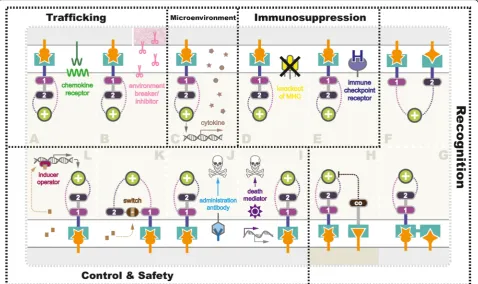
Overcoming challenges with smarter CAR designs Different from B cell malignancies, the application of the CAR-T cell strategy to non-hematopoietic cancer is faced with physical barriers as well as a variety of approaches that tumors employ to blunt host immune-surveillance [49]. Therefore, obstacle factors existing in current CAR-T trials such as fibrosis, inflammation, autoimmunity, T cell exhaustion and persistence, or recurrence must be overcome with smarter redirected T cell designs to achieve optimal therapeutic results (Fig.3).
Enhancing trafficking
Tumors are organized tissues with numerous connec-tions with surrounding cells [50], including resident im-mune system, i.e., myeloid compartment and lymphoid compartment [51], vasculature, fibroblasts, signaling molecules, and the extracellular matrix [52]. In addition, the low pH, low oxygen, low nutrients condition, and the tumor surrounding tissues, especially blood vessels and fibroblasts, make therapeutic delivery cruelly diffi-cult [53].
In current trials, homing of CAR-T cells to the tumor sites is often inefficient owing to the reasons mentioned above. The successful application of CAR-T cells against hematological malignancies is at least partially attributed to the fact that tumor and effector T cells tended to
migrate to similar sites due to their shared
hematopoietic origins. Solid tumors are known to secrete immuno-factors, including chemokine, cytokine, and growth factors, preventing effector T cells from infil-trating into the tumor bed (Fig. 3a). In 2010, a
recom-binant chemokine receptor ligand CCR2(b) was
introduced into CAR-T cells targeting MSLN [54], and GD2 [55], because the endogenous chemokine receptors on T cells adequately mismatch with the chemokines se-creted by the tumors. Beyond minimizing the effects of chemokine, strategies targeting activated surrounding fibroblast (fibroblast activation protein, FAP) in mouse models of mesothelioma and lung cancer [56,57], vascu-lature (anti-VEGFR-2) in melanoma, and renal cancer (NCT01218867) [58], as well as tumor stroma (hepara-nase) in neuroblastoma mice [59], have been tested.
migratory capacity of CAR-T cells. Qin et al. generated two versions of CAR vectors, with or without a hinge domain, targeting hematopoietic, and solid tumor anti-gens [63]. The CARs with a hinge domain demonstrated better expansion and migration capacity in vitro and CAR-T cells expressing anti-mesothelin CARs contain-ing a hcontain-inge domain showed enhanced antitumor activities.
Reforming microenvironment
Malignant transformation and the growth of tumor mass influence surrounding microenvironment, which subse-quently gives rise to peripheral immune tolerance [64]. In return, tumor microenvironment provides a driving force contributing to not only tumor invasion and metastasis but also pharmacokinetic and pharmaco-dynamics resistance [65].
In studies targeting solid tumors, CAR-T cells sur-mount challenges conferred by anti-inflammatory factors (e.g., TGF-βand IL-10), immune suppressor cells (Tregs, Bregs, myeloid cells, neutrophils, macrophages), antigen loss, tissue-specific alterations, etc., [51, 64]. Even if CAR-T cells successfully trafficked to the tumor sites, tumor microenvironment might suppress or even
inactivate them. Therefore, with the aim to equip CAR-T cells with capabilities to remodel the suppressive tumor microenvironment by secreting anti-cancer cyto-kines, strategies named TRUCK have been extensively utilized in CAR-T studies (Fig.3c). To date, several cyto-kines have been adopted into TRUCK designs, including IL-12 [66,67], IL-18 [68], and TNFRSF14 [69]. Recently, a leading-edge TRUCK system using synNotch receptor was described by Roybal et al. [70, 71], which was engi-neered to produce a range of specific payloads in response to the target antigens. In addition to inflamma-tory cytokines, the flexible synNotch system could also express pro-tumor cytokine antibodies, checkpoint anti-bodies, bispecific antianti-bodies, and adjuvants. Using syn-Notch TRUCK system, more antitumor cytokines or other factors with the potential to reform the local envir-onment based on the specific tumor heterogeneity might be exploited. For instance, reported as potential anti-cancer therapeutic candidates [72], cytokines such as IL-24 should be further tested.
Anti-immunosuppression
Immunosuppression is a great challenge to effective CAR-T therapy, as it enables the tumor cell to escape
Fig. 3Strategies being exploited to overcome challenges in CAR-T therapy in solid tumor. Various strategies are currently being tested in preclinical and
clinical studies to overcome the challenges facing CAR-T therapy for solid tumor (gray), includinga,benhancing T cell (beige) trafficking,creforming
tumor microenvironment (pink represented physiological barriers),d,eanti-immunosuppression, elevating antigen recognition towards tumor (f,g), and
healthy cells (darker beige) (h), as well asi–limproving safety control using suicide switch or on-switch. Detailed mechanisms were further illustrated in
from antitumor immune responses [73]. Various types of strategy have been exploited to engineer CAR-T cells to fight tumor immunosuppression. Manipulation of the endogenous TCR/MHC (Fig. 3d) and induction of add-itional immune checkpoint receptors (Fig. 3e) are two promising strategies investigated extensively.
Immune escape mediated by inhibitory pathways via the interaction of activated killer T cell receptors with their ligands, such as programmed cell death1 (PD-1) [74], and cytotoxic T lymphocyte antigen 4 (CTLA-4) on T cells is another major factor [75]. To overcome repressive solid tumor environments and enhance the activity and persistence of CAR-T cells, combined ther-apies using co-administration of immune checkpoint in-hibitors or cytokines with CAR-T cells have been employed. Immune checkpoint inhibitors block the im-mune checkpoint pathways by targeting key regulators in the pathways (e.g., PD-1/PD-L1, CTLA4) to enhance the immune activity of patients’effector T cells [76]. The addition of anti-PD1 monoclonal antibody has been shown to mute the inhibitory effect of such receptors and enhance the function of CAR-T cells in preclinical models [77–79]. Many groups are now attempting to generate CAR-T cells resistant to PD1-PDL1 and CTLA4-CD80/CD86 signaling [80], and some of these CAR constructs are currently under investigation in clinical trials as discussed in the previous section (Table1).
Immunosuppressive soluble factors, like TGF-β and IL10, have been demonstrated to inhibit CAR-T cell activities [81]. Specifically, TGF-β has direct negative effects on T cell differentiation and cytotoxic function, thus hampering T cell effector functions [82]. TGF-β and IL10 can also inhibit antigen-presenting cells, lead-ing to hampered activation of tumor-reactive T cells [83]. Other factors including prostaglandin E2 (PGE2) and adenosine have also been demonstrated to inhibit T cell proliferation and differentiation via signaling through G-coupled receptors [84, 85]. CAR-T cells can be potentially engineered to include the expression of a dominant-negative form of the receptor of these factors to overcome their inhibitory effects [86].
In addition to the soluble immunosuppressive factors, various suppressive surveilling immune cells within the tumor microenvironment, such as Tregs, myeloid-derived suppressor cell (MDSC), and tumor-associated macro-phage (TAM)/tumor-associated neutrophils (TAN) with the so-called M2 and N2 phenotype, present another obs-tacle against successful CAR-T treatment. Tregs have been shown to inhibit T cell activity through cell-to-cell contact inhibition and via soluble factors such as TGF-βand IL10 [87]. Tregs hamper T cell activity by producing TGF-β, IL10, and also other suppressive agents like IL35 and ad-enosine [88]. MDSC, M2-TAM, and N2-TAN inhibit
antitumor immune response by producing TGF-β, PGE2, reactive oxygen/nitrogen species, and arginase [89, 90]. M2-TAM can express high levels of PD-L1, which can interact with PD1 on CAR-T cells and inhibit them [91]. To overcome the effect of such suppressive surveilling im-mune cells, the CAR-T design has been proposed to target both the tumor cells and immune cells. Ruella et al. dem-onstrated the feasibility of targeting CD123-positive Hodgkin lymphoma cells and TAM [92]. However, whether this strategy is universally applicable to other ma-lignancies, especially for the solid tumor, still needs to be further tested.
Elevating recognition specificity
The genetic heterogeneity of solid tumors is a major issue to treat such malignancies with CAR-T. Unlike CD19 in B cell leukemia, there is no such “panacea” antigen available for solid tumors. Worse still, cross-reactions (i.e.,“OFF target”and“ON target OFF tumor”) with bystander non-tumor cells have been widely ob-served in CAR-T studies, leading to severe or even lethal adverse effects caused by T cells attack to the essential tissues.
minimize the T cell “on-target off-tumor” activity to-wards normal tissues, a NOT-gate circuit using a kill-ing CAR and an inhibitory CAR was developed [99].
Improving safety and control
Most of the efforts at CAR-T engineering has been de-voted to improve the strength of clinical response and to prolong T cell proliferation and persistence. However, the adverse effects observed in human trials suggest the urgency of additional consideration on safety control mechanisms. Utilization of control systems should be regarded as the priority in the next generation CAR-T therapy design as T cells are autonomous without regu-latory mechanisms.
To date, a growing number of user-control systems have been developed to modulate CAR-T cell expression. In 1997, Bonini et al. firstly reported successful control of graft-versus-host disease (GVHD) using a suicide gene named herpes simplex virus thymidine kinase (HSV-tk) in adoptive T cell therapy, which rendered the T cells susceptible to ganciclovir treatment [100]. In 2001, Fas intracellular domain (ΔFas) was applied as a T cell sui-cide switch to combat GVHD in marrow transplantation [101]. In 2011, Di Stasi and colleagues introduced a human caspase-9 (iCasp9) as an off-switch to condition-ally trigger apoptosis of CAR-T cells by dimerization upon the treatment of small molecules (Fig.3i) [102]. In hematopoietic stem cell transplantation recipients, 90% of iCasp9-modified T cells could be eliminated within 30 min under AP1903 administration without severe ad-verse effects or recurrence [103]. Synthetic death control switches are likely to be extensively utilized in future adoptive immunotherapy owing to their high effective-ness, easy controllability, short time of on-set, and mild adverse effects, despite that minority of the T cell popu-lations could escape from the apoptotic signal, resulting in persistent cytotoxicity [103]. Alternatively, CAR-T cells could be removed via apoptosis using antigen-specific monoclonal antibodies (Fig. 3j), such as rituxi-mab for CD20 epitope [104], and cetuximab for EGFR epitope [105]. In addition to selective elimination, expression of previously known antigens enables track-ing of CAR-T cells in vivo.
In contrast to the death switch, a distinct class of on-control mechanisms is considered as safer manipulating strategies, as CAR-T cells using on-switches are defaulted to be unresponsive. In this design, CARs are conditionally expressed or form a functional structure in the presence of the inducer, removal of which terminates the transcription or inactivates CARs. Wu et al. described an advanced on-switch CAR using a split con-struct in vivo (Fig.3k). The split CAR design distributed into two separate polypeptides, an extracellular binding domain scFv and a downstream signaling element
ITAM, two parts reassembled and activated under the treatment of a small molecule rapalog [106]. In 2016, Morsut and colleagues further developed a tissue ligand-specific on-switch CAR construct based on synNotch re-ceptors using a tetracycline (Tet)-regulated promoter (Fig. 3l) [107]. It is noteworthy that expression levels of CARs are generally dose-dependent before saturation, which allows manipulating the strength of CAR expres-sion accordingly. In Tet-on systems, CARs still exist after removal of the inducer; hence, it requires further depletion to eliminate CAR-T cells ultimately. At this stage, an antibody like myc could be conveniently ap-plied to target the tagged CARs as a safeguard factors after adoptive transfer [108]. In principle, integration of the on- and off-switch systems enables us to turn on the expression of CARs at the wanted time point, to turn off when the doses are sufficient, and to erase T cells from patients once the treatments are completed.
Strategies involving codon optimization [109–111] or construct alteration using lentivirus [112], transposon [113], or RNA [114] were also employed in the next gen-eration CAR design to ameliorate side effects resulted from T cell overactivation. Hopefully, a combination of suicide/on switches, optimized coding, relevant down-stream feedback response, and sensor circuits in the future trials could help to establish global control sys-tems to modulate the expression, strength, and timing of the engineered CAR-T cells.
Conclusion
(TGF-β, IL10), and suppressive surveilling immune cells are aspects worth to be explored. Third, the establish-ment of standard clinical protocols is needed. Improve-ments in the CAR-T cell itself will require parallel developments in clinical protocol design, including patient preconditioning, cytokine support, and other po-tential co-administered treatments. Preconditioning of the patient before adoptive cell therapy is thought to have a significant effect on the immune response, thereby producing a potential therapeutic window for CAR-T cell activity. Improvements in CAR design and better understating of the interaction between tumor and immune system will help to overcome the hurdles currently limiting the application CAR-T in solid tumor treatment.
Abbreviations
ALL:Acute lymphoblastic leukemia; BTC: Biliary tract cancer;
CAIX: Carboxyanhydrase-IX; CARs: Chimeric antigen receptors; CAR-T cells: Chimeric antigen receptor T cells; CCA: Cholangiocarcinoma; CCR: Chimeric costimulatory receptor; CEA: Carcinoembryonic antigen; cMET: Hepatocyte growth factor receptor; CTLA-4: Cytotoxic T-lymphocyte antigen 4; EGFR: Epidermal growth factor receptor; EpCAM: Epithelial cell adhesion molecule; EphA2: EPH receptor A2; FAP: Fibroblast activation
proteinα; FDA: Food and Drug Administration; FR: Folate receptor;
GD2: Disialoganglioside; GPC3: Glypican-3; GVHD: Graft-versus-host disease; HER2: Human epidermal growth factor receptor-2; HSV-tk: Herpes simplex virus thymidine kinase; iCAR: Inhibitory CAR; IL: Interleukin; L1-CAM: L1 cell adhesion molecule; MDSC: Myeloid-derived suppressor cell; MPM: Malignant pleural mesothelioma; MSLN: Mesothelin; MUC1: Mucin; NK cells: Natural killer cells; NSCLC: Non-small-cell lung carcinoma; PD-1: Programmed cell death-1; PDAC: Pancreatic ductal adenocarcinoma; PGE2: Prostaglandin E2; PR: Partial response; PSMA: Prostate-specific membrane antigen; ROR1: Receptor tyrosine kinase-like orphan receptor 1; scFV: Single chain variable fragments; TAA: Tumor-associated antigens; TAM: Tumor-associated macrophage; TAN: Tumor-associated neutrophils; TCR: T cell receptor; Tet: Tetracycline; Treg cells: Regulatory T cells; TRUCK: T cell redirected for universal cytokine-mediated killing; VEGFR: Vascular endothelial growth factor receptor
Acknowledgements
We apologize for not being able to cite all the related work due to page limit.
Funding
This work was supported by the National Natural Science Foundation of China (no. 31500657) and the International Collaboration Projects from the Science and Technology Bureau of Sichuan Province (no. 2017HH0097) to Jian Li. Wenwen Li was supported by the China Scholarship Council.
Availability of data and materials
Data sharing is not applicable to this article as no datasets were generated or analyzed during the current study.
Authors’contributions
JL, WL, GK, and QZ conceived the study and wrote the manuscript. JL, WL, KH, and YZ performed the literature search. All authors read and approved the final manuscript.
Ethics approval and consent to participate
Not applicable.
Consent for publication
All authors have read and approved the final manuscript for publication.
Competing interests
The authors declare that they have no competing interests.
Publisher’s Note
Springer Nature remains neutral with regard to jurisdictional claims in published maps and institutional affiliations.
Author details
1School of Medicine, Chengdu University, Chengdu 610106, China.2UCL Institute of Ophthalmology, 11-43 Bath Street, London EC1V 9EL, UK. 3
Sichuan Industrial Institute of Antibiotics, Chengdu University, Chengdu 610052, China.4Section of Hematology-Oncology, Department of Pediatrics, Yale University School of Medicine, New Haven, CT 06520, USA.5College of Pharmacy and Biological Engineering, Chengdu University, Chengdu 610106, China.
Received: 1 December 2017 Accepted: 6 February 2018
References
1. Panel OKs CAR T therapy for leukemia. Cancer Discov. 2017;7(9):924.
2. Grupp SA, et al. Chimeric antigen receptor-modified T cells for acute
lymphoid leukemia. N Engl J Med. 2013;368(16):1509–18.
3. Gross G, Waks T, Eshhar Z. Expression of immunoglobulin-T-cell receptor
chimeric molecules as functional receptors with antibody-type specificity.
Proc Natl Acad Sci U S A. 1989;86(24):10024–8.
4. Ahmad ZA, et al. scFv antibody: principles and clinical application. Clin Dev
Immunol. 2012;2012:980250.
5. Dotti G, Savoldo B, Brenner M. Fifteen years of gene therapy based on
chimeric antigen receptors:“are we nearly there yet?”. Hum Gene Ther.
2009;20(11):1229–39.
6. Hombach A, et al. Human CD4+ T cells lyse target cells via granzyme/
perforin upon circumvention of MHC class II restriction by an antibody-like
immunoreceptor. J Immunol. 2006;177(8):5668–75.
7. Fesnak AD, June CH, Levine BL. Engineered T cells: the promise and
challenges of cancer immunotherapy. Nat Rev Cancer. 2016;16(9):566–81.
8. Irving BA, Weiss A. The cytoplasmic domain of the T cell receptor zeta chain
is sufficient to couple to receptor-associated signal transduction pathways.
Cell. 1991;64(5):891–901.
9. Haynes NM, et al. Redirecting mouse CTL against colon carcinoma: superior
signaling efficacy of single-chain variable domain chimeras containing
TCR-zeta vs Fc epsilon RI-gamma. J Immunol. 2001;166(1):182–7.
10. Brocker T, Karjalainen K. Signals through T cell receptor-zeta chain alone are
insufficient to prime resting T lymphocytes. J Exp Med. 1995;181(5):1653–9.
11. Finney HM, et al. Chimeric receptors providing both primary and
costimulatory signaling in T cells from a single gene product. J Immunol.
1998;161(6):2791–7.
12. Shen CJ, et al. Chimeric antigen receptor containing ICOS signaling domain
mediates specific and efficient antitumor effect of T cells against EGFRvIII expressing glioma. J Hematol Oncol. 2013;6:33.
13. Maher J, et al. Human T-lymphocyte cytotoxicity and proliferation
directed by a single chimeric TCRzeta /CD28 receptor. Nat Biotechnol.
2002;20(1):70–5.
14. Savoldo B, et al. CD28 costimulation improves expansion and persistence of
chimeric antigen receptor-modified T cells in lymphoma patients. J Clin
Invest. 2011;121(5):1822–6.
15. Kofler DM, et al. CD28 costimulation impairs the efficacy of a redirected
t-cell antitumor attack in the presence of regulatory t t-cells which can be
overcome by preventing Lck activation. Mol Ther. 2011;19(4):760–7.
16. Koehler H, et al. CD28 costimulation overcomes transforming growth
factor-beta-mediated repression of proliferation of redirected human CD4+ and
CD8+ T cells in an antitumor cell attack. Cancer Res. 2007;67(5):2265–73.
17. Pule MA, et al. A chimeric T cell antigen receptor that augments cytokine
release and supports clonal expansion of primary human T cells. Mol Ther.
2005;12(5):933–41.
18. Hombach AA, et al. OX40 costimulation by a chimeric antigen receptor
abrogates CD28 and IL-2 induced IL-10 secretion by redirected CD4(+) T
cells. Oncoimmunology. 2012;1(4):458–66.
19. Chmielewski M, Hombach AA, Abken H. Of CARs and TRUCKs: chimeric
antigen receptor (CAR) T cells engineered with an inducible cytokine to
modulate the tumor stroma. Immunol Rev. 2014;257(1):83–90.
20. Chmielewski M, Abken H. TRUCKs: the fourth generation of CARs. Expert
21. Lopez-Albaitero A, et al. Overcoming resistance to HER2-targeted therapy with a novel HER2/CD3 bispecific antibody. Oncoimmunology. 2017;6(3):e1267891.
22. Yu S, et al. Chimeric antigen receptor T cells: a novel therapy for solid
tumors. J Hematol Oncol. 2017;10(1):78.
23. Liu B, Song Y, Liu D. Clinical trials of CAR-T cells in China. J Hematol Oncol.
2017;10(1):166.
24. Hartmann J, et al. Clinical development of CAR T cells—challenges and
opportunities in translating innovative treatment concepts. EMBO Mol Med.
2017;9(9):1183–97.
25. Gargett T, Brown MP. The inducible caspase-9 suicide gene system as a
“safety switch”to limit on-target, off-tumor toxicities of chimeric antigen
receptor T cells. Front Pharmacol. 2014;5:235.
26. Diaconu I, et al. Inducible caspase-9 selectively modulates the toxicities of
CD19-specific chimeric antigen receptor-modified T cells. Mol Ther. 2017;
25(3):580–92.
27. Chen N, et al. CAR T-cell intrinsic PD-1 checkpoint blockade: a two-in-one
approach for solid tumor immunotherapy. Oncoimmunology. 2017;6(2): e1273302.
28. Ahmed N, et al. Human epidermal growth factor receptor 2 (HER2)-specific
chimeric antigen receptor-modified T cells for the immunotherapy of
HER2-positive sarcoma. J Clin Oncol. 2015;33(15):1688–96.
29. You F, et al. Phase 1 clinical trial demonstrated that MUC1 positive
metastatic seminal vesicle cancer can be effectively eradicated by modified anti-MUC1 chimeric antigen receptor transduced T cells. Sci China Life Sci.
2016;59(4):386–97.
30. Brown CE, et al. Bioactivity and safety of IL13Ralpha2-redirected chimeric
antigen receptor CD8+ T cells in patients with recurrent glioblastoma. Clin
Cancer Res. 2015;21(18):4062–72.
31. Katz SC, et al. Phase I hepatic immunotherapy for metastases study of
intra-arterial chimeric antigen receptor-modified T-cell therapy for CEA+ liver
metastases. Clin Cancer Res. 2015;21(14):3149–59.
32. Petrausch U, et al. Re-directed T cells for the treatment of fibroblast
activation protein (FAP)-positive malignant pleural mesothelioma (FAPME-1). BMC Cancer. 2012;12:615.
33. Brown CE, et al. Regression of glioblastoma after chimeric antigen receptor
T-cell therapy. N Engl J Med. 2016;375(26):2561–9.
34. Brentjens RJ, et al. Safety and persistence of adoptively transferred
autologous CD19-targeted T cells in patients with relapsed or
chemotherapy refractory B-cell leukemias. Blood. 2011;118(18):4817–28.
35. Kershaw MH, et al. A phase I study on adoptive immunotherapy using
gene-modified T cells for ovarian cancer. Clin Cancer Res. 2006;12(20 Pt 1):
6106–15.
36. Lamers CH, et al. Treatment of metastatic renal cell carcinoma with CAIX
CAR-engineered T cells: clinical evaluation and management of on-target
toxicity. Mol Ther. 2013;21(4):904–12.
37. Lamers CH, et al. Treatment of metastatic renal cell carcinoma (mRCC) with
CAIX CAR-engineered T-cells––a completed study overview. Biochem Soc
Trans. 2016;44(3):951–9.
38. Park JR, et al. Adoptive transfer of chimeric antigen receptor re-directed
cytolytic T lymphocyte clones in patients with neuroblastoma. Mol Ther.
2007;15(4):825–33.
39. Beatty GL, et al. Mesothelin-specific chimeric antigen receptor
mRNA-engineered T cells induce anti-tumor activity in solid malignancies. Cancer
Immunol Res. 2014;2(2):112–20.
40. Maus MV, et al. T cells expressing chimeric antigen receptors can cause
anaphylaxis in humans. Cancer Immunol Res. 2013;1(1):26–31.
41. Beatty GL, et al. Safety and antitumor activity of chimeric antigen receptor
modified T cells in patients with chemotherapy refractory metastatic pancreatic cancer. J Clin Oncol. 2015;33(15_suppl):3007.
42. Feng K, et al. Phase I study of chimeric antigen receptor modified T cells in
treating HER2-positive advanced biliary tract cancers and pancreatic
cancers. Protein Cell. 2017.https://doi.org/10.1007/s13238-017-0440-4.
43. Ahmed N, et al. Autologous HER2 CMV bispecific CAR T cells are safe and
demonstrate clinical benefit for glioblastoma in a phase I trial. J Immunother Cancer. 2015;3(Suppl 2):O11.
44. Feng K, et al. Chimeric antigen receptor-modified T cells for the
immunotherapy of patients with EGFR-expressing advanced relapsed/
refractory non-small cell lung cancer. Sci China Life Sci. 2016;59(5):468–79.
45. Feng KC, et al. Cocktail treatment with EGFR-specific and CD133-specific
chimeric antigen receptor-modified T cells in a patient with advanced cholangiocarcinoma. J Hematol Oncol. 2017;10(1):4.
46. Louis CU, et al. Antitumor activity and long-term fate of chimeric antigen
receptor-positive T cells in patients with neuroblastoma. Blood. 2011;
118(23):6050–6.
47. Tchou J, et al. Safety and efficacy of intratumoral injections of chimeric
antigen receptor (CAR) T cells in metastatic breast cancer. Cancer Immunol
Res. 2017;5(12):1152–61.
48. Junghans RP, et al. Phase I trial of anti-PSMA designer CAR-T cells in
prostate cancer: possible role for interacting interleukin 2-T cell pharmacodynamics as a determinant of clinical response. Prostate. 2016;
76(14):1257–70.
49. Hanahan D, Weinberg RA. Hallmarks of cancer: the next generation. Cell.
2011;144(5):646–74.
50. Stuelten CH, et al. Breast cancer cells induce stromal fibroblasts to express
MMP-9 via secretion of TNF-alpha and TGF-beta. J Cell Sci. 2005;118(Pt 10):
2143–53.
51. Palucka AK, Coussens LM. The basis of oncoimmunology. Cell. 2016;164(6):
1233–47.
52. Friedl P, Alexander S. Cancer invasion and the microenvironment: plasticity
and reciprocity. Cell. 2011;147(5):992–1009.
53. Newick K, Moon E, Albelda SM. Chimeric antigen receptor T-cell therapy for
solid tumors. Mol Ther Oncolytics. 2016;3:16006.
54. Moon EK, et al. Expression of a functional CCR2 receptor enhances tumor
localization and tumor eradication by retargeted human T cells expressing a mesothelin-specific chimeric antibody receptor. Clin Cancer Res. 2011;17(14):
4719–30.
55. Craddock JA, et al. Enhanced tumor trafficking of GD2 chimeric antigen
receptor T cells by expression of the chemokine receptor CCR2b. J
Immunother. 2010;33(8):780–8.
56. Schuberth PC, et al. Treatment of malignant pleural mesothelioma by
fibroblast activation protein-specific re-directed T cells. J Transl Med. 2013; 11:187.
57. Lo A, et al. Tumor-promoting desmoplasia is disrupted by depleting
FAP-expressing stromal cells. Cancer Res. 2015;75(14):2800–10.
58. Chinnasamy D, et al. Gene therapy using genetically modified lymphocytes
targeting VEGFR-2 inhibits the growth of vascularized syngenic tumors in
mice. J Clin Invest. 2010;120(11):3953–68.
59. Caruana I, et al. Heparanase promotes tumor infiltration and antitumor
activity of CAR-redirected T lymphocytes. Nat Med. 2015;21(5):524–9.
60. Garin-Chesa P, Old LJ, Rettig WJ. Cell surface glycoprotein of reactive
stromal fibroblasts as a potential antibody target in human epithelial
cancers. Proc Natl Acad Sci U S A. 1990;87(18):7235–9.
61. Beyer I, van Rensburg R, Lieber A. Overcoming physical barriers in cancer
therapy. Tissue Barriers. 2013;1(1):e23647.
62. Beyer I, et al. Controlled extracellular matrix degradation in breast cancer
tumors improves therapy by trastuzumab. Mol Ther. 2011;19(3):479–89.
63. Qin L, et al. Incorporation of a hinge domain improves the expansion of
chimeric antigen receptor T cells. J Hematol Oncol. 2017;10(1):68.
64. Balkwill FR, Capasso M, Hagemann T. The tumor microenvironment at a
glance. J Cell Sci. 2012;125(Pt 23):5591–6.
65. Khawar IA, Kim JH, Kuh HJ. Improving drug delivery to solid tumors: priming
the tumor microenvironment. J Control Release. 2015;201:78–89.
66. Kerkar SP, et al. Tumor-specific CD8+ T cells expressing interleukin-12
eradicate established cancers in lymphodepleted hosts. Cancer Res. 2010;
70(17):6725–34.
67. Pegram HJ, et al. Tumor-targeted T cells modified to secrete IL-12 eradicate
systemic tumors without need for prior conditioning. Blood. 2012;119(18):
4133–41.
68. Avanzi MP, et al. IL-18 secreting CAR T cells enhance cell persistence,
induce prolonged B cell aplasia and eradicate CD19+ tumor cells without need for prior conditioning. Blood. 2016;128(22):816.
69. Boice M, et al. Loss of the HVEM tumor suppressor in lymphoma and
restoration by modified CAR-T cells. Cell. 2016;167(2):405–18. e13
70. Roybal KT, et al. Engineering T cells with customized therapeutic response
programs using synthetic notch receptors. Cell. 2016;167(2):419–32. e16
71. Roybal KT, Lim WA. Synthetic immunology: hacking immune cells to
expand their therapeutic capabilities. Annu Rev Immunol. 2017;35:229–53.
72. Dent P, et al. MDA-7/IL-24 as a cancer therapeutic: from bench to bedside.
Anti-Cancer Drugs. 2010;21(8):725–31.
73. Beatty GL, Moon EK. Chimeric antigen receptor T cells are vulnerable to
74. Chevolet I, et al. Characterization of the in vivo immune network of IDO, tryptophan metabolism, PD-L1, and CTLA-4 in circulating immune cells in melanoma. Oncoimmunology. 2015;4(3):e982382.
75. Ewing MM, et al. T-cell co-stimulation by CD28-CD80/86 and its
negative regulator CTLA-4 strongly influence accelerated atherosclerosis
development. Int J Cardiol. 2013;168(3):1965–74.
76. Hegde UP, Mukherji B. Current status of chimeric antigen receptor
engineered T cell-based and immune checkpoint blockade-based cancer
immunotherapies. Cancer Immunol Immunother. 2017;66(9):1113–21.
77. Condomines M, et al. Tumor-targeted human T cells expressing
CD28-based chimeric antigen receptors circumvent CTLA-4 inhibition. PLoS One. 2015;10(6):e0130518.
78. Prosser ME, et al. Tumor PD-L1 co-stimulates primary human CD8(+)
cytotoxic T cells modified to express a PD1:CD28 chimeric receptor.
Mol Immunol. 2012;51(3–4):263–72.
79. John LB, et al. Anti-PD-1 antibody therapy potently enhances the
eradication of established tumors by gene-modified T cells. Clin Cancer
Res. 2013;19(20):5636–46.
80. Pegram HJ, Park JH, Brentjens RJ. CD28z CARs and armored CARs.
Cancer J. 2014;20(2):127–33.
81. Pardoll DM. The blockade of immune checkpoints in cancer immunotherapy. Nat
Rev Cancer. 2012;12(4):252–64.
82. Massague J. TGFbeta in cancer. Cell. 2008;134(2):215–30.
83. Enblad G, Karlsson H, Loskog AS. CAR T-cell therapy: the role of physical barriers
and immunosuppression in lymphoma. Hum Gene Ther. 2015;26(8):498–505.
84. Newick K, et al. Augmentation of CAR T-cell trafficking and antitumor
efficacy by blocking protein kinase a localization. Cancer Immunol Res.
2016;4(6):541–51.
85. Su Y, et al. Cooperation of adenosine and prostaglandin E2 (PGE2) in
amplification of cAMP-PKA signaling and immunosuppression. Cancer
Immunol Immunother. 2008;57(11):1611–23.
86. Foster AE, et al. Antitumor activity of EBV-specific T lymphocytes transduced
with a dominant negative TGF-beta receptor. J Immunother. 2008;31(5):
500–5.
87. Sakaguchi S, et al. FOXP3+ regulatory T cells in the human immune system.
Nat Rev Immunol. 2010;10(7):490–500.
88. Roychoudhuri R, Eil RL, Restifo NP. The interplay of effector and regulatory T
cells in cancer. Curr Opin Immunol. 2015;33:101–11.
89. Gabrilovich DI, Nagaraj S. Myeloid-derived suppressor cells as regulators of
the immune system. Nat Rev Immunol. 2009;9(3):162–74.
90. Mussai F, et al. Neuroblastoma arginase activity creates an
immunosuppressive microenvironment that impairs autologous and
engineered immunity. Cancer Res. 2015;75(15):3043–53.
91. Ostuni R, et al. Macrophages and cancer: from mechanisms to therapeutic
implications. Trends Immunol. 2015;36(4):229–39.
92. Ruella M, et al. Overcoming the immunosuppressive tumor
microenvironment of Hodgkin lymphoma using chimeric antigen receptor
T cells. Cancer Discov. 2017;7(10):1154–67.
93. Kloss CC, et al. Combinatorial antigen recognition with balanced signaling
promotes selective tumor eradication by engineered T cells. Nat Biotechnol.
2013;31(1):71–5.
94. Wilkie S, et al. Dual targeting of ErbB2 and MUC1 in breast cancer using
chimeric antigen receptors engineered to provide complementary
signaling. J Clin Immunol. 2012;32(5):1059–70.
95. Roybal KT, et al. Precision tumor recognition by T cells with combinatorial
antigen-sensing circuits. Cell. 2016;164(4):770–9.
96. Grada Z, et al. TanCAR: a novel bispecific chimeric antigen receptor for
cancer immunotherapy. Mol Ther Nucleic Acids. 2013;2:e105.
97. Hegde M, et al. A bispecific chimeric antigen receptor molecule enhances T
cell activation through dual immunological synapse formation and offsets antigen escape in glioblastoma. J Immunother Cancer. 2015;3(Suppl 2):O3.
98. Zah E, et al. T cells expressing CD19/CD20 bispecific chimeric antigen
receptors prevent antigen escape by malignant B cells. Cancer Immunol
Res. 2016;4(6):498–508.
99. Fedorov VD, Themeli M, Sadelain M. PD-1- and CTLA-4-based inhibitory
chimeric antigen receptors (iCARs) divert off-target immunotherapy responses. Sci Transl Med. 2013;5(215):215ra172.
100. Bonini C, et al. HSV-TK gene transfer into donor lymphocytes for control of
allogeneic graft-versus-leukemia. Science. 1997;276(5319):1719–24.
101. Thomis DC, et al. A Fas-based suicide switch in human T cells for the
treatment of graft-versus-host disease. Blood. 2001;97(5):1249–57.
102. Di Stasi A, et al. Inducible apoptosis as a safety switch for adoptive cell
therapy. N Engl J Med. 2011;365(18):1673–83.
103. Zhou X, et al. Long-term outcome after haploidentical stem cell transplant and infusion of T cells expressing the inducible caspase 9 safety transgene.
Blood. 2014;123(25):3895–905.
104. Vogler I, et al. An improved bicistronic CD20/tCD34 vector for efficient purification and in vivo depletion of gene-modified T cells for adoptive
immunotherapy. Mol Ther. 2010;18(7):1330–8.
105. Wang X, et al. A transgene-encoded cell surface polypeptide for selection, in vivo tracking, and ablation of engineered cells. Blood.
2011;118(5):1255–63.
106. Wu CY, et al. Remote control of therapeutic T cells through a small molecule-gated chimeric receptor. Science. 2015;350(6258):aab4077. 107. Morsut L, et al. Engineering customized cell sensing and response
behaviors using synthetic notch receptors. Cell. 2016;164(4):780–91.
108. Kieback E, et al. A safeguard eliminates T cell receptor gene-modified autoreactive T cells after adoptive transfer. Proc Natl Acad Sci U S A.
2008;105(2):623–8.
109. Kuball J, et al. Facilitating matched pairing and expression of TCR
chains introduced into human T cells. Blood. 2007;109(6):2331–8.
110. Cohen CJ, et al. Enhanced antitumor activity of T cells engineered to express T-cell receptors with a second disulfide bond. Cancer Res. 2007;
67(8):3898–903.
111. Provasi E, et al. Editing T cell specificity towards leukemia by zinc finger
nucleases and lentiviral gene transfer. Nat Med. 2012;18(5):807–15.
112. Porter DL, et al. Chimeric antigen receptor-modified T cells in chronic
lymphoid leukemia. N Engl J Med. 2011;365(8):725–33.
113. Jin Z, et al. The hyperactive sleeping beauty transposase SB100X improves the genetic modification of T cells to express a chimeric
antigen receptor. Gene Ther. 2011;18(9):849–56.
114. Singh N, Barrett DM, Grupp SA. Roadblocks to success for RNA CARs in solid tumors. Oncoimmunology. 2014;3(12):e962974.
115. Lamers CH, et al. Gene-modified T cells for adoptive immunotherapy of renal cell cancer maintain transgene-specific immune functions in vivo.
Cancer Immunol Immunother. 2007;56(12):1875–83.
116. Lamers CH, et al. Immune responses to transgene and retroviral vector in patients treated with ex vivo-engineered T cells. Blood. 2011;117(1):
72–82.
117. Lamers CH, et al. Treatment of metastatic renal cell carcinoma with autologous T-lymphocytes genetically retargeted against carbonic
anhydrase IX: first clinical experience. J Clin Oncol. 2006;24(13):e20–2.
118. Abate-Daga D, et al. A novel chimeric antigen receptor against prostate stem cell antigen mediates tumor destruction in a humanized mouse
model of pancreatic cancer. Hum Gene Ther. 2014;25(12):1003–12.
119. Morgan RA, et al. Case report of a serious adverse event following the administration of T cells transduced with a chimeric antigen receptor
recognizing ERBB2. Mol Ther. 2010;18(4):843–51.
120. Morgan RA, et al. Recognition of glioma stem cells by genetically modified T cells targeting EGFRvIII and development of adoptive cell therapy for
glioma. Hum Gene Ther. 2012;23(10):1043–53.
121. Saied A, et al. Neutrophil:lymphocyte ratios and serum cytokine changes after hepatic artery chimeric antigen receptor-modified T-cell infusions for
liver metastases. Cancer Gene Ther. 2014;21(11):457–62.
122. Katz SC, et al. Regional CAR-T cell infusions for peritoneal carcinomatosis are
superior to systemic delivery. Cancer Gene Ther. 2016;23(5):142–8.
123. Golubovskaya V, et al. CD47-CAR-T cells effectively kill target cancer cells and block pancreatic tumor growth. Cancers (Basel). 2017;9(10):139. 124. Pule MA, et al. Virus-specific T cells engineered to coexpress tumor-specific
receptors: persistence and antitumor activity in individuals with
neuroblastoma. Nat Med. 2008;14(11):1264–70.
125. Heczey A, et al. Invariant NKT cells with chimeric antigen receptor provide a novel platform for safe and effective cancer immunotherapy. Blood. 2014;
124(18):2824–33.
126. Tanaka M, et al. Vaccination targeting native receptors to enhance the function and proliferation of chimeric antigen receptor (CAR)-modified T
cells. Clin Cancer Res. 2017;23(14):3499–509.
127. Stroncek DF, et al. Elutriated lymphocytes for manufacturing chimeric antigen receptor T cells. J Transl Med. 2017;15(1):59.
129. Gohil SH, et al. An ROR1 bi-specific T-cell engager provides effective targeting and cytotoxicity against a range of solid tumors. Oncoimmunology. 2017;6(7):e1326437.
130. Koneru M, et al. A phase I clinical trial of adoptive T cell therapy using IL-12 secreting MUC-16(ecto) directed chimeric antigen receptors for recurrent ovarian cancer. J Transl Med. 2015;13:102.
• We accept pre-submission inquiries
• Our selector tool helps you to find the most relevant journal
• We provide round the clock customer support
• Convenient online submission
• Thorough peer review
• Inclusion in PubMed and all major indexing services
• Maximum visibility for your research
Submit your manuscript at www.biomedcentral.com/submit