Programmed Cell Death in the Developing
Kidney and the Ubiquity of the Programme
Harriet S. R. Coles
Thesis submitted to University College, London
for the degree of Doctor of Philosophy
August 1994
1-ProQuest Number: 10044606
All rights reserved
INFORMATION TO ALL USERS
The quality of this reproduction is dependent upon the quality of the copy submitted. In the unlikely event that the author did not send a complete manuscript and there are missing pages, these will be noted. Also, if material had to be removed,
a note will indicate the deletion.
uest.
ProQuest 10044606
Published by ProQuest LLC(2016). Copyright of the Dissertation is held by the Author. All rights reserved.
This work is protected against unauthorized copying under Title 17, United States Code. Microform Edition © ProQuest LLC.
ProQuest LLC
789 East Eisenhower Parkway P.O. Box 1346
Abstract
It has recently been suggested (Raff 1992) th a t all cells except
blastomeres die by programmed cell death unless signalled to survive.
In this thesis 1 explore implications of this idea by asking w hether
there are cases of developmental cell death th a t have been overlooked,
and w hether cell death is induced in m any cell types by blocking
protein kinase activity and protein synthesis.
Normal cell death was not considered to be im portant in mammalian
kidney development. 1 have found, however, th a t cell death occurs
with distinct time courses, in the nephrogenic region and medullary
papilla of the developing r a t kidney. Up to 3% of cells in these areas
are apoptotic, and are cleared w ithin 1-2 hours by phagocytosis.
These values are sim ilar to those in vertebrate neural tissues where
50% or more of the cells die during normal development, suggesting
th a t large scale death is a normal feature of kidney development. In
vivo treatm ent w ith epidermal growth factor or insulin-like growth
factor inhibits kidney cell death suggesting th a t this normal cell death
may reflect insufficient survival factors.
Raff (1992) suggested th a t all cells depend on survival factors in order
to avoid cell death. Blocking protein kinase activity (and consequently
cell signalling) and protein synthesis in a variety of neonatal ra t tissue
explants and preim plantation blastocysts induces 90% cell death
w ithin 18 hours. In contrast, blocking protein kinases and protein
synthesis in 2-4 cell stage blastomeres does not induce apoptosis.
constitutively express the protein components of the cell death
program m e.
1 conclude th a t cell death during vertebrate development is
more extensive th an was previously thought, th a t normal cell death
may often reflect lim iting supplies of survival factors, and th a t
blastomeres differ from even their earliest derivatives in the way cell
survival and death are controlled. These findings support the idea
th a t all cells except blastomeres require constant signalling from
other cells in order to avoid programmed cell death.
-"Oh well, no m atter w hat happens, there's always death."
Napoleon, 1817
"If there w asn't death, I think you couldn't go on."
Stevie Smith, 1969
4-Contents
Abstract 2
List of Figures 6
Acknowledgements 7
1 General Introduction 8
2 Programmed CeU Death in the Developing Rat
Kidney 23
Introduction 24
Results 26
Discussion 41
Methods 46
3 Factors Affecting Kidney Cell Survival 51
Introduction 52
Results 55
Discussion 64
Methods 71
4 The Ubiquity of the Cell Death Programme 7 6
Introduction 7 7
Results 81
Discussion 94
Methods 100
5 General Discussion 103
Bihhography 119
-Figures
1.1 Early nephrogenesis in the rat metanephric kidney 18 1.2 Preimplantation development of the mouse embryo 20-1 2.1 Propidium iodide staining of nuclei 31 2.2 Electron micrographs of apoptotic bodies in the rat kidney 32 2.3 End-labelling of nicked DNA in situ, in the nephrogenic zone
of neonatal rat kidney 33 2.4 Pyknotic and mitotic indices during kidney development 34-5 2.5 Anti-Pax-2 antibody and propidium iodide double-labelling of 36-7
rat kidney
2.6 Localisation of propidium-iodide-stained pyknotic nuclei in
the nephrogenic zone 38
2.7 Confocal fluorescence micrographs of
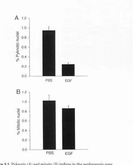
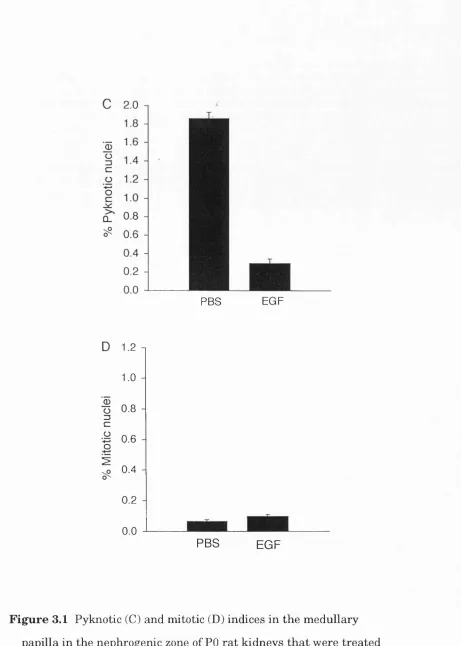
propidium-iodide-stained pyknotic nuclei 39-40 3.1 Pyknotic and mitotic indices in rat kidneys treated with EGF
or PBS 60-1
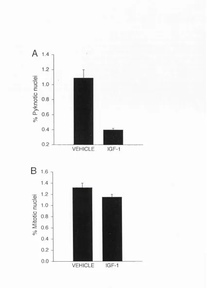
3.2 Pyknotic and mitotic indices in the nephrogenic zone of rat
kidneys treated with IGF-1 or PBS 62 3.3 Change in the pyknotic index in the nephrogenic zone of rat
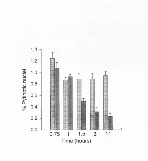
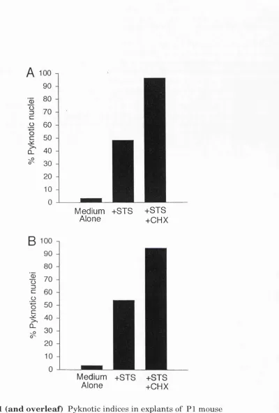
kidneys treated with EGF 63 4.1 Pyknotic indices in explants of mouse kidney, muscle, lung,
and pancreas 84-5
4.2 Fluorescence micrographs of propidium-iodide-stained nuclei
in mouse explants 86
4.3 End-labelling of nicked DNA in situ in explants of mouse
kidney 87
4.4 Survival of 2-4 cell-stage blastomeres 88 4.5 End-labelling of nicked DNA in situ in2-4 cell stage
blastomeres 89-90
4.6 Survival of 8-16 cell stage blastomeres 91 4.7 Survival of inner cell mass cells 92 4.8 Survival of trophéetoderm cells 93
6-A cknowledgem ents
I am grateful to everyone who has discussed this work with me at
University College, London, and at other research institutions. In
particular, I should like to thank: Barbara Barres, Ju lia Burne,
Yasuki Ishizaki, Mike Jacobson and Jim Voyvodic for discussion,
advice and companionship in the laboratory; Richard G ardner and
Tim Davies a t the ICRF Developmental Biology Unit in Oxford for
a fruitful collaboration; and Anne Mudge for discussions and
advice. I owe my greatest thanks to Martin Raff for inspiring and
guiding me throughout my Ph.D., and for teaching me about
science writing. Finally, I am indebted to John for help, support,
encouragement and everything else.
-C hapter 1
G eneral Introduction
-Introduction
Vertebrate development, from the fertilised egg to the adult animal, has
heen described mainly in terms of cell proliferation and differentiation;
there is, however, increasing evidence th a t programmed cell death (PCD)
has an equally important role in this process. The research community
has been slow to consider PCD as a part of the cell biology of multiceUular
organisms and it is likely th a t there are still cases of PCD during
development th a t have been overlooked. In this thesis I describe one
such case - PCD in the development of the ra t kidney. I also ask whether
all cells in developing multiceUular animals are capable of undergoing
PCD.
Apoptosis and cell necrosis
Cells th a t undergo PCD usually die w ith morphological features
collectively called apoptosis. This type of cell death is morphologically
distinct from most cell deaths th a t result from acute injury, a process
caRed cell necrosis (Kerr et al., 1972; Wyllie et al., 1980). In necrosis, the
cell, its organeUes and nucleus usually swell and lyse, spilling cytosolic
components into the extracellular space, which elicits an inflammatory
response in which the ceUular debris is phagocytosed by macrophages. In
apoptosis, the ceU and its nucleus shrink and often fragment, and the cell
or its fragments (called apoptotic bodies) are rapidly phagocytosed by
either neighbouring ceUs or macrophages before there is any leakage of
cytosolic contents. Thus in apoptosis unw anted cells are removed
discretely and tissue integrity is maintained. While necrosis usually
affects a group of cells in the affected region, apoptosis often occurs
selectively in cells scattered throughout a tissue.
-Introduction
The term "apoptosis" was introduced to distinguish between
accidental cell death th at results from injury (cell necrosis) and normal
active, or programmed cell death (Kerr et al., 1972). Considerable
variations in the morphology of cells undergoing PCD have been reported
(Clarke, 1990; Lockshin and Zakeri, 1991). In some cell types, for
example, the nuclear DNA is cleaved into oligonucleosomal fragments
(Wylhe 1980; Wylhe et al., 1984), while in other cell types it is not (Howell
and Martz, 1987; Lockshin and Zakeri, 1991, M artin 1993). These
variations are likely to be due to different cell phenotypes and tissue
organisations, although they could, in principle, reflect distinct
mechanisms of cell death. Therefore, I shall use "normal cell death"
(NCD), "apoptosis" and "programmed cell death" (PCD) as general and
interchangeable terms for cells undergoing active cell death.
The study of normal cell death: an overview
Compared with the extensive research on the control of cell
proliferation, attention has only recently been turned to the control of cell
survival and death. For the past 100 years, the potential relevance of
cell death to the control of body size has been debated, and researchers in
developmental biology, pathology and histology have noticed predictable
and discrete cell death occurring throughout vertebrate development. In
1951, Glucksmann catalogued the diverse observations th a t implicated
normal cell death in almost all developing vertebrate tissues; his aim was
to "stimulate interest in the study of these degenerations as one of the
mechanisms of the integration of cells into tissues and organs". He also
em phasised the significance of normal cell death as a controlled
phenomenon. In 1966, Saunders raised the im portant question of
-Introduction
whether these predictable cell deaths were executions or suicide. In 1972,
Kerr, WyUie and Currie defined a group of morphological characteristics
th a t distinguish cells dying by normal cell death from cells dying by
necrotic cell death. Thus they provided a framework for the systematic
investigation of cell death in the absence of a molecular m arker for
normal cell death; also, they named the process "apoptosis" and proposed
th at it is an active form of ceU death - a cell suicide.
In the absence of a molecular markers for apoptotic cells the
evidence for a suicide programme in verteb rate cells rem ained
circumstantial consisting mainly of the predictable timing, location and
common morphology of apoptotic cells, and, in some cases th e
dependence of normal cell death on de novo protein synthesis (Wyllie et
al., 1980; M artin et al 1988; Oppenheim et al., 1990; Schwartz et al.,
1990). Furthermore, the pattern and extent of developmental cell death
varies between closely related species, indicating th a t the regulation of
cell death is open to selection during evolution and therefore has a genetic
basis (Snow 1987). In contrast, studies on nematodes clearly defined the
genetic basis of program m ed cell d eath in in v erte b ra tes. In
Caenorhabditis elegans, 131 of the 1090 somatic cells in hermaphrodites
undergo program m ed cell deaths, which are cell autonomous,
morphologically similar to apoptosis, and affect the same cells in all
individuals(SulstonandHorvitz, 1977; Hedgecock et al., 1983). M utants
defective in these cell deaths have identified 14 genes th at are involved in:
(1) the specification of cells to the cell death pathway (Trent et al., 1983;
Ellis and Horvitz, 1986); (2) the mechanism and regulation of cell death
(EUis and Horvitz, 1986; Hengartner et al., 1992); and (3) the clearance of
dead cells by phagocytosis and degradation (Hedgecock et al., 1983; Elhs
-Introduction
et al., 1991). The discovery of mammalian homologues of one of the
genes required for normal cell death in worms (Tsujimoto et al., 1985;
Yuan et al., 1993) and of a family of mammalian, cell death suppressor
genes (Boise et al., 1993; Oltavi et al., 1993) has confirmed th a t normal
cell death in vertebrates depends on an intrinsic suicide programme th at
has been highly conserved in evolution from worms to humans.
Recently it has been proposed th a t the programme for cell
death is a fundamental feature of the cells of multiceUular animals, and
th at all cells (except blastomeres) will kiU themselves unless continuously
signalled not to do so (Raff* 1992). The implications of this idea are th a t
PCD may occur more extensively and on a larger scale th an previously
thought and th a t limiting supplies of survival factors may be a common
mechanism for regulating ceU populations. The experiments presented in
this thesis explore these possibilities.
The significance of programmed cell death
Programmed cell death occurs in m any animal tissues a t some time in
their development (Glucksmann, 1951), where it serves to eliminate
unwanted cells (EUis et al., 1991). This programmed cell death underlies
the fusion or separation of tissues such as n eu ral tube closure
(Glucksmann 1951) and palatal shelf formation (Hassell and P ratt,
1977; Shah 1979), the sculpting of digits from the limb-bud of amniotes
(Saunders and Fallon, 1967), the numerical m atching of interacting
populations of cells such as the innervation of muscle by motor neurones
(Hamburger and Levi-Montalcini, 1949; Oppenheim 1985), the restriction
of ceU location such as during the migration of primordial germ ceUs along
the gonadal ridge (Godin et al., 1991), the negative selection of
Introduction
reactive lymphocytes (Kappler et al., 1987; Rislielow et al., 1988), the
regression of exclusively embryonic or vestigial structures such as the
pronephros and metanephros and the Wolffian duct in females (Clarke
1982), and the reabsorption of larval structures during metamorphosis in
holometabolous insects and amphibia (Goldsmith 1966; Kerr et al., 1974).
PCD also occurs in adult organisms where it also serves to
eliminate unwanted cells. It is im portant in lim iting inflammatory
responses (Savill et al., 1993), in cytotoxic T-lymphocyte killing of target
cells (Russell et al., 1983; Golstein 1987), in the prevention of
oncogenesis, in the disposal of virally infected cells (Bursch et al., 1992),
and in the preparation of the uterine epithelium for implantation of the
embryo and placenta formation (El-Shershaby and Hinchliffe, 1974;
Enders et al., 1981). PCD also underlies the involution of cyclically
stimulated endocrine tissues, such as the involution of the endometrium
during the menstrual cycle (Claman 1972; Verhage et al., 1984). It may
also be im portant in the homeostasis of organ size in general: for
instance, the liver, after experimentally induced hyperplasia, returns to
its original size by a large increase in PCD (Bursch et al., 1984;
Columbano et al., 1985). PCD in the adult organism has, however, been
less intensively studied than in the embryo, and many examples of PCD
may have been overlooked.
The diversity of functions to which PCD has been applied
during evolution underlies its importance in the biology of multiceUular
organisms. An inevitable consequence of this, however, is the subversion
of PCD in the evolutionary arms race between parasite and host. Ju st as
there are viral genes th a t hijack the cell cycle to aUow successful viral
reproduction in host cells, so there are viral genes, such as BHRFl that
-Introduction
suppress PCD and so prolong the life-span of its host cell (Henderson et
al, 1991). Moreover, while some oncogenes promote cell proliferation thus
contributing to oncogenesis, others, such as bcl-2 suppress PCD thus
contributing to both oncogenesis (Vaux et al., 1988; S tra sser et al.,
1991a) and possibly to métastasés (Raff 1992).
The cell death programme and its intracellular regulation
Studies on both nematodes and vertebrates are beginning to sketch a cell
death pathway within dying cells. Two nematode genes - ced-3 and ced-4 -
are essential for PCD since loss of function mutations in either, block all
PCDs (Ellis and Horvitz 1986); genetic mosaic analysis shows th a t both
act in the dying cell or its mother cell (Yuan and Horvitz, 1990; Yuan and
Horvitz, 1992; Yuan et al., 1993). A third gene - ced-9 - is a suppressor of
PCD, since gain-of-function mutations of ced-9 block all PCDs. Normally,
ced-9 acts to prevent ced-3 and ced-4 activity: loss of function ced-9
mutations are lethal if ced-3 and ced-4 are intact (Hengartner et al.,
1992). Details of the cell death pathway are being investigated using
nematodes in which ced-9, ced-3 and ced-4 have been inactivated by
mutation. Ced- 3 or ced-4 is then expressed ectopically under the control
of th e mec-2 prom oter, which targ ets expression of these genes
specifically to touch cells. In these transgenic worms ced-3 expression
kills the cells, while ced-4 does not; therefore, ced-3 apparently acts
dow nstream of ced-4 in the PCD pathw ay (Horvitz and Yuan,
unpubhshed results).
Mammalian homologues of both ced-3 and ced-9 have been
found: ced-3 is homologous to the human cysteine protease interleukin-ip
converting enzyme (ICE) (Yuan et al., 1993; Miura et al., 1993) and ced-9
Introduction
has homology with the protoncogene bcl-2 (Hengartner and Horvitz,
1994). A mammalian homologue of ced-4 has not heen found yet; the
nematode gene, however, encodes a protein with 2 possible calcium-
binding domains (Yuan and Horvitz, 1992). The beginnings of a cell death
pathway are now emerging: a cysteine protease {ced-3 / ICE) may either
activate other cell death proteins or cleave proteins required for cell
viability. In either case, the protease seems to he either directly or
indirectly negatively regulated by Ced-9 / bcl-2 family members and
positively regulated by Ced-4. The findings th a t the hum an bcl-2 gene
acts in nematode cells as it does in human cells to suppress PCD (Vaux
et al., 1992; Hengartner and Horvitz, 1994), and th a t the nematode gene
ced-3 acts in mammalian cells as it does in nematode cells to induce cell
death (Miura et al, 1993), show th a t the cell death programme has been
highly conserved through evolution from nematodes to mammals, and
confirms th a t programmed cell death is a fundam ental feature of
multiceUular animal ceUs.
The control of programmed cell death by extracellular signals
The regulation of normal cell death by extracellular signals was first
demonstrated by Hamburger and Levi-Montalcini in their work on nerve
growth factor (NGF) (Levi-Montalcini and Hamburger, 1949; Hamburger
and Yip, 1984). As further examples of cell death have been discovered
some general principles of the extraceUular regulation of ceU survival and
cell death are now emerging. Cells seem to die in vivo either due to the
lack of survival factors - for example, in development of the vertebrate
CNS (Cowan et al., 1984) - or due to the presence of PCD-inducing
factors - for example during the reabsorption of the Mullerian duct in
Introduction
male mammalian embryos (Clarke 1982). In general, cell death seems to
be regulated by lim iting supplies of survival factors during tissue
homeostasis and histiogenesis, when only a proportion of the total ceUs
die; while cell death is induced by "killing" signals either when whole
structures are reabsorbed, or when cell deletion m ust over-ride other
circumstances - for example, when cytotoxic-T-lymphocytes (CTL) kill
target cells (Russell 1983; Golstein 1987).
It is likely th at the capacity to die by PCD is widespread among
animal ceUs. Virtually any vertebrate cell can be induced to die by CTLs
with no requirement for new gene expression in the target cell (Russell
1983; Landon et al., 1990; Ucker 1991). Moreover, specific signalling
proteins (survival factors) seem to be required by many cell types in
order to avoid PCD in vitro and recent studies show th a t long-term
survival of cells may require more than one survival factor, in vitro at
least (Bottenstein et al., 1980; Arakawa et al., 1990; Barres et al., 1993).
It has been suggested th a t PCD is the default state of all cells (except
blastomeres) and therefore th a t cell survival is a continuously signalled
event (Raff 1992). This would allow constant regulation of cell
populations by limiting supplies of survival factors and the rapid and
discrete disposal of potentially misplaced cells th a t would fail to get the
survival factors th a t they require to live. Signalling molecules th a t
induce cell survival or cell death in one cell type can, however, have
different effects on other cell types. Thyroid hormone induces apoptosis
and therefore tail reabsorption during amphibian metamorphosis, while
simultaneously stimulating the differentiation of skin, gut and liver cells
from larval to adult types (Nishikawa et al., 1989).
-Introduction
An introduction to kidney development
In chapters 2 and 3 I describe studies on PCD in the developing kidney;
the following is a brief account of kidney development.
On day 11 of ra t development, a caudal outgrowth of the Wolffian
duct - the ureteric bud - invades the caudal end of the nephric cord - the
metanephric mesenchyme. The metanephric mesenchyme induces the
ureteric bud to branch repeatedly; in turn, the ureteric hud induces
groups of mesenchymal cells to condense, divide and differentiate into
epithehal cells, which then assemble into nephrons. Thus, the nephrons
are derived from metanephric mesenchyme which undergoes conversion
into epithehal cells, while the collecting ducts are derived from the ureteric
bud (Ekhlom et al., 1987). The nephrogenic zone is the region of active
nephron induction and is found a t the outer edge of the developing kidney.
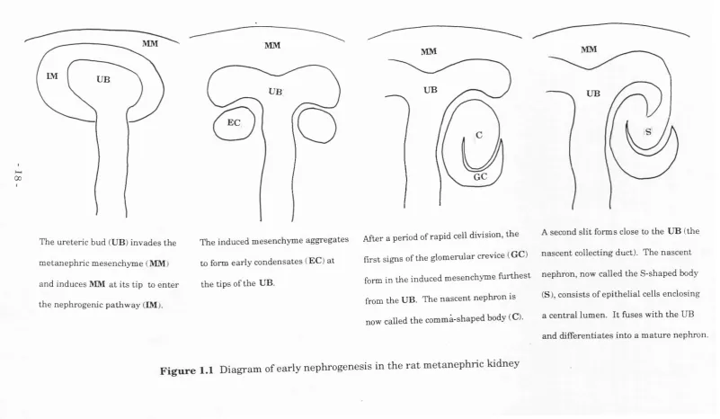
During the earhest stages of nephron formation, the tips of the branching
ureteric bud induce metanephric mesenchyme cells to undergo a series of
clearly identifiable morphological changes (Fig 1.1): they condense to form
a renal vesicle which then epitheliahses, elongates (to form a comma-
shaped body), and hollows to form a folded tube (the s-shaped body). At
one end, this tube fuses to the tip of the ureteric bud (which forms a
collecting duct), and at the other end it interacts with capillaries to form a
glomerulus (Saxèn, 1987; Bard, 1992).
M any grow th factors have been im plicated in kidney
development, including epidermal growth factor (EGF), insuhn-hke growth
factors (IGFs), transforming growth factor p (TGFp), hepatocyte growth
factor (HGF), platelet-derived growth factor (PDGF), and fibroblast
growth factors (FGFs) (for a review see Mendley and Toback, 1988). This
diversity reflects the m any cell types found in th e kidney and
17-MM
IM UB
00
MM
MM
The ureteric bud (UB) invades the
metanephric mesenchyme (MM)
and induces MM at its tip to enter the nephrogenic pathway (IM).
UB
EC
UB
GC
UB
The induced mesenchyme aggregates After a period o f rapid cell division, the A second slit forms close to the UB (the
to form early condensates (EC) at first signs of the glomerular crevice (GC) nascent collecting duct). The nascent form in the induced mesenchyme furthest nephron, now called the S-shaped body
from the UB. The nascent nephron is (S), consists of epithelial cells enclosing
now called the commà-shaped body (C). a central lumen. It fuses with the UB and differentiates into a mature nephron. the tips of the UB.
Introduction
experiments suggest distinct roles for each factor. For example, PDGF
and FGF may be important in angiogenesis (Mendley and Toback, 1988);
HGF may stim ulate branching of the ureteric hud (Montesano et al.,
1991) and the mesenchymal to epithelial transition (Tsarfarty et al.,
1994)'; and TGF p may be important in transforming mitogenic stimuli
into hypertrophic stimuli (Fine et al., 1985). While EGF and IGFs are
traditionally regarded as mitogens for several renal cell types (Fisher et
al., 1989; Rogers et al., 1991), in Chapter 3 I suggest a new role for both
EGF and IGFs.
An introduction to pre-implantation mammalian development
In C hapter 4 I describe experim ents on cell types from th e
preimplantation mouse embryo; the following is a brief account of early
mammalian development.
Soon after fertilisation, the vertebrate egg undergoes repeated
cleavage divisions to produce successively sm aller cells, called
blastomeres, which are contained within the zona pellucida (Fig. 1.2).
These blastomeres are equipotent until the early 8-cell stage: isolated 2-
and 4-cell stage blastomeres can form a whole mouse, while early 8-cell
stage blastomeres can contribute to a wide range of tissues in chimeras
(Kelly, 1977). During subsequent development there is a progressive
restrictio n of cell potency. Late 8-cell stage embryos undergo
compaction: blastomeres increase intercellular contact and become
polarised with the formation of distinct apical and basal surfaces. During
the 16-cell stage 2 populations of cells develop: the outer cells form the
trophectoderm and the inner cells, with no contact with the zona
pellucida, become the inner cell mass. At this stage the embryo - or
-Figure 2.1 (overleaf) Diagram showing preimplantation development of
the mouse embryo
h o u rs p o st fertilisation
0
24
27
72
84
95
98-108
cell#
16-32
blastomere polar body. maternal mRNA breaks down
M
O
R UL
A
com pactiontight junctions form between outer blastomeres sealing off interior of em bryo
■trophectoderm
blastocoelic cavity form s
- - inner cell m ass.
gap junctions between ICM s and TE
com plete com partm ental isatio n
of ICM from TE
B L A S T
O
C Y S T^ - zona pellucida breaks down
ICM becomes outer rind of primitive endoderm and inner core
of primitive ectoderm
21-Introduction
blastocyst - appears as a hollowed sack of cells th a t contains a small
group of ICM cells; within 1 cell cycle, the ICM differentiates into an
outer rind of endoderm which surrounds an inner core of primitive
ectoderm. This is followed by the eventual breakdown of the zona
pellucida and the implantation of the emhryo.
As soon as two cell types are apparent in the embryo,
intercellular interactions seem to become important: in the absence of
contact with ICM cells the trophectoderm cells (TE) stop growing and
become giant cells, while isolated ICM cells become TE. The nature of
these interactions is unknown, but there are gap junctions between the
TE and ICM cells up until 95 hours postfertilization, after which time
these connections are lost.
In this thesis, I report an example of developmental cell death
th a t has hitherto been overlooked - PCD during metanephric kidney
development; and in this new system I look at the regulation and extent
of PCD. The possibility th a t kidney PCD is regulated by Hmiting supplies
of survival factors strengthens the hypothesis th a t all cells die by PCD
unless signalled to survive (Raff, 1992). I have, therefore, also looked at
whether all cells have the programme for cell death and whether they
need to synthesise new proteins to execute the programme.
-C hapter 2
Program m ed C ell D eath in th e D evelop in g
R at K idney
-Kidney Cell Death
Introduction
PCD is only now becoming accepted as p art of the repertoire of animal
cells along with division and differentiation. U ntil recently, PCD was
thought to occur only in highly specialised circumstances such as in
negative selection in the thymus (Kislielow et al., 1988; Kappler et al.,
1987) and in metamorphosis (Goldsmith 1966; Kerr et al., 1974). In
developmental biology, for example, cell death had only been studied in
detail in the developing nervous and immune systems, and in a few cases
of morphogenesis in the early embryo; this was in spite of Glucksmann's
review (1951) im plicating cell death in th e development of most
vertebrate tissues and organs. Now, however, it is becoming clear th a t
most, and perhaps all, cells can kill themselves by PCD (Raff et al.,
1993).
Why has it taken so long for normal cell death to be viewed as
a general property of animal cells? Cell death in vitro has often been
regarded as an artefact of culture conditions. Cell death in vivo was also
initially viewed as an artefact, in this case, of tissue preparation; more
recently, although considered real, it has often been regarded as
quantitatively insignificant, since in most cases the dead cells are rare.
However, this is because cells th at undergo PCD are cleared so rapidly by
phagocytosis, th a t there is no leakage of cytosolic contents and hence no
inflammatory response. Therefore, even large scale normal cell death is
usually histologically inconspicuous. Only over the last 20 years or so, for
example, has it gradually been recognised th a t many types of vertebrate
neurones are overproduced and up to 50% or more of them die during
normal development (Barde, 1989; Cowan et al., 1984; Hamburger and
Levi-Montalcini, 1949; Oppenheim, 1991; Purves, 1988). And only in
-Kidney Cell Death
th e p a s t two y ears h as it been recognised th a t 50% of the
oligodendrocytes produced in the developing rat optic nerve normally die
(Barres et al., 1992). This massive death of newly formed neurones and
oligodendrocytes was initially missed because the dead cells constitute
less than 1% of the cells in the developing tissue. As the proportion of
dead cells in the developing nervous system is not very different from
th at seen in many other developing vertebrate organs, it is possible th at
large scale normal cell death occurs in many non-neural organs, even if it
has not yet been recognised.
In the present study I have looked for cell death in the normal
developing ra t kidney, where cell death was not thought to play an
im portant role, despite intense study of kidney development. In this
chapter I show th a t cell death plays an important part in normal kidney
development. It occurs in two regions, the nephrogenic zone of the cortex
and the medullary papilla, and in each it follows a distinct developmental
time course. The dead cells have a typical apoptotic morphology, as
shown by electron, fluorescence, and phase contrast microscopy. DNA
end-labelling studies in situ show th at the DNA in the dead cells is nicked.
The majority of the dead cells in the cortex of the kidney seem to be either
uninduced hlastem al cells or strom al cells; a m inority are cells of
developing nephrons.
25-Kidney Cell Death
Results
Apoptotic cells in the developing kidney
To identify apoptotic cells in the developing kidney, I stained frozen
sections of embryonic and postnatal perfusion-fixed ra t kidneys with
propidium iodide to label nuclei and then examined the sections in a
fluorescence microscope. Pyknotic nuclei, which often appeared as a
cluster of 2 or more brightly stained fragments, were readily recognised in
such sections (Fig. 2.1A, B). Whereas normal nuclei were generally oval
in shape and had a mean diameter of 8 ± 0.2 mm (n = 83), pyknotic
nuclei, or their fragments, were roughly spherical, had a mean diameter of
3 ± 0.2 mm (n = 27) and were more brightly stained than normal nuclei.
W ith phase co n trast optics, pyknotic nuclei and th e ir fragm ents
appeared darker th an normal or mitotic parenchymal cell nuclei (Fig.
2.1C). In embryonic non-perfused kidneys it was necessary to
differentiate between nucleated red blood cells and pyknotic cells
(Coggeshall e t al., 1993): the red blood cell nuclei were readily
distinguished as they were more spherical, larger, more textured, and less
brightly stained than apoptotic nuclei, and they were never fragmented
(Fig. 2.1 D). Mitotic figures were also clearly identifiable (not shown).
Apoptosis is characterised by profound ultrastructural
changes, including condensation of the chromatin and fragmentation of
the cell to form small, membrane-bound apoptotic bodies (Wyllie et al.,
1980; Clarke, 1990). To confirm th at the cells th a t die in normal kidney
development do so by apoptosis, I examined thin sections from newborn
ra t kidneys by electron microscopy (EM). All the dead cells seen had the
-Kidney Cell Death
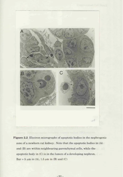
characteristic features of apoptosis. Of the 22 apoptotic cells examined
hy EM, 20 had the form of typical apoptotic bodies contained within
normal cells (Fig. 2.2A, B), while 2 were in the lumen of developing
nephrons (Fig. 2.2C). The cells th a t contained apoptotic bodies were
morphologically indistinguishable from their neighbours. Thus most of
the apoptotic cells in th e developing kidney fragm ent into apoptotic
bodies, which are very quickly phagocytosed, by neighbouring
parenchymal cells rather than by macrophages.
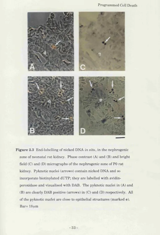
A typical, but not invariable, characteristic of apoptosis is
internucleosomal cleavage of nuclear DNA by endonucleases (Wyllie,
1984). To determine whether the pyknotic nuclei seen in the kidney had
fragmented DNA, I used the terminal transferase-mediated dUTP-biotm
nick end-labelling (TUNEL) method on PO rat kidney sections (Gavrieli et
al., 1992). Biotinylated dUTP was incorporated into the 3' cut ends of
genomic DNA; the incorporated nucleoside was visualised with avidin-
peroxidase, using DAB as a substrate. All the cells th a t appeared
pyknotic by phase-contrast microscopy had DAB+ nuclei (Fig 2.3),
confirming th a t these cells die by apoptosis.
Timing of cell death in the developing kidney
I found pyknotic nuclei m ainly in two regions of the embryonic and
postnatal ra t kidney: in the nephrogenic zone, which is the region of the
developing kidney cortex where new nephrons are produced, and in the
developing medullary papilla (Fig. 2.4A). The time course of cell death
was different in the two regions. In the nephrogenic zone, cell death was
highest (pyknotic index of 2.7%) in embryonic kidneys and decreased
thereafter, reaching a low basal level (pyknotic index of 0.15%) at
-Kidney Cell Death
postnatal day 14 (P14), where it rem ained for a t least 50 days
postnatally (Fig. 2.4B, open circles). In the most proximal region of the
papilla, cell death peaked at around P6-7 (pyknotic index of 3.2%) and fell
to less th an 0.1% by P14 (Fig. 2.4C, open circles). In the most distal
regions of the papilla, cell death was especially high in the embryo, b ut I
did not quantitate the death in this region.
Identity of the dead cells in the nephrogenic zone
The nephrogenic zone contains epithelial cells of the ureteric bud,
epithelial cells derived from the metanephric mesenchyme and uninduced
mesenchymal cells. To get an idea of which cell types were dying in the
nephrogenic zone, frozen sections of P3 ra t kidney were double-labelled
with PI and anti-Pax-2 antibodies, which specifically recognise the nuclei
of early nephrogenic cells (condensing mesenchymal cells and their early
epithelial derivatives) and of ureteric bud cells (Dressier and Douglass,
1992). It is important to use a nuclear and not a cytoplasmic m arker to
avoid ambiguity, since most dead cells in the kidney are present as
phagosomes inside other cells. I found th a t 70% of the pyknotic nuclei in
the nephrogenic zone were Pax-2 “ (n=50; Fig 2.5). This suggests th a t the
majority of dead cells were hlastemal or stromal, rather than epithelial.
Antigenic m arkers may be degraded early in the cell death
programme. In the developing optic nerve, for example, where 90% of the
dead cells are newly-formed ohgodendrocytes, the oligodendrocyte-specifrc
monoclonal antibody RIP labelled only 15% of the dead cells, whereas a
different oligodendrocyte-specific monoclonal antibody th a t recognises
galactocerebroside (GC) labelled 90% of the dead cells (Barres e t al.,
1993). Therefore, I examined propidium-iodide-stained cryosections of
-Kidney Cell Death
kidneys from E19 rats and recorded the location of over 400 pyknotic
nuclei in the nephrogenic zone. Most of the pyknotic nuclei (60%) were
found among metanephric mesenchymal cells, many of which were close
to, but not in, nephrogenic structures (Fig. 2.6A); they were not seen
among the densely packed metanephric mesenchymal cells a t the outer
edge of the embryonic kidney. The remaining 40% of pyknotic nuclei were
m ainly found in condensed mesenchymal and tu b u la r epithelial
structures, which could be readily identified as developing nephrons; they
were either in the walls of the developing nephrons or in their lumen (Fig.
2.6B, see also Fig. 2.2C). It was common to see dead cells among the
proximal-most cells of s-shaped bodies (Fig. 2.6C), easily recognisable
epithehal structures th a t wiU eventually form glomeruli. Pyknotic nuclei
were rarely seen in the branches of the ureteric bud in the nephrogenic
zone. In the maturing cortex at the inner edge of the nephrogenic zone,
the incidence of cell death decreased abruptly.
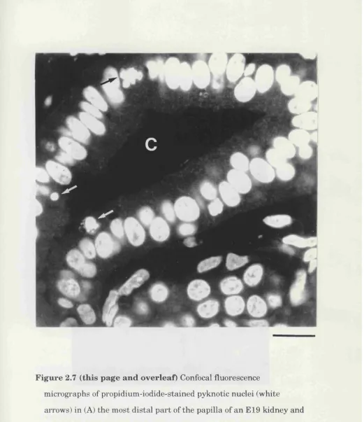
The papilla of the developing kidney contains epithelial cells of
both the ureteric bud and the loops of Henle, as well as interstitial cells.
In embryonic animals, when cell death is highest in the most distal region
of the papilla, the majority of dead cells were in the walls or lumen of
tubular structures, at least some of which seemed to be branches of the
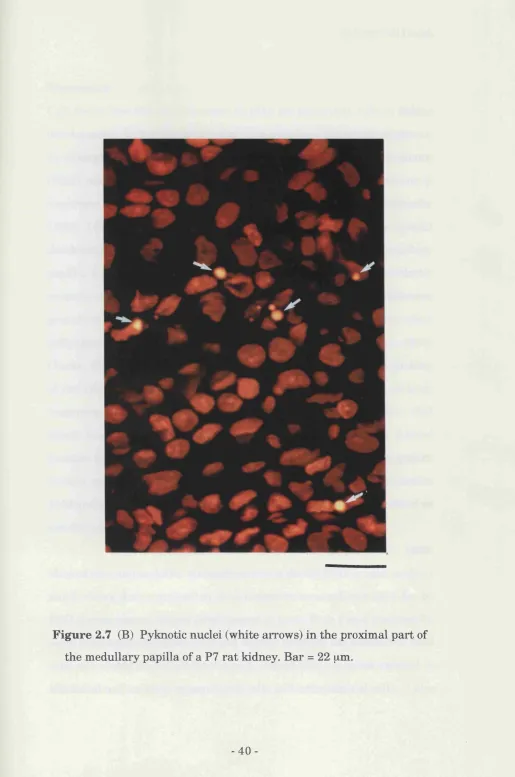
ureteric bud (Fig. 2.7A). In postnatal animals, when the majority of cell
death in the papilla is in the most proximal region, (where tubule
structures were less clearly defined), dead cells were scattered throughout
the tissue (Fig. 2.7B).
At all ages examined I found mitotic figures mainly in the nephrogenic
zone, where their proportion paralleled th at of pyknotic nuclei in this zone
(Fig. 2.4B, closed circles). By contrast, mitotic figures were rare in the
-Kidney Cell Death
papilla a t all ages examined (Fig. 2.40, closed circles). The close
relationship between mitosis and apoptosis in the nephrogenic zone (Fig.
2.4B) suggests th a t many of the cells th a t die in this region are newly
generated (while the cells th a t die in the papilla apparently are not). To
examine further the relationship between cell division and cell death, I
injected th e thym idine analogue bromodeoxyuridine (Brd-U, 4
intraperitoneal injections given 8 hours apart) into PO animals. Two
hours after the last injection, the rats were perfusion-fixed and frozen
sections of their kidneys were prepared and stained with both propidium
iodide and monoclonal anti-Brd-U antibody, as described previously
(Barres at al., 1992; Gonchoroff et al., 1985). In these experiments about
50% of the five cells and 25% of the pyknotic cells in the nephrogenic zone
were Brd-U labelled (data not shown), indicating th at at least one quarter
of the dead cells had synthesised DNA sometime in the 26 hour period
before their death. Unfortunately this experiment could not be extended
for a longer period as Brd-U was toxic to kidney cells at later time points
(data not shown).
-r
4
€
F ig u r e 2.1 P ropidium iodide sta in in g of nuclei in developing r a t kidney. (A) an d (C) Fluorescence and phase c o n tra st m icrographs, respectively, of th e sam e field, show ing a pyknotic nucleus (arrow ) in th e nephrogenic zone of a P I kidney. (B) Confocal fluorescence
m icrograph of a frag m en ted pyknotic nucleus in th e m e d u llary pap illa of a n E19 kidney. (D) Confocal fluorescence m icrograph of a norm al n u cleated red hlood cell in th e p ap illa of a n E19 kidney. B ar = 20 fim in (A) and (C), 6 pm in (B), 4 pm (D).
-F ig u re Electron micrographs of apoptotic bodies in the nephrogenic
zone of a newborn rat kidney. Note th at the apoptotic bodies in (A)
and (B) are within neighbouring parenchymal cells, while the
apoptotic body in (C) is in the lumen of a developing nephron.
Bar = 5 gm in (A), 1.5 \im in (B) and (C)
-Programmed Cell Death
F igure 2.3 E nd-labelling of nicked DNA in situ , in th e nephrogenic zone of n eo n atal r a t kidney. P h ase c o n tra st (A) an d (B) an d b rig h t field (C) and (D) m icrographs of th e n ephi ogenic zone of PO r a t kidney. Pyknotic nuclei (arrow s) contain nicked DNA and so incorporate biotinylated dUTP; th e y are labelled w ith avidin-
peroxidase and visualised w ith DAB. The pyknotic nuclei in (A) and (B) are clearly DAB positive (arrow s) in (C) an d (D) respectively. All of th e pyknotic nuclei are close to epith elial s tru c tu re s (m arked e).
Bai'= ISjim
33-F ig u re 2.4 (overleaf) Pyknotic a n d m itotic indices in th e nephrogenic zone (B) a n d m e d u lla ry p a p illa (C) of th e developing r a t Iddney a t d ifferen t ages. A sch em atic d raw in g of a m id sag g ital section th i'o u g h a developing k id n e y is show n in (A) to in d ic a te th e regions w h ere n o rm a l cell d e a th w as assessed . In (B) th e n u clei in th e o u ter-m o st 100 \im o f cortex w ere counted in ev ery second field (to tal of 10-30 fields) of 3 n o n -se ria l sections for each pup. In (C) th e nuclei in a 300 X 300 p,m g rid a t each side of th e p ro x im al p a p illa (show n in A) w ere counted in 4 n o n -serial sections for each pu p . T he re s u lts a re
p re se n te d as m e a n + s.e.m . of th e re s u lts from 4-5 p u p s a t each age.
[x] nephrogenic zone
g proxim al papilla
distal papilla
Age (postnatal days)
B 3.0 -1
Pyknotic Mitotic
0) Ü3
z
(0
0.5
-0.0
-10 0 10 20 30 40 50 60
Pyknotic Mitotic
3.0
-2.0
0.5
-0.0
-10 0 10 20 30 40 50 60
Age (postnatal days)
-35-F ig u re 2.5 (overleaf) Pax-2 and propidium-iodide double labelling of
the nephrogenic zone of developing ra t kidney. Phase contrast (A)
and fluorescence micrographs (B) of the same field of propidium-
iodide-stained sections of PO ra t kidney showing pyknotic nuclei
(arrows) next to an epithelial structure in a P3 ra t kidney. (C)
fluorescence micrograph of the same field stained with an anti-Pax-2
antibody and visualised with a FITC-conjugated secondary antibody,
showing th a t the pyknotic nuclei in (A) and (B) are not Pax-2 positive
(arrows). Bar= 22 ^im
\ y j ...
37-F ig u r e 2.6 Localisation of propidium -iodide-stained pyknotic nuclei in th e nephrogenic zone of a P I kidney. In (A) m ost of th e pyknotic nuclei are close to, b u t not in, developing nephrons. In (B), w hich is a confocal m icrograph, and (C), th e pyknotic nuclei a re in th e w all or lum en of developing nephrons. Note th e pyknotic nucleus (arrow) in th e proxim al p a rt of a n s-shaped body in (C).
B ar = 25 pm in (A) and (C), 7 pm in (B).
38-Figure 2.7 (this page an d overleaf) Confocal fluorescence m icrographs of propidium -iodide-stained pyknotic nuclei (w hite
arrows) in (A) th e m ost d istal p a rt of th e p apilla of a n E19 kidney and (B) th e proxim al p a r t of th e p apilla in a P7 kidney. In (A) a m itotic nucleus (hlack arrow ) is also seen and th e tu h u la r epithelial stru ctu re shown is a collecting duct (m arked e).
B ar =15 pm in (A) and 22 pm in (B).
-39-1
H
g
Figure 2.7 (B) Pyknotic nuclei (w hite arrow s) in th e proxim al p a rt of th e m edullary papilla of a P7 r a t kidney. B ar = 22 ^m.
-Kidney Cell Death
Discussion
Cell death has not been thought to play an im portant role in kidney
development. In his comprehensive monograph on kidney development,
for example, Saxèn (1987) does not mention cell death, and Glucksmann
(1951) refers only to a study th a t implies th a t the first generations of
nephrons degenerate during metanephric development (Kampmeier
1926). I find, however, th a t there is substantial cell death in the normal
developing ra t kidney, both in the nephrogenic zone and in the medullary
papilla. It can be readily detected by electron microscopy in thin plastic
sections, or in fi^ozen sections, either by fluorescence microscopy following
propidium iodide staining or by phase contrast microscopy. The dead
cells show the characteristic features of apoptosis (Wyllie et al., 1980;
Clarke, 1990), including nuclear condensation and fragmentation, nicking
of the DNA, and cell shrinkage. Furthermore, the dead cells or their
fragments are phagocytosed by neighbouring parenchymal cells. Cell
death during renal development may have been previously missed
because many of the recent significant studies on kidney development
(review see Saxèn 1987) have used the transfilter technique (Grobstein
1953 and 1956), in which dead cells could easily have been interpreted as
artefactual.
Previous studies (Weller et al., 1991; Koseki et al., 1992)
showed th at metanephric mesenchyme cells die by PCD in vitro and my
study shows th a t a proportion of metanephric mesenchyme cells die by
PCD during normal kidney development in vivo. Both Pax-2 staining for
early induced nephrogenic cells and studies in which the location of dead
ceUs was scored show th a t the majority of dead cells are either stromal or
hlastemal metanephric mesenchyme cells and not epithelial cells. I also
41-Kidney Cell Death
find th a t a considerable proportion (40%) of the dead cells are in
nephrogenic structures.
Glucksmann (1951) distinguished three kinds of normal cell
death during development: (1) phylogenetic death, associated with the
loss of vestigial structures or of larval organs during metamorphosis; (2)
morphogenetic death, associated w ith the sculpting of specific structures such as digits or the separation or fusion of epithelia; and (3)
histiogenetic death, associated with cell differentiation. Most of the
deaths th at I see in the nephrogenic zone in the developing kidney fit best
into the category of histiogenetic deaths: they are scattered and seem to
occur mainly in m etanephric mesenchymal cells a t a tim e when a
proportion of these cells is being induced to differentiate into nephrogenic
epithelial cells. Furthermore, our findings suggest th at many of the cells
th at die in the nephrogenic zone have recently divided: there is a strong
temporal correlation in this region between the pyknotic and mitotic
indices (see Fig. 4B), and Brd-U incorporation studies indicate th a t at
least 25% of the dead cells in this region synthesised DNA sometime in
the 26 hour period before they died. This observation could not be
extended to cells th a t had divided within the last two days as Brd-U was
toxic at longer time points.
W hat is the function of metanephric mesenchyme cell death
in kidney development? Cell death in the developing nervous system is
thought to help match both the numbers of neurones to the numbers of
target cells they innervate (Cowan et al., 1984) and the number of
oligodendrocytes to the number (and length) of axons th a t require
myelination (Barres et al., 1992; Barres and Raff*, 1993). In the kidney,
cells of two lineages are involved in early nephrogenesis - nephrogenic
42-Kidney Cell Death
mesenchymal cells and epithelial cells of the ureteric bud - and it is
possible th a t cell death helps to match the numbers of cells in these two
lineages.
It is less clear how to categorise the cell deaths in the
developing nephrons. Developmental cell death contributes to the fusion
of epithelia in, for example, palatal shelf formation (Farbman, 1986; Shah
1979) and neural tube closure (Glucksmann 1951); it is also suggested
th a t cell death could contribute to lumen formation (Snow 1987), by
hollowing out structures. The dying cells th a t I observe at the ampullae
(the tips of the ureteric bud) may be instrum ental in the fusion of two
epithelia - the nascent nephrons and the collecting ducts. Although it is
not known how the glomerular crevice is formed in the S-shaped body, it
is thought th a t changes in cell adhesion may play a role (Saxèn 1987).
My observation th a t dying cells are commonly found in the proximal tip of
the S-shaped body raises the possibility th at cell death may participate
in glomerular crevice formation. Finally the dying cells in the medullary
papilla may be involved in the fusion of the major collecting ducts (Potter,
1972). Thus the epithehal cell deaths seen are probably best considered
examples of morphogenetic deaths.
De-regulation of genes involved in cell death may contribute to
some diseases. The m am m alian gene bcl-2, for example, was first
described as an oncogene in follicular B-cell lymphomas (Tsujimoto et al.,
1985): translocation brings the gene under the control of an Ig enhancer
so th a t it is over-expressed in B-cells, where it suppresses PCD, thereby
promoting B-cell survival and tumorigenesis by increasing the probability
of further oncogenic mutations (Vaux et al. 1988; Chen-Levy et al., 1989;
Strasser et al., 1990). Experimental over-expression of the bcl-2 gene
-Kidney Cell Death
product also prevents PCD in many other vertebrate cell types (Vaux et
al., 1988; Sentman et al., 1991; Strasser et al., 1991; Garcia et al., 1992).
My finding th a t cell death is a normal feature of kidney development
raises the possibility th at defects in cell death might play a part in some
developmental abnormalities of the kidney. This was first suggested by
Kampmeier (1923 and 1926), who proposed th a t congenital cysts arose
from vestigial renal vesicles th at did not degenerate during development.
The Bcl-2 protein is expressed in the developing kidney where it is
preferentially located in the m etanephric cap and the developing
nephrons (Veis and Korsmeyer, 1993; my unpublished observations). In
homozygous bcl-2 knockout mice, polycystic kidney disease is an
invariable feature and may he the cause of death in at least some of the
mice (Veis et al., 1993), supporting the idea th a t de-regulated PCD may
be involved in some developmental kidney disorders.
Wilms' tumour is a second kidney disease th a t may involve
abnormal renal PCD. It is a nephroblastom a in which control of
nephrogenesis is aberrant (van Heyningen and H astie, 1992). The
Wilms' tumour-suppressor gene wt-1 is a zinc-ftnger, DNA-binding protein
th a t acts as a putative repressor of various genes, including the IGF-2
gene (Drummond et al., 1992) and possibly the IGF-1 receptor genes
(Werner et al., 1993). wt-1 is often inactivated in Wilms' tumours (van
Heyningen and Hastie, 1992), which may result in increased expression
of IGF-2 (Drummond et al., 1992) and the IGF-1 receptor (Werner et al.,
1993). My finding th a t IGF-1 decreases the pyknotic index in the
nephrogenic zone of the kidney (see chapter 3) raises the possibility th at
decreased PCD may contribute to Wilms tum our form ation and
Kidney Cell Death
progression, ju st as it does in follicular B-cell lymphoma (Vaux et al.,
1988; Strasser et al., 1990).
-Kidney Cell Death
Methods
Tissue preparation
Sprague-Dawley rats were bred in the UCL animal facility. Postnatal
ra ts were deeply anaesthetised with pentobarbitone and perfused
through the heart with phosphate buffered saline (PBS), followed by 4%
paraformaldehyde in 0.1 M sodium phosphate buffer (PBS), pH 7.4. The
kidneys were removed and fixed over night at 4°C and cryoprotected with
30% sucrose in PBS until equilibrated. Embryonic kidneys were dissected
from embryos th a t had been removed from the u teru s and put
immediately on ice; they were then fixed as above. Whole kidneys were
frozen in OCT compound (Miles), and 6.5 pm cryosections were cut on a
Bright cryostat. Sections were collected on gelatinised glass microscope
shdes, air dried, and post-fixed with 70% ethanol at -20°C for 10 minutes.
Propidium iodide labelling
Sections were incubated with 4 pg/ml propidium iodide (Sigma) and 100
pg/ml RNase (Sigma; DNase-free) in PBS for 30 minutes at 37°C (Barres
et al., 1992; Rodriguez-Tarduchy et al., 1990). The slides were washed in
PBS, mounted in Citifluor (City University, London), examined in a Zeiss
Universal fluorescence microscope using a 40x oil immersion phase
contrast objective, and photographed with Tri-X film, ASA 400. In some
cases sections were examined with a Biorad MRC-600 laser-scanning
confocal im aging system in conjunction w ith a Nikon O ptiphot
microscope and the images were printed on a Mitsubishi CP 100 printer.
Pyknotic nuclei in propidium-iodide-labelled sections were
readily recognised by fluorescence microscopy: they were smaller and
Kidney Cell Death
more brightly stained th an norm al nuclei, and th ey w ere often
fragmented. Clusters of nuclear fragments or apoptotic bodies occurring
within one normal nuclear diam eter were counted as one pyknotic
nucleus (Wyllie, 1975). Pyknotic nuclei (or apoptotic bodies) could also be
readily recognised by phase contrast microscopy by their small, phase
dark appearance. Propidium-iodide-labelled mitotic figures were also
easily distinguished by fluorescence microscopy.
For each age examined, 3 non-serial 6.5 pm m idsagittal
sections were examined from a t least 3 animals. In each section
pyknotic nuclei were counted in every second field. In the kidney, cells
were counted in the outer-most 100pm of the cortex and in a 300 x 300
pm area on each side of the proximal part of the medullary papilla (see
figure 3A). Both pyknotic and mitotic nuclei were counted and the
numbers obtained were corrected for split cell counts (Abercrombie,
1946) and expressed as a percent of total nuclei.
Abercrombie correction
Propidium iodide-stained cryosections of PO ra t kidney (6.5 pm) were
prepared. To measure the diameters of pyknotic and normal nuclei, I
traced nuclear profiles on a digitising tablet (Summa graphics) and used
cigal software (J. Voyvodic, unpublished) for the morphometry. The
average diameter of non-dividing, non-pyknotic nuclei was 8 pm + 0 . 2
(n=83) compared to th at of pyknotic nuclei which was 3 pm + 0.2 (n= 27)
and mitotic nuclei which was 10 pm + 3.2 (n=5). To determ ine the
relationship between profile and particle numbers in the kidney and
thymus, I obtained correction factors for split cell counts using the
equation derived by Abercrombie (1946):
-Kidney Cell Death
P = A M L + M where:
P = corrected number of nuclei A = raw count (profile count) M = section thickness
L = average diameter of the nucleus
Correction Factor
Normal
Pyknotic Mitotic
Kidney
0.45
0.687
0.45
Tissue
Thymus
0.46
0.59
Immunohistochemistry
Cryosections of paraformaldehyde-fixed P I and P6 ra t kidney were
rehydrated with PBS, permeabilised with 70% ethanol at -20°C for 10
minutes and then washed in PBS containing 0.05% Triton-XlOO. Non
specific antibody binding was blocked by incubation the sections in 50%
goat serum and 20 mM L-lysine in PBS for 30 minutes. After washing,
the sections were labelled with Rabbit anti-Pax-2 IgG antibody (10 jig/inl;
Dressier and Douglas, 1992) in 2% goat serum, followed by flourescien-
coupled goat-anti-rabbit antibody (Welcome, diluted 1:100), both for 1
hour at room temperature. The slides were stained with PI, mounted and
examined as described above.
Brd-U incorporation
Five new born (PO) ra t pups were given 4 intraperitoneal injections of
Brd-U (0.1 mg/g body weight; Boehringer Mannheim) at 8 hour intervals.
Kidney Cell Death
Two hours after the last injection, the pups were perfused and the
kidneys fixed and processed as described above. Air dried sections were
rehydrated with PBS and post-fixed with 70% ethanol for 10 minutes a t
-20°C. The DNA was denatured by incubation in 2 M HCL for 10
minutes and the and was then neutrahsed by incubation in 0.1 M sodium
borate (pH 8.5) for 10 minutes. To block non-specific binding, the
sections were incubated in 50% sheep serum, containing 1% bovine
serum albumin (Sigma) and 100 mM l-lysine . The sections were then
labelled with BU-1, a monoclonal anti-Brd-U antibody (Gonchoroff et al.,
1985; Greipp et al., 1985; concentrated su p ern atan t diluted, 1:10),
followed by biotinylated sheep anti-mouse IgG (Amersham; diluted 1:50)
and then fluorescein-coupled strepavidin (Amersham; diluted 1:100).
Finally, the sections were stained with propidium iodide and examined as
described above. The pyknotic index in the kidneys was not increased in
Brd-U-treated rats, suggesting th at the Brd-U treatm ent was not toxic to
kidney cells at the concentrations used.
Electron microscopy
PO ra t pups were deeply anaesthetised with pentobarbitone and perfused
with PB, followed by 1% glutaraldehyde in 0.1 M phosphate buffer, pH 7.2
(Yun and Kenney, 1976). The tissue was cut into 1 mm slices and fixed
for a further 2.5 hours at room temperature and then post-fixed with 1%
osmium tetroxide in PB for 1 hour. After dehydrating in acetone and
embedding in Epon, thin sections were cut on an LKB Ultratome, counter
stained with uranyl acetate and then lead citrate, and examined in a
JEOL 100-CXII electron microscope at 80kV.
Kidney Cell Death
The TUNEL technique
To determine whether pyknotic cells in the kidney have fragmented DNA,
6.5 pm cryosections of PO ra t kidney were collected on APES treated
slides, air-dried and rehydrated with 10 mM Tris.HCL pH8. Proteins were
stripped from the sections with proteinase K (20 pg/ml, Sigma) for 15
m inutes a t room tem perature, and endogenous peroxidases were
inactivated with 3% hydrogen peroxide. After extensive rinsing in double
distilled water, the shdes were treated with biotinylated dUTP (Bio-16-
dUTP, 3 nM, both Boehringer Mannheim UK) in the presence of terminal
deoxynucleotidyl transferase (TDT, 10 u / 1 in TDT buffer, both GIBCO
BRL) a t 37^0 for 1 hour to end-label any nicked DNA in the sections.
The reaction was term inated by 300 mM NaCl and 30 mM sodium
citrate.
The slides were washed with PBS for 5 m inutes a t room
tem perature, blocked with 2% BSA for 10 minutes and then washed
again. Biotinylated-dUTP endlabeUed DNA was labelled with and Avidin
DHrbiotinylated horseradish peroxidase H complex for 30 m inutes at
room temperature. Labelled nuclei were then visualised by a 7 minute
incubation with the peroxidase substrate diaminobenzidine tetra-
hydrochloride (DAB, Amersham UK) in the presence of 30% peroxide.
The slides were then rinsed, lightly counter stained with Toll Blue, C
mounted in citifluor (City University, London) and examined with phase
contrast and bright field microscopy. Pretreatm ent of sections with
DNase 1 (0.2 mg/ml, 10 minutes at room temperature) or omission of
the dUTP from the end-labelling reaction destroyed all DAB reactivity
in the sections.
-C hapter 3
F actors A ffecting K idney C ell Su rvival
Factors Affecting Kidney Cell Survival
Introduction
The extensive death of developing vertebrate neurones is thought to
result, a t least in part, from a competition among neurones for limiting
amounts of survival (neurotrophic) factors secreted by the target cells
th a t they innervate. The best documented example of this is nerve-
growth-factor-dependent sympathetic and sensory neurones; increasing
the availability of either target tissue or nerve growth factor (NGF;
Hamburger and Yip 1984) in vivo decreases the amount of neuronal cell
death; conversely, decreasing the availability of either target tissue (Levi-
Montalcini, 1987) or NGF (Levi-Montalcini, 1972; Gorin and Johnson,
1979) in vivo increases the amount of neuronal cell death. It is thought
th a t many other cases of neuronal cell death in the developing central
nervous system are due to a similar mechanism (Cowan et al., 1984;
Purves 1988; Oppenheim 1991); for example, hrain-derived neurotrophic
factor (BDNF) rescues quail nodose ganglion neurones in vivo (Hofer and
Barde, 1988). Recently it was found th a t the death of newly formed
oligodendrocytes in the developing optic nerve may also reflect a
competition for limiting amounts of survival factors (Barres et al., 1992),
suggesting th at this cause of cell death is not confined to neurones.
The overproduction of neurones and th eir subsequent
selection due to limiting availability of survival factors is thought to
m atch the sizes of synaptically connected populations of cells and
eliminate erroneous neuronal projections during vertebrate neural
development (Cowan et al., 1984; Purves 1988; Oppenheim, 1991).
Similarly, normal cell death of oligodendrocytes may match the numbers
of these cells to the size and length of the axon th a t they myelinate
(Barres et al., 1992). It seems unlikely th at such an elegant mechanism
-Factors AfFecting Kidney Cell Survival
for regulating the co-ordinated development of cell populations is unique
to the developing nervous system. Indeed, it has recently been proposed
th a t all cells (other than blastomeres) in higher animals may require
survival factors to avoid killing themselves and th a t many cases of
normal cell death may reflect limiting supplies of survival factors (Raff
1992). I have looked at the possible role of survival factors in normal cell
death in the developing kidney in order to test this possibihty.
Regulation of cell survival has not been considered to be a
p art of normal kidney development, and therefore most studies have
investigated the mitogenic but not survival effects of growth factors on
kidney ceUs. Epidermal growth factor (EGF), for example, is known to be
im portant in m etanephric development and has been generally
considered to be a co-mitogen (Mendley and Toback, 1988, Fisher et al.,
1989). However, EGF m aintains the integrity of kidney rudiment
explants grown in serum- and inducer-free conditions without stimulating
DNA synthesis ( Weller et al., 1991). Furthermore, EGF decreases the
amount of DNA degradation seen in early kidney rudiments cultured in
serum- and inducer-free medium (Koseki et al., 1992). These studies
suggest th a t EGF may act as a survival factor for m etanephric
mesenchyme in vitro.
A second growth factor of interest is insulin-like growth factor
1 (lGF-1), which is the main mediator of the effects of growth hormone in
the body (Mathews et al., 1988). While IGFs 1 and 2 are known to be
important in the regulation of mammalian growth, it has been assumed
th at they promote growth by promoting cell proliferation. However, IGF-
1 promotes survival and not proliferation in vertebrate CNS neurones
(Svrzic and Schubert, 1990), Balb/c 3T3 murine fibroblasts (Tamm and