Original Research Article.
93 |P a g e Int J Med Res Prof.2018 May; 4(3); 93-100. www.ijmrp.com
Unusual Histopathology in Malignant Phyllodes Tumours Causing
Diagnostic Difficulties in Cytology: A Retrospective Study
Bandyopadhyay Goutam
1, Mukhopadhyay Sabuj Ghana
2*, Raina Rimjhim
31MD Pathology (AIIMS), Professor and Head,
2*MD Pathology (IPGMER, Kolkata), RMO cum Clinical Tutor, 3Post Graduate Trainee (MD Pathology),
Department of Pathology, Burdwan Medical College, Burdwan, West Bengal, India.
ABSTRACT
Introduction: Phyllodes tumour of breast, previously
designated as “Cystosarcoma Phylloides,” later classified as “Benign” or “Malignant” according to their histologic appearance and beheaviour. Varied Stromal changes like
lipomatous, osseous, cartilaginous, myosarcomatous,
angiosarcomatous and epithelial changes like apocrine and squamous metaplasia are observed in benign and malignant counterparts. Though FNAC is the first line of diagnosis in breast neoplasm, but rare unusual pattern may limit the cytological diagnosis and necessitate the histopathological final conclusion.
Methods: Nine cases with final histopathological diagnosis of Malignant Phyllodes, based on morphology alone, in last four years are retrieved and studied vigorously for evaluation of cytological pitfalls in such lesions.
Results: All cases of this study are female ranging from 45- 54 yrs and having huge mass in one breast without any axillary lymph nodes. 33.3% (n=3) cases were cytologic category 5 (Malignant), but the cytological diagnosis was not corroborated on histology. Cytology of 22.2% (n=2) reported as category 4(suspicious of malignancy), with incorrect provisional diagnosis of Borderline Phyllodes. 11.1% (n=1) case reported on cytology as Category 3 (Atypical probably benign) due to extensive squamous metaplasia obscuring underlying
chondrosarcomatous differentiation. Another two(22.2%) was Inadequate for opinion in cytology (CategoryC1) due to repeated hemorrahagic aspirate from different sites which was due to specific stromal changes in the phyllodes stroma.
Conclusion: Unusual stromal component with lack of
epithelial component and/ or metaplasia of epithelial component may lead to improper erroneous cytologic evaluation, particularly in huge breast masses.
Key words: Phyllodes Tumour, Epidermal Cyst,
Chondrosarcoma, Granular Cell Change.
*Correspondence to:
Dr. Sabuj Ghana Mukhopadhyay,
MD Pathology (IPGMER, Kolkata), RMO cum Clinical Tutor,
Department of Pathology,
Burdwan Medical College, Burdwan, West Bengal, India.
Article History:
Received: 15-11-2017, Revised: 12-12-2017, Accepted: 29-04-2018
Access this article online
Website:
www.ijmrp.com
Quick Response code
DOI:
10.21276/ijmrp.2018.4.3.018
INTRODUCTION
Phyllodes Tumour (PT), first described as Cystosarcoma Phyllodes is a fibroepithelial neoplasm of breast identified by characteristic leaf-like arrangement of spindle shaped stromal cells along with elongated epithelial component on histopathology. Phyllodes tumours are diagnosed when the fibroepithelial architecture shows an exaggerated intracanalicular pattern with leaf-like fronds protruding into cystically dilated spaces accompanied by stromal hypercellularity.1 Diagnosis of phyllodes
tumour should always be accompanied by a sub-classification into benign borderline and malignant categories due to largely different behavior (local spread and metastasis) and prognosis.
Fine needle Aspiration Cytology (FNAC) is first line investigation of any breast lump. In case of benign phylloides tumour, the stromal component, represented by stromal fragments, isolated
stromal cells, and naked stromal nuclei are found to be more numerous than the epithelial one in most of the cases. The malignant ones show stromal fragments of variable dimensions, with moderate cellularity, made of discohesive spindle cells, with atypical nuclei, minimal/no epithelial elements and presence of atypical multinucleated giant cells.
At histopathological analysis, final and definitive grading of PT into benign, borderline and malignant grade is done on the basis of a constellation of histological parameters like the degree of stromal cellularity and atypia, mitotic count, stromal overgrowth, and the nature of their tumour borders.2
fibroepithelial proliferative diseases of breast-like fibroadenomas. It is easier to differentiate malignant from benign Phyllodes tumour on FNAC.3 But in the presence of atypical changes in the stroma
of a malignant phyllodes tumour, diagnosis can be very difficult. This gives rise to widely varying diagnosis ranging from C1 [Inadeqate] to C5[Malignant] in cytologic scale of malignancy4
according the areas sampled by the passing needle, confusing the surgeon and delaying prompt treatment. This problem persists in limited breast biopsies like core needle biopsies too.
In this article we have retrospectively assessed the cytological diagnosis of nine cases of malignant phyllodes tumour received over the span of three years and compared them with final histopathological diagnosis.
MATERIALS AND METHODS
This study was conducted on female patients of unilateral breast mass diagnosed histopathologically as malignant phyllodes tumour in Department of Pathology of Burdwan Medical college, Burdwan, West Bengal, India from April 2014 to March 2017 (3 years). Total number of cases identified were 19. Only those patients, who had a preoperative cytological examination of the breast mass in the same institution were included in the study. Nine patients were identified matching the inclusion criteria. The participating pathologists have retrieved full history, performed clinical examination and Fine Needle Aspiration Cytology (FNAC) of the patients. The cytological findings were
node status.
Reports of radiological investigations were retrieved from surgical records section when available.
FNAC was done under aseptic precaution by standard procedure with a 23G needle and a FNAC handle. Multiple passes from at least two areas of the mass was conducted and smears were prepared. Half of the smears were dry-fixed for Leishman Giemsa (LG) staining and half of them were immediately wet-fixed for Papanicolaou stain (PAP) in each case. They were reviewed by two pathologists and graded independently. A consensus diagnosis and grading were reached and the final report was dispatched. After mastectomy the specimen received were grossed carefully (Fig 1) under the following headings: Measurement in three dimensions, weight, status of skin and nipple, measurement of the lesion (tumour), surface of the tumour, character of cut surfaces, presence of satellite nodules and axillary lymph nodes if any.
The grossed tissue was processed in standard processing method and stained with Hematoxylin and Eosin (H and E). They were described microscopically under the following headings: Description of tumour proper (with special emphasis to tumour cell description, degree of mitosis, nuclear atypia), status of surgical margins, evidence of lymphovascular invasion, amount of haemorrhage and necrosis and presence of heterologous elements. Final histopathological conclusive opinion was reached by consensus agreement among two author pathologists.
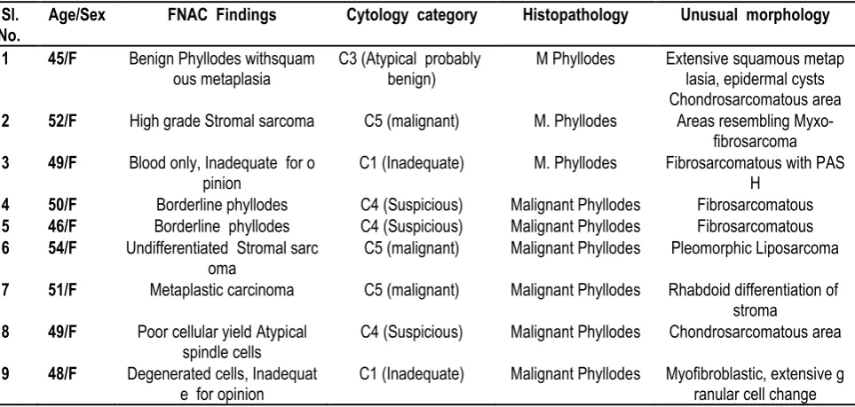
Table 1: Nine cases with Cytological and histopathological findings with pertinent unusual features.
Sl. No.
Age/Sex FNAC Findings Cytology category Histopathology Unusual morphology
1 45/F Benign Phyllodes withsquam
ous metaplasia
C3 (Atypical probably benign)
M Phyllodes Extensive squamous metap
lasia, epidermal cysts Chondrosarcomatous area
2 52/F High grade Stromal sarcoma C5 (malignant) M. Phyllodes Areas resembling
Myxo-fibrosarcoma
3 49/F Blood only, Inadequate for o
pinion C1 (Inadequate) M. Phyllodes Fibrosarcomatous with PASH
4 50/F Borderline phyllodes C4 (Suspicious) Malignant Phyllodes Fibrosarcomatous
5 46/F Borderline phyllodes C4 (Suspicious) Malignant Phyllodes Fibrosarcomatous
6 54/F Undifferentiated Stromal sarc
oma C5 (malignant) Malignant Phyllodes Pleomorphic Liposarcoma
7 51/F Metaplastic carcinoma C5 (malignant) Malignant Phyllodes Rhabdoid differentiation of
stroma
8 49/F Poor cellular yield Atypical
spindle cells C4 (Suspicious) Malignant Phyllodes Chondrosarcomatous area
9 48/F Degenerated cells, Inadequat
Figure 1: Gross Specimen with areas of sectioning marked by arrow.
Figure 2: Case graded as C1. Different areas of Pseudo Angiomatous Stromal Hyperplasia (PASH) which caused bleeding during FNAC, (H & E x 100)
2a
2b
Figure 3: Case graded as C1. Myofibroblastic differentiation with extensive granular cell change. Benigh ductal epithelium is also visible (arrows)( H & E x 100 and 400)
RESULTS
Nine cases matching the inclusion criteria had an age range between 45 years to 54 years and have reached menopause. One of them were childless and rest of them had 1 to 3 children. All of them have breastfed their children to some extent. No significant history of past breast disease, family history of breast pathology were elicited in the subjects.
All of them presented with a history of rapidly growing unilateral breast mass with an average duration of 3.2 months.(1 month to 6.5 months). The lesions originated in any quadrants and quickly enlarged to involve the entire affected breast causing significant cosmetic disfigurement. One of the patients had a prominent skin involvement with petechiae and ulceration of the skin. In one patient, the lesion was also fixed to chest wall. Nipple was not involved in any of the patients. Axillary lymphadenopathy was reported in the same patient who had ulceration of the overlying skin. Average size of the mass was 10.4 cm(8.3cm to 16.9 cm). The mass were firm and lobulated. In two cases the mass had a cystic feel.
Two patients had an ultrasonographic examination of breast which has given a provisional diagnosis of Phyllodes tumour.
Cytology findings (n=9) were categorized as [Table 1] : C1: 2 cases (22.2%) Inadequate for conclusive opinion C3: 1 case (11.1%) Cytologic atypia,probably benign C4: 3 cases (33.3%) Suspicious of Malignancy. C 5: 3 cases (33.3%) Malignant.
In two cases no opinion was possible as there was not enough material to make a conclusive diagnosis (C1). On histopathology one of them showed areas of fibrosarcoma with Pseudoangiomatous Stromal Hyperplasia (PASH) along with typical phyllodes morphology of overgrown malignant stroma and very scanty trapped ductal elements. The PASH was responsible for too much blood in the slides obscuring diagnostic details (Fig 2). In another case, the histopathology revealed typical malignant phyllodes morphology admixed with myofibroblastic changes in the stroma and extremely rare granular cell change surrounded by benign ductal elements, which, have not been reported in any phyllodes tumour till date.(Fig 3)
One case was reported as cytologic category C3, as there was benign ductal elements and numerous nucleated and anucleate squamous cells, some of them showing cytologically atypical nuclear features. The cytology report was dispatched as extensive squamous metaplasia in a benign cystic disease of breast. The histopathology also revealed extensive squamous metaplasia, epidermal cyst formation with keratin pearls. But one area also showed a malignant spindle cell stroma with prominent well differentiated chondrosarcomatous area. The ducts had dual lining and normal cytomorphology. So, It was reported as a Malignant Phyllodes tumour.(Fig 4)
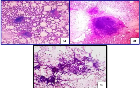
Three cases were reported cytologically as Suspicious of malignancy (C4). Two of them were reported as Borderline phyllodes tumour, as there was stromal overgrowth and paucity of ductal elements but lack of definitely malignant spindle cells.(Fig 5) On histopathology both of them showed, malignant phyllodes morphology with well differentiated fibrosarcomatous changes which caused difficulty in diagnosis by FNAC. One of the C4 cases had a very poor cellular yield even after repeated aspiration and as reported as atypical spindle cell lesion, suspicious of malignancy. Histopathology of that case revealed extensive chondrosarcomatous areas (Fig 6), nearly replacing the normal breast architecture with some areas of malignant spindle cell and scanty trapped benign ductal elements.
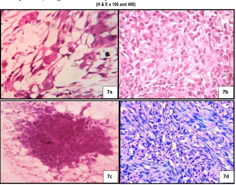
The three remaining cases were cytologically diagnosed as malignant lesions of breast (C5). But in two of the cases no benign ductal elements were found in the cytological smears. But presence of highly malignant atypical spindle cells has resulted in diagnosis of High Grade Stromal Sarcoma of breast. Interestingly, in one of the C5 case there were malignant spindle cells admixed with malignant epithelial looking cells and was reported as Metaplastic Carcinoma of Breast. On histopathology two cases reported as stromal sarcoma revealed, benign ductal elements and were reclassified as malignant phyllodes tumour. Whereas the case reported as metaplastic carcinoma was found to be as malignant phyllodes tumour with extensive rhabdoid change in the stroma which have an epitheloid look in the FNAC smears causing incorrect cytological conclusion.(Fig 7)
Figure 4; Case graded as C3. 4a, b: FNAC smears showing nucleated and anucleate squamous cells along with benign dual population ductal and myoepithelial cells.(LG x 100) 4c,d: Histopathology of same case
showing epidermoid cysts and chondrosarcomatous area.(H & E x 100 and 400)
Figure 5: Case graded as C4. Cytological features reported as borderline phyllodes owing to contoured cellular stromal fragments, mild nuclear atypia in spindle cells and traversing capillaries in the stromal fragments. (LG x 100 and 400)
4A
4B
4C
4D
5A
5B
Figure 6: Histopathology of the case shown in figure 5 shows well differentiated chondrosarcomatous areas (H & E x 100 and 400)
Figure 7: Cases graded as C5. 7a Cytologically reported as metaplastic carcinoma. 7b same case histological finding shows rhabdoid differentiation in a malignant phyllodes tumour.7C, Cytologically only malignant spindle cells were
noted without any ductal component and reported as primary breast sarcoma.7d, same case histopathology shows fibrosarcomatous areas in a malignant phyllodes tumour.(PAP and H & E x100 and 400
DISCUSSION
Phyllodes tumors are rare among fibroepithelial lesions of breast. They make up to 0.5% of female breast tumors and have an incidence of about 2.1 per million, the peaking in women aged 45 to 49 years.5 The term Phyllodes Tumour first appeared as an
entity in WHO classification of 1981.6 A clear majority of the
cases(35 - 64%) are benign7 and amenable to cure after wide
excision or mastectomy. Patients are usually postmenopausal, median age of diagnosis being 45 years. This is sharply in
contrast with fibroadenomas, which tends to happen in young women. The diagnosis of PT is fraught with difficulty. Clinical signs of a unilateral rapidly growing breast tumour often alerts the surgeon of the possibility of a phyllodes tumour. There is no specific radiological feature. After detection of a breast lump, FNAC from the lesion is a routine procedure in most parts of the world, usually associated with ultrasonography and/or mammography. The FNAC smears are celluar and diagnostic in most cases. The role of a cytopathologist is to confirm the
6a
6b
7a
7b
suspicion of the PT and grade it benign, low grade or high grade which corresponds to histological classification of benign, borderline and malignant defined by WHO.8
Cytologically, it is easier to differentiate benign from malignant phyllodes tumors than to separate benign phyllodes tumors from fibroadenomas. In the correct setting, the presence of both epithelial and stromal elements within the cytological smear supports the diagnosis of PT. Epithelial cells may be absent from specimens taken from malignant lesions causing confusion with primary breast sarcomas. The presence of cohesive stromal cells (phyllodes fragments), isolated mesenchymal cells, clusters of hyperplastic duct cells, foreign body giant cells, blood vessels crossing the stromal fragments, and bipolar naked nuclei and the absence of apocrine metaplasia are highly suggestive of a phyllodes tumor.7 However, the value of FNAC in the diagnosis of
phyllodes tumor remains controversial, with an overall accuracy of about 63%.9,10 Core tissue biopsy is an attractive alternative to
FNAC because of the architectural information provided by histology compared with cytology. Komenaka et al.11 found the
sensitivity of core needle biopsy to be 99% and negative predictive value and positive predictive value 93% and 83%, respectively, for the diagnosis of PT.
Histopathology following biopsy or surgery is still gold standard in diagnosis and classification of PT. Microscopic diagnosis of phyllodes tumour rests on two basic principles of stromal hypercellularity and the presence of benign glandular elements as an integral component of the neoplasm. A benign phyllodes tumour shows mildly increased stromal cellularity compared to a fibroadenoma, and has minimal nuclear atypia, pushing borders,
and mitoses of ≤4/10 high-power fields (HPFs). Stromal overgrowth (defined as the presence of stroma without epithelium in at least one low-power field as observed with a × 4 microscope objective) is not present. A malignant phyllodes tumour shows marked stromal cellularity and atypia, has permeative margins, and has mitotic activity of at least 10/10 HPFs. Stromal overgrowth is usually easily identified. Phyllodes tumours with intermediate features are assigned to the borderline category. Previous grading schemes have assessed similar histological parameters, including that described by Azzopardi in 1979, which incorporated the nature of the tumour edge, stromal overgrowth, mitotic activity, and cellular atypia.12
A high-grade spindle cell neoplasm of the breast invokes different diagnostic considerations, namely malignant phyllodes tumour with sarcomatous overgrowth, spindle cell metaplastic breast carcinoma, and primary or secondary breast sarcoma.
The architectural hallmark of leaf-like fronds surrounded by benign glandular epithelium serves to delineate phyllodes tumour from its mimics.7 In some malignant phyllodes tumours, diagnostic
dilemma arises when the stromal overgrowth is so prominent that epithelial elements are difficult to find. Extensive sampling (up to one section per cm of tumour tissue) may be required for their confirmation. Metastasis and tumour necrosis are important feature of this subtype. The stroma of a malignant phyllodes tumour may, on occasion, show heterologous sarcomatous differentiation, most frequently liposarcoma, but also including
myosarcoma, angiosarcoma, chondrosarcoma, and
osteosarcoma.13 A metaplastic breast carcinoma with spindle cells
usually contains varying proportions of a malignant epithelial counterpart, which may be of squamous, glandular or
adenosquamous type. Metaplastic carcinomas can also be entirely devoid of frank epithelial elements. Heterologous mesenchymal differentiation may also be noted there resulting in confusion with Malignant PT. The presence of in situ component of ductal carcinomas (DCIS) adjacent to a malignant spindle cell tumour of breast is much helpful for a diagnosis of metaplastic carcinoma.13 The demonstration of diffuse cytokeratin or p63
immunoreactivity in the malignant spindle cells supports a diagnosis of metaplastic carcinoma, although interpretation must be tempered in cases of focal keratin or p63 expression, as such reactivities have been described in stromal cells of phyllodes
tumours. Primary breast sarcomas, are distinctly
uncommon14, and sarcomas metastatic to the breast are
exceptionally rare. They have no special histological features of either phyllodes tumour or metaplastic breast carcinoma, presenting with histological picture reminiscent of sarcoma from any other site and according to the primary line of differentiation. A clinical history and imaging of previously diagnosed sarcoma, may be helpful. The diagnostic difficulties with these stromal changes are two-fold. Firstly, they are sometimes very prominent so as to obscure the typical phyllodes morphology. Secondly, they are not always of the usual types mentioned above. There may be a myriad of changes ranging from squamous metaplasia to atypical ductal and lobular cells, even frank carcinoma.10 Such cases when
arising in conjunction with a malignant phyllodes tumour is named carcinosarcoma. FNAC can suspect a presence of phyllodes morphology in most cases, but overgrowth of an atypical sarcomatous element can give a different diagnosis ranging from inadequate for opinion to primary sarcoma (ranging from C1 to C5 in cytologic scale of behaviour).7
Our preliminary study has shown that FNAC may not be as reliable as believed to diagnose malignant phyllodes tumour especially having myriad stromal changes. Large multi-institutional studies are necessary to impart statistically significant data regarding incidence and fate of cytological findings in such cases. However, it may be postulated that, biopsy and histopathology
should always be attempted in case of ‘inconclusive’ cytology in a
rapidly growing unilateral breast mass.
CONCLUSION
Malignant phyllodes Tumour usually runs a fatal course due to rapid growth of the tumour and metastasis. Pathologists should be aware of the atypical changes in the stroma and limitation of FNAC in such cases. Clinicians should be conversant with such changes in stroma, which will help them to manage similar cases by early biopsy and a more adequate surgery.
ACKNOWLEDGEMENTS
We are indebted to All non-teaching technical staffs of Department of Pathology and Medical Records Section of Burdwan Medical College, Burdwan.
REFERENCES
1. Tan BY, Acs G, Apple SK, et al. Phyllodes tumours of the breast: a consensus review. Histopathology. 2016;68(1):5-21. doi:10.1111/his.12876.
of Breast: A Review Article. ISRN Surgery. 2013;2013:361469. 8. Tavassoli F.A., Devilee P. (Eds.): World Health Organization Classification of. Tumours. Pathology and Genetics of Tumours of the Breast and Female Genital Organs. Fibroepithelial tumours, IARC Press: Lyon 2003
9. Chhieng DC, Cangiarella JF, Waisman J et al. Fine-needle aspiration cytology of spindle cell lesions of the breast. Cancer. 1999;87:359–371.
10. Simi U, Moretti D, Iacconi P et al. Fine needle aspiration cytopathology of phyllodes tumor. Differential diagnosis with fibroadenoma. Acta Cytologica. 1988;32(1):63–66.
11. Komenaka IK, El-Tamer M et al. Core needle biopsy as a diagnostic tool to differentiate phyllodes tumor from fibroadenoma. Archives of Surgery. 2003; 138(9): 987–90.
Copyright: © the author(s) and publisher. IJMRP is an official publication of Ibn Sina Academy of Medieval Medicine & Sciences, registered in 2001 under Indian Trusts Act, 1882. This is an open access article distributed under the terms of the Creative Commons Attribution Non-commercial License, which permits unrestricted non-commercial use, distribution, and reproduction in any medium, provided the original work is properly cited.