©2014 IJMSER, All Rights Reserved
Available at http://www.ijmser.com/ (ISSN 2349 – 6037)
1088Int. J. of Multidisciplinary and Scientific Emerging Research, Vol.4, No.2 (June 2015)
Antimicrobial Activity of Sixteen Medicinal Plants against Oral Flora and its
Efficacy Comparison with 2% Chlorhexidine
Preeti Gauniyal1 and Dr. Udaivir Singh Teotia2
1Research scholar of Shri Venkateshwara University, Gajraula, Amroha Uttar Pradesh (India) Shri Venkateshwara University, Gajraula, Amroha, Uttar Pradesh (India)
Accepted 24 May 2015, Available online 17 June 2015, Vol.4, No.2 (June 2015)
Abstract
The present study was carried out to evaluate the phytochemical and antimicrobial activity of sixteen medicinal plants against five microbial strains causing oral infections. The phytochemical analysis carried out revealed the presence of alkaloids, flavonoids, glycosides, tannins, saponins, reducing sugar and steroids in most of the medicinal plants. The antimicrobial activity of ethanolic extract of sixteen medicinal plants were evaluated using well diffusion method against Streptococcus mutans, Enterococcus faecalis, Lactobacillus acidophilus ,Candida albicans and Candida tropicalis. Ethanolic extracts of Calendulla officinalis and Mangifera indica were not effective against Streptococcus mutans and Enterococcus faecalis respectively. However, Azadirachta indica, Centella asiatica, Lannea coromandelica, Rosa centifolia, were showing week and the extract of Acacia nilotica, Citrus limon, Citrus sinesis, Emblica officinalis ,Glycyrrhiza glabra, Juglans regia, Ocimum sanctum, Mentha piperita and Psidium guajava displaying strong antimicrobial activity against most of the test species. The ethanol extracts of Syzygium aromaticum showing strong antimicrobial activity against all test species. The results provide justification for the use of the medicinal plants to treat various oral infections.
Keywords: Medicinal plants, well diffusion method, Antimicrobial activity.
1. INTRODUCTION
Periodontal diseases, Endodontic, dental caries and oral candidiasis are common oral pathologies affecting human community1. These diseases are caused by some plaque forming bacteria and fungus, which reside in the oral cavity. Periodontal diseases have mainly caused by
Streptococcus and Candida species 2. Candida albicans
and Candida tropicalis are not cariogenic, but were included in this work because these are pathogenic fungus causing oral thrush particularly in immunocompromised person 3.
Dental caries is a microbial pathology caused by, bio film consisting of microbes present on tooth surface 4. It is a disease that has been associated with cariogenic species of Streptococcus, mainly Streptococcus mutans and
Lactobacillus spp5.In India, 60-70% of the children are affected by dental caries 6. Fermentation of carbohydrate by acidogenic oral microbes, play important role in dental plaque, the pH decreases below to 5.5 and 6.0 for enamel and dentin, respectively and it causes demineralization of the underlying enamel or dentin, it is the key for initial development of dental caries 7. Streptococcus mutans and
Lactobacillus acidophilus can colonize on the tooth
surface and initiate formation of the plaque by synthesizing extracellular polysaccharide from sucrose8. Periodontitis is caused primarily by anaerobic bacteria microbes Porphyromonasgingivalis, Peptostreptococcus micros and Prevotellainter media as well as, by facultative anaerobic bacteria 9. Periodontal pathology, are a group of diseases which affects one or more of the periodontal tissues (i.e. alveolar bone, cementum, periodontal ligament and gingiva)10.
Oral candidiasis is the most common, treatable fungal infection in the early and late life 11. Oral candidiasis is also known as oral thrush, oral candidosis, 12oropharyngeal candidiasis, moniliasis13candidal
1089Int. J. of Multidisciplinary and Scientific Emerging Research, Vol. 4, No.2 (June 2015)
parapsilosis, C. krusei, C. pseudotropicalis and C. stellatoidea but C. albicans is most often causes dental diseases 15.Enterococcus faecalis is the most commonly implicated bacteria in asymptomatic persistent disease. The highly complex nature of the bacteria poses a great challenge in endodontics16.It is a predominant organisms implicated in the root canal failures and persistent infections 17, 18. In post treatment of apical periodontitis the prevalence ranges is from 24% to 77% 19.
Several drugs and antibiotics, such as chlorhexidine, ampicillin and quaternary ammonium-antiseptics, have been very effective in preventing oral infections 20
.However, various side effects such as- tooth and restoration staining, diarrhea, increasing of calculus formation and disarrangements of the intestinal and oral flora has been associated with the use of these chemicals 21.
Use of medicinal plants can be a useful as alterative measure. Medicinal plants products have been used since ancient times in folk medicine, involving both eastern and western communities22. Many plants and plant-derived
antimicrobial and medically bioactive components are used in therapeutics for the treatment of oral hygiene23. During past few years, the development of antibiotic resistance as well as the appearance of undesirable side effects of certain drugs has lead to the search of new antimicrobial agents mainly among plant extracts with the goal to discover new phytochemicals, which reduces side effects 24, 25.
This study have been designed to evaluate the antimicrobial activity of sixteen medicinal plants against oral flora and its efficacy comparison with 2% Chlorhexidine
2. MATERIAL AND METHOD
2.1 Plant Materials
The fresh air dried plant parts were collected from the different forest and market of Himachal Pradesh and Uttrakh and. These were authenticated by Dr. A.S. Sandhu, National Institute of Pharmaceutical Education and Research (NIPER), Chandigarh, India.
Sr. No.
Botanical name of
Medicinal Plants
Common name Family Part Used Herbarium/
Museum No.
1. Acacia nilotica Kikar, Babul Fabaceae Stem NIP-H-205
2. Azadirachta indica Neem Meliaceae Leave NIP-H-207
3. Calendulla officinalis Pot marigold Asteraceae Flower NIP-H-208
4. Centella asiatica Brahmi Mackinlayaceae Leave NIP-H-209
5. Citrus limon Lemon Rutaceae Fruit peel NIP-H-211
6. Citrus sinesis Orange Rutaceae Fruit peel NIP-H-212
7. Emblica officinalis Amla Phyllanthaceae Fruit NIP-H-213
8. Glycyrrhiza glabra Mulethi Leguminosae Root NIP-NPM-CD-162
9. Juglans regia Walnut Juglandaceae Bark NIP-H-214
10. Lannea coromandelica Jhingangummi Anacardiaceae Twig NIP-H-215
11. Mangifera indica Mango Anacardiaceae Stem NIP-H-216
12. Mentha piperita Peppermint Labiatae Leaves NIP-H-217
13. Ocimum sanctum Tulsi Lamiaceae Leave NIP-H-218
14. Psidium guajava Guava Myrtaceae Twig NIP-H-219
15. Rosa centifolia Red Rose Rosaceae Flower NIP-H-220
16. Withania somnifera Ashwagandha Solanaceae Root NIP-NPM-CD-165
Table 1: List of Medicinal Plants Used in Study.
The details of the medicinal plant/plant parts screened their families Herbarium / Museum number were mentioned in Table 1.Fresh plant materials were washed in tap water, air shaded dried and then powered in homogenizer and stored in airtight bottles.
2.2 Preparation of Extracts
Air shade dried powdered parts of medicinal plants material (100gm) of table no. 1, were ethanol extracted (500ml) separately by soaking, for 48hrs at room temperature. The solvent were removed under reduced pressure to obtain crude ethanol extract of different
plants. The extracts were dried and stored in a glass bottle and kept at 4-60C for further use of antimicrobial and phytochemical screening.
2.3 Phytochemical Screening
1090Int. J. of Multidisciplinary and Scientific Emerging Research, Vol. 4, No.2 (June 2015) 2.3.1Test for Alkaloids
0.5 g of the extract was diluted to 10 ml with acid alcohol, boiled and filtered. 2 ml of dilute ammonia was added to 5 ml of the filtrate, followed by the addition of 5 ml of chloroform. The mixture was shaken gently to extract the alkaloid base, and the chloroform layer was extracted with 10 ml of acetic acid. The chloroform layer was divided into two portions. Mayer’s reagent was added to one portion and Drag gendorff’s reagent to the other. The formation of a cream (with Mayer’s reagent) or reddish brown precipitate (with Draggendorff’sreagent) was regarded as positive for the presence of alkaloids.
2.3.2Test for Cardiac Glycosides
0.5 g of extract was diluted to 5 ml in water and 2 m of glacial acetic acid containing one drop of ferric chloride solution was added to it. 1 ml of concentrated sulphuric acid was added to form a layer, and the colour at the interphase was recorded. A brown colour ring at the interface indicated the presence of a deoxy sugar characteristic of cardenolides. A violet ring may appear below the brown ring, while in the acetic acid layer; a greenish ring may form just above the brown ring and gradually spread throughout this layer.
2.3.3Test for Terpenoids
2 ml of chloroform was added to 0.5 g of the extract. Concentrated H2SO4 (3 ml) was carefully added to form a
layer, and the solution was observed for a reddish brown coloration at the interface, which indicated the presence of terpenoids.
2.3.4Test for Steroids
Extracts were separately evaporated on water bath and residue was formed. A few mg of residue was taken in 2ml of chloroform. To this 2ml of concentratedH2SO4 was
added by the side of the testy tube. The test tube was shaken for few minutes. A red colour developed in the chloroform layer and lower layer of acid gave greenish yellow fluorescence. This colorization and fluorescence is due to presence of steroids.
2.3.4Test for Flavonoids
Three methods were used to test for flavonoids. (i) Dilute ammonia (5 ml) was added to a portion of an aqueous filtrate of the extract. Concentrated sulphuric acid (1 ml) was then added. A yellow colouration that disappeared on standing indicated the presence offlavonoids. (ii) A few drops of 1% aluminium solution were added to a portion of the filtrate. A yellow colouration indicated the presence of flavonoids. (iii) A portion of the extract was heated with 10 ml of ethyl acetate over a steam bath for 3 min. The mixture was filtered, and 4 ml of the filtrate was shaken with 1 ml of dilute ammonia solution. A yellow
colouration indicated the presence of flavonoids.
2.3.5 Test for Tannins
About 0.5 g of the extract was boiled in 10 ml of water in a test tube and then filtered. A few drops of 0.1% ferric chloride were added, and the solution was observed for brownish green or a blue-black colouration.
2.3.6 Test for Reducing Sugars
The ethanol extract (0.5 g in 5 ml of water) was added to boiling Fehling’s solution (A and B) in a test tube. The solution was observed for a colour reaction (a purple ring at the junction of two liquids).
2.3.7 Test for Saponins
5 ml of distilled water was added to 0.5 g of extract in a test tube. The solution was shaken vigorously and observed for a stable persistent froth. The froth was mixed with three drops of olive oil and shaken vigorously, after which it was observed for the formation of an emulsion.
2.4 Antimicrobial Activity
2.4.1 Preparation and Standardization of Microbial Inoculum
All test microbial strains used in the antimicrobial assay were procured from Institute of Microbial Technology (IMTECH), Chandigarh, India- Lactobacillus acidophilus
(MTCC 10307), Enterococcus faecalis (MTCC 439)
Streptococcus mutans (MTCC 890), Candida tropicalis (MTCC 184) and Candida albicans (MTCC 854). The microbes were sub cultured on the specific culture media recommended for different microbe such as- Lactobacillus MRS broth (Lactobacillus acidophilus), Brain heart infusion broth (Streptococcus mutans and Enterococcus faecalis), and Sabouraud’s Dextrose broth (Candida albicans and Candida tropicalis) incubated at 37°C. Turbidity produced was adjusted to match 0.5 McFarland standard (10 8 cfu/ml) which was further adjusted 10 cfu/ml28.
2.4.2 Agar well diffusion method
1091Int. J. of Multidisciplinary and Scientific Emerging Research, Vol. 4, No.2 (June 2015) veneer callipers.
2.5 Statistical Analysis
The results of antimicrobial analysis were subjected to statistical analysis. The values of growth inhibitory zones expressed in mean ± SD (standard deviation) of three triplicates.
3. RESULT & DISCUSSION
The sixteen ethanolic extracts of medicinal plants were tested against oral microbes, Streptococcus mutans and
Lactobacillus acidophilus the most common bacterial strains, that causes dental plaque and caries; Enterococcus faecalis associated with various periradicular diseases including- asymptomatic chronic periradicular, primary endodontic infections and persistent infections, Candida albicans and Candida tropicalis are some other pathogenic fungal species that knowingly cause several oral diseases,
such as oral thrush and Candidiasis.
The antibacterial properties of sixteen medicinal plants may be due to presence of different medically active agents which were classified as bioactive antimicrobial compounds29. Constituents of secondary metabolites- such as alkaloids, tannins, steroids, glycosides, flavonoids, terpenoids, saponin, reducing sugar and several other compounds are phytochemicals of plants that serve as a defence mechanism against many microbes, insects and other herbivores. This work revealed the presence of bioactive compounds like alkaloids, flavonoids, glycosides, tannins, terpenoids, steroid etc, in most of the selected plants which could be responsible for their antibacterial and antifungal property.
The phytochemical constituents of the selected medicinal plants are summarized in table 2. These medically active constituents are known to act by different ways and exert antimicrobial property.
Sr. No
Ethanolic extract of Medicinal Plants
Alkaloids Glycosides Terpenoids Steroids Flavonoids Tannins Reducing
Sugars
Saponin
1. Acacia nilotica + + + + + + + +
2. Azadirachta indica + + + + + - + +
3. Calendulla
officinalis
+ + + + + - - +
4. Centella asiatica + + + + + + + -
5. Citrus limon + - + + + + + -
6. Citrus sinesis + - + + + + + +
7. Emblica officinalis + + - - + + + +
8. Glycyrrhiza glabra - - - + + - - +
9. Juglans regia + + + - + + - +
10. Lannea
coromandelica
_ _ + _ + + - -
11. Mangifera indica + - + - + + + -
12. Mentha piperita + - + + + + + +
13. Ocimum sanctum + + + + + + + +
14. Psidium guajava + + + + - + + +
15. Rosa centifolia + + + - + + + +
16 Withania somnifera + + + - - - + +
Table 2: Phytochemical Activity of Ethanolic Extract Medicinal Plants.
In this study Acacia nilotica and Ocimum sanctum were showing strong phytochemical activity were as, the most of phytochemicals were found in Azadirachta indica, Centella asiatica, Mentha piperita and Psidium guajava.
The minimum numbers of secondary metabolites were
observed in Glycyrrhiza glabra,Lannea coromandelica
and Withania somnifera.
1092Int. J. of Multidisciplinary and Scientific Emerging Research, Vol. 4, No.2 (June 2015) activity30. In this work alkaloids are present in all ethanolic
extract of twenty medicinal plants except Glycyrrhiza glabra and Lannea coromandelica. Antimicrobial property of saponins is due to, its ability to, cause leakage of certain enzymes from the cell and proteins31.All medicinal plants except Centella asiatica, Citrus limon, Lannea coromandelica and Mangifera indica have saponins.
Glycosides serve as defence mechanisms against predation by many microorganisms, insects and herbivores32. Glycosides were present in most of the plants except,
Citrus limon, Citrus sinesis, Glycyrrhiza glabra, Lannea coromandelica, Mangifera indica and Mentha piperita.
Flavonoids forms complex with soluble proteins, extra cellular and with bacterial cell walls33. Except Psidium guajava and Withania somnifera all medicinal plants have flavonoids, in this study. Steroids have been reported, to
have the correlation between membrane lipids, antibacterial properties and sensitivity for steroidal compound indicate, the mechanism in which the steroids specifically associate with membrane lipid and exerts its action by through leakages from liposomes 34. In this present work Acacia nilotica, Azadirachta indica, Calendulla officinalis, Centella asiatica, Citrus limon, Citrus sinesis, Glycyrrhiza glabra, Mentha piperita, Ocimum sanctum and Psidium guajava have Steroids. Tannins bind to proline rich proteins and interfere with the protein synthesis35. Acacia nilotica, Centella asiatica, Citrus limon, Citrus sinesis, Emblica officinalis, Juglans regia, Lannea coromandelica, Mangifera indica, Mentha piperita, Ocimum sanctum, Psidium guajava and Rosa centifolia were the medicinal plants having Tannins in this study.
Sr no.
Medicinal Plants S. mutans E. faecalis L. acidophilus C. albicans C.tropicalis
Zone of Inhibition in Millimeters
1. Chlorhexidine ( + ve control) 30.3 ± 2.0 30 ± 3 25 ± 1 20 ± 2.4 19 ± 1
2. Distil water (-ve control) - - - - -
3. Acacia nilotica 24.6 ± 1.1 24.3 ± 0.5 22.6 ± 2.08 22.6 ± 1.5 19.3 ± 2.08
4. Azadirachta indica 17.6 ± 2.0 17.3 ± 1.5 22.3 ± 2.08 20.3 ± 3.05 19.3 ± 0.5
5. Calendulla officinalis - - 12.3 ± 1.5 18 ± 2 -
6. Centella asiatica 15 ± 1 10 ± 2 15 ± 3 15 ± 4 14.6 ± 1.1
7. Citrus limon 19.3 ± 1.1 14.3 ± 1.5 30.3 ± 0.5 20.3 ± 2.5 20 ± 1
8. Citrus sinesis 20 ± 2 - 28.3 ± 1.5 18.6 ± 0.5 20 ± 2
9. Emblica officinalis 24.6 ± 0.5 22.6 ± 1.5 26.6 ± 1.5 18.6 ± 1.5 22.3 ± 2.08
10. Glycyrrhiza glabra 20 ± 2 25 ± 3 19.3 ± 1.5 17 ± 2 18 ± 1
11. Juglans regia 19.6 ± 1.5 20 ± 2.6 19 ± 3 20.6 ± 1.1 19.6 ± 2.5
12. Lannea coromandelica 16.3 ± 1.5 12.3 ± 1.1 18.3 ± 1.5 15.3 ± 1.1 -
13. Mangifera indica - - - 15 ± 2 12 ± 1
14. Mentha piperita 19.6 ± 1.5 19.6 ± 2.08 25.6 ± 1.1 24.6 ± 1.5 16 ± 1
15. Ocimum sanctum 20 ± 2 22 ± 2 17.6 ± 2.0 17 ± 2 16 ± 2
16. Psidium guajava 20.6 ± 1.5 19.6 ± 0.5 20.6 ± 1.5 18.6 ± 0.5 20 ± 1
17. Rosa centifolia 15 ± 1 - 11 ± 2 16.3 ± 1.5 12 ± 3
18. Withania somnifera 22 ± 2 18.3 ± 1.5 19.3 ± 2.0 22 ± 2 18.6 ± 0.5
Table 3: Antimicrobial activity of Medicinal plants expressed in mean ± SD (standard deviation).
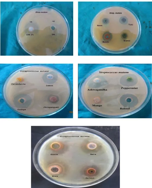
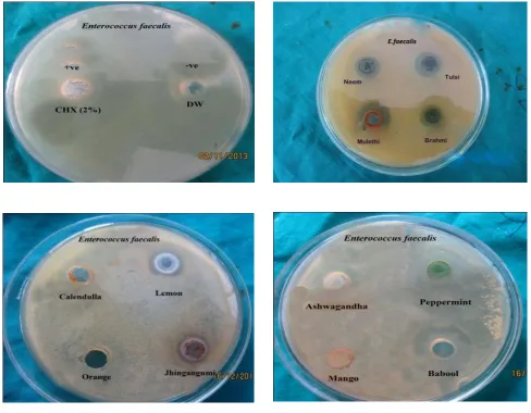
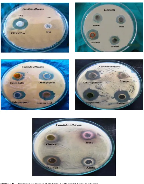
Evaluation of antibacterial and antifungal activities of the sixteen medicinal plants extracts are summarized in Table 3 and figure 1.1, 1.2, 1.3, 1.4, 1.5. Sixteen medicinal plants tested for antimicrobial activity, all ethanolic extracts showed antimicrobial activity, by inhibiting one or more test microbial species. The zone of inhibition by oral species against sixteen ethanolic extract shows that the extracts of Calendulla officinalis and Mangifera Indica
were not effective against Enterococcus faecalis and
Streptococcus mutans respectively. The extracts, of
Acacia nilotica, Citrus sinesis, Citrus limon, Emblica
officinalis, Juglans regia, Glycyrrhiza glabra,Ocimum sanctum, Mentha piperita, Psidium guajava and Withania Somnifera were displaying strong antimicrobial activity, against all the test oral microbes. However, Centella asiatica, Azadirachta indica, Lannea coromandelica and
Rosa centifolia were showing week. Some medicinal plants, Citrus limon , Citrus sinesis, Emblica officinalis, Mentha piperita have potency higher than 2% Chlorhexidine against Lactobacillus acidophilus and
1098Int. J. of Multidisciplinary and Scientific Emerging Research, Vol. 4, No.2 (June 2015) 4. CONCLUSION
The present study helps to establish some compounds of natural origin that could be used to formulate new and more potent antimicrobial agent, which act on some pathogenic micro-organisms associated with human diseases. Some medicinal plants have potency higher than 2% Chlorhexidine against some test oral microbes. This study has provided a documented scientific evidence of the important role that medicinal plants play as antimicrobial agent in the treatment of oral diseases, thereby explaining their popular application as traditional remedies.
5. ACKNOWLEDGEMENTS
The authors acknowledge their profound gratitude to Dr. Gaurav Gupta, Director of Himachal Institute of Dental Sciences, H.P, India, for providing facilities for research work.
REFERENCES
[1] Marsh, P., Martin, M., 1992. Oral Microbiology, 3rd ed. Chapman and Hall, London, pp. 131–136.
[2] Van Oosten, M.A., Mikx, F.H., Rengi, H.H. (1987), Microbial and clinical measurement of periodontal pockets during sequential periods of non-treatment, mechanical debridement and metronidazole therapy. Journal of Clinical Periodontology, v.14, pp.197–204.
[3] L.P Samaranayake (2000), Essential Microbiology for Dentistry; with a Contribution by B.M. Jones; Foreword by Crispian Scully Edinburgh. Churchill Livingstone, New York.
[4] R.P. Allaker, C.W.I Douglas (2008), Novel anti-microbial therapies for dental plaque-related diseases.” International Journal of Antimicrobial Agents, in press.
[5] Chung , J H Choo , M H Lee , J K Hwang (2006), Anticariogenic activity of macelignan isolated from Myristica fragrans (nutmeg) against Streptococcus mutans , Phytomedicine, v. 13 , pp. 261- 266.
[6] Shobha Tandon, Kunal Gupta, Sugandhi Rao and KJ Malagi (2002), Effect of Triphala mouthwash on the caries status, International Journal of
Ayurveda Research, v. 1, no.2, pp. 93-99.
[7] Stephan RM (1940), Changes in hydrogen-ion concentration on tooth surfaces and in carious lesions, Journal of the American Dental Association, v. 27, no.5, pp. 718-723.
[8] M. Hirasawa, K. Takada (2002), Susceptibility of
Streptococcus mutansand Streptococcus
sobrinusto cell wall inhibitors and development of a novel selective medium for Streptococcus sobrinus, Caries research, v. 36, pp. 155-160.
[9] Von Troil-B Linden , H Torkko , S Alaluusua , J Wolf , Jousimies-H Somer , S. Asikainen (1995), Periodontol findings in spouses. A clinical, radiographic and microbiological study, Journal
of Clinical Periodontology, v. 22, pp. 93-99.
[10] GC Armitage (2004), Periodontal diagnoses and classification of periodontal diseases ,
Periodontology, v. 34, pp. 9–21.
[11]J. BEpstein (1990), Antifungal therapy in oropharyngealmycotic infections, Oral Surgery,
Oral Medicine, Oral Pathology, v.69, pp.32-41.
[12] James, D. William, Berger, G. Timothy (2006), Andrews' Diseases of the Skin: Clinical Dermatology. Philadelphia: Saunders Elsevier. pp. 308.
[13]Scully, Crispian (2008), Oral and maxillofacial medicine: the basis of diagnosis and treatment (2nd ed.). Edinburgh: Churchill Livingstone. pp. 191–199.
[14]D. Greenspan (1994), Treatment of oral candidiasis in HIV infection, Oral Surg Oral Med Oral Pathology, v.78, pp.211‐5.
[15]C. Scully, M. Kabir, L. P. Samaranyake (1994), Candida and oral candidosis: A review, Crticial Reviews inOral Biology & Medicine, v.5, no.2, pp.1251-57.
[16]I.N. Rôças, J.F Siqueira, K.R.N. Santos (2004), Association Of Enterococcus Faecalis With Different Forms Of Periradicular Diseases,Journal of Endodontics,v.30, no.5,pp. 315–20.
[17]A. Molander, C. Reit, G. Dahlen, T. Kvist (1998), Microbiological Status Of Root Filled Teeth With Apical Periodontitis, InternationalJournal of Endodontics, v. 31, pp.1-7.
[18]G. Sundqvist, D. Figdor, S. Persson, U. Sjogren (1998), Microbiologic Analysis Of Teeth With Failed Endodontic Treatment And The Outcome Of Conservative Re-Treatment, Oral Surgery,
1099Int. J. of Multidisciplinary and Scientific Emerging Research, Vol. 4, No.2 (June 2015) [19]H. Charles, Stuart, A. Scott Schwartz, B.
Thomas, B. Cristopher (2006), Enterococcus faecalis: Its Role In Root Canal Treatment Failure And Current Concepts In Retreatment,
Journal of Endodontics, v.32, no.2, pp. 93-98.
[20]V.W.K Tsui, R.W.K Wong, A.B.M. Rabie (2008), The inhibitory effects of narigin on the growth of periodontal pathogens in vitro, Phytotherapy, v.22,pp. 401-406.
[21]G. More, T.E. Tshikalange, N. Lall, F. Botha, J.J.M. Meyer (2008), Antimicrobial activity of medicinal plants against oral microorganisms ,
Journal of Ethnopharmacology ,v. 119, no. 3, pp. 473–477.
[22]F. C. Groppo, C.C Bergamaschi, K. Cogon, M Franz-Montana, R.H.L Motta, E. D Andrade (2008), Use of phototherapy in dentistry: A review article, Phytotherapy Research,v. 22, pp. 993-998.
[23]J. Tichy, J Novak (1998), Extraction, Assay, and Analysis of Antimicrobials from Plants with Activity against Dental Pathogens (Streptococcus
sp.).Journal of Alternative and Complementary Medicine, v. 4, no.1, pp. 39-45.
[24]V.I Enne, D.M Livermore, P. Stephens, L.M.C Hall (2001), Persistence of sulphonamide resistance in Escherichia coli in the UK despite national prescribing restriction, Lancet, v.28,p.p1325-1328.
[25]A. Marchese, G.C. Schito (2001), Resistance pattern of lower respiratory tract pathogens in Europe,International Journal of Antimicrobial Agents. V.16, no.1, pp. 25-29.
[26]C. K. Kokate, K.R. Khandelwal, A.P. Pawar, S.B. Gokhale (1995), Practical Pharmacognosy 3rd ed., Nirali Prakashan Pune. p.137.
[27]Trease and Evans (1989), Text Book of Pharmacognosy 12th ed., ELBS Publications. pp.49, 126, 132-137, 205, 248.
[28]A. Odebiyi and A.E. Sofowora (1979), Antimicrobial alkaloids from a Nigeria chewing stick Fagara zanthozyboides. Plantamedica, v. 40, pp. 204-207.
[29]S. Arulmozhi, P. M Mazumder, P. Ashok, L.S. Narayanan (2007), Pharmacological activities of Alstoniascholaris Linn. (Apocynaceae)- A review, Pharmacological Review, v.1, pp. 163 165.
[30]D. Mantle, F. Eddeb, A.T Pickering (2000), Comparison of relative antioxidant activities of British medicinal plant species in vitro, Journal of Ethnopharmacology, v.72, pp. 47 -51.
[31]R. M. Zablotowicz, R.E Hoagland, S.C. Wagner(1996), Effect of saponins on the growth and activity of rhizosphere bacteria, Advances in Experimental Medicine and Biology, v. 405, p.p 83 95.
[32]M. L Dhar, M.M Dhar, B.N. Dhawan, C. Ray (1979), Screening of Indian plants for biological activity. Indian Journal of Biology, v.6, pp. 232- 234.
[33]C. Marjorie (1999), Plant products as antimicrobial agents, Clinical Microbiology Review, v.12, pp. 564- 582.
[34]F. Raquel, Epand (2007), Bacterial lipid composition and the antimicrobial efficacy of cationic steroid compounds, Biochimicaet Biophysica Acta, pp. 2500 -2509.