root canal. Considering incomplete removal of bacteria from the canal by usual methods, lasers have been suggested as a new modality. Despite their anti-bacterial properties, lasers can cause thermal changes. This study assessed the thermal changes of root surface in pulpectomy of primary teeth following the use of Er:YAG laser.
Materials and Methods: Sixty primary anterior teeth were collected and prepared by K-file up to number 50. Then, they were randomly divided into two groups and were irradiated with Er:YAG laser. The first group was irradiated with 1 W laser and the second group with 1.5 W laser. The laser irradiation time was two 10-second cycles with a 2-second interval in both groups. Thermal changes were measured by a thermometer in the apical and coronal areas per second. The results were analyzed by repeated measures ANOVA considering the laser power as between-subject variable.
Results: There was a temperature increase in the coronal and apical areas in use of 1 W power. There was a temperature rise in the coronal and apical areas in use of 1.5 W power. The temperature rise in the apical third was more than that in the coronal third; also, the average temperature rise was more in use of 1.5 W power than 1 W power.
Conclusions: As the average temperature increase was not more than 7°C in any group, this type of laser seems to be suitable for root treatment of primary anterior teeth.
Key words: Lasers, Solid-State; Pulpectomy; Tooth, Deciduous; Thermal Conductivity Journal of Dentistry, Tehran University of Medical Sciences, Tehran, Iran (2018; Vol. 15, No. 3) Corresponding author:
E. Foroughi, Department of Pediatric Dentistry, School of Dentistry, Arak University of Medical Sciences, Arak, Iran [email protected] Received: 29 November 2016 Accepted: 17 April 2017
INTRODUCTION
Early loss of primary teeth can cause malocclusion, unesthetic appearance, speech problems and temporary or permanent functional impairment. It is imperative to preserve pulp vitality as much as possible [1]. However, root canal therapy in primary teeth may be required in some cases such as irreversible pulpitis and pulp necrosis in order to prevent damage to permanent teeth [2]. In some occasions, pulpectomy alone is not sufficient [3], and cleaning, shaping and filling of the canal with absorbable pastes yields successful results [4-6].
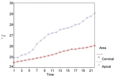
Fig. 1: Thermal changes in the apical and cervical thirds of the root surface caused by 1 W laser when the preliminary temperature was 22ºC
Laser is another modality suggested for activation of irrigating solutions in 2009 [14]. Laser has many advantages such as no mechanical contact and not creating smear layer, and can be used for decontamination of root canal and control of bleeding [15-17]. Birang et al, [18] in their study on single-rooted permanent teeth showed that Er:YAG laser results in better smear layer removal than a simple rinse with sodium hypochlorite. Also, Noiri et al. [19] indicated the effectiveness of Er:YAG laser for elimination of endodontic pathogens. Many types of lasers are used in pediatric dentistry [20]. Soares et al. [21] showed that using Er,Cr:YSGG laser for cleaning of the root canals of primary teeth had similar canal cleaning efficacy compared to rotary instruments and was superior to manual instruments. However, some controversial results have also been reported. For instance, Liu [22] indicated higher success rate for treatment with Nd:YAG laser in
Fig. 2: Thermal changes in the apical and cervical thirds of the root surface caused by 1.5 W laser when the preliminary temperature was 22ºC
Eriksson et al. [30] concluded that temperature increase of 10ºC for one minute was high enough for alveolar bone necrosis. Generally, 7ºC increase in temperature is considered as the tolerance threshold for periodontal tissues [33]. Regarding the morphologic and structural differences between primary teeth and permanent teeth, they may also be different in terms of thermal changes [34, 35]. Another disadvantage of laser in dentistry could be the possibility of apical perforation of curved canals because of straight line of laser beam. According to Li et al, [36] Er:YAG laser could ablate dentin and enamel; thus, if the pulse reaches the dentin surface in a wrong angle, it could cause apical perforation; this limits the laser application to straight canals only. The effect of thermal changes caused by laser during pulpectomy of primary teeth has not yet been studied; thus, this study was conducted aiming to assess the effect of thermal changes caused by laser during pulpectomy of primary anterior teeth.
MATERIALS AND METHODS
Sample preparation:
This study was conducted on 115 hopeless, severely decayed, primary anterior teeth. The study protocol was approved in the ethical committee of our university (code:32085). Sixty teeth with root resorption less than one-fourth of the root length were selected. The crowns of the teeth were cut by a diamond fissure bur such that the remaining root length was 10 mm. Then, the roots were cleaned and shaped to 9 mm working length using a K-file (Kerr, Orange, CA, USA) up to number 50. During cleaning and shaping, the canals were rinsed with 5.25% sodium hypochlorite. After preparation, the teeth were autoclave-sterilized at 134ºC for 15 minutes [37]. The teeth were kept in distilled water at room temperature after extraction.
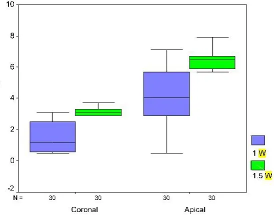
Fig. 3: Boxplot of the mean thermal changes after 1 W and 1.5 W laser irradiation in the coronal and apical thirds
They were placed in a Teflon mold with a height of 5 cm and diameter of 10 cm. A digital thermometer (ST-8891E; Standard, Hong Kong, China) with an accuracy of 0.1ºC and range of 30 to 550ºC, which had data logger was used. One of the thermometer probes was in contact with the root surface in the coronal third and the other probe was in contact with the apical third to measure the temperature in these areas. The other probe of the device was placed at room temperature to compare the thermal changes in root surface with the room temperature changes. The samples were irradiated with Er:YAG laser (Fidelis, Fotona, Slovenia) at a wavelength of 2940 nm, fiber length of 20 mm and diameter of 300 µ in spiral motion. Laser irradiation was started from the apical third 1 mm away from the radiographic apex and 9 mm away from the preparation endpoint. In the first group, laser with 1 W power, 100 mJ energy, 10 Hz frequency and energy density of 70.77 J/cm2 in short pulse mode (250 ms) was irradiated for 20 seconds (two 10-second cycles with a 2-second time interval). In the second group, laser with 1.5 W power, 150 mJ energy, 10 Hz frequency and energy density of 106.15 J/cm2 in short pulse
mode (250 ms) was irradiated for 20 seconds (two 10-second cycles with a 2-second time interval). The temperature was recorded at each second, sent to the computer automatically and saved in Thermometer-E software. The results were analyzed by repeated measures ANOVA considering the laser power as between-subject variable using SPSS version 22 (SPSS Inc., IL, USA).
RESULTS
Figures 1 and 2 show the recorded temperatures from the root surface during laser irradiation (for two 10-second rounds with a 2-second time interval) with 1 and 1.5 W powers.
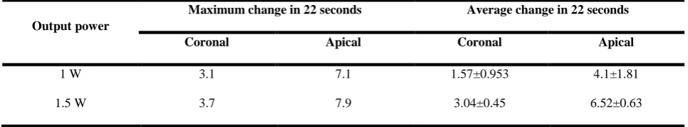
the temperature increase by 1.5 W power was significantly more than that by 1 W power. The maximum temperature increase by 1 and 1.5 W laser during 22 seconds in the apical third was 7.1ºC and 7.9ºC, respectively (Table 1). The maximum temperature increase for 1 and 1.5 W laser during 22 seconds in the coronal third was 3.1 and 3.7ºC, respectively (Table 1). Figure 3 shows the range of thermal changes during 22 seconds for the apical and coronal thirds caused by 1 and 1.5 W laser.
DISCUSSION
Success of root canal treatment is directly related to the removal of microorganisms from the root canal system [38]; but complete removal of microorganisms from the root canal system is not possible after cleaning, shaping and irrigation in all cases [39-41]. Recently, laser was introduced as a method of decontamination and removing the canal debris [9, 39-41]. Many different types of lasers are used in pediatric dentistry [20]. Besides the many advantages of laser, it may cause some undesirable effects [42].
One of these undesirable effects is the thermal damage following temperature increase [43]. It seems that these thermal effects are different based on different wavelengths of laser. For instance, Schoop et al. [44] assessed the thermal effects of Er;Cr:YSGG and Nd:YAG lasers on permanent teeth and indicated that using Er;Cr:YSGG laser in five cycles of five seconds each with a time interval of 15 seconds between each two cycles caused an average thermal increase of about 8.3±0.7ºC for 1 W power and
of 15 seconds between each two cycles caused an average thermal increase of about 5.4±0.6ºC for 1 W power and 8.2±0.4ºC for 1.5 W power. Generally, lower thermal damage by Er:YAG has been previously documented. For instance, Barcellos et al. [45] indicated that Er:YAG laser caused lower temperature increase in comparison with Nd:YAG laser. Also, Wigdor et al. [46] showed that the thermal damage caused by Er:YAG laser is less than that by Nd:YAG and CO2 lasers. Therefore, considering thermal changes, Er:YAG laser seems to be more applicable and a safer choice.
One of the effective factors to prevent thermal damage is water or air spray [9, 47, 48]. Theodoro et al. [49] concluded that using laser for 30 seconds accompanied by the use of water spray as a coolant would decrease the temperature (-2.2±1.5ºC). In this study, after applying laser, temperature increased; these thermal changes may be related to the use of cooling system. But it should be noted that too much spraying of water and air causes energy absorption and reduces the ablative effect of laser [50].
root length. Barcellos et al. [45] showed that temperature increase in the apical third was more than that in the cervical third for 1.4 W power. Our study also showed that temperature increase in the apical third was more than that in the cervical third. This is important because in the apical region, there is only a thin layer of dentin and cementum between the root canal and periodontal ligament (PDL) and the adjacent bone [45]. However, in this study, temperature increase by the Er:YAG laser was not higher than the threshold of 7ºC in the apical third.
Among the adjustable laser parameters, the average power and irradiation time have important roles in safety of clinical application of laser [45]. Based on the studies by Takeda et al, [51-53] 1-2 W power is sufficient for removing the smear layer. Also, according to Cecchini et al, [54] 1.2 W power would be enough for canal decontamination. In order to examine the thermal effect of suitable powers for removing the smear layer and canal decontamination in primary teeth, among the different laser powers, 1 and 1.5 W powers were examined in this study. According to the results of this study, temperature increase was not higher than 7ºC in the samples. This shows the safety and applicability of Er:YAG laser for primary teeth in 1 and 1.5 W powers. About the duration of laser irradiation, based on the study by Soares et al, [21] increasing the duration of laser irradiation would not improve the cleaning efficacy.
Determining the time required for canal
decontamination in primary teeth is also important since longer duration of laser irradiation would result in higher temperature rise. More research is required on this field. Another way to prevent temperature increase in the PDL is to use laser periodically and not continuously [45]. In this study, laser irradiation was done in two 10-second cycles with a time interval of two seconds, which did not cause average temperature increase over the threshold of tissues. It should be mentioned that the temperature did not decrease in the time interval; however, different results may be obtained in the human body because of the blood vessels in the PDL [32]. Further studies are required to assess the suitable duration of laser irradiation.
CONCLUSION
According to the results, it can be concluded that using 1 and 1.5 W powers for 20 seconds (two 10-second cycles) is safe in root canal therapy. The thermal change was 4.1±1.81ºC and 1.57±0.953ºC for 1 W power and 6.25±0.63ºC and 3.04±0.45ºC for 1.5 W power in the apical and coronal thirds, respectively.
REFERENCES
1- Fuks A, Kupietzki A. Pulp therapy for the primary dentition. In: Casamassimo PS FH, McTigue DJ, Nowak AJ, editors. Pediatric dentistry: Infancy through adolescence. 5th ed., St. Louis, Missouri, Elsvier Saunders, 2013:333-52.
2- Mariela Rodriguez Cordeiro M, Jose de Carvalho Rocha M. The effects of periradicular inflamation and infection on a primary tooth and permanent successor. J Clin Pediatr Dent. 2005 Apr;29(3):193-200.
3- Gould J. Root canal therapy for infected primary molar teeth--preliminary report. ASDC J Dent Child. 1972;39(4):269.
4- Mortazavi M, Mesbahi M. Comparison of zinc oxide and eugenol, and Vitapex for root canal treatment of necrotic primary teeth. Int J Clin Pediatr Dent. 2004 Nov;14(6):417-24.
5- Moskovitz M, Sammara E, Holan G. Success rate of root canal treatment in primary molars. J Dent. 2005 Jan;33(1):41-7.
6- Primosch RE, Ahmadi A, Setzer B, Guelmann M. A retrospective assessment of zinc oxide-eugenol pulpectomies in vital maxillary primary incisors successfully restored with composite resin crowns. Pediatr Dent. 2005 Nov;27(6):470-7.
LinerBond-M. Tissue dissolution by sodium hypochlorite: effect of concentration, temperature, agitation, and surfactant. J Endod. 2010 Sep;36(9):1558-62.
14- De Moor RJG, Blanken J, Meire M, Verdaasdonk R. Laser induced explosive vapor and cavitation resulting in effective irrigation of the root canal. Part 2: evaluation of the efficacy. Lasers Surg Med. 2009 Sep;41(7):520-3.
15- Olivi G, Genovese M, Caprioglio C. Evidence-based dentistry on laser paediatric dentistry: review and outlook. Eur J Paediatr Dent. 2009 Mar;10(1):29. 16- Ramazani N, Ahmadi R, Daryaeian M. Oral and dental laser treatments for children: Applications, advantages and considerations. J Lasers Med Sci. 2012 Jan;3(1):44-9.
17- Tate AR. Formecresol performs better than calcium hydroxide as a pulpotomy technique over 2-year period. J Evid Based Dent Pract. 2011 Mar;11(1):65-6.
18- Birang R, Hasheminia M, Fakhari E, Nasouri M, Nasouri S, Birang E. Evaluation of root canal smear layer removal by two types of lasers: A scanning electron microscopy study. European J Gen Dent. 2013 May;2(2):151.
19- Noiri Y, Katsumoto T, Azakami H, Ebisu S. Effects of Er:YAG laser irradiation on biofilm-forming bacteria associated with endodontic pathogens in vitro. J Endod. 2008 Jul;34(7):826-9. 20- Ghadimi S, Chiniforush N, Bouraima SA, Johari M. Clinical approach of laser application in different aspects of pediatric dentistry. J Lasers Med Sci. 2012 Apr;3(2):84-90.
separating science from hype. J Am Dent Assoc. 2004 Feb;135(2):204-12.
25- Kuvvetli SS, Sandalli N, Topcuoglu N, Kulekci G. Antibacterial efficacy of diode and Er: YAG laser irradiation in experimentally contaminated primary molar root canals. J Clin Pediatr Dent. 2009 Sep;34(1):43-8.
26- Armengol V, Jean A, Marion D. Temperature rise during Er: YAG and Nd: YAP laser ablation of dentin. J Endod. 2000 Mar;26(3):138-41.
27- Attrill D, Davies R, King T, Dickinson M, Blinkhorn A. Thermal effects of the Er: YAG laser on a simulated dental pulp: a quantitative evaluation of the effects of a water spray. J Dent. 2004 Jan;32(1):35-40.
28- Raucci-Neto W, De Castro LM, Corrêa-Afonso AM, Da Silva RS, Pécora JD, Palma-Dibb RG. Assessment of thermal alteration during class V cavity preparation using the Er: YAG laser. Photomed Laser Surg. 2007 Aug;25(4):281-6. 29- Zach L, Cohen G. Pulp response to externally applied heat. Oral Surg Oral Med Oral Pathol. 1965 Apr;19(4):515-30.
30- Eriksson R, Albrektsson T, Magnusson B. Assessment of bone viability after heat trauma: a histological, histochemical and vital microscopic study in the rabbit. Scand J Plast Reconstr Surg Hand Surg. 1984 Jan;18(3):261-8.
Oral Pathol Oral Radiol Endod. 2002 Jun;93(6):730-5. 32- Saunders E. In vivo findings associated with heat generation during thermomechanical compactionof gutta‐percha. Part II. Histological response to temperature elevation on the external surface of the rootInt Endod J. 1990 Sep;23(5):268-74.
33- Sauk J, Norris K, Foster R, Moehring J, Somerman M. Expression of heat stress proteins by human periodontal ligament cells. J Oral Pathol Med. 1988 Nov;17(9‐10):496-8.
34- Hirayama A, Yamada M, Miake K. An electron microscopy study on dentinal tubules of human deciduous teeth. Shikwa Gakuho. 1986 Jun;86(6):1021. 35- Sumikawa DA, Marshall G, Gee L, Marshall S. Microstructure of primary tooth dentin. Pediatr Dent. 1999 Nov;21(7):439-44.
36- Li ZZ, Code JE, Van de Merwe WP. Er:YAG laser ablation of enamel and dentin of human teeth: Determination of ablation rates at various fluences and pulse repetition rates. Lasers Surg Med. 1992 Jan;12(6):625-30.
37- Carvalho CA, Xavier AC, Valera MC, Jorge AO, Ferraz MM, Oliveira LD. Morphological and chemical changes of dentin after applying different sterilization methods. Rev Odontol UNESP. 2015 Jun;44:131-6.
38- Estrela C, Bammann L, Pimenta F, Pécora J. Control of microorganisms in vitro by calcium hydroxide pastes. Int Endod J. 2001 Jul;34(5):341-5. 39- Harashima T, Takeda F, Zhang C, Kimura Y, Matsumoto K. Effect of argon laser irradiation on instrumented root canal wall. Dent Traumatol. 1998 Feb;14(1):26-30.
40- Önal B, Ertl T, Siebert G, Müller G. Preliminary report on the application of pulsed CO2 laser radiation on root canals with AgCl fibers: A scanning and transmission electron microscopic study. J Endod. 1993 Jun;19(6):272-6.
41- Pashley D, Michelich V, Kehl T. Dentin permeability: effects of smear layer removal. J Prosthet Dent. 1981 Nov;46(5):531-7.
42- Zhang S, Chen T, Ge L. [Scanning electron microscopy was used to observe dentin morphology in primary and permanent teeth treated by erbium:
yttrium-aluminum-garnet laser]. Beijing Da Xue Xue Bao. 2011 Oct;43(5):766-9.
43- Wang X, Sun Y, Kimura Y, Kinoshita JI, Ishizaki NT, Matsumoto K. Effects of diode laser irradiation on smear layer removal from root canal walls and apical leakage after obturation. Photomed Laser Surg. 2005 Dec;23(6):575-81.
44- Schoop U, Kluger W, Moritz A, Nedjelik N, Georgopoulos A, Sperr W. Bactericidal effect of different laser systems in the deep layers of dentin. Lasers Surg Med. 2004 Aug;35(2):111-6.
45- Barcellos DC, Carvalho CAT, Torres CRG, Pucci CR, Azuma CRS, Pugliesi EN. Assessment of external root temperature during root canal irradiation by Nd: YAG and Er: YAG lasers. J Laser Appl. 2009 Aug;21(3):119-23.
46- Wigdor H, Abt E, Ashrafi S, Walsh JT. The effect of lasers on dental hard tissues. J Am Dent Assoc. 1993 Feb;124(2):65-70.
47- Levy G. Cleaning and shaping the root canal with a Nd: YAG laser beam: a comparative study. J Endod. 1992 Mar;18(3):123-7.
48- Takeda F, Harashima T, Kimura Y, Matsumoto K. A comparative study of the removal of smear layer by three endodontic irrigants and two types of laser. Int Endod J. 1999 Jan;32(1):32-9.
49- Theodoro LH, Haypek P, Bachmann L, Garcia VG, Sampaio JE, Zezell DM, et al. Effect of Er: YAG and diode laser irradiation on the root surface: morphological and thermal analysis. J Periodontol. 2003 Jun;74(6):838-43.
50- Lee BS, Jeng JH, Lin CP, Shoji S, Lan WH. Thermal effect and morphological changes induced by Er: YAG laser with two kinds of fiber tips to enlarge the root canals. Photomed Laser Surg. 2004 Jun;22(3):191-7.
51-Takeda F, Hatashima T, Eto J, Kimura Y, Matsumoto K. Effect of Er: YAG laser treatment on the root canal walls of human teeth: an SEM study. Dent Traumatol. 1998 Dec;14(6):270-3.