O R I G I N A L A R T I C L E
Fibrin tissue adhesive reduces postoperative blood loss in total
knee arthroplasty
Luigi Sabatini•Andrea Trecci•Daniele Imarisio •
Marco Davide Uslenghi•Giuseppe Bianco•
Roberto Scagnelli
Received: 19 November 2010 / Accepted: 18 April 2012 / Published online: 16 May 2012 ÓThe Author(s) 2012. This article is published with open access at Springerlink.com
Abstract
Background Blood transfusion is often required in total knee replacement; various methods of blood preservation have been studied. The best solution is to reduce the loss of blood during and after surgery.
Materials and methods We designed this study to eval-uate the hemostatic efficacy and safety of fibrin tissue adhesive (Quixil) in patients receiving total knee arthro-plasty [low contact stress (LCS, DePuy, Warsaw, IN, US) cementless total knee replacement (TKR)] with a pro-spective, randomized, standard treatment controlled study. Thirty-five patients were randomized to receive treatment with fibrin tissue adhesive (treatment group), and 35 were randomized to be managed with postoperative blood recovery and reinfusion (control group). Blood loss in suction drain, decrease in hemoglobin values, and trans-fusions were recorded.
Results A significant reduction in apparent total blood loss was detected in the treatment group compared with the control group. There was also a lower decrease in hemoglobin level, although this difference was not significant. When fibrin tissue adhesive was administered, the need for transfusions was lower. No major adverse events were recorded in our series. Conclusions Fibrin tissue adhesive reduced blood loss in TKR and seemed to significantly reduce the need for blood transfusion. Fibrin tissue adhesive can be an appropriate solution to enhance hemostasis and vessel sealing at the operative site in TKR, in order to reduce blood loss after surgery and the risk of complications.
Keywords Fibrin tissue adhesiveBlood loss
Total knee arthroplastyFibrin sealant
Introduction
Total knee replacement (TKR) is associated with major intra- and postoperative blood loss (approximately 800–1,400 ml), and blood transfusion is frequently required [1]. Use of tourniquet may reduce intraoperative blood loss, but postoperative blood loss can still be considerable.
Decreasing this postoperative blood loss may reduce patient morbidity [2,3], length of hospitalization, and costs by eliminating the need for transfusion.
Various methods of blood preservation have been studied, in order to avoid transmission of viral diseases and transfusion reactions; the various perioperative methods include hemodilution, intraoperative and postoperative blood salvage and reinfusion [4], hypotensive or epidural anesthesia, and transfusion of predonated autologous blood [5].
Obviously, the most appropriate solution is to enhance hemostasis and vessel sealing at the operative site. Plasma proteins were used in the past. Modern treatment with fibrin tissue adhesive, also known as fibrin glue or fibrin sealant, consists in application of plasma fibrino-gen mixed with thrombin to form an adhesive fibrin clot. Since 1972, fibrin sealants have been increasingly used as hemostatic and sealing agents in a variety of surgical specialties, including, recently, total knee replacement [6].
Because of a lack of solid evidence concerning safety and efficacy, blood-bank products and bovine thrombin concentrates have been used extensively for many years. L. Sabatini (&)A. TrecciD. Imarisio
M. D. UslenghiG. BiancoR. Scagnelli
Orthopedics and Traumatology Department, Ospedale Civile di Saluzzo, Via Spielberg 58, 12037 Saluzzo, CN, Italy
Fibrin tissue adhesive is composed of two main com-ponents: fibrinogen and thrombin. When mixed together, they mimic the last step of the coagulation cascade: thrombin activates fibrinogen to polymerize to an unstable clot, and factor XIII, which is present in the fibrinogen concentrate and is activated by thrombin (factor XIIIa), stabilizes the clot by catalyzing cross-links between the fibrin molecules. Factor XIIIa also forms cross-links between natural plasmin inhibitors (which copurify with fibrinogen) and the fibrinogen mash to enhance clot resis-tance against fibrinolysis. Some products contain additional fibrinolytic inhibitors, such as bovine aprotinin or tranex-amic acid [7], although the contribution of such additives is controversial [8].
The desire to avoid any bovine products led to the development of a second-generation fibrin glue which replaced bovine aprotinin with tranexamic acid as the antifibrinolytic agent; safety issues concerning tranexamic acid led the manufacturer to develop a modified version without the antifibrinolytic agent [9]. In addition, suc-cessful application of tissue adhesive needs experience and training [10].
There are numerous publications concerning good results of use of fibrin tissue adhesive in all fields of sur-gery, but many reports are uncontrolled studies and some trials show no benefits but harmful effects [11]. In ortho-pedic surgery, the literature is poor and there are few reports of total knee replacement, for which fibrin glue is not yet used routinely.
Materials and methods
We designed this study to evaluate the hemostatic efficacy of fibrin tissue adhesive in patients receiving total knee arthroplasty. This is a prospective, randomized, standard treatment controlled study, where the control group had postoperative blood recovery and reinfusion. Both fibrin tissue adhesive and postoperative blood reinfusion were already approved for use in our hospital. We decided to evaluate the differences between the two procedures, and the efficacy of fibrin adhesive [5]. The protocol used con-formed to the Declaration of Helsinki, and we notified the Ethical Review Board. All patients signed informed written consent to either procedure before being included in the study. All patients were enrolled in a program of autolo-gous blood recovery; for each patient in good health con-ditions and with good hemoglobin values, we prepared two units of autologous blood.
We evaluated 70 patients treated for osteoarthritis of the knee with total knee cementless arthroplasty from April 2009 to April 2010. Exclusion criteria were cemented TKR, current infection, any kind of cancer, and coagulation
pathology, in order to create two similar groups: treatment and control.
Thirty-five patients were randomized to receive treat-ment with fibrin tissue adhesive (the treattreat-ment group), and 35 were randomized to be managed with postoperative blood recovery and reinfusion (control group, Di-deco).
The mean age in the fibrin tissue adhesive group was 70.7±6.4 years, while in the Di-deco group it was 70.4±6.7 years (p =0.84).
In our series, 54 patients were female and 16 were male (10 males in the treatment group, 6 in the control group).
The mean presurgery hemoglobin value in the treatment group was 13.5±1.5 g/dl, and in the control group it was 13.2±1.3 g/dl (p=0.34).
The mean surgical time was 79±16 min for the treatment group and 78.2±14.3 min for the control group (p =0.84).
The fibrin tissue adhesive used was Quixil (Omrix Biopharmaceuticals, Belgium), formed by two 5-ml com-ponents: cryoprecipitated fibrinogen, and a high concen-tration of human thrombin dissolved in calcium chloride; an antifibrinolytic agent, tranexamic acid, is added to the fibrinogen as a stabilizer. Both components undergo double viral inactivation. The constituents are placed in separate syringe tubes and mixed by connecting them together to a single lumen, through which the glue is expelled as a high-pressure spray.
All total knee arthroplasties were managed by the same surgical team; a LCS total knee prosthesis cementless procedure was used for all operations [LCS rotating plat-form (RP) or LCS anterior–posterior glide (APG) with posterior cruciate sparing], all performed by medial para-patellar approach in a bloodless field with use of tourniquet until prosthesis insertion (Tables1,2).
In the treatment group, after the preparation of the femur and the tibia and before insertion of the prosthesis, the operative field was rinsed of any debris and meticulously dried, then the fibrin tissue adhesive (half of 5 ml of each product component, corresponding to half of the kit) was applied to the back of the knee cavity, the posterior recess, the gutters, and the exposed surfaces of the femur and tibia by topical air pressure spraying with use of a double-syr-inge spray device from a distance of approximately 15 cm. Then, after prosthesis insertion, remaining product was applied over soft tissues, extensor mechanism, and prepa-tellar bursa, to cover as much surface area as possible with glue film.
After the tourniquet was deflated (2 min after glue application), we performed hemostasis of major vessels using electrocautery. We then placed two drains: one inside and the other outside of the knee joint.
same two drains to a Di-deco blood recovery device without fibrin tissue adhesive application.
Hemoglobin and hematocrit values were determined preoperatively (the day before operation) and on the first and third postoperative days. Preoperative platelet count,
prothrombin time, and activated partial thromboplastin time were determined for all patients.
Intraoperative blood loss was similar in the two groups because of the use of tourniquet and the same operative technique.
Table 1 Group features
Patient no. Di-deco Quixil
Sex Age (years) Hb before surgery (g/dl) Sex Age (years) Hb before surgery (g/dl)
1 F 67 11.2 F 78 10.5
2 F 69 12 F 78 12.9
3 F 76 13.5 M 69 12.9
4 F 79 11.1 F 78 12.7
5 M 67 12.8 F 63 13.4
6 F 64 13.8 F 53 13.6
7 F 65 12.2 F 73 12.4
8 F 71 11.9 F 75 15.8
9 F 86 15 M 74 15.4
10 F 74 14.7 F 74 15.5
11 F 70 11.7 M 77 13.4
12 F 70 13.9 M 82 14.1
13 F 59 13.1 F 69 12.4
14 F 76 11.7 M 63 15.2
15 F 73 12.3 M 74 15
16 F 79 13.3 F 77 13.3
17 M 68 14.4 m 78 17.5
18 F 71 13.8 F 64 13.1
19 F 73 13.8 F 66 12
20 F 56 11.9 F 75 15.2
21 F 80 13.6 F 78 13.6
22 F 69 11.7 M 77 13.6
23 F 66 11.5 M 60 12.2
24 F 79 14.6 F 67 12.6
25 M 76 13.6 F 71 12
26 M 75 14.4 F 68 11.4
27 M 54 13.2 F 62 14.8
28 F 78 14.2 F 69 13.8
29 F 70 15.8 F 61 14.2
30 F 66 12.4 F 70 13.6
31 F 70 14.4 F 72 10.2
32 F 66 12.9 M 73 14.7
33 M 70 15.6 F 69 13.8
34 F 65 14.6 F 68 14.2
35 F 67 11.7 F 70 12.6
6 males 10 males
Mean 70.4 13.2085714 70.7142857 13.5314286
Median 70 13.3 71 13.6
SD 6.78753182 1.29555154 6.4606111 1.51167446
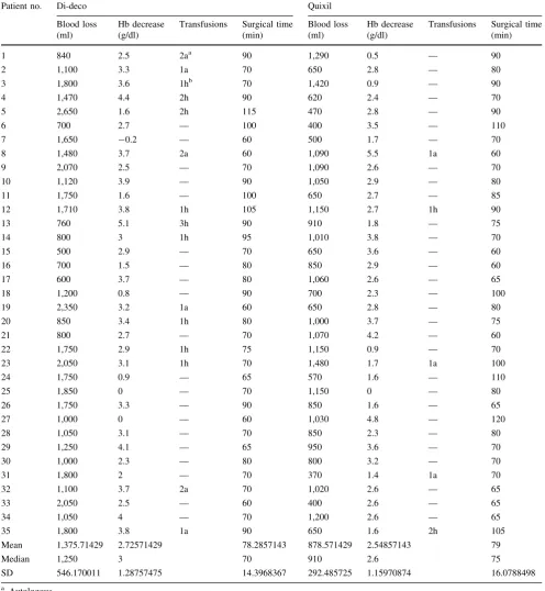
Table 2 Postoperative findings
Patient no. Di-deco Quixil
Blood loss (ml)
Hb decrease (g/dl)
Transfusions Surgical time (min)
Blood loss (ml)
Hb decrease (g/dl)
Transfusions Surgical time (min)
1 840 2.5 2aa 90 1,290 0.5 — 90
2 1,100 3.3 1a 70 650 2.8 — 80
3 1,800 3.6 1hb 70 1,420 0.9 — 90
4 1,470 4.4 2h 90 620 2.4 — 70
5 2,650 1.6 2h 115 470 2.8 — 90
6 700 2.7 — 100 400 3.5 — 110
7 1,650 -0.2 — 60 500 1.7 — 70
8 1,480 3.7 2a 60 1,090 5.5 1a 60
9 2,070 2.5 — 70 1,090 2.6 — 70
10 1,120 3.9 — 90 1,050 2.9 — 80
11 1,750 1.6 — 100 650 2.7 — 85
12 1,710 3.8 1h 105 1,150 2.7 1h 90
13 760 5.1 3h 90 910 1.8 — 75
14 800 3 1h 95 1,010 3.8 — 70
15 500 2.9 — 70 650 3.6 — 60
16 700 1.5 — 80 850 2.9 — 60
17 600 3.7 — 80 1,060 2.6 — 65
18 1,200 0.8 — 90 700 2.3 — 100
19 2,350 3.2 1a 60 650 2.8 — 80
20 850 3.4 1h 80 1,000 3.7 — 75
21 800 2.7 — 70 1,070 4.2 — 60
22 1,750 2.9 1h 75 1,150 0.9 — 70
23 2,050 3.1 1h 70 1,480 1.7 1a 100
24 1,750 0.9 — 65 570 1.6 — 110
25 1,850 0 — 70 1,150 0 — 80
26 1,750 3.3 — 90 850 1.6 — 65
27 1,000 0 — 60 1,030 4.8 — 120
28 1,050 3.1 — 70 850 2.3 — 80
29 1,250 4.1 — 65 950 3.6 — 70
30 1,000 2.3 — 80 800 3.2 — 70
31 1,800 2 — 70 370 1.4 1a 70
32 1,100 3.7 2a 70 1,020 2.6 — 65
33 2,050 2.5 — 60 400 2.6 — 65
34 1,050 4 — 70 1,200 2.6 — 65
35 1,800 3.8 1a 90 650 1.6 2h 105
Mean 1,375.71429 2.72571429 78.2857143 878.571429 2.54857143 79
Median 1,250 3 70 910 2.6 75
SD 546.170011 1.28757475 14.3968367 292.485725 1.15970874 16.0788498
a Autologous
b Homologous
Studentttest (blood loss):p=1.6558910-5
Studentttest (Hb decrease):p=0.54736077
Studentttest (Hb levels before surgery):p=0.34083659
Fisher test (blood units administered): 15 patients (Di-deco) versus 6 patients (Quixil):p=0.03
Blood loss at the end of the operation was recorded by measuring the volume in the suction apparatus without washing solution; apparent postoperative blood loss was recorded by measuring the volume in the suction drain bottles on the evening of the operation and the first, second, and third postoperative days (until drain removal).
In the control group, the Di-deco device recovered the blood loss for the first 6 h after operation; if the blood amount in this time was sufficient to be washed and pre-pared (400 ml), it was reinfused. At the end of 6 h, the Di-deco device was replaced with a suction drain. All Di-Di-deco reinfusions were recorded.
In all cases (in the treatment group, and in the control group after Di-deco reinfusion), decisions regarding blood transfusion were based on hemoglobin value (measured 4–6 h after operation, and on the first, third, and fifth post-operative days) and clinical conditions, including also car-diovascular history and patient age, always being used for hemoglobin values lower than 8 g/dl [12]. The decision to administer blood was taken by a surgeon blinded to the patient’s group.
All patients received subcutaneous injection of low-molecular-weight heparin every evening of the period of hospitalization, starting the day before surgery.
Any complications, such as wound complications, fever, prolonged drainage from drain site, and adverse events, were recorded.
After the first postoperative day, patients began contin-uous passive motion; all patients were allowed to get out of bed after the third postoperative day and started physio-therapy the same day.
The end points of this study are evaluation of the effi-cacy of fibrin tissue adhesive in reducing blood loss, decrease of hemoglobin on the first postoperative day, and the number of transfused units needed by patients. To evaluate these values we used Student’sttest and Fisher’s exact test.
Results
The treatment and control groups were comparable in terms of patient characteristics such as general health conditions, age, presurgery hemoglobin level (p=0.34), and surgery time. As previously stated, intraoperative blood loss was similar for the two groups because of tourniquet use.
The median apparent postoperative blood loss at drain removal (third postoperative day) was 910±292 ml in the fibrin tissue adhesive group compared with 1,250±546 ml in the control group, and the difference was highly statisti-cally significant (p=0.0000165).
The median decrease in hemoglobin concentration on the first postoperative day was 2.6±1.16 g/dl in the fibrin
tissue adhesive group and 3±1.28 g/dl in the control group. The difference in this case was not statistically significant (p=0.55).
The blood transfusion requirements in the fibrin tissue adhesive group also were found to be significantly (p =0.03) lower than those in the control group; only 5 patients in the fibrin tissue adhesive group required blood transfusion, and only 1 patient required two units, whereas 15 patients in the control group required blood transfusion, with 5 requiring two units and 1 requiring three units (for more than one transfusion unit,p =0.0057).
Adverse events
Fever was the most common adverse event associated with surgery, with no difference in frequency between the two group (six patients in the treatment group, six in the control group). Hematoma was recorded in two cases in the fibrin adhesive group and six patients in the control group (one in the control group needing surgical drainage).
We recorded no superficial wound infection or deep vein thrombosis. We recorded no embolism in our series after application of fibrin tissue adhesive by topical spraying with use of the double-syringe spray device.
Discussion
Many bleeding problems are involved in TKR, not only intraoperatively but also postoperatively. The amount of blood loss after total knee arthroplasty is often underesti-mated because the apparent rate is considered instead of the calculated blood loss [13].
Intraoperative use of tourniquet (prolonged ischemia in the limb increases fibrinolytic activity) and suction drain-age use can increase blood loss.
In many cases it is necessary to transfuse blood units to avoid a large decrease in hemoglobin values [14,15]. So, it appears very useful to adopt the most appropriate solution to enhance both hemostasis and safety of recovery and reinfusion [16].
factors most frequently associated with immediate post-transfusion death) are comparable to those associated with use of homologous blood [17].
Through the use of fibrin sealant we hoped to prevent the need for recovery and reinfusion due to enhanced hemostasis in the operative field; use of Quixil was our choice, so we evaluated in this study the efficacy and safety of fibrin tissue adhesive in knee replacement as an alter-native to use of the Di-deco device.
Blood loss in our patients (in both groups) was similar to mean blood loss reported in literature; a significant reduction in apparent total blood loss was found in the group treated with fibrin tissue adhesive compared with the control group, where the Di-deco device was used. It is important to underline that, in the control group, not all patients reached the amount of blood loss needed to trigger reinfusion until 6 h; in our series, 23 patients (66 %) received blood recovery from Di-deco.
The treatment group had a smaller postoperative mean decrease in hemoglobin level than the control group on the first postoperative day, but this difference was not significant.
Use of fibrin tissue adhesive significantly reduced the total number of units of blood transfused postoperatively to almost one-third of the value in the control group (8 versus 22); it reduced also the rate of patients requiring transfu-sion to almost one-third (6 versus 15), with only 2 patients requiring more than one unit, compared with 6 in the control group.
Since the decision regarding transfusion and the number of blood units transfused are unreliable values, being influenced by patient conditions and medical decisions, we did not consider these parameters as significant.
Prevention of blood loss, including prevention of con-comitant compartmental shift in body fluid, is definitely superior to replacement of lost blood. It is much safer for a patient to receive a multidonor viral-inactivated blood product (fibrin tissue adhesive) than homologous blood that cannot be viral inactivated [18].
The results of our study suggest that use of Quixil in total knee arthroplasty reduces postoperative apparent blood loss, also compared with use of the Di-deco device. The Italian Agency of Drugs (AIFA) had informed that use of a spray device to apply fibrin tissue adhesive can produce massive embolism (two cases, one fatal, having been reported). They recommend use of a spray device with pressure less than 2.0–2.5 bar, to apply Quixil from a minimal distance of 10–15 cm, and to monitor patients during spray application. We were informed of these rec-ommendations after our study, but we did not find any clinical signs of pulmonary embolism.
The mean cost for a Quixil dose is comparable to the use of one Di-deco device for each patient; in addition, one
operator is necessary to wash and prepare blood units from the Di-deco.
We therefore believe that use of fibrin tissue adhesive is advantageous, as it reduces blood loss; avoiding bleeding has been shown to be safer than reinfusion, since even autologous blood is at risk of contamination. Furthermore, one operator is needed to prepare units, increasing theo-retical costs.
In conclusion, use of fibrin tissue adhesive was found to significantly reduce apparent blood loss, also decreasing costs. On comparison of fibrin glue use with postoperative blood reinfusion from the Di-deco device in TKR, we noted a decrease of apparent blood loss, a similar decrease of hemoglobin values, and transfusions of fewer blood units, even if it is not possible to define a real decrease of transfused units due to the variability of patient features.
We believe that fibrin tissue adhesive can be an appro-priate solution to enhance hemostasis and vessel sealing at the operative site in order to reduce blood loss and patient morbidity after surgery.
Conflict of interest None.
Open Access This article is distributed under the terms of the
Creative Commons Attribution License which permits any use, dis-tribution, and reproduction in any medium, provided the original author(s) and the source are credited.
References
1. Berman AT, Geissele AE, Bosacco SJ (1988) Blood loss with total knee arthroplasty. Clin Orthop 234:137–138
2. Everts PA, Davilee RJ, Oosterbos CJ et al (2007) Autologous platelet gel and fibrin sealant enhance the efficacy of total knee arthroplasty: improved range of motion, decreased length of stay and a reduced incidence of arthrofibrosis. Knee Surg Sports Traumatol Arthrosc 15(7):888–894
3. Curtin WA, Wang GJ, Goodman NC, Abbott RD, Spotnitz WD (1999) Reduction of hemorrhage after knee arthroplasty using cryo-based fibrin sealant. J Arthroplasty 14(4):481–487 4. Flynn JC, Csenesitz TA (1979) Present status of intraoperative
blood recovery during orthopaedics surgery. Jefferson Orthop J 8:22–25
5. Cowell H.R (1937) Editorial. Prior deposit of autologous blood for transfusion. J Bone Joint Surg 69A:319
6. Wang GJ, Hungerford DS, Savory CG (2001) Use of fibrin sealant to reduce bloody drainage and haemoglobin loss after total knee arthroplasty. J Bone Joint Surg 83A(10):1503–1505 7. Hynes M, Calder P, Scott G (2003) The use of tranexamic acid
to reduce blood loss during total knee arthroplasty. Knee 10:375–377
8. Levy O, Martinowitz U, Oran A (1999) The use of fibrin tissue adhesive to reduce blood loss and the need for blood transfusion after total knee arthroplasty. J Bone Joint Surg 81A(11):1580–1588 9. Patel S, Rodriguez Merchan EC, Haddad FS (2010) The use of
fibrin glue in surgery of the knee. JBJS (Br) 92B:1325–1331 10. Wang GJ, Goldthwaite CA Jr, Burks SG, Spotniz WD (2003)
arthroplasty: implication for surgical education. J Long Term Eff Med Implants 13(5):389–397
11. Kohno H, Nagasue N, Chang YC et al (1992) Comparison of topical hemostatic agents in elective hepatic resection: a clinical prospective randomized trial. World J Surg 16:966–970 12. Blackwell Science Ltd (2001) Guidelines for the clinical use of
red cell transfusions. Br J Haematol 113:24–31
13. Good L, Peterson E, Lisander B (2003) Tranexamic acid decreases external blood loss but not hidden blood lossin total knee replacement. Br J Anaesth 90:596–599
14. Sehat KR, Evans R, Newman JH (2000) How much blood is really lost in total knee arthroplasty: correct blood loss manage-ment should take hidden loss into account. Knee 7:151–155
15. Sehat KR, Evans R, Newman JH (2004) Hidden blood loss fol-lowing hip and knee arthroplasty: correct management of blood loss should take hidden loss into account. JBJS (Br) 86B:561–565 16. Everts PAM, Devilee RJJ, Brown Mahoney C (2006) Platelet gel and fibrin sealant reduce allogenic blood transfusions in total knee arthroplasty. Acta Anaesthesiol Scand 50:593–599 17. Birkmeyer JD, Goodnough LT, AuBuchon JP, Noordail PG
(1993) the cost-effectiveness of postoperative autologous blood donation for total hip and knee replacement. Transfusion 33:544–551