INTRODUCTION
Opioid-induced hyperalgesia (OIH) is the paradoxical increase in pain percep-tion that may become manifest during opioid treatment for acute and chronic pain (1–4). OIH is characterized by in-creased sensitivity to painful stimuli (hy-peralgesia) and nonpainful stimuli (allo-dynia) and compromises adequate pain treatment (1–4). Morphine is the proto-typical µ-opioid receptor (MOR) agonist,
acts primarily at this receptor subtype and is considered the gold standard for treatment of moderate to severe pain (1). In humans, morphine is metabolized in the liver by UDP-glucuronosyl trans-ferase (UGT) into two major metabo-lites: M3G (60–70%) and glucuronide (M6G, 5–10%) (5,6). The larger proportion to M3G metabolism is because glucuronidation at the aromatic hydroxyl group (that is, the third C
atom) is easier than at the alicyclic hy-droxyl group (that is, the sixth C atom) (7). For this reason, most rodents, in-cluding rats, do not form M6G but only form M3G (7). The two metabolites also have distinct actions. Whereas M6G has proven analgesic actions, there is evi-dence that M3G is pronociceptive (5). For example, a systemic M3G injection increases pain sensitivity in mice, and relatively small intracerebroventricular, intrathecal or systemic M3G doses in rats evoke a general state of neuroexcita-tion with agitaneuroexcita-tion to innocuous touch (8–12). In cancer patients treated with morphine, a cerebrospinal fluid ratio of M3G/M6G concentrations of <1 coin-cides with effective analgesia, whereas a ratio >1 is associated with ineffective analgesia (13). We interpret these data to suggest a role for M3G not only in
µ
-Opioid Receptor or Morphine-3-glucuronide
Maarten Swartjes,
1René A G Mooren,
1Amanda R Waxman,
2Caroline Arout,
2Koen van de Wetering,
3Jan den Hartigh,
4Jos H Beijnen,
5Benjamin Kest,
2and Albert Dahan
11Department of Anesthesiology, Leiden University Medical Center, Leiden, the Netherlands; 2Neuropsychology Program, Queens College, City University of New York, Flushing, and Department of Psychology and Center for Developmental Neuroscience, The College of Staten Island, City University of New York, Staten Island, New York, United States of America; 3Division of Molecular Oncology, The Netherlands Cancer Institute, Amsterdam, the Netherlands; 4Department of Clinical Pharmacy and Toxicology, Leiden University Medical Center, Leiden, the Netherlands; and 5Department of Pharmacy and Pharmacology, Slotervaart Ziekenhuis, Amsterdam, the Netherlands
Opioid-induced hyperalgesia (OIH) is a paradoxical increase in pain perception that may manifest during opioid treatment. For morphine, the metabolite morphine-3-glucuronide (M3G) is commonly believed to underlie this phenomenon. Here, in three separate studies, we empirically assess the role of M3G in morphine-induced hyperalgesia. In the first study, CD-1 mice injected with morphine (15 mg/kg subcutaneously) after pretreatment with the opioid receptor antagonist naltrexone (NTX) (15 mg/kg) showed tail withdrawal latency reductions indicative of hyperalgesia (2.5 ± 0.1 s at t = 30 min, P < 0.001 versus baseline). In these mice, the morphine/M3G concentration ratios versus effect showed a negative correlation (rp= –0.65, P < 0.001), indicating that higher morphine relative to M3G concentrations are associated with increased OIH. In the second study, similar hyperalgesic re-sponses were observed in mice lacking the multidrug resistance protein 3 (MRP3) transporter protein (Mrp3–/–mice) in the liver
and their wild-type controls (FVB mice; latency reductions: 3.1 ± 0.2 s at t = 30 min, P < 0.001 versus within-strain baseline). In the final study, the pharmacokinetics of morphine and M3G were measured in Mrp3–/–and FVB mice. Mrp3–/–mice displayed a
sig-nificantly reduced capacity to export M3G into the systemic circulation, with plasma M3G concentrations just 7% of those ob-served in FVB controls. The data confirm previous literature that morphine causes hyperalgesia in the absence of opioid receptor activation but also indicate that this hyperalgesia may occur without a significant contribution of hepatic M3G. The relevance of these data to humans has yet to be demonstrated.
Online address: http://www.molmed.org doi: 10.2119/molmed.2012.00244
Address correspondence toAlbert Dahan, Department of Anesthesiology, Leiden
University Medical Center, P5-Q, PO Box 9600, 2300 RC Leiden, the Netherlands. Phone: +31-71-526-2301; Fax: +31-71-526-6230; E-mail: a.dahan@lumc.nl.
ineffective analgesia but also in morphine-induced hyperalgesia.
Importantly, M3G effects are not di-minished by the general opioid antago-nist naloxone (14,15), indicating that M3G does not act via opioid receptor ac-tivation. Consistent with the hypothesis that M3G contributes to induced hyperalgesia, there is ample evi-dence that the opioid receptors are not involved in OIH. Morphine and other MOR agonists, such as fentanyl, induce OIH during systemic blockade of the opioid receptors with the nonselective opioid receptor antagonist naltrexone (NTX) (16–19). Furthermore, OIH in-duced by morphine and fentanyl is ob-served in triple knockout mice com-pletely lacking µ-, κ- and δ-opioid
receptors and their subtypes (17,19). Thus, M3G is widely believed to under-lie hyperalgesia consequent to morphine exposure.
The aim of the current study was to empirically assess the putative role of M3G in morphine-induced hyperalgesia. To that end, we injected outbred CD-1 mice with morphine during treatment with NTX or saline and related plasma concentrations of morphine and M3G to the observed pharmacodynamic effects on a standard thermal nociceptive assay (tail-withdrawal test). Next, we com-pared the pharmacodynamic effects of morphine in mice lacking the multidrug resistance protein 3 (MRP3) with their wild-type FVB controls during systemic NTX exposure. MRP3 is a protein in-volved in transporting glucuronidated substances, such as M3G, from the cyto-plasm of the hepatocytes into the sys-temic circulation (20). MRP3-deficient mice (Mrp3–/–) consequently have low and no detectable M3G concentrations in plasma and brain, respectively (20). Our hypothesis is that M3G formed in the liver plays a major role in induced hyperalgesia with (a) a signifi-cant correlation between M3G plasma concentrations and magnitude of hyper-algesia and (b) absence of hyperhyper-algesia in Mrp3–/–mice treated with morphine and NTX. Only when both assumptions
are met are we able to accept our hypothesis.
MATERIALS AND METHODS
Animals
Experiments were performed after ap-proval of the protocol by the local Ani-mal Ethics Committee. Adult Ani-male CD-1 mice were purchased from Charles River (Maastricht, the Netherlands). Mrp3–/– and FVB mice were generated in the lab-oratory (20); the Mrp3–/–mice have a FVB genetic background. The animals re-ceived water and food ad libitumand were housed in groups in individually ventilated, constant-temperature cages and kept in rooms with 12-h light/12-h dark cycles (lights on/lights off at 7:00 AM/PM). The experimental studies were performed in accordance with insti-tutional guidelines and the guidelines of the International Association for the Study of Pain (21).
Nociceptive Assay
Nociception was assessed by measur-ing tail withdrawal latencies (TWLs) from a hot water bath as described previ-ously (17,22). TWLs were obtained in triplicate (at 30-s intervals) with a cutoff value of 30 s to prevent tissue damage to the tail. Bath temperature was set to 47.5 ± 0.2°C to obtain a pretreatment TWL between 9 and 11 s to avoid possi-ble floor effects in hyperalgesic mice. The TWLs were averaged and recorded into a digital datasheet for further analysis. All experiments were performed near mid-photophase to reduce circadian effects.
Drugs
Morphine hydrochloride 3·H2O (MOR) at a solution of 20 mg/mL was obtained from the local hospital pharmacy. NTX hy-drochloride powder was purchased from Sigma-Aldrich (Zwijndrecht, the Nether-lands) and dissolved in saline (0.9% NaCl) to obtain a 20 mg/mL solution.
Study Design
Study 1.Morphine and M3G pharma-cokinetics and morphine
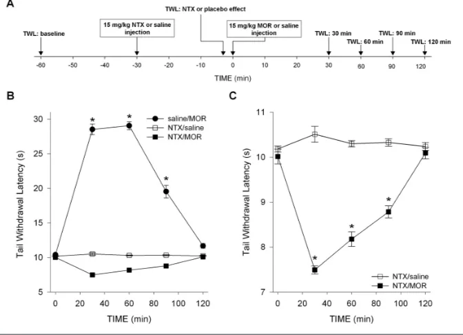
pharmacody-namics were tested in 72 CD-1 mice. The effect of morphine in CD-1 mice during treatment with NTX or saline was as-sessed using the tail-withdrawal test as described above. Each animal was tested once. The groups were randomly divided in three sets of 24 animals that received subcutaneous injections of 15 mg/kg NTX, followed 30 min later by 15 mg/kg morphine (NTX/MOR), saline followed 30 min later by 15 mg/kg morphine (saline/MOR) and 15 mg/kg NTX fol-lowed 30 min later by saline (NTX/ saline), respectively. TWLs were obtained 30 min before NTX, 3 min before the MOR or saline drug injections (NTX/ placebo effect) and at times 30, 60, 90 and 120 min after these injections (MOR/saline effect; see Figure 1A for an illustration of the study design). Each group was subdivided in four sub-groups of six animals with different times for sacrificing and extraction of blood for pharmacokinetics (PK) analysis (at t= 30, 60, 90 and 120 min, directly after the nociceptive test).
Study 2.Morphine pharmacodynam-ics was tested in Mrp3–/–and FVB mice. Mice of each strain were tested either after the subcutaneous injection of 15 mg/kg morphine, 30 min after 15 mg/kg NTX pretreatment (NTX/ MOR) or after the subcutaneous injection of saline, 30 min after 15 mg/kg NTX pretreatment (NTX/saline) (n = 6/ strain/ condition). The injections and nocicep-tive testing protocols were identical to those in study 1.
Analysis of Morphine and M3G Concentrations
In study 1, plasma proteins of all plasma samples were precipitated with 700 µL acetonitrile, 100 µL of 1 mmol/L zinc sulfate and an adequate amount of internal standards. A total of 200 µL of the supernatant was transferred in a glass tube and dried; the residues were reconstituted in 100 µL of 0.1 % (v/v) formic acid in water. Then, 20 µL of the sample was injected by an Ultimate 3000 autosampler (Dionex, Amsterdam, the Netherlands) and pumped on a 3-µm, 120Å, 50 ×2.1 mm YMC pack ODS-AQ
column (YMC Inacom, Overberg, the Netherlands). The eluent was monitored by a Quattro microAPI tandem mass spectrometer (Waters, Etten-Leur, the Netherlands). Peak areas of reaction ions from morphine, M3G, M6G and the in-ternal standards were obtained in the multiple reaction mode and integrated by data software Masslynx 4.1 (Waters, Etten-Leur, the Netherlands). All ana-lytes were measured in one run. In study 3, morphine and M3G concentrations in urine and plasma were determined as described by Rook et al.(23).
Statistical Analysis
Behavioral data of studies 1 and 2 were analyzed by two-way repeated-measure analysis of variance for the main effect with a post hocTukey test for treatment and time. Morphine and M3G concentrations obtained in study 3 were compared between genotypes with the Mann-Whitney Utest (plasma) and two-tailed ttest (urine). All statistical tests were performed using SigmaPlot version 12 for Windows (Systat Software, Chi-cago, IL, USA). Pvalues <0.05 were con-sidered significant. Data are expressed as mean ± standard error of the mean (SEM).
RESULTS
Study 1
TWL baseline values were 10.1 ± 0.05 s (NTX/MOR), 10.3 ± 0.05 s (NTX/saline) and 10.2 ± 0.08 s (saline/MOR). These
values did not differ significantly from values obtained after NTX or saline injec-tions (as measured just before morphine or saline injections). The effects of MOR or saline treatments on TWL are given in Figures 1B and C. No effect on TWL was observed from the combination NTX/ saline. Morphine combined with saline produced profound antinociception dur-ing the entire testdur-ing period (increase in TWL at t= 30 min: 19.1 ± 0.6 s, P< 0.001 versus baseline; main effect: P< 0.001 versus NTX/saline). In contrast, combin-ing morphine with NTX decreased TWL by 2.5 ± 0.1 s at t= 30 min compared with baseline values (P< 0.001). Hyper-algesia persisted during the entire testing period (main effect: P= 0.004 versus NTX/saline and P< 0.001 versus saline/ MOR).
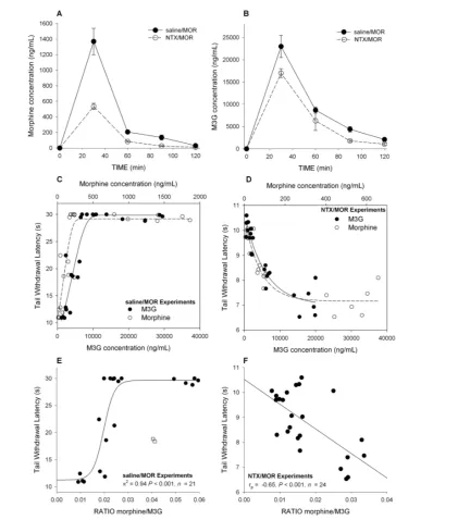
NTX pretreatment had an impact on both morphine and M3G PK, causing a reduction in maximum concentrations (Maximum plasma concentration (CMAX);
Figures 2A, B). After saline/ MOR, mor-phine and M3G CMAX(reached at t= 30 min) were 1,364 ± 170 ng/mL and 23.2 ± 2.5 µg/mL, respectively. After
NTX/MOR, morphine and M3G CMAX (reached at t= 30 min) were 535 ± 40 ng/mL (P= 0.002 versus MOR/saline) and 18 ± 1 µg/mL (P= 0.002 versus MOR/saline), respectively.
In Figures 2C and D, the
pharmacokinetic–pharmacodynamic (PK–PD)relationships between mor-phine, M3G and TWL are given for ani-mals treated with saline/MOR and those treated with NTX/MOR. This PK-PD analysis shows that analgesic TWLs cor-related well with morphine and M3G plasma concentrations, with linear dose-dependent increases in TWL until the cutoff value of 30 s was reached (Fig-ure 2C). Similarly, hyperalgesic TWLs correlated well with morphine and M3G plasma concentrations with dose-depen-dent decreases in TWLs, which were well
Figure 2.Morphine and M3G pharmacokinetics and pharmacodynamics in CD-1 mice (study 1). (A) Mean morphine plasma concentra-tions versus time in mice treated with saline followed by morphine (saline/MOR) and mice treated with NTX followed by morphine (NTX/MOR). (B) Mean M3G plasma concentrations versus time in mice treated with saline followed by morphine (saline/MOR) and mice treated with NTX followed by morphine (NTX/MOR). (C) Individual morphine (open circle) and M3G (closed circle) plasma concentra-tions after saline pretreatment versus effect (TWL). To guide the eye, a sigmoid function is fitted through the two data sets. (D) Individual morphine (open circle) and M3G (closed circle) plasma concentrations after NTX pretreatment versus effect (TWL). To guide the eye, an exponential function is fitted through the two data sets. (E) The ratio of the plasma concentrations morphine and M3G (morphine/M3G) versus TWL for animals treated with saline/MOR. There is a clear positive correlation for ratios <0.03 (rp= 0.72). At ratios >0.03, no further increase in TWL was observed because of the preset cutoff value of 30 s. The line through the data is a sigmoid fitted to the whole data set (R2= 0.94). Two outliers (open squares) were not taken into account in the analysis. Each data point is one morphine/M3G
described by an exponential function (Figure 2D). The morphine/M3G concen-tration ratios versus effect (TWL) are given in Figure 2F for animals treated with NTX/MOR, showing a negative correlation (Pearson correlation coeffi-cient, rp= –0.65, P< 0.001), indicating that higher morphine levels relative to M3G concentrations are associated with increased hyperalgesia.
Study 2
The effects of treatment on TWL in FVB and Mrp3–/–mice are given in Fig -ure 3. NTX/saline was without effect on TWL (P= 0.44) in FVB and Mrp3–/–mice. In contrast, NTX/MOR caused hyperal-gesia in both strains. In FVB mice, TWL decreased by 3.9 ± 0.2 s at t= 30 min (P< 0.001 versus baseline), and hyperalgesia was present throughout the test period (treatment effect: P< 0.001 versus NTX/ saline treatment). Similarly, in Mrp3–/– mice, TWL decreased by 3.1 ± 0.2 s at t= 30 min (P< 0.001 versus baseline) and hyperalgesia was present throughout the test period (treatment effect: P< 0.001 versus NTX/saline treatment; strain comparison: P= 0.10).
Study 3
Morphine plasma concentrations 30 min after 15 mg/kg morphine did not differ between Mrp3–/–and FVB mice. In contrast, Mrp3–/–mice generated M3G plasma levels, just 7% of those measured in FVB mice: 0.9 ± 0.08 µg/mL at t= 30 min versus 12.5 ± 0.5 µg/mL (P= 0.03; Figure 4). The data also show that Mrp3–/–mice excreted more morphine in their 24-h urine than FVB mice (MRP3–/– 39.2 ± 2.8 µg/24 h versus FVB 22.6 ± 3.3 µg/24 h, P= 0.003), whereas M3G excretion was significantly reduced by 88% in MRP3–/–(60.3 ± 6.9 µg/24 h) rela-tive to FVB (532 ± 97 µg/24 h; P= 0.004) mice.
DISCUSSION
Opioids are widely used in the man-agement of moderate to severe pain. De-velopment of hyperalgesia was acknowl-edged as an important factor in the
limitation of efficacy of opioid therapy in some patients. Elucidating the mecha-nism of hyperalgesia and identifying compounds that prevent or reverse its manifestation might result in better opioid-based interventions for treatment
of pain. Over the years, several mecha-nisms of OIH have been proposed, in-cluding the accumulation of the pronoci-ceptive morphine metabolite M3G (8–12), activation of the N-methyl-D-aspartate receptor (NMDAR) by morphine or M3G
Figure 3.Pharmacodynamic results of study 2. Effect of NTX pretreatment on morphine or
saline induced changes in TWL in FVB and Mrp3–/–mice. NTX pretreatment caused
mor-phine-induced hyperalgesia in both genotypes. The combination NTX/saline had no ef-fect on the measured latencies. Treatment efef-fect (NTX/MOR versus NTX/saline) is shown in FVB mice (P < 0.001) and Mrp3–/–mice (P < 0.001). Strain comparison, P = 0.10. *P < 0.001
versus baseline. Values are means ± SEM. t = 0 is the time of the morphine or saline subcu-taneous injection.
Figure 4.Pharmacokinetic results of study 3. (A) Morphine and M3G plasma
concentra-tion in FVB and Mrp3–/–mice. (B) The 24-h morphine and M3G excretion in FVB and
(16–19) and activation of TLR4 receptors by morphine or M3G (24,25). These mechanisms may not be mutually exclu-sive. This study focused on the possible involvement M3G in the development of OIH after morphine treatment and as-sessed further whether the MOR is the molecular site of OIH.
The first set of experiments that we performed showed that OIH is rapidly induced in CD-1 mice injected with mor-phine after pretreatment with high-dose NTX, a nonselective antagonist of the opioid receptors (Figures 1B, C). These data indicate that OIH is exposed by blocking the opioid receptors to mor-phine and subsequent analgesia, which confirm earlier findings showing that morphine and the phenylpiperidines (for example, fentanyl) induce OIH via non-opioid receptor–related pathways (16–19,26). Whether these hyperalgesia or excitatory pathways are activated by morphine or its major metabolite in ro-dents (M3G) is unknown. There is evi-dence from experimental and clinical studies that M3G has excitatory proper-ties and hence may be involved in OIH after morphine treatment (8–12). To ex-plore this further, we measured mor-phine and M3G concentrations in plasma of the CD-1 mice. Depending on the treatment, both compounds showed a high degree of correlation to either anal-gesia after saline/MOR (Figure 2C) or hyperalgesia after NTX/MOR (Fig -ure 2D). The MOR/M3G concentration ratios versus effect (TWL), obtained in mice treated with NTX/MOR, show a negative correlation (Figure 2E), indicat-ing that with relative greater morphine than M3G concentrations, the magnitude of hyperalgesia increases. This favors a role for morphine rather than M3G in the induction of OIH in our model.
An interesting observation in study 1 is that NTX pretreatment affected mor-phine’s pharmacokinetics, causing lower morphine and M3G concentrations com-pared with saline pretreated animals (Figures 2A, B). NTX enhances morphine glucuronidation (27), which may be ex-plained by induction of the liver UGT
enzyme system and possibly causes some greater excretion of M3G via gut and kidney. Because of these effects, the MOR/M3G concentration ratios were smaller in NTX relative to saline-pre-treated animals. Assuming a role for morphine in OIH, we may have under -estimated the absolute magnitude of OIH in the NTX-pretreated animals. Irre-spective, the relationship between the MOR/M3G ratios versus TWLs will not be affected. Hence, we argue that the in-fluence of NTX on morphine’s pharma-cokinetics did not affect the outcome of our studies.
We further explored the role of M3G by testing the effect of NTX/MOR in mice lacking the MRP3gene. The MRP3 gene product, a multidrug resistance transporter present on the sinusoidal membrane of the hepatocyte, is involved in the efflux of glucuronides, including M3G, from the hepatocyte into the bloodstream (20). Mice that lack the MRP3gene have a greatly reduced ca-pacity to export M3G into the systemic circulation and consequently M3G accu-mulates in the liver, as the metabolic pro-cess in the liver (converting morphine into M3G) remains intact. Indeed, M3G pharmacokinetic analysis in Mrp3–/– mice showed low amounts of M3G in plasma (7% of control) and urine (12% of control; Figure 4) after a morphine injec-tion. We did not measure M3G in brain tissue in the current study, but we previ-ously were unable to detect any M3G in brain tissue of Mrp3–/–mice, 30 min after injection of the identical 15 mg/kg mor-phine dose injection here (20). This result suggests that the low plasma concentra-tions of M3G do not result in detectable concentrations of M3G in the brain of Mrp3–/–mice (see below).
The pharmacodynamic data obtained in the Mrp3–/–mice showed that, like FVB controls, they manifest induced hyperalgesia (after NTX/MOR) of substantial and significant magnitude of the same duration (Figure 3). Hence, morphine-hyperalgesia developed de-spite low M3G concentrations in urine, plasma and brain. These data suggest
that morphine rather than M3G is the main cause of OIH in this model. In-deed, in humans and mice, after sys-temic administration, the glucuronides cross the blood-brain barrier poorly or not at all (5,6,20), sug-gesting that morphine is more likely in-volved in OIH than M3G. For example, in wild-type mice, after systemic M3G administration, brain concentrations re-main low at levels more than 50 times less than those observed in the rest of the system (liver, plasma) (20). Nonethe-less, we cannot exclude the possibility that some morphine is metabolized at central sites into M3G. Supportive data, however, suggest that these brain M3G levels are low, and, consequently, this pathway will be of minor importance (28). Further brain morphine and M3G concentration response studies are needed to quantify a possible role of central morphine metabolism on morphine-induced hyperalgesia. Irre-spective, it is unlikely that M3G from the hepatic metabolism of morphine played a role in the observed morphine-induced hyperalgesia in Mrp3–/–mice.
consequently pronociception, including opposition of acute and chronic analge-sia, opioid analgesic tolerance and also OIH. Recent studies indicate involve-ment of nonclassical opioid receptors, in-cluding the toll-like receptor 4 (TLR4) on glia cells in this process. Both morphine and M3G display significant TLR4 activ-ity (24,25).
CONCLUSION
The role of M3G in morphine-induced hyperalgesia was extensively investi-gated through a series of experiments that involved both pharmacodynamic and pharmacokinetic measurements. We confirm the presence of OIH in mice in-jected with morphine after pretreatment with NTX and show for the first time the manifestation of morphine-induced hyperalgesia in mice lacking the MRP3 protein, despite the very low plasma M3G concentrations in these animals due to the inability to release M3G from hepatocytes. Collectively, these data suggest that morphine itself is responsi-ble for inducing hyperalgesia through non- opioidergic pathways and that he-patic M3G is not involved in morphine hyperalgesia. The relevance of these murine data to humans has yet to be demonstrated.
DISCLOSURE
The authors declare that they have no competing interests as defined by Molecu-lar Medicine, or other interests that might be perceived to influence the results and discussion reported in this paper.
REFERENCES
1. Ballantyne JC, Mao J. (2003) Opioid therapy for chronic pain. N. Engl. J. Med.349:1943–53. 2. Angst MS, Clark JD. (2006) Opioid-induced
hy-peralgesia: a qualitative systematic review. Anes-thesiology.104:570–87.
3. van Elstraete AC, Sitbon P, Trabold F, Mazoit X, Benhamou D. (2005) A single dose of intrathecal morphine induces long-lasting hyperalgesia: the protective effect of prior administration of keta-mine. Anesth. Analg. 101:1750–6.
4. Yaksh TL, Harty GJ, Onofrio BM. (1986) High dose of spinal morphine produce a nonopiate re-ceptor–mediated hyperesthesia: clinical and the-oretic implications. Anesthesiology. 64:590–7.
5. Christrup LL. (1997) Morphine metabolites. Acta Anaesthesiol. Scand.41:116–22.
6. Sarton E, et al.(2000) Sex differences in morphine analgesia: an experimental study in healthy vol-unteers. Anesthesiology.93:1245–54.
7. Lötsch J. (2009) Pleiotropic effects of morphine-6β-glucuronide. Anesthesiology. 110:1209–10. 8. Bartlett SE, Cramond T, Smith MT. (1994) The
ex-citatory effects of morphine-3-glucuronide are at-tenuated by LY274614, a competitive NMDA re-ceptor antagonist, and by midazolam, an agonist at the benzodiazepine site on the GABAA recep-tor complex. Life Sci.54:687–94.
9. Labella FS, Pinsky C, Havlicek V. (1979) Mor-phine derivatives with diminished opiate recep-tor potency show enhanced central excitarecep-tory ac-tivity. Brain Res.174:263–71.
10. Gong QL, Hedner J, Bjorkman R, Hedner T. (1992) Morphine-3-glucuronide may functionally antagonize morphine-6-glucuronide induced an-tinociception and ventilatory depression in the rat. Pain.48:249–55.
11. Woolf CJ. (1981) Intrathecal high dose morphine produces hyperalgesia in the rat. Brain Res.
209:491–5.
12. Yaksh TL, Harty GJ, Onofrio BM. (1986) High dose of spinal morphine produce a nonopiate receptor-mediated hyperesthesia: clinical and theoretic implications. Anesthesiology.64:590–7. 13. Dennis GC, et al.(1999). Analgesic responses to intrathecal morphine in relation to CSF concen-trations of morphine-3,β-glucuronide and mor-phine-6,β-glucuronide. Life Sci.64:1725–31. 14. Lipkowski AW, Carr DB, Langlade A, Osgood PF,
Szyfelbein SK. (1994) Morphine-3-glucuronide: silent regulator of morphine actions. Life Sci.
55:149–54.
15. Ekblom M, Gardmark M, Hammarlund-Udenaes M. (1993) Pharmacokinetics and pharmacody-namics of morphine-3-glucuronide in rats and its influence on the antinociceptive effect of mor-phine. Biopharm. Drug Disp.14:1–11.
16. Juni A, Klein G, Kest B. (2006) Morphine hyperal-gesia in mice is unrelated to opioid activity, anal-gesia, or tolerance: evidence for multiple diverse hyperalgesic systems. Brain Res.1070:35–44. 17. Juni A, Klein G, Pintar JE, Kest B. (2007)
Nocicep-tion increases during opioid infusion in opioid receptor triple knock-out mice. Neuroscience.
147:439–44.
18. Juni A, Klein G, Kowalczyk B, Ragnauth A, Kest B. (2008) Sex differences in hyperalgesia during morphine infusion: effect of gonadectomy and es-trogen treatment. Neuropharmacology.54:1264–70. 19. van Dorp E, et al.(2009) Morphine-6β
glucuronide rapidly increases pain sensitivity in-dependently of opioid receptor activity in mice and humans. Anesthesiology. 110:1356–63. 20. Zelcer N, et al.(2005) Mice lacking multidrug
re-sistance protein 3 show altered morphine phar-macokinetics and decreased antinociception by morphine-6-glucuronide. Proc. Natl. Acad. Sci. U. S. A.102:7274–9.
21. Zimmermann M. (1983) Ethical guidelines for in-vestigations of experimental pain in conscious animals. Pain.16:109–10.
22. Swartjes M, Morariu A, Niesters M, Aarts L, Dahan A. (2011) Non-selective and NR2B-selec-tive NMDA receptor antagonists produce an-tinociception and long-term relief of allodynia in acute and neuropathic pain. Anesthesiology.
115:165–74.
23. Rook EJ, Hillebrand MJX, Rosing H, van Ree JM, Beijnen JH. (2004) The quantitative analysis of heroin, methadone and their metabolites and the simultaneous detection of cocaine, acetylcodeine and their metabolites in human plasma by high-performance liquid chromatography coupled with tandem mass spectrometry. J. Chromatogr. B Analyt. Technol. Biomed. Life Sci. 824:213–21. 24. Wang X, et al.(2012) Morphine activates
neuroin-flammation in a manner parallel to endotoxin.
Proc. Natl. Acad. Sci.U. S. A. 109:6325–30. 25. Hutchinson MR, et al.(2010) Evidence that
opi-oids may have toll-like receptor 4 and MD-2 ef-fects. Brain Behav. Immun.24:83–95.
26. Waxman AR, Arout C, Caldwell M, Dahan A, Kest B. (2009) Acute and chronic fentanyl admin-istration causes hyperalgesia independently of opioid receptor activity in mice. Neurosci. Lett.
462:68–72.
27. Antonilli L, Petecchia E, Capriolo D, Badiani A, Nencini P. (2005) Effect of repeated administra-tions of heroin, naltrexone, methadone, and alco-hol on morphine glucuronidation in the rat. Psy-chopharmacology. 182:58–64.
28. Wahlström A, Winblad B, Bixo M, Rane A. (1988) Human brain metabolism of morphine and naloxone. Pain. 35:121–7.
29. Chizh BA. (2007) Low dose ketamine: a thera-peutic and research tool to explore N -methyl-D-aspartate (NMDA) receptor-mediated plasticity in pain pathways. J. Psychopharmacol.21:259–71. 30. Sigtermans M, et al.(2009) Ketamine produces
ef-fective and long-term pain relief in patients with Complex Regional Pain Syndrome Type 1. Pain.
145:304–11.
31. Sánchez-Blázquez P, Rodríguez-Muñoz M, Garzón J. (2010) Mu-opioid receptors transiently activate the Akt-nNOS pathway to produce sus-tained potentiation of PKC-mediated NMDAR-CaMKII signaling. PLoS One.5:311278. 32. Hemstapat K, Monteith GR, Smith D, Smith MT.