R E S E A R C H
Open Access
A model to explain specific cellular
communications and cellular harmony:- a
hypothesis of coupled cells and interactive
coupling molecules
Cyril J Craven
Correspondence:
Queensland University of Technology (QUT), Brisbane, Australia
Abstract
Background:The various cell types and their relative numbers in multicellular organisms are controlled by growth factors and related extracellular molecules which affect genetic expression pathways. However, these substances may have both/either inhibitory and/or stimulatory effects on cell division and cell differentiation depending on the cellular environment. It is not known how cells respond to these substances in such an ambiguous way. Many cellular effects have been investigated and reported using cell culture from cancer cell lines in an effort to define normal cellular behaviour using these abnormal cells.
A model is offered to explain the harmony of cellular life in multicellular organisms involving interacting extracellular substances.
Methods:A basic model was proposed based on asymmetric cell division and evidence to support the hypothetical model was accumulated from the literature. In particular, relevant evidence was selected for the Insulin-Like Growth Factor system from the published data, especially from certain cell lines, to support the model. The evidence has been selective in an attempt to provide a picture of normal cellular responses, derived from the cell lines.
Results:The formation of a pair of coupled cells by asymmetric cell division is an integral part of the model as is the interaction of couplet molecules derived from these cells. Each couplet cell will have a receptor to measure the amount of the couplet molecule produced by the other cell; each cell will be receptor-positive or receptor-negative for the respective receptors. The couplet molecules will form a binary complex whose level is also measured by the cell. The hypothesis is heavily supported by selective collection of circumstantial evidence and by some direct evidence. The basic model can be expanded to other cellular interactions.
Conclusions:These couplet cells and interacting couplet molecules can be viewed as a mechanism that provides a controlled and balanced division-of-labour between the two progeny cells, and, in turn, their progeny. The presence or absence of a particular receptor for a couplet molecule will define a cell type and the presence or absence of many such receptors will define the cell types of the progeny within cell lineages.
A model of life
A simple model is offered to explain the requisite harmony of multicellular life. From this basic model, complexity needs to be added to explain the abundance, profusion and variety of life and the sophistication of human existence.
The adult worm Caenorhabditis eleganshas exactly 959 cells in the hermaphrodite, having lost exactly 131 defined cells by apoptosis and fusion during ontogenesis [1,2]. Could we expect the same organised, awe-inspiring exactitude of proliferation, differen-tiation, apoptosis etc. for a human with 50–100 × 1012 cells? The current model offers the reciprocal interactions of coupled cells which have been derived from asymmetric cell division, as the basis for this exactitude of multicellular life.
(A) Background:- questions within existing knowledge
The model offered here relates to the regulation of cell division by extracellular mes-sages and relates to questions as to when and why a growing cell decides to divide sym-metrically or asymsym-metrically and what particular type of symmetric or asymmetric division occurs.
When will a cell proliferate, differentiate or apoptose or otherwise live or die?
Chemical messages will be an integral part of this decision-making
A unicellularorganism is a fully-armoured, selfish, intelligent cell. It is often in an anarchistic milieu, an unpredictable and fickle environment [3], within which it needs to respond appropriately to an array of gross changes and predicaments which are monitored by the cell for the cell’s sole information. In a favorable en-vironment, an increase in effector molecules (e.g. nutrients) will induce appropriate enzymes for their own catabolism, thus to increase metabolism from quiescence [4], perhaps to increase cell size (hyperplasia) and perhaps to proliferate by cell division. In an adverse environment, with nutritional deprivation, senescence [5] or sporulation may be the response.
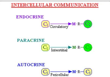
If an Autocrine interaction is considered to be the reception of a ligand signal produced by exactly the same cell, then a Paracrine interaction could be considered to be either Homoty-pic or HeterotyHomoty-pic, where a HomotyHomoty-pic one refers to the reception of the ligand signal by a cell of the same type that produced it. Thus this would be referred to as a Homocrine inter-action and the Heterotypic Paracrine interinter-action referred to as simply Paracrine [6].
A Juxtacrine interaction is a more intimate type of cellular communication where the chemical message is passed between two cells that are in physical contact with each other, being received directly or via secreted extracellular matrix (ECM). It involves a receptor on one cell and a ligand from another cell which is anchored or fixed as part of the cell it-self [7]. (If the communication is bidirectional, then perhaps both act as ligand and recep-tor simultaneously–each perhaps a ligeptor). Another interaction is via intercytoplasmic conduits,“Gap junctions”, which are channels made of connexin proteins between neigh-bouring cells, allowing small-molecule exchange and perhaps signalling [8]. Further, there are reciprocal/bidirectional communications as observed in pre-and post-synaptic chem-ical signalling in neurons [9], and with inside-out and outside-in signalling via integrins [10]. The focus of this discussion is on chemical interactions of ligand and receptor be-tween cells and the consequent intelligent use of these messages.
Reception and interpretation of chemical messages will give intelligence to cells for this decision-making
Within tissues and organs, these messenger molecules will often be present within the extracellular matrix which, while organised, will also contain a veritable soup of
Figure 1Types of communication between cells.This illustrates the terms used to describe communication between cells (C1and C2are cells) where a message (M), carried by a molecule (eg. a Ligand) released from one cell
metabolic intermediates, end-products of metabolism, nutrients, vitamins, false mes-sengers from foreign cells or invasive molecules that would redirect the genetic mani-festation of that cell and, in some circumstances, other molecules which may be toxic to metabolism - lytic or necrotic.
In order to respond selectively to the coordinating messages from self-type cells, the target cell needs specific cell-membrane receptors which will receive only the specific signal for that cell type. The cell can then interpret each message accurately via coup-ling of the receptor to intracellular molecules which transmit the extracellular signal to the nucleus, and thus the cell can respond appropriately. The cell exists “in the dark” and can only gain knowledge of the extracellular environment by receptors. The receptor-mediated response may be a signal to induce a change in protein synthesis to alter metabolic reactions with adaptation and/or to cause hyperplasia, migration, prolif-eration, transformation, differentiation, apoptosis or other. The signal may be necessary but not sufficient and the cell may only respond provided other requirements are met. A message received or interpreted falsely may lead to a cell not synchronized or not in harmony with its neighbours or it may cause the cell to transform to one which is un-controlled in its growth, to the detriment of the whole organism.
Membrane receptors translate the external environment to cause intracellular reac-tion and a specific set of receptors will control and define the inherent properties of a cell. Indeed, cells have long been identified by cell markers - proteins on the cell membrane - many of which are receptors. For example, the Cluster of differenti-ation (CD) is a group of membrane proteins used to define immunologically the cell sur-face molecules of blood cells, especially white blood cells. Other cell markers also exist e.g. Lin (a marker used to detect lineage commitment), Sca (Stem cell antigen), c-kit (the receptor for stem cell factor) The presence or absence of the various CD proteins and other markers that act as receptors, ligands, enzymes or adhesion molecules, is used to identify specific cells. For example, two subsets of murine pluripo-tent hematopoietic stem cells exist, one with the phenotype Lin(−) Sca(+) kit(+) CD38(+) CD34(−), the other Lin(−) Sca(+) kit(+) CD38(−) CD34(+) [11]. While there are over 300 CD proteins already detected, it has been estimated that there would be between 2,400 and 5000 cell-surface molecules on leucocytes [12]. Many of these would be receptors. A cell is then defined by these receptors it uses to receive messages and to appropriately re-spond. While the number of molecules of particular receptors might vary with adaptation or maturation of a cell, a major change in the types of cell-membrane receptors is consid-ered here to indicate a change of the cell’s type and the change in receptors would be a critical part of a differentiation process. (See Additional file 1,1“Terminology”for Defini-tions). Cells are then defined by their receptors and their messages will control both the metabolic pathways of the cell and the cell’s decision to“proliferate, differentiate or apo-ptose or otherwise live or die”. Receptor-mediated uptake of signal-carrying molecules is an integral part of the model presented herein.
uses ratios of concentrations rather than absolute numbers. This intelligent use of this information received resides in all cells allowing both local and whole-of-body decisions to be made and appropriate changes to ensue. In multicellular organisms, local deci-sions are supervised by cells that reside within the specialised and integrated cells (e.g. within the brain) which produce the whole-of-body intellect. For Insulin-Like Growth Factor-I (IGF-I), this growth factor may be either locally produced to have a paracrine action [13] or it may be derived from hepatocytes that secrete it into the systemic system under the control of pituitary-derived growth hormone (GH). In turn, GH secretion is controlled by the GH-releasing-hormone released from the hypothalamus to stimulate the pituitary.
Information may then be transferred via receptor-mediated uptake of growth factors, cytokines, adipokines, chemokines, hormones and other bioactive substances. As a group, these are referred to herein as generic Information-Carrying Molecules and Inter-Cellular Messengers(ICMs) (See Additional file 1,1“Terminology”).
Simply put, the information carried by ICMs derived from one cell type will be inter-pretated by a second cell via specific receptors and consequent intracellular changes, thus providing intelligence to this second cell.
What were the transition steps in the evolution from a unicellular entire (e.g. a Protozoa) to an organised but internally symbiotic multicellular organism (e.g. a metazoan)?
The development of the original eukaryotic cell types may have been based on asym-metric cell division and on common cellular interactions [14]. The former underlies the fundamental basis for the developmental evolution of organisms and for the function-ing of totipotent or multipotent stem cells [15]. An example of the latter is that the same molecules (e.g. phorbol esters, diacylglycerol, tetrapyrroles) that stimulate cell division in two unicellular eukaryotes (the ciliate Tetrahymena thermophila and the yeast Saccharomyces cerevisiae) also cause cell division and other activities in multicel-lular mammalian cells [16].
Asymmetric cell division, with division of labour, is likely a part of this transition process Cooperativity is part of earliest life, where some cells of a unicellular group reacted to the environment and evolved, with cell division, to cooperate metabolically with their predecessors. (See Additional file 1,2“Change in Cellular Characteristics with or with-out Differentiation (with Cell Division), with Division of Labour” for examples of this). Asymmetric cell division is considered to be an extension of this, to separate clearly the metabolic labour of survival and growth. A cell community becomes more efficient by allocating some tasks to specific cells; each cell does not then need to utilise its whole armamentarium of genes.
where there are two types of haploid cells, the alpha-cell and the a-cell, whose forma-tion involves asymmetric cell divisions. The alpha-cell secretes the alpha-factor which binds to a specific receptor on a-cells to transmit a signal and, in a reciprocal way, the a-cell secretes an a-factor which binds to a receptor on alpha-cells. Each factor or pheromone induces hyperexpression of genes specific for the opposite cell type [17]. A similar system occurs in other fungi [18].
These examples in fungi and the following example of pheromones, are the early pro-totypes of the current “CTC Model” proposed in Section B of this article, involving asymmetric cell division (AsCD) with reciprocal interactions.
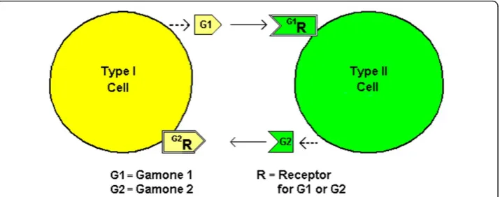
Pheromones, also known as Gamones, are secreted by gametes for sexual reproduction. In an ancestral ciliate, Blepharisma japonicum, two mating cells (Types I and II) are formed as offspring cells from AsCD [19]. Type I cells secrete Gamone 1 (a glycoprotein) and Type II cells secrete Gamone 2 (a trp derivative) and it is thought that Gamone 1 is recognized by putative Gamone 1 receptors on Type II cells and Gamone 2 is recognized by putative Gamone 2 receptors on Type I cells [20] (See Figure 2). This is a similar recip-rocal arrangement as forS. cerevisiae. In the ciliate,Euplotes raikovi,however, a receptor for the pheromone Er-1 has been identified in Type I cells although the this particular pheromone stimulates cells that produce it i.e. it appears to act autocrinely [21,22]. An al-ternate explanation, consistent with the model offered herein, would be that the cells are in fact of two types–one producing, the other internalizing the pheromone.
Division of metabolic labour may be the reason for such formation of coupled cells, with AsCD as the mechanism. Many different biological species are syntrophic in that one species lives off the products of another species while others are symbiotic with mutual advantage by interaction between two different species. A third situation ap-pears for a archeal biofilm where two sets of cells, physiologically and possibly genetic-ally differentiated with respect to each other but derived from a single-species, are involved in mutual syntrophic reactions. It seems that one cell type of the archea duces methane from hydrogen (methanogenic) and the other uses the methane to pro-duce perhaps acetate or formate (methanotrophic) which is used by the first methanogenic cell [23]. This could be interpreted as a division of labour (DOL) result-ing from a sresult-ingle precursor cell, by AsCD. DOL has also been described in Additional file 1,2 forCyanobacteriaand algae but for a more complex multicellular organism, the
total sum of catabolic and anabolic reactions for the whole is enormous. Many descrip-tions of metabolism impose a complex interplay of reacdescrip-tions on a single cell, whereas the evolution of multicellular organisms has been towards specialisation and division of metabolic labour. Indeed early cells were more complex with more diverse constituents than modern cells [3]. The specialisation is evidenced by certain cells of the human body producing metabolites which are released into the extracellular fluid, taken up by other cells via transporters/receptors and used metabolically by these cells. Certain hu-man cells that have stored sources of energy (glycogen and triacylglycerols) release glu-cose and fatty acids for other cells. Products of metabolism, alanine and lactate, are transferred from muscle to specific liver cells and used for energy or glucose production. Also, lactate is produced in one cell (astrocyte), secreted and taken-up by a paracrine cell (a neuron) for fuel [24] and for long-term memory formation [25]. Adipocytes also have a specific G-protein-coupled receptor for lactate [26]. Glutamine is produced in one cell (glial), secreted and then taken-up by another type of cell (GABAergic neurons) for the production of GABA (gamma-amino butyric acid) [27]. Extracellular glutamine is particu-larly relevant for cancer cells with a glutamine-stimulated anabolic state [28,29]. Citrate is secreted by epithelial cells of the prostate [30], while other cells (sperm) have influx citrate transporters and these cells use citrate for fuel and for the production of long-chain fatty acids, cholesterol and steroids [31]. These are then examples of cooperativity, possibly or-chestrated by a cell-fate plan involving asymmetric cell division with consequent inter-dependence of cells associated with division of labour.
While DOL will obviously reduce the number of genes expressed in a single cell, it has been estimated that even a dedicated cell such as a B-cell lymphocyte, will express more than 10 000 genes [32]. A dedicated cell with responsive receptors is still a com-plex entity even with DOL.
Of course, not all of the examples of division of labour involve a direct AsCD or mu-tual survival. Erythrocytes, terminally differentiated cells which lack a nucleus, mito-chondria, lysosomes, endoplasmic reticulum and Golgi apparatus, are dedicated to serving others by supplying oxygen and removing tissue acidity via their haemoglobin content. These are “singlet” cells with no coupled cells needed to balance this forma-tion of erythrocytes, but their own survival for about 110 days is dependent on a variety of other cells for nutrients. Although being dedicated and loaded with haemoglobin, one red blood cell still contains more than 750 proteins for its survival [33]. A very dedicated cell without responsive receptors is still somewhat complex.
As opposed to the division of labour by a fully deployed cell using AsCD, it is also relevant to acknowledge the possible function of opposing cell actions such as fusion, engulfment and endosymbiosis in both evolution [34,35] and in the growth of modern multicellular organisms.
The CTC model proposed in this hypothesis evolves from a consideration of division of labour with fewer genes expressed, involving asymmetric cell division with reciprocal interactions as indicated between fungi cells and for pheromone interactions.
A balance of cells in a multicellular organism is achieved by complementation of the types of cell division (CD) - Symmetric (SCD) versus Asymmetric (AsCD)
many biology textbooks focus on mitosis with symmetric division or it may be based on evidence from light microscopy where there is no obvious physical change in cellu-lar characteristics. However, despite the latter, there may have been asymmetric cell division with many possible outcomes as discussed in the next section. Of course, there could be a combination of SCD and AsCD in these cultures just as there is for growth and development of a whole organism [36].
A classical line of thought has been that, after SCD, one cell could be transformed by the environment (heat, dehydration, presence of an interacting ICM) into a“new” cell and this cell could produce duplicated progeny of this new cell by SCD. Alternatively, one cell of a pair could be altered occultly (or be primed) by the environment such that a new cell type only becomes obvious after another SCD. As such, there was no asym-metric cell division.
However, currently, while the environment is still considered a critical part of the de-cision making, AsCD is known to be widespread in many aspects ofLife–even ubiqui-tous. AsCD may be produced in several ways and survival of multicellular organisms is a balance of SD versus AsCD. AsCD, controlled by events that occur pre-cytokinesis, can be classified as either intrinsic, involving an inherited determinant, or cell-extrinsic, involving intercellular communication or environmental factors. In addition, the determination of asymmetry could be post-cytokinesis and extrinsic, following an initial symmetric distribution of cellular constituents. (See Additional file 1,3 Note 1
“The Roles of Symmetric versus Asymmetric Cell Division” for evidence and causes of AsCD and factors affecting AsCD/SCD balance).
Stem cells are thought to balance self-renewal and differentiation through judicious variation of asymmetric and symmetric divisions. The balance is not hardwired into the genes but is responsive to extrinsic and intrinsic cues [37]. Hemopoietic stem cells will form different progeny (erythrocyte, monocyte, lymphocyte etc.) based on the exposure to a variety of ICMs such as interleukins, colony-stimulating factors (CSFs) and erythropoeitin [38]. The balance of AsCD versus SCD can depend on these types of extracellular factors and also on cellular molecules such as p53 protein, cAMP or GMP, or on the lineage, the stage of growth or even light exposure (See Additional file 1,3 Note 1).
Modelling has been used to determine the relative contributions of the various op-tions of AsCD and SCD. Mathematical modelling and lineage studies to explain neuron formation within the mouse neocortex have focused on the relative amounts of three types of CD - AsCD, SCD with “terminal” differentiated cells produced and SCD with “non-terminal” proliferative cells produced [39]. The conclusion was that asymmetric and both types of symmetric cell divisions coexist during the entire period of neurogen-esis. Other models of cell division, in hematopoietic stem cells, indicate a need to con-sider the time taken for, and the frequency of, cell division by the AsCD and SCD of stem and progenitor cells and a need for control by external signals [40]. In the Drosophila optic lobe, four signalling pathways control the sequential transition of symmetrically-dividing neuroepithelial cells into asymmetrically-dividing neuroblasts as the proneural wave progresses across the neuroepithelium [41].
Overall, the balance of cells in a multicellular organism is achieved by both SCD and AsCD as directed by the cell-fate plan of the organism with control by external signals. The CTC model proposed in this hypothesis explains how external signals control cel-lular balance by appropriate selection of SCD or of AsCD as required.
Cells within a multicellular organism may be able to select from a portfolio of mechanisms to divide in order to produce a required outcome
Within the types of symmetric and asymmetric cell divisions, there are many theoretical possibilities. These are depicted and discussed in Additional file 1,3Note 2“Categories of Symmetric and Asymmetrical Cell Divisions”Figure 1. This Figure lists at least ten options of cells in forming a lineage, including self-renewal and differentiation which may be terminal. Some other examples of the AsCD categories are discussed in Additional file 1,3 Note 3“Further Examples of these AsCD Categories”.
While this portfolio of options is impressive, to encompass totally the options of a cell, it would be necessary to include the potential of cells to reverse or change from a differentiated state via a cell division. Some of these extra options are included in Additional file 1,3Note 4“Change of differentiation type–options of a differentiated cell”.
The CTC model proposed in this hypothesis explains how external signals help select the appropriate type of cell division required from these options.
How does a multicellular messenger - a growth factor - affect cellular decisions?
An example of a extracellular molecule which communicates information via a plasma membrane receptor is the Insulin-Like Growth Factor-I (IGF-I) - a 70-amino acid ICM which participates in communication between cells in endocrine, paracrine and auto-crine modes [43] and whose internalization may lead to changes in hyperplasic growth or to proliferation etc. The“IGF system”is complex, with several essential components including ligands; IGF-I and IGF-II, cell membrane-bound receptors (e.g. IGF-IR), at least six soluble “binding proteins” (e.g. IGFBP-1…-6) which form binary complexes with IGFs in the extracellular environment, plus even a ternary complex in serum, form the system. Also, IGFBP proteases will play a part [44].
In our current understanding of this system, the transfer of information from one cell to another by IGF involves receptor binding, subsequent kinase activation, followed by increased metabolism and a decision to divide, perhaps to differentiate or not to apo-ptose. This IGF receptor-binding is reduced by the limited availability of the free IGF due to its binding by the IGF binding proteins (IGFBPs). The IGFBPs have other sug-gested functions e.g. reducing the loss of IGF via the kidney due to the larger size of the complex, decreasing proteolytic cleavage and increasing storage. Modified IGFs that don’t bind IGFBPs {e.g. des(1–3)IGF-I}, have a higher activity than IGF itself and this is explained by the increased availability of the free modified IGF [45]. While these expla-nations seem valid, other explaexpla-nations are required to explain fully the IGF system, es-pecially the IGF-independent effects of IGFBPs [46]. The CTC model proposed in this hypothesis offers an alternate explanation.
This understanding is limited. What are the unanswered questions?
be interpreted to produce a life change to the cell. The former metaboliceffect (in-volving phosphorylation of IGF-IR and activation of the mitogen-activated-protein-kinase path and/or of the phosphatidylinositol-3-mitogen-activated-protein-kinase path) may be a prerequisite for the cellular effect (involving nuclear interactions, transcription effects, chromo-somal reorganization and other whole-of-cell decisions). For the latter, how does the cell know which switches are to be activated or deactivated; will the cell divide, differentiate, transform or undergo apoptosis or otherwise change its status? What intelligence is required to make these decisions; what is the messenger molecule and how does the message reach and be interpreted by the nucleus?
How is IGF-I mostly the well-known stimulator of growth [47] but sometimes an in-hibitor of growth (in cultured smooth muscle cells [48], lymphocytes [49] and Wilm’s tumour cells [50])? How also is a particular IGF-BP an inhibitor of growth (of a breast cancer cell line [51] and a fibroblast cell line [52]) and, at other times, a stimulator of growth (in cultured fibroblasts [53] and osteoblast-like bone cells [54])? How can it be explained that an IGFBP may have an ability to stimulate cells in an IGF-independent fashion [53,54] if the current model has the IGFBP solely as a binder of IGF thereby restricting IGF loss and IGF activity? Overall, how does the whole organism know if there is a balance of the cells producing IGFs and those producing IGFBPs? Some of the answers may be associated with the discovery of receptors for certain IGFBPs and the presence of such receptors becomes part of the current CTC model, described in Section (B), used to answer these questions.
Similar questions apply to other growth factors and related molecules (ICMs) which have both stimulatory and/or inhibitory effects depending on cell-type used, the con-centration of ICM, the presence of other ICMs, the time of exposure and the develop-mental state of the cells. {See Additional file 1,4 “The Effects of Growth Factors and other Bioactive Molecules (collectively referred to here as ICMs - Information-Carrying Molecules and Inter-Cellular Messengers)” for the effects of some fifty ICMs on cell proliferation, both stimulatory and inhibitory).
How do these ICMs have opposite effects on different cells and/or in different circum-stances? Of particular note is that a number of binding proteins (BPs) (besides the IGFBPs) have both stimulatory and inhibitory effects e.g. FGF Binding Protein, Sex-Hormone Binding Globulin, Kallistatin (Kallikrein Binding Protein), Corticosteroid Binding Globulin. Again, how can these BPs that bind ICMs be stimulants of cell growth if they re-duce the amount of the biologically active Free ICM? Again, the current model relies on receptors for these BPs to explain their conflicting effects.
(B) Results and discussion:- a new model–a simplistic basic model
1. Description of the essentials of the basic model–a balance of Trefones and couplet cells
(a) Primary couplet cells and Trefone couplets–the CTC Model with the IGF system as a prime example
receives its message. In this model, an IGF-I could be a Trefone for a cell which pro-duces it and which also has a receptor for its couplet molecule – a specific IGFBP. Equally an IGF-BP could be a Trefone for a cell which produces it and has a receptor for IGF-I. Each Trefone-receptor complex would be internalised into the responding cell, to be part of nuclear messages to affect metabolic and cellular-life decisions. The IGF-I and the IGFBP would be classed as “Couplet Trefones” as they form their own complex (IGF-I:IGFBP) –a complex herein referred to as a Trefone Couplet Complex (TCC). (See Additional file 1,5 Note 1,“Additional Information on the Definition of a Trefone”for more on the properties of a Trefone and Additional file 1,5Note 2“ Clarifica-tion on Ligand and Binding Protein InteracClarifica-tion; the TCC” for more discussion on the complex of ligand and binding protein).
One Trefone is produced and secreted by each of two separate cells and these initial two cells are coupled by their prior formation via asymmetric cell division of their par-ent cell. These two “Couplet Cells” are also coupled in another way in that each cell type has a receptor for the Trefone produced by the other and each cell is altered in re-sponse to the signal tranduced from the Trefone of the other cell. The Trefone signal from one cell will transfer a message for the other cell (i) normally to increase metabol-ism and to stimulate production of the other Trefone and (ii) if necessary, to stimulate cell division or induce some other“whole-of-cell, life/death”action to balance the activ-ity of total “a”cells and“i” cells. Lower levels of the Trefone would induce a metabolic effect while higher levels, prolonged [55], would induce cell division or other action. For example, insulin from a beta-islet cell, when endocytosed at a low level by an insu-lin receptor into a specially receptive cell (a couplet cell, perhaps an alpha-pancreatic cell), would stimulate metabolism and the secretion of the couplet Trefone (perhaps glucagon), while at a high level, upon prolonged exposure, insulin would stimulate the proliferation of the receptive alpha-cell, to produce even more Trefone (glucagon) se-cretion. (Note that these effects are separate from the receptor-mediated effect of insu-lin on carbohydrate and on other metabolic activities of cells generally). For IGF-I signalling, the metabolic and the whole-of-cell effects use the same pathway; in human intestinal smooth muscle cells, the metabolic effect of IGF-I (e.g. to regulate IGFBP production) is mediated by activation of distinct MAP kinase and PI 3-kinase pathways, the same pathways through which IGF-I stimulatesgrowth[56].
The “Couplet Cells” would have been produced by asymmetric cell division of an o-Cell (the original cell - a stem or progenitor cell) to produce two cells committed to or already differentiated. This is referred to as Dual LCDf in Additional file 1,3 Note 2
“Categories of Symmetric and Asymmetrical Cell Divisions”Figure 1, B(iv), where non-identical C/D1 and C/D2cells are produced by AsCD. (Subsequent cell divisions may
be symmetrical (SCD, SRE) to produce more of each of these two types of cells). The coupled progeny cells will be referred to herein as the “a”Cell type (a-Cell or aC) and the“i”Cell type (i-Cell oriC), rather than C/D1and C/D2. (See Additional file 1,5Note
simplicity, this is not shown in Figure 3(a). A specific example is given in Figure 3(b) for the IGF-I and IGFBPn couplet. This system of Coupled Trefones and Cells is re-ferred to as the“CTC”model.
The purpose of Trefones is to maintain a balance of cells. This can be understood by a simple example:- If the number of cells of one type (a-Cells) is in excess (or if they have a high metabolic rate) of that of cells of the couplet (i-Cells), then the tion of (Free) a-Trefone will be in excess of that of i-Trefone, with a limited concentra-tion of TCC. Free aT then stimulates the i-Cells initially to increase the biosynthetic rate ofiT production, and then stimulates the i-Cells to divide symmetrically (SRE) so as to produce moreiT to match the concentration ofaT. This stimulation of the i-Cells is also dependent on a signal that relates to the concentration of TCC and continues until the latter is high and the Free nT concentrations, [aT]F and [iT]F, are low and
equal. In the same period, the a-cells would be quiescent or perhaps undergo cell divisions, of the types indicated in Additional file 1,5Note 3,Table S1, to produce more i-Cells. For the IGF system, IGF-I from the a-Cell will stimulate the i-Cell and an IGFBP from the i-Cell would stimulate the a-Cell but each response would be modified by the level of the IGF-I:IGFBP complex. Each cell would thus measure these external Trefones to assess the activity/number of couplet cells and would respond to maintain a balance.
The situation is similar if the number of a-Cells and i-Cells are equal and in harmony in a mix and thenaT is added in excess. The i-Cells will undergo cell division to replen-ish rapidly these cells to balance the perception of a-Cell excess. In the same period, this would produce reducing-to-zero stimulation (i.e. inhibition) of a-Cell metabolism plus stimulation of some a-Cells to produce two i-Cells and perhaps some a-Cells
Figure 3Couplet Trefones and cells and their membrane receptors. (a)A couplet of cells (aC andiC) with
each producing a soluble Trefone and each having a receptor (R) to bind the soluble Trefone produced by the other cell.aR oniC bindsaT andiR onaC bindsiT.(b)A couplet of cells with the a-Cell producing the“a”Trefone, IGF-I, the
would divide to produce more i-Cells while maintaining a-Cells. This latter scenario would also occur if only cells of one type (e.g. a-Cells) are present initially and the cor-responding Trefone that they produce is added. Again, these types of cell divisions for the a-Cell are indicated in Additional file 1,5Note 3, Table S1.
Thus, Trefones would control an individual cell’s metabolic activity and also cell numbers and types by regulation of SCD and AsCD. a-Cells need to balance i-Cells be-cause the initial AsCD (dual LCDf ) of the o-Cell was designed to allow a “division of labour”(DOL) with consequent increased efficiency of production and use of metabo-lites. The function of the this reciprocal relationship of Trefones with receptors is to balance mutually the activity and number of Couplet Cells produced by this AsCD and by subsequent cell divisions (symmetric or asymmetric) in a particular cell lineage. The presumption here is that the total body of cells of an organism is in harmony and bal-ance and that this living equilibrium is maintained by metabolic induction/inhibition within cells and/or by an increase/decrease in cell number and/or by variation in the types of cell produced by cell division and associated differentiation.
A potential CTC system exists in Drosophilaand this is examined in Additional file 1,5Note 4“Drosophila–Potential Trefones and Asymmetric Cell Division”.
In the CTC model, the two Trefones that are newly expressed by the two progeny following a specific cell division are primary Trefones. Secondary Trefones will be dis-cussed later.
(b) The reason for a cell’s need to measure the Trefone couplet complex (TCC)
How does a cell know if one Trefone is in excess of the other? If the cells have been produced from the progenitor cell for“division of labour”then each cell couplet needs to know (by“counting”) the state of the other couplet and to be able enhance or reduce its own metabolism or cell number or status in response to signals. The two cells of the couplet need to be in harmony or more exactly the two groups of a-Cells and i-Cells need to be in harmony. Their end product(s) within the inferred DOL agreement need to balance and each individual cell of the two groups of cells plays a part by responding to the Trefone from the other group. How does a cell decide, based on one measured Trefone level, whether there is a balance of the Trefone produced by one group versus the Trefone produced by the other group cell?
to a common compartment wherein a new equilibrium ofaT,iT and the TCC complex is established. These new equilibrium levels of Free Trefone and Bound Trefone (i.e.TCC) will allow each cell to“know”the balance of the external two Trefones. The cell can then respond to any imbalance by the interaction of Free Trefone of TCC with specific signal transducers or directly with transcription factors within the nucleus to change gene expression.
This latter is analogous to a model in Drosophila for the Patched receptor which binds the morphogen-ligand Hedgehog; it is the ratio of the internalized Free Patched (i.e. with no bound ligand) to the internalized Bound Patched (i.e. a Patched-Hedgehog complex) which determines cell response, not just the absolute number of Free Patched receptor molecules internalized [57].
A receptor for the TCC (the complex of the Trefones) on each cell of a couplet, is one option of this CTC model. Alternatives to this option of a receptor for the TCC will be described later. Figure 4 illustrates this generically and for the IGF system.
How does the cell then interpret the combined signals from the nT and the TCC? The signals could be interpreted to our understanding, based on an association con-stant (Ka) where Ka equals [aT:iT]/[aT].[iT] for the reversible formation of the TCC (aT:
i
T) fromaT andiT i.e.aT +iT⇆aT:iT. If the total concentrations ofaT andiT were ini-tially the same, and if the Ka value was assumed to be 1 × 10¹⁰, then this allows calcula-tion of the Free concentracalcula-tions of the Trefones at a range of given Total Trefone concentrations. From this input, the importance of knowing the concentration of Free levels of Trefones and of the TCC, plus the value of just having aT bind toiT, can be appreciated. (See Additional file 1,6 Table S1,“Calculated Levels of Trefones and TCC, Connected to Cellular Actions” for possible cellular reactions to varying concentrations
Figure 4Couplet Cells with membrane TCC receptors. (a)A couplet of cells (aC andiC) with each
producing a Trefone and each having a receptor (R) to bind the Trefone produced by the other cell. This is as in Figure 3 but with a TCC receptor (TCCR) on each cell.(b)A couplet of cells with the a-Cell producing the“a”
of Trefones). An association can be made between the variation in concentrations of Free Trefones and of TCC with cellular responses, where the predicted response fo-cuses on whole-of-cell decisions rather than metabolic changes. This Table is a critical part of the CTC model.
Depending on the concentration of FreeiT oraT and of TCC, theaCells andiCells re-spectively, will respond. From the messages received, the consequent cell action may be symmetric cell division (SCD), asymmetric cell division (AsCD), transdifferentiation (TD)/SCD, apoptosis (APO), quiescence (QSC), differentiation or dedifferentiation to a progenitor to restart the lineage. The two examples described in Additional file 1,6 of quantitative and objective interpretations from the data in this Table show how the cells are able to make intelligent decisions based on their Trefone environment. In one case, the concentration of TCC is critical for decision-making where the concentration of Free Trefone is identical in three different situations which require different cellular reactions; in the other case, the presence of the complex amplifies the change in con-centration of the Free Trefone. A simple example would be that if Free aT were defi-cient, possibly indicating a deficiency of a-Cells, then existing a-Cells would proliferate by SCD (SRE) to double their number and some i-Cells would divide to form an a-Cell (plus an i-Cell) or even to produce two a-Cells by mechanisms explained in Additional file 1,5Note 3, Table S1.
Each couplet cell will have internalized a certain amount of one Trefone and the TCC. As an example of an abnormal situation, let us assume thataT is being normally produced by the a-Cell but the externalaT is not producing a response ataR of the i-Cell because aT is being somehow acutely destroyed or diverted elsewhere and the ef-fective level of aT is Lo (but not due to lack of response by a variant aR or lowaR on the i-Cell). Residual extracellularaT would form LoaT:iT complex and the i-Cell would therefore lack aT stimulus so that decreasediT production and i-Cell proliferation are expected. The extracellulariT level would be initially in excess ofaT but slowly decreas-ing as it continues to stimulate the a-Cell. Intracellularly, the a-Cell, which producesaT, has acquired a Hi level of iT but a Lo level of TCC from receptor-mediated uptakes. Upon release of iT andaT:iT from receptors in a communal endosomal compartment, there would be a mixing and re-equilibration of the internalizediT andaT:iT so that the final level of the intracellular TCC and of free iT oraT would reflect the extracellular balance ofiT andaT.
In the cell itself, this mathematical interpretation would be replaced by a concentra-tion dependent activaconcentra-tion and/or inhibiconcentra-tion of specific genes to direct proliferaconcentra-tion (or other) by its innate, inherited intelligence [58]; knowledge which will allow it to know whether there is balance between the two Trefones and thus between the two types of cells. The balance of extracellular Trefones, as determined by intracellular equilibrium levels, could then control cell decisions by altering the level of a transcription factor; for example, during mammalian embryo development, increased Cdx2 levels are associated with more SCDs, while downregulation of Cdx2 is associated with more AsCDs [59].
production of its own specifically producednT which is secreted by its whole family of n-cells and not just by the individual cell.
The reasons that a cell-membrane receptor for extracellular TCC has not been de-tected include the lack of a convincing search and the fact that the number of receptor molecules may need only be very low to sample the large concentration of TCC. This will be addressed later.
(c) How does this model change our understanding?
The CTC model includes a controversial issue; cultured cells are not homogeneous but will contain a-Cells and i-Cells in variable amounts depending on the medium used to isolate them. The heterogeneity of cultured cells is well-known and will be heavily veri-fied later. On the other hand, the difference between a-Cells and i-Cells may be min-imal with no obvious physical transformation. The cells may be microscopically identical and may require microarray, mass cytometry or similar techniques [60] to de-tect genetic expression differences associated with the division of labour. The Couplet Cells may differ only in minor detail - the octet of cells in embryonic development was once believed to consist of identical cells.
An example of how the CTC model changes our understanding relates to the mol-ecule derived from IGF-I by the loss of three N-terminal amino acid residues, the highly active des(1–3)IGF-I. The current reason accepted for the higher activity of compared to IGF-I, is that, because the desIGF-I does not bid IGFBPs very well, there is more free desIGF-I to act on the IGF-I receptor, thus producing a high receptor-mediated response.
In a CTC model, cultured cells would have been isolated in a medium containing a significant amount of IGF-I from the fetal calf serum or equivalent. If the model in Figure 4(b) is used here, the cells isolated would likely be predominantly i-Cells because a-Cells are not stimulated to grow by IGF-I. Some a-Cells may be present and they can be generated at any time by a change in the relative amounts of IGF-I and the corresponding IGFBPn Trefone which form the IGF-I:IGFBPn com-plex. The a-Cells potentially would be stimulated by the IGFBPn produced by the i-Cells but this BP would be completely bound by the IGF. DesIGF is then not a potent stimulator of a cell because of its high free concentration per se, rather, at the same time, the cell monitors/detects a low concentration of TCC because of the low association of desIGF and the IGFBP. Consequently, the cell interprets this as a low concentration of IGFBPn (normally produced by that cell), so there is a potent stimulation of growth of the i-Cells to divide by SCD to produce more IGFBPn. As the time of growth (in days) continues and the IGF-I decreases slowly and the IGFBPn increases slowly, there will be further changes in the cell compos-ition as various SCD and AsCD options eventuate. It follows that with added IGF-I
or added IGFBPn to a cell culture, the cell type which is in the minority – and
there will always be some of both types present because of basal AsCD – may be-come the majority cell type. The minority type would be stimulated to SCD to in-crease its numbers, while the majority type would undergo AsCD to inin-crease also the minority type.
with high levels of cell associated IGFBP-3; it is the presence of cell surface-associated IGFBP-3 (not the secreted IGFBP) which reduces IGF-I-induced IGF-IR-signalling but not that of des(1–3)IGF-I [61]}.
Another dilemma, mentioned previously, is the number of growth factors and other ICMs which have both stimulatory and inhibitory effects on growth. At least 50 ICMs have this dual activity and these are listed in Additional file 1,4. While there may be many reasons to explain some of these dual actions, as outlined in the File, a complete explanation is lacking. The explanation from the perspective of the CTC model is that one Trefone will stimulate one cell type of the couplet cells and may inhibit the other cell type e.g. the a-Trefone will stimulate i-Cells but may inhibit a-Cells. The concentra-tion of the Trefone complex, the TCC, will delineate effects.
(2) Supporting evidence for the model
(a) Binding proteins as Trefone
In the CTC model, soluble binding proteins are part of the Trefone couplet –they are Trefones that carry a message of their own, separate from the message of the other interacting Trefone. An IGFBPn, once internalised, carries a message just as IGF-I does. Specific cells will have receptors for a specific IGFBP; perhaps for only IGFBP-1 or for only IGFBP-5. While the Trefone carries a message, the responding cell will specify the outcome of the consequent signalling, rather than the specific Trefone. Specifi-city is principally a product of the transcription factor repertoire of a given cell at the time of signalling. While it is the state of the cell rather than the nature of the signal itself that determines the outcome of signalling [62], more specifically, the concentration of Trefones, their relative ratio and the specific transcription factors will control the cell’s reaction to the message. Trefone activation of a receptor (e.g. the EGF receptor) controls the binding of two transcription factors in theDrosophilaeye to affect EGF signalling [63].
(b) Evidence for cell-membrane receptors for binding proteins/Trefones
(i) PreambleThe presumption here is that a receptor for a particular substance would mean that the cell has a particular purpose for that substance–it may be a an essential metal ion, a favoured energy source, a substance that is required for metabolism that the cell is incapable of synthesizing because it hasn’t the genes (e.g. a vitamin), or it may be a messenger molecule. For the latter, if the molecule is not internalised, then the receptor would need to be very specific to translate and transfer the message; if it is to be internalised, the receptor would not need to be specific in that once the molecule is intracellular then it can deliver its specific message directly or by formation of some complex. In the latter case, the message becomes more specific and accurate in that it is now contingent on the presence of two Trefones.
independent effects of a particular IGFBP. While some IGFBPs may be transiently bound to the cell-membrane as they are being secreted from a cell at low temperatures, there is now ample evidence to support the presence of a real receptor which binds an IGFBP. A receptor for each IGFBP has been reported with some 35 reports of evidence relating specifically to IGFBP receptors. See Additional file 1,7 “A Listing of Receptors for the Six IGFBPs with Associated Cellular Effects, especially of Proliferation” for this evidence. An IGFBP may then be a Trefone for specific cells.
This receptor binding may be specific for one or more of the IGFBPs or perhaps not so specific. Again, this lack of specificity is not critical as the specificity of action can be determined by the specific IGFBP once internalised.
The reception of an IGFBP message produces an intracellular activation of pathways and signalling independent of IGF-I. IGFBP-5 stimulates secretion of IGF-I and growth in human intestinal smooth muscle cells by activation of p38 MAP kinase-dependent and Erk1/2-dependent pathways, not reliant on IGF-I [64].
Note that an IGFBP can both produce an intracellular metabolic response and play a role in regulating cell proliferation. IGFBP-5 stimulates phosphorylation of the IGFBP-5 receptor [65] just as IGF-I stimulates the phosphorylation the Type 1 IGF receptor [66]. IGFBP-3 also stimulates the activity of an intracellular phosphotyrosine phosphatase ac-tivity that deactivates insulin receptor substrate-1. This down-regulates the IGF-I signal-ling pathway suggesting a major role for IGFBP-3 in regulating cell proliferation [67].
The CTC model would require that specific cells would react to only one Trefone that bound IGF-I. There is ample evidence of this IGFBP specificity: In a breast cancer cell line (MCF-7), IGFBP-3 stimulates a phosphotyrosine phosphatase activity that down-regulates the IGF-I signalling pathway, This is specific to IGFBP-3 since IGFBP-5 (structurally the closest to IGFBP-3), had no such effect and, of the cells tested, the IGFBP-3 effect occurs only in MCF-7 cells indicating that the stimulation is cell-type specific [68].
Other examples of IGFBP specificity are seen with normal calvaria bone cells and an osteosarcoma cell line. In calvaria cells, which produce IGF- I, only IGFBP-5 (of IGFBP-2 to −6) stimulates DNA synthesis while in Saos-2 cells, which produce little IGF I, only IGFBP-6 stimulates [69].
Overall, then, a specific IGFBP may be a Trefone for specific cells with an IGF the couplet Trefone.
(iii) Receptors for other binding proteins/Trefones, including“Soluble Receptors”
Secondly, some of these binding proteins/Trefones have themselves been shown to have cell receptors. Additional file 1,8Note 2, Table S1“Cell Receptors for binding Pro-teins and/or Soluble Receptors” lists 14 binding proteins for which a receptor has been detected, again excluding the IGFBPs. IGFBPs are not alone in being binding proteins that may be Trefones.
(c) Evidence for receptors for extracellular binary complexes - ternary membrane complex formation
(i) The IGF system–receptors for the binary complexes IGFI/II:IGFBPn–TCC re-ceptors The presence of a membrane receptor for the extracellular complex of two Trefones is one possible mechanism by which a cell could acquire the intelligence re-quired for the assessment of the balance or imbalance of couplet cells. (An intracellular complex is another option and this will be discussed later). For the IGF system, a mem-brane receptor which internalizes the IGF:IGFBP complex would provide the necessary information.
Indirect evidence for this was reported in proliferating cultured opossum kidney cells, where, in separate experiments, labeled IGF-I and labeled IGFBP-3 were internalized and rapidly transported directly to the nucleus. When cells were treated with both la-beled molecules simultaneously, both localized to the nucleus in a synchronous man-ner. The best interpretation was that there was an internalization of an IGF-I:IGFBP-3 complex and then nuclear translocation of the complex. IGFBP-3, which contains a nu-clear localization signal (NLS), could act as a carrier for the complex, as IGF-I does not have a NLS. In the individual cases, there was probably endogenous unlabeled IGFBP-3 and IGF-I present (respectively) to allow formation of a complex. The fact that des(1–3) IGF-I, which binds the IGF-I receptor but not IGFBP-3, was not transported to the nu-cleus supports the interpretation [70].
Other evidence supporting the presence of a TCC receptor for the IGF system is pre-sented in Additional file 1,9Note 1.“Further Evidence for the Existence of a Receptor for TCC”.
Because of this indirect evidence for a receptor and because its existence could ex-plain some other experiments, attempts have been made to identify positively this puta-tive TCC receptor but without success [71,72]. Attempts may have failed because of technical reasons: if the IGF is first bound to IGFBP by the crosslinker DSS (disuccini-midyl suberate), then the amino groups through which the DSS crosslinks the TCC may not be available for subsequent cross-linking of the TCC with the receptor. See Additional file 1,9Note 2“Problems with Cross-linking Experiments”for further discus-sion on this and other reasons for non-successful detection of receptors.
Overall, the reasons that a cell membrane receptor for extracellular TCC has not been detected include the lack of a convincing search and the fact that the number of receptor molecules need only be very low to sample the large concentration of TCC that is present extracellularly compared to the individual Trefones.
receptor accessory protein (such as for IL-1 [73]) or the megalin complex [74]. It is ac-knowledged that some internalizations may be involved in simple removal of “waste” products and that evidence of a signalling response is needed following the internaliza-tion of the complex. However, even an assumed waste product, the heme-hemopexin complex with LRP/alpha 2-MR as its receptor, induced heme-oxygenase 1 mRNA tran-scription and protein synthesis in cultured monocyte [75]. Further evidence supporting the presence of receptors for a variety of complexes in other systems is presented in Additional file 1,10“Evidence for Binary Receptors aside from the IGF System”.Some 30 examples are given. Internalization of the TCC is therefore very feasible and it is the favoured but not critical component of this current model because, as an alternative, the TCC could form intracellularly, in addition to its extracellular for-mation. [See Section 3 (a)].
(iii) Two receptors The CTC model explains the variability of the effects of Trefones by the presence of two receptors - one cell-membrane receptor for one Trefone (either
a
T or iT) and one for the extracellular (EC) complex of the two couplet Trefones (for the TCC i.e. aT:iT). The relative intracellular amounts of one Trefone and the TCC would direct cell stimulation or inhibition.
For the external-membrane receptor for the TCC, its binding site could be very simi-lar in structure to the receptor that binds one or the other free Trefone. Just as an anti-body prepared against aT could be expected also to bind theaT:iT complex (unless the epitope ofaT is obscured by the binding site ofaT toiT), so there could be some recep-tor cross-reactivity between a free Trefone and the bound Trefone in the TCC complex. Receptor A may bind theaT with high affinity, while Receptor B, which bindsaT:iT with high affinity, could bindaT with low affinity. The evidence for two receptors, one with high affinity to bind one Trefone and one with high affinity to bind the TCC, is not dir-ectly available but there is ample evidence for the existence of two receptors (at least) for many ICMs. Often the receptors are shown to have a high-affinity and low-affinity binding sites for a specific Trefone. While the presence of these two (or more) recep-tors can be explained in other ways (Trefone or cell specificity; synergistic, permissive or modulatory effects, glycosylation differences), the view here is that the receptor with high affinity is the true receptor for the Trefone and the one with low affinity is in fact the TCC receptor. Further evidence supporting the concept of a receptor for a TCC is presented in Additional file 1,11 “Evidence for the Presence of Two Receptors for a Par-ticular Trefone, outside the IGF System”with reference to some 30 ICMs.
While more defined knowledge of the receptors is known for some of the cases in Additional file 1,11 the emphasis is on the existence of one receptor which binds one Trefone with high affinity and some other receptor that binds it with low affinity, pos-sibly indicating the binding by this latter receptor of the TCC (with high affinity). The existence of a TCC receptor is then not inconsistent with the known variability of the number of receptors or of receptor sites.
(d) Evidence for internalization, intracellular signal transfer and nuclear location
(i) Endocytosis of ligands, receptors and their nuclear localization (NL) Firstly, a
subsequent transmission of a message to activate or inhibit pathways of metabolism perhaps via a receptor kinase and downstream phosphatidylinositide 3-kinases (PI3Ks) and/or mitogen-activated protein kinases (MAPKs). Secondly, a Trefone can also affect
cellular proliferation which is also regulated by these specific activated PI3Ks and/or MAPKs which translocates the message into the nucleus. The message is then trans-lated into gene expression effects. The concentration of certain intracellular enzymes may then be altered by modifications to the level of their DNA transcription and/or RNA translation firstly to maintain or enhance a specific proteomic state for the cell and secondly, if necessary, to direct the choice of type of cell cycle dependent on the ratio of the signals, via DNA/chromosomal changes. Nuclear transfer of the message carried by each Trefone is critical for both functions.
In some cases, it has been shown that activation of receptor tyrosine kinase, internal-ization and nuclear localinternal-ization of a Trefone (e.g. FGF-1) are required for stimulation of cellular proliferation [76,77]. However, while receptor autophosphorylation is part of the metabolic effect, it may be dissociated from a growth-factor mediated mitogenic re-sponse in the case of PDGF [78]. Again, this nuclear localization seems to be necessary for the mitogenic response of Schwannoma-derived growth factor (of the EGF family), but not for the early response involving the activation of“early”genes (NGFI-A and c-fos) [79].
Information in Additional file 1,12 Note 1“Mechanisms of Nuclear Localization” ex-pands on the evidence for NL with emphasis on possible mechanisms.
(ii) Evidence for NL relating to the Trefones IGF-I/II Proliferating opossum kidney cells internalize IGF-I and transport it directly to the nucleus, in contrast to resting cells where IGF-I is internalized by a clathrin-coated pit pathway and conveyed to endosomes [70].
IGF-I has been shown to translocate into the nucleus of epithelial cells from chicken embryonic lens but not into the nucleus of fiber cells that were derived from the differ-entiation of those epithelial cells [80]. There is also evidence that IGF-I induces nuclear translocation of IRS-1 (a down-stream effector of IGF-I) to the nucleoli within special fibroblast cells transfected with IGFI-R [81]. In addition, IGF-I does cause nuclear ef-fects relating to cell division; in oligodendrocyte progenitor cells, IGF-I promotes nu-clear localization of cyclin B/cdk1, a complex that regulates progression through G2 and entry into mitosis, as well as of Cdc25C, an activator of cdk1 which then enhances progression through G2/M to cell division [82].
(iii) Evidence for NL relating to the Trefone receptor, IGF-I receptor A receptor that is found to be localized in the nucleus may have translocated there of its own ac-cord or it may have been co-transferred with a Trefone/ligand. The experimental ap-proach has often focused on the detection of only the receptor and not both the receptor and Trefone/ligand so that there is no way of knowing if the complex is present or not. On the other hand, a receptor molecule alone may carry a message transferred from a Trefone molecule and may simply transfer the signal into the nu-cleus in the absence of the Trefone itself.
in a perinuclear location whereas, in the absence of IGF-I, receptors were located at the cell surface [83]. At the same time, IGF-I receptors were detected in the nuclei of ham-ster kidney cells [84].
Regulated by IGF levels, cell-surface IGF-IR translocated to the nucleus following clathrin-mediated endocytosis. Nuclear IGF-IR was present in renal and breast cancer cells and in nonmalignant tissues characterized by a high proliferation rate. In addition, the nuclear IGFR was phosphorylated in response to IGF-I, it was bound to chromatin and acted directly as a transcriptional enhancer [85].
IGF-I dependent nuclear translocation of receptor was also reported in melanoma cells but the receptor was first modified by small ubiquitin-like modifier protein-1 (SUMO-1) before its association with enhancer-like elements to increase transcrip-tion [86].
In contrast to the above, it has been shown that IGF-IR localizes to the nucleus of corneal epithelial cells and is associated with nuclear chromatin, but this ap-peared to be independent of IGF-I in that neither IGF-I withdrawal nor IGF-I stimulation altered nuclear IGF-IR [87]. Again, IGF-IR translocates to the nucleus in breast cancer cells and IGF-IR stimulates IGF-IR gene expression [88]. In orbital fibroblasts from patients with Graves’ disease, IGF-I and just the alpha subunit of the IGF-I receptor {derived from the full receptor (α, β) by a membrane protease} appear in the nucleus [89].
Overall, the evidence for receptors in the nucleus could be interpreted as being a feed-forward effect where, if there is more ligand/receptor internalization and inad-equate recycling of receptor, then there will be more stimuli to produce more receptor de novo.
(iv) Evidence for NL relating to the Trefones IGFBP-1…IGFBP-6 There have been several reports of nuclear localization of IGFBPs, specifically relating to IGFBP-2, −3 and−5. Of course, nuclear localization is necessary but not sufficient to prove intranuc-lear activity affecting cell division; functional roles of nucintranuc-lear localization needs to be established. See Additional file 1,12Note 2“NL of IGFBPs”.
(v) Evidence for NL relating to the other Trefones and ICMs and their receptors
There are numerous reports of NL for a variety of ICMs with and without their recep-tors. The cellular effects are often mediated by chromatin interactions but it is not pos-sible to deduce that it is just the receptor or just the ligand that influences these effects. See Additional file 1,12Note 3“NL of Potential Trefones other than those of the IGF System”for evidence relating to these issues.
(e) Evidence for couplet cells
(i) A Mechanism for the formation of couplet cells – asymmetric cell division (AsCD) AsCD is widespread in nature (as documented in (A) Section 2) and with dual LCDf (Lineage Commitment to Differentiation), couplet cells could readily be pro-duced. Cell types are usually identified by the use of membrane proteins and/or use Cluster of Differentiation (CD) markers so there is an expectation that progeny cells from AsCD will differ in their membrane proteins, and it is plausible that their types of membrane receptor molecules will differ.
Precarious equilibrium of life is sustained by individual cellular adaptation plus vari-ation in cell numbers by SCD and AsCD both of which also balance cell deaths. The key to life may be in the balance and harmony of cell types with each type performing specific functions to make the multicellular organism a multifunctional entity. This CTC model suggests the formation of two coupled cells (an “a-Cell” and an “i-Cell”) from the parent“o-Cell”by AsCD. For the IGF system (See Figures 3 and 4), this could involve IGF-I (an aT, produced and secreted by a-Cells) and one IGFBP (IGFBP-n, an
i
T, produced and secreted by i-Cells). i-Cells would have a receptor for IGF-I, while the a-Cell would have a receptor for the IGFBP-n. Receptors for the TCC (the IGF-I: IGFBP-n binary complex) would be on both cells but because of the predominance of the complex, compared to the concentration of free IGF-I and free IGFBP, very few TCC receptors would be needed to monitor the extracellular concentration of TCC.
(ii) Examples of potential “a” Cells and “i”cells as evidence for this CTC model
Cells controlled by the IGF system
The literature data on the effects of components of the IGF system are enormous and confounding as documented partially above. Our understanding is limited to se-lective pockets of information and there is no all-embracing umbrella theory to explain the myriad and maze of confusing results and some examples follow. IGFs are often stimulators of proliferation but sometimes inhibitors [90]. With a cell line from a Wilms's tumour, the addition of IGF-I or -II inhibits growth over four days and this is apparently specific as the addition of IGFBP-2 removes this growth inhibition [50]. IGFBPs are also inhibitors of growth by virtue of their ability to complex IGFs and pre-vent their stimulatory properties, but, independent of this binding, IGFBPs may inhibit or stimulate, perhaps by their own receptors as described in Additional file 1,7 Table S1. IGFBP-5 is an inhibitor of proliferation of mink lung epithelial cells [91] yet it is a stimulator of osteoblast-like bone cells [54]. IGFBP-2 exhibits both properties also [92], with opposing effects on different cell types - sometimes pro-proliferative, sometimes an inhibitor of proliferation [93], with conflicting roles in suppressing the growth of normal prostate epithelial cells, while enhancing the growth of prostate cancer cells [94].
homogeneous perhaps initially and probably not after 1–6 days of growth. The asser-tion here is that the cells are initially, predominantly of one type, the a-Cell type or the i-Cell type. With growth in the 1–6 days of experimental observations, a mixture of asymmetric and symmetric cell divisions may create a heterogeneous mixture of cells. Dependent on the presence or absence of the Trefones either provided in the medium or produced by the predominant cell type, a predominantly a-Cell population may be converted to a predominantly i-Cell population or vice versaor conversion may occur to produce a final cell population of 50:50 or anywhere in-between. The following Sec-tion (iii) details evidence of heterogeneity of cells cultures.
The CTC model is again described in Figure 5. Some preliminary examples and ex-pectations of this “a-Cell and i-Cell” model follow, with respect to Trefones IGF-I/II and IGFBP-n, before a detailed description of evidence is provided. In the following, both a-Cells and i-Cells are present but one cell type predominates. With cell division by appropriate SCD and/or AsCD, cells of one predominant type can decrease with in-crease of the other or both can growth. If the a-Cell cannot produce the i-Cell by AsCD or SCD(or vice versa)as possibly in a cancer cell line, then anomalies will be observed.
a-Cell Predomination–Proliferating of a-Cells occurs with IGFBP-n as a stimulant and growth should be inhibited by the presence or addition of IGF which would reduce the Free IGFBP-n by formation of the complex IGF:IGFBP-n.
The IGFBP-n is then a stimulant if the IGF:IGFBP-n concentration is limited. For ex-ample,IGFBP-3 induces cell growth in a dose- and time-dependent mannerin vitro,in three metastatic/highly aggressive colon carcinoma cell lines [95].
The IGFI/II inhibition should be blocked by IGFBP-n. This is observed when growth of a colon carcinoma cell line is inhibited by IGF-I/II and the inhibition is blocked by concurrent addition of IGFBP-3 [96]. High levels of the TCC would inhibit growth as high TCC signals will pass a message that high growth of both cell types has occurred.
i-Cell Predomination-Proliferating occurs with IGFI/II as the stimulant and growth should be inhibited by the presence or addition of IGFBP-n which would reduce the Free IGF by formation of the complex IGF:IGFBP-n.
IGFI/II is then a stimulant if the IGF:IGFBP-n concentration is limited. This is the normal“growth factor”effect of IGFI/II for which it was named
The IGFBP-n inhibition should be blocked by IGFI/II. Prior inhibition of IGF-responsive colon tumor cell line by IGFBP-2 was reversed by the addition of IGF, while
the lower growth activity, measured in transfected embryonic kidney clones secreting IGFBP-2, was compensated in great part by the administration of IGF-I or –II [97]. The addition of IGFBP-2 produces less proliferation by binding IGF-I. This is the inter-pretation of results of studies with an intestinal epithelial cell line derived from rat je-junal crypts. Two IGF-I analogs which have a reduced affinity for IGFBPs, exhibited 10-fold greater potency than IGF-I, presumably because, with growth, endogenously se-creted IGFBP-2 depresses the Free IGF-I available to bind cell receptors and cause cell division [98].
For the IGF system, the a-Cell Type and the i-Cell Type could be identified by the following list of properties.
(1) For thea-Cell Type:
(i) The cells will have receptors for the IGFBPn but will not produce the IGFBPn (iT). (ii)The cells will produce and secrete IGF-I/II (aT).
(a) And have receptors for the specific IGFBP-n. (b) But have no receptors for IGF-I/II.
(c) But will not produce nor secrete IGFBP-n.
(iii) IGFBP-n will stimulate IGF-I/II protein/mRNA production by a-Cells; IGFBP-n receptor is presumed present.
(iv) IGFBP-n will normally stimulate proliferation of a-Cells; IGFBP-n receptor is presumed present.
(v) IGFBP-n will inhibit proliferation of a-Cells if in excess of a high level of IGF-I/II. (vi) The cells will have receptors for the specific IGFBP-n (iT) but not for IGF-I/II;
IGF-I/II will inhibit proliferation of a-Cells by binding IGFBPn. (vii) The cells will have no IGF-I receptor and not secrete IGFBPn.
(2) For thei-Cell Type;
(i) The cells will have receptors for IGF-I/II but will not produce the IGF-I/II (aT). (ii) The cells will produce and secrete the specific IGFBP-n (iT).
(A Specific IGFBP may be assumed to be the specific Trefone for the cell). (a) And have receptors for IGF-I/II.
(b) But have no receptors for the specific IGFBP-n. (c) But will not produce and secrete IGF-I/II.
(iii) IGF-I/II will stimulate IGFBP-n protein/mRNA production by i-Cells; IGF-I/II receptor is presumed present.
(iv) IGF-I/II will stimulate proliferation of i-Cells; IGF-I/II receptor is presumed present.
(v) IGF-I/II will inhibit proliferation of i-Cells if in excess of a high level of IGFBPn.
(vi) The cells will have receptors for IGF-I/II but not for the specific IGFBP-n; IGFBP-n will inhibit proliferation of i-Cells by binding IGF-I/II.
(vii) The cells will have no IGFBPn receptor and will not secrete IGF-I.
i-Cells, with reference to the above list of expected properties. The selective evidence is consistent with the proposed model and is chosen from a variety of cell types from a wide range of published information. The evidence has been pieced together by select-ively picking snapshots of cellular activities to form a whole picture - a montage, from a mountain of often disparate and incongruous activities of a variety of often cancer-derived cells which have been stimulated to function perhaps in unnatural circum-stances. The selection of data has been biased in order to produce the CTC model of what normal cells may do in normal organisms.
With the information from Additional file 1,13 it is possible to allocate cells into an a-Cell type or an i-Cell type. This is presented in Additional file 1,14 “Summary of Candidates with mainly a-Cell-Type or i-Cell-Type Characteristics” with additional Notes 1–11. It is noted that some of the differences in results could be due to a change in the cell types within the course of the experiment due to vari-ation in cell-division types. The extent of change would depend on the content of the media and the experimental additives.
While an a-Cell and the couplet i-Cell may exist in close proximity in a tissue, they may also be separated within the body. One cell could be localized in the thyroid, pi-tuitary gland or endometrium and the other in the liver. Certainly hepatocytes and other liver cells are very heterogeneous [99-101] and a specific individual Trefone could be produced by a specific cell type within the liver and other organs/tissues; there may be specific cells for the various binding proteins produced by the liver e.g. corticosteroid-, thyroxine- and sex hormone- binding globulins (CBG, SHBG, TBG). This separated locality could occur if the couplet cells were formed early in embryo-genesis leading to separation within the adult body. Mobile cells in the blood and lymphatic system could also be identified as a-Cells or i-Cells.
Cells controlled by systems of ICMs other than the IGF system
There are many examples of cells and cellular properties that respond to other potential Trefones outside of the IGF system. Evidence that supports the CTC model is presented:-anaT is produced in one cell type and the receptor is present in a different cell type; aniT is produced by, and the receptor for the aT is present in, the same cell type; the receptor for anaT is present in a cell type but the cell does not produce theaT; one Trefone added to a cell type stimulates the production of the couplet Trefone. There are also some specific examples of Couplet Cells and Couplet Trefones in the evidence presented in Additional file 1,15“Interacting Cells and Trefones not associated with the IGF System”.
(iii) Reservations concerning the validity of experimental interpretations derived from cell-culture studies
Heterogeneity/variability of cells in culture
How is it known that a pure culture of cells is used in an experiment?