INTRODUCTION
The forkhead transcription factors, denoted as Forkhead-box (Fox) factors, belong to a group of nuclear proteins that contain conserved winged helix/ forkhead DNA binding domain (1). More than 100 forkhead transcription factors have been identified in different animal species and are further classified into subfamilies. Expressed in a variety of tissues, forkhead transcription fac-tors have been shown to mediate vari-ous cellular functions, especially in con-trolling embryonic developmental processes, metabolism, and cell growth (1-3). In addition, certain forkhead tran-scription factors participate in the regu-lation of immune functions, as
exempli-fied by the function of Foxp3 in pro-gramming the development of naturally occurring CD4+ CD25+ regulatory T cells and by the role of Foxn1 in thymic de-velopment (4,5).
The members of the forkhead box class O (FOXO) subfamily of forkhead tran-scription factors, FOXO1, FOXO3a, and FOXO4, have been shown to mediate various cellular functions, including cell proliferation, tumor suppression, metab-olism, and oxidative stress responses (6,7). The activities of these FOXO factors are tightly regulated at the protein level via posttranslational modification, in-cluding phosphorylation by kinases, such as protein kinase B (also known as Akt, a downstream mediator of
phos-phatidylinositol 3-kinase signaling) and inhibitor of nuclear factor-κB kinase (8–12). Transcriptional activity of FOXO factors can be inactivated through phos-phorylation. FOXO factors can be trans-ported from the nucleus to the cytoplasm and subjected to proteasomal degrada-tion (10). Recently, accumulated evidence has suggested the important role of FOXOs in controlling lymphocyte activa-tion and immune funcactiva-tions, as demon-strated in recent studies on human or mouse primary lymphocytes and lym-phocyte cell lines, which showed that the inactivation of FOXO activity via phos-phatidylinositol 3-kinase/Akt signaling activation leads to cell proliferation and activation, cell survival, and B-cell class-switch recombination (13–20). More evi-dence for the important role of FOXOs in regulating lymphocyte homeostasis comes from the findings in FOXO gene-knockout mice, in which Foxo1-deficiency results in embryonic lethality. Nevertheless, the expression of a domi-nant-negative mutant Foxo1 transgene in
Mononuclear Cells of Systemic Lupus Erythematosus and
Rheumatoid Arthritis Patients
Address correspondence and reprint requests toShih-Chang Lin, Division of Allergy and Immunology, Department of Internal Medicine, Cathay General Hospital, Taipei, Taiwan; 280, Jen-Ai Rd Section 4, Taipei, Taiwan. Phone: 27082121 ext 3683; Fax: 886-2-27082121 ext 3211; E-mail: sclin@cgh.org.tw.
Submitted March 11, 2007; Accepted for publication August 20, 2007.
Chia-Chen Kuo
1and Shih-Chang Lin
21Division of Allergy and Immunology, Department of Internal Medicine, Cathay General Hospital, Taipei, Taiwan; 2
Division of Allergy and Immunology, Department of Internal Medicine, Cathay General Hospital, Taipei, Taiwan;
Laboratory of Allergy and Immunology, Cathay Medical Research Institute, Hsichih, Taipei Shien, Taiwan; Department of Medicine, School of Medicine, Fu Jen Catholic University, Hsinchuang, Taipei Shien, Taiwan
FOXO forkhead transcription factors play an important role in controlling lymphocyte activation and proliferation. To evaluate the possibility that FOXO transcriptional expression is dysregulated in systemic lupus erythematosus (SLE) and rheumatoid arthritis (RA) patients, we determined the quantities of FOXO1, FOXO3a, and FOXO4 transcripts in peripheral blood mononuclear cells (PBMCs) from normal controls as well as from SLE and RA patients. Results showed that FOXO1 and FOXO3a are dominant FOXO factors at the transcript level in PBMCs. Statistical analysis showed that the FOXO1 transcript levels in RA patients and in SLE pa-tients with active disease activity were significantly lower than those in normal controls, and the FOXO1 transcript levels were in-versely correlated with lupus disease activity. In contrast, the differences in FOXO3a and FOXO4 transcript levels between normal controls and patients were not significant. These data suggest that the transcriptional dysregulation in FOXO1 is possibly linked to the pathogenesis of SLE and RA.
T cells has been shown to affect thymo-cyte survival in mice (21–23). Foxo3a-deficient mice developed mild autoim-mune diseases during old age, with spontaneous T-cell activation, lympho-proliferation, and increased NF-κB acti-vation (24). Foxo4-deficient mice were found to have a normal phenotype, a finding that, together with the mild auto-immune phenotype in Foxo3a-deficient mice, can be explained by the joint action of FOXO1, FOXO3a, and FOXO4 to guard lymphocyte activation (12,21,25,26).
Due to the critical role of FOXO factors in the control of lymphocyte activation demonstrated in mice and in human lymphocyte in vitro, we hypothesized that the dysregulation in FOXO gene ex-pression may be connected to the devel-opment of systemic lupus erythematosus (SLE) and rheumatoid arthritis (RA), two human autoimmune diseases with tissue damages mediated by the overactivation of autoreactive lymphocytes. In this study, we examined our hypothesis by comparing the transcript levels of FOXO1, FOXO3a, and FOXO4 in human peripheral blood mononuclear cells (PBMCs) from healthy individuals with those in PBMCs from SLE and RA pa-tients. We then performed statistical analysis to evaluate the possible dysreg-ulation of the FOXO gene transcript lev-els in SLE and RA patients, and we also designed experiments to profile the rela-tive transcript abundance of FOXO genes in human PBMCs.
MATERIALS AND METHODS
Study Populations
We recruited 16 healthy individuals, 30 SLE patients, and 16 RA patients into this study from Cathay General Hospital, Taipei, Taiwan. The diagnoses of RA and SLE were based on the classification cri-teria of American College of Rheumatol-ogy (27,28). The disease activities of SLE patients were evaluated with the Sys-temic Lupus Erythematosus Disease Ac-tivity Index (SLEDAI) (29). Based on the SLEDAI scores, SLE patients were
as-signed to one of two study groups, the active SLE group (14 patients with active disease and SLEDAI score >3) and the inactive SLE group (16 patients with in-active disease and SLEDAI score ≤3). Based on the presence or absence of treatment with etanercept (a TNF-α-blocking agent), RA patients were as-signed to one of two study groups, the RA without etanercept group (11 pa-tients) and the RA with etanercept group (five patients with the effective control of joint inflammation by the etanercept treatment). This study was approved by the institutional review board for re-search ethics at Cathay General Hospital, Taiwan. Informed consent was provided from all blood donors.
Preparation of PBMCs and Total RNA Isolation
Whole blood samples in heparin-containing tubes were collected from healthy individuals, SLE patients, and RA patients, and PBMCs were isolated immediately by Ficoll-Hypaque density centrifugation. Total RNA was then ex-tracted from PBMCs by using the RNeasy mini-kit (Qiagen, Valencia, CA, USA), ac-cording to the manufacturer’s protocol.
Semiquantitative Reverse Transcrip-tion-Polymerase Chain Reaction
Semiquantitative reverse transcription-polymerase chain reaction (RT-PCR) was performed to determine the transcript levels of FOXO1, FOXO3a, and FOXO4 in human PBMCs, and the amplification of GAPDH transcripts was used as the control to normalize the transcript levels of these FOXOs. RT was performed by using the oligo-dT primer (Invitrogen,
Carlsbad, CA, USA), and 0.5 μg total RNA in a 25-μL reaction mixture, con-taining 50 mM Tris-HCl (pH 8.3), 75 mM KCl, 10 mM DTT, 5 mM MgCl2, 2.5 mL dNTP (10 mM), 10 U RNAsin, and 200 U MMLV reverse transcriptase (Invitrogen). The RT reaction was carried out at 39 °C for one hour to synthesize cDNAs. Then, PCR was performed to amplify cDNAs in a 25-μL reaction mixture containing 50 nmol of each gene-specific primer, 3 mL RT product, 2.5 mM dNTP, 1X PCR buffer (5 mM Tris-HCl pH 8.3, 42.5 mM KCl, 0.1% Triton X-100), 0.5 mL Taq poly-merase (Promega, Madison, WI, USA), and 2 mM MgCl2. The sequences of gene-specific primers used in PCR are presented in Table 1. The cDNAs of FOXO1, FOXO3a, and FOXO4 transcripts were all amplified for 19 cycles (1 min at 94 °C, 1 min at 59 °C, and 1 min at 72 °C), and the cDNAs of GAPDH transcripts were amplified for 17 cycles (1 min at 94 °C, 1 min at 55 °C, and 1 min at 72 °C). The PCR cycling numbers had been opti-mized to avoid the amplification satura-tion. Five microliters of the RT-PCR product was separated on 2% agarose gels, which were subsequently stained with ethidium bromide. DNA fragments in gels were then visualized and quanti-fied by using the ChemiGenus2 imaging system (Syngene, Frederick, MD, USA), based on the known concentrations of DNA markers. The specificity of RT-PCR DNA fragments was determined by re-striction fragment length polymorphism analysis and by direct sequencing of some RT-PCR DNA fragments purified from agarose gels with the use of a GeneClean kit (Qiagen).
Table 1.Sequences of Gene-Specific Primer Sets Used in PCR
Genes Primer sequences
FOXO1 Forward: 5′-GCAGATCTACGAGTGGATGGTC-3′ Reverse: 5′-AAACTGTGATCCAGGGCTGTC-3′ FOXO3a Forward: 5′-CTTCAAGGATAAGGGCGACAG-3′
Reverse: 5′-TCGTCCTGGACTTCATCCAAC-3′ FOXO4 Forward: 5′-AGCGACTGACACTTGCCCAGAT-3′
Reverse: 5′-AGGGTTCAGCATCCACCAAGAG-3′ GAPDH Forward: 5′-ATGACCCCTTCATTGACCTCA-3′
Western Blot Analysis
Whole cell lysates obtained from PBMCs were fractionated on a 10% re-ducing sodium dodecyl sulfate-polyacry-lamide gel and then transferred to an Im-mobilon-P membrane (Millipore,
Bedford, MA, USA) according to the manufacturer’s instructions. The mem-brane was incubated overnight with a mouse anti-FOXO1 monoclonal antibody (Sigma, St Louis, MO, USA) in the TBST buffer (20 mM Tris-HCl pH 7.6, 137 mM NaCl, and 0.5% Tween 20) with 5% skim milk. After being washed with the TBS-T buffer, the membrane was incubated with HRP-conjugated anti-mouse IgG antibody for five hours. Immunoreactive protein bands on the membrane were then visualized by using an ECL West-ern-blotting detection reagent (Pierce, Rockford, IL, USA) and an image pro-cessing system (ChemiGenius XE2). The actin protein in cell protein extracts was used as the internal control for FOXO1 in the Western blot analysis.
Statistical Analysis
We used the nonparametric Mann-Whitney rank-sum test to test the signifi-cance of the difference in the transcript levels of FOXO1, FOXO3a, and FOXO4 between the control group and each of the disease groups (RA without etaner-cept, RA with etaneretaner-cept, active SLE, and inactive SLE groups). Simple linear re-gression was used to test the correlation between the FOXO1 transcript levels and the SLEDAI scores. P-values were calcu-lated and considered significant if less than 0.05.
RESULTS
Profiling FOXO Gene Transcripts in PBMCs
To gain a better insight into the rela-tive transcript abundance of FOXO1, FOXO3a, and FOXO4 genes in human PBMCs, the transcript levels of these FOXOs in PBMCs of healthy individuals were evaluated by RT-PCR experiments with the same PCR-cycling conditions for amplifying each FOXO gene
tran-scripts. Some representative RT-PCR re-sults are shown in Figure 1, and it ap-pears that all FOXO1, FOXO3a, and FOXO4 transcripts can be detected in PBMCs of healthy individuals with slight interindividual variation. The transcript levels of FOXO genes in PBMCs were de-termined by quantifying the amounts of DNA products derived from the RT-PCR amplification of FOXO transcripts and were normalized according to GAPDH transcript levels, and FOXO1 and FOXO3a transcripts appeared to be more abundant than FOXO4 transcripts in nor-mal PBMCs (Table 2). When we used the same experimental methods to assay the relative transcript abundance of FOXO1, FOXO3a, and FOXO4 genes in PBMCs from SLE and RA patients, we obtained results similar to those for PBMCs from healthy individuals, except that the in-terindividual variation in the FOXO tran-script levels was more obvious in SLE and RA patients, as interpreted by the wide distribution of the amounts of
gene-specific RT-PCR DNA products (Figure 2 and Table 2). These data sug-gest that in human PBMCs FOXO1 and FOXO3a are the dominant FOXO mem-bers at the mRNA level.
Decreased FOXO1 Expression in SLE and RA Patients
To evaluate the possibility that the transcriptional expression of FOXO genes is dysregulated in SLE and/or RA patients, the quantities of FOXO1, FOXO3a, and FOXO4 transcripts in PBMCs isolated from controls and from SLE and RA patients were determined by quantifying DNA products produced by the RT-PCR amplification of FOXO tran-scripts. Statistical analysis was then con-ducted to determine the significance of the differences in the quantities of FOXO1, FOXO3a, and FOXO4 transcripts between controls and patients. Mean-while, to evaluate the influence of lupus disease activity and treatment with a TNF-α-blocking agent on the FOXO tran-Figure 1.RT-PCR experiments were performed to amplify FOXO1, FOXO3a, FOXO4, and GAPDH transcripts in PBMCs isolated from normal controls, from RA patients with or with-out the etanercept treatment, and from SLE patients with active or inactive disease (19 PCR amplification cycles for FOXO1, FOXO3a, and FOXO4 transcripts; 17 cycles for GAPDH transcripts). Shown are some representative results of RT-PCR DNA products visu-alized in gels.
Table 2. Means and Standard Errors of the Quantities of DNA Products Produced by the RT-PCR Amplification of FOXO Transcripts for Different Study Groups.
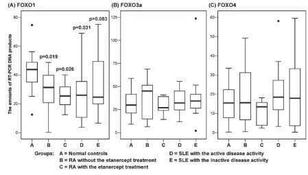
script levels, SLE patients were divided into active SLE and inactive SLE groups and RA patients into RA with etanercept and RA without etanercept groups in the statistical comparisons. From the statisti-cal comparison for the FOXO1 transcript levels, we found that RA patients with or without etanercept treatment and SLE patients with the active disease activity were significantly different from controls (all Pvalues less than 0.05), that the sig-nificance for the difference between nor-mal controls and SLE patients with the inactive disease activity was marginal (P=0.083), and that there was no signifi-cant difference between different disease groups. It appears that RA patients and SLE patients with active disease have de-creased FOXO1 transcript levels in PBMCs, and even SLE patients with the inactive disease also tend to have this phenomenon (Table 2, Figure 2). On the other hand, when different pairs of study groups were statistically analyzed for the difference in the FOXO3a and FOXO4
transcript levels, no significant differ-ences were found. These data indicate that FOXO1 transcripts in PBMCs are down-regulated in SLE and RA patients, and that treatment with the TNF-α-blocking agent appears to have no affect
on the FOXO transcript levels in RA pa-tients. In addition to the transcript levels, the protein levels of FOXO1 in PBMCs of SLE and RA patients were also found to be down-regulated, as demonstrated by Western blot analysis (Figure 3). Figure 2.The quantities of FOXO1, FOXO3a, and FOXO4 transcripts in PBMCs of normal controls and SLE and RA patients were deter-mined by RT-PCR and the subsequent quantification of gene-specific RT-PCR DNA products. The FOXO transcript levels were normalized according to the GAPDH transcript levels. The distributions (boxplots with medians and quartiles) of the quantities of FOXO1, FOXO3a, and FOXO4 transcripts for various study groups are presented. P values of the statistical comparisons for the FOXO1 transcript levels are indicated. P values of the statistical comparisons for the FOXO3a and FOXO4 transcript levels are all more than 0.5 (data not shown).
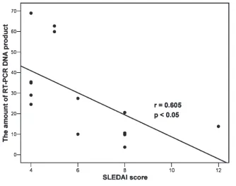
Inverse Correlation of FOXO1 Transcript Levels with SLE Disease Activity in PBMCs
We then determined whether the FOXO1 transcript levels in PBMCs are correlated with the lupus disease activity in SLE patients with active disease. Therefore, the quantities of FOXO1 tran-scripts in PBMCs determined by quanti-fying DNA products from the RT-PCR amplification of FOXO1 transcripts for SLE patients with active disease were used in the simple linear regression anal-ysis to test their correlation with SLEDAI scores. As shown in Figure 4, quantities of FOXO1 transcripts were found to be inversely correlated with SLEDAI scores, suggesting a negative correlation of FOXO1 transcript levels in with lupus disease activity in PBMCs from SLE pa-tients. We also found that the FOXO3a and FOXO4 transcript levels were not significantly correlated with SLEDAI scores (data not shown).
DISCUSSION
The FOXO forkhead factors appear to critically regulate lymphocyte activation. In this study, we evaluated the transcript profiles of FOXO1, FOXO3a, and FOXO4 genes in PBMCs from healthy individuals as well as in PBMCs from SLE and RA pa-tients. FOXO1 and FOXO3a seem to be the dominant FOXO members expressed at the mRNA level in PBMCs of healthy individuals, and FOXO1 transcripts are slightly more abundant than FOXO3a transcripts. Similar transcript patterns were also found in PBMCs of SLE and RA patients, albeit with increased interindi-vidual variation. These results are some-what discordant with the finding in mice that FOXO3a is the dominant FOXO member expressed in spleen (30,31), and the discrepancy may be due to the differ-ence in tissues used to assay FOXO tran-scripts. One the other hand, the finding that FOXO4 is less abundant than FOXO1 and FOXO3a seems to parallel the finding in mice, suggesting that FOXO4 plays a relatively minor role in mediating FOXO activity in human immune cells, as ex-trapolated by the observation of normal
phenotype in FOXO4-deficient mice (21). However, further study on the transcript and protein levels of each FOXO member in different immune cell subpopulations may be needed to elucidate the relative roles of each FOXO protein in controlling human immune functions, although it has been reported that FOXO1 is the domi-nant FOXO member, with very low levels of FOXO3 and FOXO4 occurring in human peripheral blood T cells (16).
The important role of FOXOs in con-trolling lymphocyte activation has prompted us to hypothesize that the FOXO gene expression is dysregulated in SLE and RA patients. In this study, we examined this hypothesis by comparing FOXO1, FOXO3a, and FOXO4 transcript levels in PBMCs from healthy individu-als with those in PBMCs from SLE and RA patients, and the results showed that that the transcript levels of FOXO1, but not FOXO3a or FOXO4, in SLE and RA patients are significantly different from those in healthy individuals. The FOXO1
simply analyzed the FOXOs transcript levels in PBMCs in this study, and there-fore it remains to be seen whether the ex-pression of FOXOs in different immune cell populations is dysregulated in SLE and RA patients, as shown in lupus-prone mice, in which FOXO activity is down-regulated in helper T cells (24).
The mechanism that mediates the down-regulation of FOXO1 transcript levels in PBMCs of SLE and RA patients remains to be clarified. However, because it has recently been shown in mouse B cells that BCR signaling down-regulates FOXO1 transcriptional expression (32), it is possible that FOXO1 transcripts are down-regulated in lymphocytes after ac-tivation and therefore are found to be de-creased in PBMCs of RA and SLE pa-tients with enhanced lymphocyte activation. In addition, although TNF-α has been reported to activate the FOXO1 activity in human fibroblasts (33), our re-sults suggest that the transcript levels of FOXO1 as well as FOXO3a and FOXO4 in PBMCs are not influenced by TNF-αin RA patients. The lack of the reversal of FOXO1 transcript down-regulation in RA patients receiving anti-TNF-αtherapy also suggests that the down-regulation of the FOXO1 transcript levels is not likely to be related to joint inflammation.
Taken together, our results provide the first evidence that FOXO1 gene expres-sion in PBMCs is down-regulated in SLE and RA patients, suggesting that the reg-ulation of FOXO1 at the transcript level may be linked to the pathogenesis of SLE and RA. Thus FOXO1 transcript levels in PBMCs may serve as a biomarker of lupus disease activity in SLE patients. Further investigation into the expression patterns of FOXO factors in different im-mune cell subpopulations may improve our insight into the role of different FOXO members in the pathogenesis of SLE and RA.
ACKNOWLEDGMENTS
We thank Yanfeng Lu for review of the manuscript. This project is supported by a grant from the National Science Coun-cil of Taiwan (Grant# 94-2314-B-281-003).
REFERENCES
1. Carlsson P, Mahlapuu M. (2002) Forkhead tran-scription factors: key players in development and metabolism. Dev. Biol.250:1-23. 2. Kaestner KH, Knöchel W, Martínez DE. (2000)
Unified nomenclature for the winged helix/fork-head transcription factors. Genes Dev.14:142-6. 3. Burgering BM, Kops GJ. (2002) Cell cycle and
death control: long live Forkheads. Trends Biochem. Sci.27:352-60.
4. Fontenot JD, Gavin MA, Rudensky AY. (2003) Foxp3 programs the development and function of CD4+CD25+regulatory T cells. Nat. Immunol. 4:330-6.
5. Nehls M, Pfeifer D, Schorpp M, Hedrich H, Boehm T. (1994) New member of the winged-helix protein family disrupted in mouse and rat nude mutations. Nature372:103-7.
6. Accili D, Arden KC. (2004) FoxOs at the cross-roads of cellular metabolism, differentiation, and transformation. Cell117:421-6.
7. Huang H, Tindall DJ. (2006) FOXO factors: a matter of life and death.Future Oncol.2:83-9. 8. Biggs WH 3rd, Meisenhelder J, Hunter T,
Cave-nee WK, Arden KC. (1999) Protein kinase B/Akt-mediated phosphorylation promotes nuclear ex-clusion of the winged helix transcription factor FKHR1. Proc. Natl. Acad. Sci. USA96:7421-6. 9. Takaishi H, Konishi H, Matsuzaki H, et al. (1999)
Regulation of nuclear translocation of Forkhead transcription factor AFX by protein kinase B.
Proc. Natl. Acad. Sci. USA96:11836-41. 10. Van der Heide LP, Hoekman MF, Smidt MP.
(2004) The ins and outs of FoxO shuttling: mech-anisms of FoxO translocation and transcriptional regulation. Biochem. J.380:297-309.
11. Hu MC, Lee DF, Xia W, et al. (2004) IκB kinase promotes tumorigenesis through inhibition of forkhead FOXO3a. Cell117:225-37.
12. Coffer PJ, Burgering BM. (2004) Forkhead-box transcription factors and their role in the im-mune system. Nat. Rev. Immunol.4:889-99. 13. Brunet A, Bonni A, Zigmond MJ, et al. (1999) Akt
promotes cell survival by phosphorylating and inhibiting a Forkhead transcription factor. Cell
96:857-68.
14. Medema RH, Kops GJ, Bos JL, Burgering BM. (2000) AFX-like Forkhead transcription factors mediate cell-cycle regulation by Ras and PKB through p27kip1. Nature404:782-7.
15. Dijkers PF, Medema RH, Lammers JW, Koender-man L, Coffer PJ. (2000) Expression of the pro-apoptotic Bcl-2 family member Bim is regulated by the forkhead transcription factor FKHR-L1.
Curr. Biol.10:1201-4.
16. Stahl M, Dijkers PF, Kops GJ, et al. (2002) The forkhead transcription factor FoxO regulates transcription of p27Kip1and Bim in response to IL-2. J. Immunol.168:5024-31.
17. Yusuf I, Zhu X, Kharas MG, Chen J, Fruman DA. (2004) Optimal B-cell proliferation requires phos-phoinositide 3-kinase-dependent inactivation of FOXO transcription factors. Blood104:784-7. 18. Fabre S, Lang V, Harriague J, et al. (2005) Stable
activation of phosphatidylinositol 3-kinase in the T cell immunological synapse stimulates Akt
sig-naling to FoxO1 nuclear exclusion and cell growth control. J. Immunol.174:4161-71. 19. Chen J, Yusuf I, Andersen HM, Fruman DA.
(2006) FOXO transcription factors cooperate with δEF1 to activate growth suppressive genes in B lymphocytes. J. Immunol.176:2711-21. 20. Omori SA, Cato MH, Anzelon-Mills A, et al.
(2006) Regulation of class-switch recombination and plasma cell differentiation by phosphatidyli-nositol 3-kinase signaling. Immunity25:545-57. 21. Hosaka T, Biggs WH 3rd, Tieu D, et al. (2004)
Disruption of forkhead transcription factor (FOXO) family members in mice reveals their functional diversification. Proc. Natl. Acad. Sci. USA101:2975-80.
22. Furuyama T, Kitayama K, Shimoda Y, et al. (2004) Abnormal angiogenesis in Foxo1 (Fkhr)-deficient mice. J. Biol. Chem.279:34741-9. 23. Leenders H, Whiffield S, Benoist C, Mathis D.
(2000) Role of the forkhead transcription family member, FKHR, in thymocyte differentiation.
Eur. J. Immunol.30:2980-90.
24. Lin L, Hron JD, Peng SL. (2004) Regulation of NF-κB, Th activation, and autoinflammation by the forkhead transcription factor Foxo3a. Immu-nity21:203-13.
25. Birkenkamp KU, Coffer PJ. (2003) FOXO tran-scription factors as regulators of immune homeo-stasis: molecules to die for? J. Immunol.171:1623-9. 26. Jonsson H, Peng SL. (2005) Forkhead
transcrip-tion factors in immunology. Cell. Mol. Life Sci.
62:397-409.
27. Tan EM, Cohen AS, Fries JF, et al. (1982) The 1982 revised criteria for the classification of systemic lupus erythematosus. Arthritis Rheum. 25:1271-7. 28. Arnett FC, Edworthy SM, Bloch DA, et al. (1988) The American Rheumatism Association 1987 re-vised criteria for the classification of rheumatoid arthritis. Arthritis Rheum.31:315-24.
29. Bombardier C, Gladman D, Urowitz M, Caron D, Chang C. (1992) Derivation of the SLEDAI. A dis-ease activity index for lupus patients. The Com-mittee on Prognosis Studies in SLE. Arthritis Rheum.35:630-40.
30. Furuyama T, Nakazawa T, Nakano I, Mori N. (2000) Identification of the differential distribu-tion patterns of mRNAs and consensus binding sequences for mouse DAF-16 homologues.
Biochem. J.349:629-34.
31. Biggs WH 3rd, Cavenee WK, Arden KC. (2001) Identification and characterization of members of the FKHR (FOX O) subclass of winged-helix transcription factors in the mouse. Mamm. Ge-nome12:416-25.
32. Hinman RM, Bushanam JN, Nichols WA, Sat-terthwaite AB. (2007) B cell receptor signaling down-regulates forkhead box transcription factor class O 1 mRNA expression via phosphatidyli-nositol 3-kinase and Bruton’s tyrosine kinase.
J. Immunol.178:740-7.