R E S E A R C H
Open Access
Endothelial progenitor cells and neural
progenitor cells synergistically protect
cerebral endothelial cells from Hypoxia/
reoxygenation-induced injury via activating
the PI3K/Akt pathway
Jinju Wang
1†, Yusen Chen
2†, Yi Yang
3†, Xiang Xiao
1, Shuzhen Chen
1, Cheng Zhang
1, Bradley Jacobs
4, Bin Zhao
2,
Ji Bihl
1*and Yanfang Chen
1,2,4,5*Abstract
Background:Protection of cerebral endothelial cells (ECs) from hypoxia/reoxygenation (H/R)-induced injury is an important strategy for treating ischemic stroke. In this study, we investigated whether co-culture with endothelial progenitor cells (EPCs) and neural progenitor cells (NPCs) synergistically protects cerebral ECs against H/R injury and the underlying mechanism.
Results:EPCs and NPCs were respectively generated from inducible pluripotent stem cells. Human brain ECs were used to produce an in vitro H/R-injury model. Data showed: 1) Co-culture with EPCs and NPCs synergistically inhibited H/R-induced reactive oxygen species (ROS) over-production, apoptosis, and improved the angiogenic and barrier functions (tube formation and permeability) in H/R-injured ECs. 2) Co-culture with NPCs up-regulated the expression of vascular endothelial growth factor receptor 2 (VEGFR2). 3) Co-culture with EPCs and NPCs
complementarily increased vascular endothelial growth factor (VEGF) and brain-derived neurotrophic factor (BDNF) levels in conditioned medium, and synergistically up-regulated the expression of p-Akt/Akt and p-Flk1/VEGFR2 in H/ R-injured ECs. 4) Those effects could be decreased or abolished by inhibition of both VEGFR2 and tyrosine kinase B (TrkB) or phosphatidylinositol-3-kinase (PI3K).
Conclusions:Our data demonstrate that EPCs and NPCs synergistically protect cerebral ECs from H/R-injury, via activating the PI3K/Akt pathway which mainly depends on VEGF and BDNF paracrine.
Keywords:EPCs, NPCs, Cerebral ECs, Co-culture, PI3K/Akt signal pathway, Hypoxia-reoxygenation injury, VEGFR2, VEGF, BDNF
Background
Brain endothelial cells (ECs) are critical components of the blood brain barrier (BBB). Increased BBB permeabil-ity leads to the development of tissue swelling, inflam-matory cell infiltration and subsequently exaggerate
injury in ischemic stroke [1]. Therefore, protection of ECs and BBB function should be an important strategy for reducing ischemic injury. On the other hand, endo-thelial progenitor cells (EPCs) have been suggested to participate in EC protection, repair and angiogenesis [2]. Transplantation of EPCs is a promising cell therapy for ischemic diseases such as acute myocardial infarction and stroke [3–5]. Our previous studies have shown that EPC infusion promotes angiogenesis in mouse ischemic stroke models [5, 6]. EPCs released angiogenic growth factors, such as vascular endothelial growth factor * Correspondence:ji.bihl@wright.edu;yanfang.chen@wright.edu
†Equal contributors
1Department of Pharmacology & Toxicology, Boonshoft School of Medicine,
Wright State University, 3640 Colonel Glenn Hwy, Dayton, OH 45435, USA 2Department of Neurology, Affiliated Hospital of Guangdong Medical
College, Zhanjiang 524001Guangdong, China
Full list of author information is available at the end of the article
© 2016 Wang et al.Open AccessThis article is distributed under the terms of the Creative Commons Attribution 4.0 International License (http://creativecommons.org/licenses/by/4.0/), which permits unrestricted use, distribution, and reproduction in any medium, provided you give appropriate credit to the original author(s) and the source, provide a link to the Creative Commons license, and indicate if changes were made. The Creative Commons Public Domain Dedication waiver (http://creativecommons.org/publicdomain/zero/1.0/) applies to the data made available in this article, unless otherwise stated. Wanget al. Molecular Brain (2016) 9:12
(VEGF) and insulin-like growth factor, could be responsible for the beneficial effect of EPC conditioned medium on the viability of H2O2-compromised human umbilical vein ECs [7, 8]. Currently, we do not know whether EPCs can protect cerebral ECs against hypoxia/reoxygenation (H/R)-injury.
Transplantation of neural progenitor cells (NPCs) has also been shown to be effective for treating ische-mic stroke in animal models [9, 10]. In addition to generating neurons, grafted NPCs could promote angiogenesis in a rodent stroke model [11]. A recent report suggests that co-culture with NPCs decreases the passive permeability of brain ECs [12]. Collect-ively, these studies indicate a crosstalk between NPCs and ECs. However, it is unclear whether NPCs and EPCs have synergistic effects on EC protection.
The PI3K/Akt signal pathway participates in various cellular processes such as cell survival and prolifera-tion [13]. Previous studies have shown that activaprolifera-tion of the PI3K/Akt signal pathway promotes neuron survival [14, 15], cardiac microvascular EC migration [16], and axonal outgrowth compromised by oxygen-glucose deprivation [17, 18]. It is unknown whether this pathway is involved in the mechanism of the benefits of NPCs and EPCs.
The aims of this study were to elucidate whether EPCs and NPCs synergistically protect brain ECs from H/R-induced injury and to explore whether the effects are mediated by the PI3K/Akt signal pathway.
Results
NPCs and EPCs were successfully generated from human inducible pluripotent stem cells
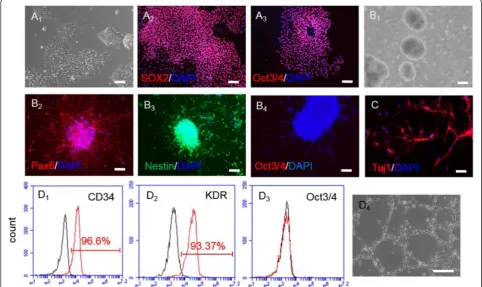
As shown in Fig. 1, the human inducible pluripotent stem cells (iPSCs) grew as colonies staining positively for pluripotent markers, Sox2 and Oct3/4. The generated NPCs grew as neurospheres after 7-day neural induc-tion, and expressed neural progenitor markers pax6 (98 ± 1 %) and nestin (96 ± 1.5 %), but not expressed Oct3/4, indicating a high differentiation efficacy. The generated NPCs had ability of differentiating into neurons, which was evidenced by expressing neuron specific marker Tuj1.
After 7-day EPC induction, approximately 48 ± 2.1 % of cells positively expressed endothelial progeni-tor marker CD34. In order to get a pure population of EPCs, we used CD34-conjugated microbeads to en-rich the generated EPCs. The CD34-conjugated microbeads purified cells positively expressed CD34
Fig. 2EPCs and NPCs promoted the survival of H/R-injured ECs via activating the PI3K pathway. MTT assay and PI/FITC-Annexin V apoptosis assay were conducted on H/R-injured ECs co-cultured with EPCs and/or NPCs for 24 h as described in Material and Methods. A1, representative morph-ology images showing the viability of ECs. A2, summarized data showing EC viability which is synergistically increased when co-cultured with the combination of EPCs and NPCs than that co-cultured with EPCs or NPCs alone. B1, representative flow plots of EC apoptotic rate. B2, summarized data of the apoptotic rate of ECs, showing that the combination of EPCs and NPCs offers better anti-apoptotic effect than EPCs or NPCs alone. Block the PI3K pathway could diminish the beneficial effects of EPCs and/or NPCs. And the PI3K pathway upstream blockers, SU1498 and K252a, reduced these effects of EPCs and NPCs. *p< 0.05, vs. Normoxia; #p< 0.05, vs. vehicle. Data are expressed as mean ± SEM,n= 6/group/measure-ment. LY294002: PI3K inhibitor; SU1498: VEGFR2 inhibitor; K252a: TrkB inhibitor
(96 ± 2.1 %) and KDR (95 ± 1.8 %). As expected, the purified EPCs did not express Oct3/4. In addition, the generated EPCs had tube formation ability as revealed by matrigel assay.
Co-culture with EPCs and NPCs synergistically protected ECs from H/R-induced apoptosis and compromised viability via activating the PI3K pathway
After exposed to the hypoxic condition for 6 h, ECs were co-cultured with EPCs and/or NPCs for 24 h, followed with apoptotic assay or MTT assay. Results (Fig. 2) showed that co-culture with EPCs and NPCs exerted a greater effect on decreasing H/R-injured EC apoptosis than that co-culture with EPCs or NPCs separately did (vehicle vs. EPCs or NPCs, p< 0.05; EPCs and NPCs vs. EPCs or NPCs, p< 0.05). Similarly, the EC viability was also synergistically increased by co-culture with EPCs and NPCs (vehicle vs. EPCs or NPCs,p< 0.05; EPCs and NPCs vs. EPCs or NPCs, p< 0.05). The synergistic ef-fects on reducing EC apoptosis and improving EC viabil-ity were achieved by an increase of approximately 24 and 28 %, respectively.
Moreover, our data showed that the PI3K inhibitor (LY294002) pre-treatment could completely abolish the abovementioned effects of EPCs and/or NPCs, suggest-ing that the beneficial effects of EPCs and NPCs are me-diated by the PI3K pathway. To define the contribution of VEGFR2 and TrkB (PI3K upstream molecules) to these effects, the respective inhibitors SU1498 and K252a were padded in the co-culture system. Our re-sults revealed that blockade of the VEGF/VEGFR2 and BDNF/TrkB signals reduced the effects of EPCs and NPCs.
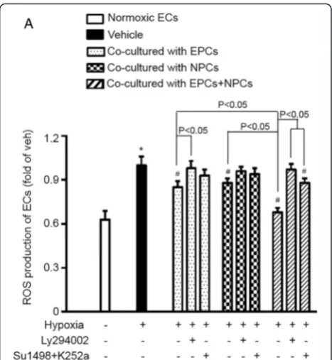
Co-culture with EPCs and NPCs synergistically decreased the oxidative stress of H/R-injured ECs via activating the PI3K pathway
As shown in Fig. 3, ROS production was decreased in H/R-injured ECs co-cultured with EPCs or NPCs (ve-hicle vs. EPCs or NPCs, p< 0.05). Moreover, co-culture with EPCs and NPCs decreased ROS production to a larger extent than that with EPCs or NPCs alone did (EPCs and NPCs vs. EPCs or NPCs,p< 0.05). The syner-gistic effect on decreasing ROS production was obtained by about 18 % increase.
As expected, pre-treatment with PI3K inhibitor, LY294002, abolished the anti-oxidative effect of EPCs and/or NPCs on H/R-injured ECs. Pre-treatment with a combination of the PI3K upstream blockers SU1498 and K252a reduced the most anti-oxidative effects of EPCs and NPCs on H/R-injured ECs. All of these data indicate that the anti-oxidative effect of EPCs and NPCs is mediated by the PI3K signal pathway.
H/R-compromised tube formation ability of ECs was synergistically improved by co-culturing with EPCs and NPCs via activating the PI3K signal pathway
We further assessed whether co-culture with EPCs and/ or NPCs altered the tube formation function of ECs ex-posed to H/R. The results (Fig. 4) showed that EPCs or NPCs alone increased the tube formation ability of H/R-injured ECs (vehicle vs. EPCs or NPCs,p< 0.05). More-over, co-culture with EPCs and NPCs exhibited a syner-gistic effect on improving the tube formation ability of ECs compromised by H/R (EPCs and NPCs vs. EPCs or NPCs,p< 0.05). The synergistic effect of EPC plus NPC co-culture on improving the tube formation ability was increased by approximately 19 %.
In order to elucidate the possible role of the PI3K pathway in the effect of EPCs and/or NPCs on EC tube formation, the pathway specific inhibitor LY294002 was used in the co-culture study. Our re-sults showed that PI3K inhibition entirely abolished this effect of EPCs and NPCs. Similarly, to further explore whether VEGF/VEGFR2 and BDNF/TrkB sig-nals could be responsible to trigger the activation of the PI3K pathway, we pre-added their respective
inhibitors SU1498 and K252a into the co-culture system. As we expected from the data of apoptotic and MTT assays, blockade of the VEGF/VEGFR2 and BDNF/TrkB signals reduced the effect on tube formation.
The endothelial permeability was improved by co-culturing with EPCs and NPCs
Under physiological conditions, the endothelial mem-brane is impermeable to macromolecules (mass weight around 70 k Dalton) [19]. We performed permeability assay to evaluate whether co-culture of EPCs and/or NPCs could improve the barrier function of ECs com-promised by H/R. As expected, H/R injury increased trans-endothelial permeability to FITC-conjugated dex-tran (mass weight around 10 k Dalton). Co-culture of EPCs or NPCs decreased the flux of FITC-dextran, and EPCs combined with NPCs was more effective in de-creasing the FITC-dextran flux through the EC mono-layer (Fig. 5a).
Co-culture with EPCs and NPCs complementarily elevated the levels of VEGF and BDNF in the conditioned medium of ECs exposed to H/R
In order to explore the mechanisms underlying the pro-tective benefits of EPCs and NPCs, we performed ELISA assay to determine the levels of VEGF and BDNF in the culture medium. As shown in Fig. 5b, c, we found that co-cultured with EPCs alone increased the VEGF level, but not the BDNF level in the EC culture medium,
whereas, co-cultured with NPCs alone raised the BDNF level, not the VEGF level. Moreover, co-culture with EPCs and NPCs increased the levels of both VEGF and BDNF in the EC medium, suggesting a complementary effect.
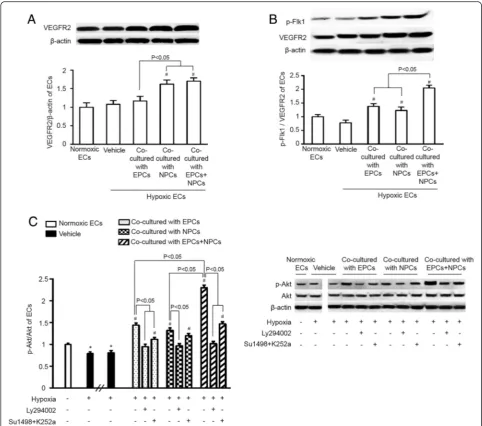
The expression of VEGFR2 was upregulated and ratios of p-Flk1/VEGFR2 and p-Akt/Akt were increased in H/R-in-jured ECs co-cultured with EPCs and NPCs
As shown in Fig. 6a, co-culture with NPCs alone or with EPCs and NPCs similarly increased the expression level of VEGFR2 in H/R-injured ECs, whereas, co-cultured with EPCs alone did not significantly change the expres-sion of VEGFR2 in ECs, indicating that interaction of NPCs with ECs.
Western blot results demonstrated that the expression ratios of p-Flk1/VEGFR2 and p-Akt/Akt in H/R-injured ECs were increased by co-culture with EPCs or NPCs alone, with a greater increase when co-cultured with both EPCs and NPCs (Fig. 6b, c). Data showed that the net increase of the synergistic effect on up-regulating the expression ratio of p-Akt/Akt was approximately 30 %. As expected, the PI3K inhibitor LY294002 abol-ished the phosphorylation of Akt, suggesting that the PI3K/Akt signal pathway is activated in ECs co-cultured with EPCs and NPCs. A combination of SU1498 and K252a decreased the phosphorylation of Akt, reflecting that it at least partially depends on the upstream mole-cules VEGFR2 and TrkB (Fig. 6b).
Fig. 4EPCs and NPCs improved the angiogenic function of H/R-injured ECs via activating the PI3K pathway.a, representative plots of tube formation. Scale bar: 200μm.b, summarized data of EC tube formation, showing that EPC and NPC co-culture offers synergistically effects on improving EC function compared to EPCs or NPCs alone. And such synergistic effect could be blocked by LY294002, or partially abolished by the combination of SU1498 and K252a. *p< 0.05, vs. Normoxia; #p< 0.05, vs. Vehicle. Data are expressed as mean ± SEM,n= 6/group/measurement. LY294002: PI3K inhibitor; SU1498: VEGFR2 inhibitor; K252a: TrkB inhibitor
Discussion
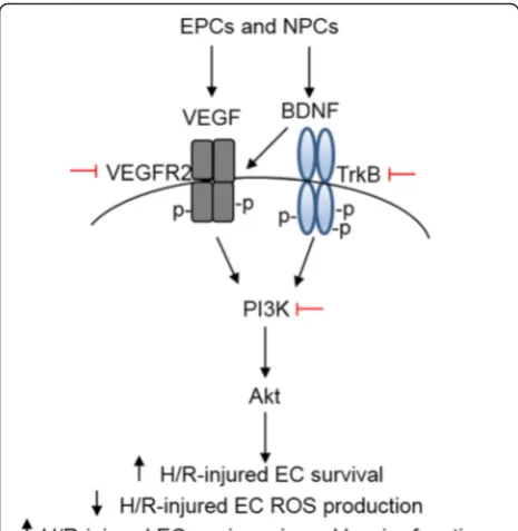
In the present study, we showed that EPCs and NPCs produced from human iPSCs had synergistic beneficial effects on H/R-injured brain ECs. The major findings in-clude: i) Co-culture with EPCs and NPCs synergistically protected ECs from H/R-induced apoptosis and dysfunc-tion; ii) The levels of VEGF and BDNF in the medium of ECs co-cultured with EPCs and NPCs were increased; iii) Co-culture with NPCs up-regulated VEGFR2 expres-sion and its phosphorylation on ECs; iv) Blockade of the VEGFR2 and Trkb or PI3K/Akt pathway inhibited or abolished the protective effects of EPCs and NPCs (Fig. 7).
ECs are unique and critical in maintaining normal BBB function [20]. Impairment of BBB occurs in the early stage of ischemic brain injury, leading to subse-quent brain swelling and inflammatory responses [21]. Thus, protecting brain ECs from H/R-induced injury will theoretically alleviate brain tissue damage in ischemic stroke. Nevertheless, there is no clinically effective strat-egy to protect ECs against H/R-induced injury in acute ischemic stroke. Transplantation of stem cells has been shown to accelerate the functional recovery of ischemic stroke by promoting angiogenesis and neurogenesis [22].
Indeed, others and our studies have demonstrated that engrafted EPCs or NPCs can alleviate acute ischemic in-jury and promote angiogenesis and neurogenesis in an ischemic stroke mouse model [5, 6, 9, 10]. However, it is unknown whether there are synergistic effects if EPCs and NPCs are combined to treat ischemic-reperfusion stroke.
In the present study, we examined the effects of EPCs and NPCs on H/R-injured brain ECs in vitro. It is well known that iPSCs have unlimited self-renewal ability and are able to differentiate to various types of cells with less ethical issues for clinical applications [23, 24]. We successfully differentiated human iPSCs into EPCs and NPCs. To mimic the status of ECs in acute ischemic stroke, we produced an in vitro model of brain EC H/R injury, characterized with decreased viability, increased apoptosis and cellular permeability, increased ROS pro-duction, as well as compromised tube formation ability [25]. By using this model, we found that co-culture with EPCs or NPCs alone had beneficial effects on protecting ECs from H/R-induced injury, including increase in apoptosis, ROS production and intercellular permeabil-ity, and decrease in viability and capillary formation. Moreover, co-culture with both EPCs and NPCs
achieved synergistic effects on those measurements by 18–28 % increase.
Numerous studies have shown that VEGF is released from EPCs and ECs, and that BDNF is released from NPCs, which are respectively responsible for the benefi-cial effects of EPCs and NPCs [26, 27]. In order to deter-mine whether EPC-derived VEGF and NPC-derived BDNF are the major factors involved in the observed ef-fects of EPCs and NPCs in this study, we have analyzed the levels of VEGF and BDNF in the culture medium of ECs. Our results showed that EPC co-culture increased VEGF, but not BDNF level in the EC medium, whereas,
NPC co-culture increased BDNF, but not VEGF level in the EC medium. More importantly, the data revealed that EPCs and NPCs complementarily increased the VEGF and BDNF levels in the co-culture medium of ECs. In the present study, we did not study the dose-dependent effects of VEGF and BDNF on ECs and not compare if co-application of VEGF and BDNF is more significant than simply increasing the dose of VEGF or BDNF alone. However, our results revealed that the combination of EPCs and NPCs have synergistic effects on ECs. For exploration of the underlying mechanism, we analyzed the expression of VEGFR2 and its
Fig. 6Co-culture with EPCs and NPCs activated the PI3K/Akt signal pathway on H/R-injured ECs.a, VEGFR2 expression was significantly
upregulated in H/R-injured ECs co-cultured with NPCs or the combination of EPCs and NPCs.bthe protein expression ratio of p-Flk1/VEGFR2 was significantly increased in H/R-injured ECs co-cultured with EPCs or NPCs, with a higher ratio in ECs co-cultured with the combination of EPCs and NPCs.cthe protein expression ratio of p-Akt/Akt was increased in H/R-injured ECs co-cultured with EPCs and NPCs, and this effect was blocked or reduced when ECs were pre-treated with PI3K inhibitor LY294002 or VEGFR2 inhibitor SU1498 or TrkB inhibitor K252a. *p< 0.05, vs. Normoxia; #p< 0.05, vs. vehicle. Data are expressed as mean ± SEM,n= 6/group/measurement
phosphorylation. The results showed that co-culture with both EPCs and NPCs synergistically increased the expression of p-Flk1/VEGFR2 in ECs. Of note, we found that co-culture with NPCs, but not EPCs, up-regulated the expression of VEGFR2 on ECs. This is supported by a previous report showing that BDNF increases the on a rat brain EC line [28], and suggests that NPCs can medi-ate the synergistic effects by its secreted BDNF and pro-vides the basis of the synergistic effects observed in the co-culture group combining EPCs and NPCs. Further-more, we examined the role of VEGF/VEGFR2 and BDNF/TrkB signal pathways in the beneficial effects of EPC/NPC co-culture. Our data showed that blockade of both signals largely decreased the abovementioned ef-fects of EPCs and NPCs, suggesting that the beneficial effects of EPCs and NPCs are mainly dependent on the VEGF/VEGFR2 and BDNF/TrkB signals. These data are in consistent with the notion that VEGFR2 and TrkB are the major modulators of endothelial survival [28]. Col-lectively, VEGF and BDNF are the major factors for re-sponsible of the synergistic effects of EPCs and NPCs in
the co-cultures, although there are unidentified factors contributing a minor part.
The PI3K is the downstream pathway molecule of VEGFR2 and TrkB, which mediates various cell activities includes cell survival, cell proliferation [29-32]. There-fore, we conducted experiments for further pathway ana-lysis. We found that both EPCs and NPCs increased the level of p-Akt/Akt in ECs. And there was a synergistic effect on level of p-Akt/Akt when EPCs and NPCs were simultaneously applied. The synergistic effect of EPC plus NPC co-culture was 30 % on up-regulating the tein expression ratio of p-Akt/Akt. Moreover, the pro-tective effects elicited by EPCs and/or NPCs were abolished by blockade of PI3K with LY294002. Taken to-gether, our data indicate that the PI3K pathway, down-stream of VEGFR2 and TrkB, is responsible for the beneficial effects of EPCs and NPCs.
Conclusion
In conclusion, our data demonstrate that EPCs and NPCs can offer synergistic benefits in protecting brain ECs against H/R injury by VEGF and BDNF paracrine-mediated activation of the PI3K/Akt signal pathway. These findings will help us to develop cell-based therapy for ischemic stroke.
Methods
Human iPSCs culture
The vector-free viral-free human iPSCs cell line (iPS-DF-19-9/7 T) was purchased from Wicell Research Insti-tute (USA). The iPSCs were cultured with mTeSR1 medium (Stem cell technology) on matrigel-coated plates (BD Bioscience), and expanded every 4–5 days ac-cording to the manufacturer’s protocol. Passage of 28– 44 of human iPSCs were used for NPC or EPC induc-tion. All experimental procedures were approved by Wright State University Institutional Biosafety Commit-tee and were in accordance with the approved guidelines.
Generation of NPCs and EPCs from human iPSCs
EPCs and NPCs were produced from human iPSCs ac-cording to previous reports with slight modifications [14, 15]. In brief, iPSC colonies were detached with dispase (2 mg/ml in DMEM/F12; Stem cells), pooled together and cultured to form embryoid bodies (EBs) in EB medium (DMEM/F12+ 20 % knock out serum + 1 % nonessential amino acid + 0.1 mM 2-mercapethonal + 1 % penicillin-streptomycin solution) in low-attachment dishes (Corn-ing) for 5–7 days. The DMEM medium and all supple-ment reagents were purchased from Gibco. The EBs were used for NPC and EPC generation.
For NPC generation, EBs were cultured in neural medium (NM: Neurobasal medium A + B27 minus
Vitamin A (1x) + N2 (1x) + FGF (20 ng/ml) + EGF (20 ng/ml) + 1 % penicillin-streptomycin solution) in low-attachment dishes for 7–9 days. The neurobasal mediumA and all supplement reagents were purchased from Gibco. The generated NPCs were propagated in free-floating aggregates (neurospheres), and used for an in vitro differentiation assay to investigate neural differ-entiation ability [33]. The generated NPCs and neurons were confirmed by immunofluorescence analysis [34].
For EPC generation, EBs were cultured on gelatin (0.1 %) coated plates with EPC medium (EBM-2 medium + growth factor mixture + 5 % FBS + VEGFA (50 ng/ml) + FGF (25 ng/ml) for 7–9 days [14]. The EBM-2 medium and all supplement reagents were pur-chased from Gibco. The generated EPCs were purified by magnetic activated cell sorting (MACS) with CD34-microbeads according to the manufacture’s protocol (Miltenyi Biotec), and assessed by flow cytometry and matrigel assay.
Characterization of generated NPCs
For immunofluorescence analysis, generated NPCs were fixed, permeabilized and blocked with blocking buffer (PBS with 1 % BSA and 1 % Triton-100), then incubated with neural progenitor specific markers nestin (1:100; Pierce), pax6 (1:50; Pierce) and pluripotent specific marker Oct3/4 (1:200; Abcam). Then the cells were in-cubated with Cy-3 or Alexa fluo 488-conjugated second-ary antibodies (1:150; Jackson ImmunoResearch) for 2 h at room temperature. DAPI was used to counterstain nuclei.
In order to determine the neuron differentiation cap-ability of the produced NPCs, the generated NPCs were cultured in neuron differentiation medium (NPBM medium + BDNF (20 ng/ml) + FGF (20 ng/ml) + EGF (20 ng/ml) + 1 % penicillin-streptomycin solution) for 3 weeks. The NPBM medium and all supplement re-agents were purchased from Gibco. The differentiated cells were permeabilized, incubated with neuron specific markerβ-tubulin (1:100; Pierce) and followed by incuba-tion with Cy3-conjugated secondary antibody (1:150; Jackson ImmunoResearch). DAPI were used to counter-stain nuclei. All images were taken under an inverted fluorescence microscope (EVOS, Life Technologies).
Purification and characterization of generated EPCs
In order to exclude the contamination of human iPSCs, the generated EPCs were purified by using MACS ac-cording to the manufacture’s instruction. In brief, the differentiated cells (107cells) were incubated with 10μl CD34-conjugated microbeads (Miltenyi Biotec Inc) in the refrigerator for 20 min, followed by wash in PBS for twice. The beads-binding cells were separated using an autoMACS separator (Miltenyi Biotec Inc). All CD34+
cells were resuspended with EPC culture medium and cultured in a regular humidified incubator.
For flow cytometry analysis, the purified EPCs were in-cubated with FITC-conjugated CD34, FITC-conjugated KDR, FITC-conjugated Oct3/4 or FITC-conjugated IgG for 30 min (5μl, eBioscience) in the dark. All antibodies were purchased from eBioscience. After incubation, all samples were analyzed under flow cytometry (Accuri C6 flow cytometer). 10,000 events were collected for data analysis.
H/R-injury model of brain ECs
Cerebral EC cell line was purchased from Cell Systems (Kirkland, WA) and cultured according to the manufac-turer’s protocol. Passages 4–13 of ECs were used for ex-periments in this study. The H/R-injury model of ECs was produced as previously described [25]. Briefly, ECs were changed with fresh culture medium and cultured for 6 h in a hypoxic chamber (1 % O2, 5 % CO2, and 94 % N2; Biospherixhypoxia chamber), then reoxyge-nated by incubation in a standard 5 % CO2incubator for 24 h. During the reoxygenation period, ECs were co-cultured with EPCs and/or NPCs as described below.
Co-culture brain ECs with EPCs and/or NPCs
The co-culture system was set up according to a previ-ous report with minor modifications [35]. In brief, the day before co-culture, NPCs (4 × 10⁵), or EPCs (4 × 10⁵), or NPCs (2 × 10⁵) and EPCs (2 × 10⁵) were plated into transwell membrane inserts (pore size, 0.4 μm; polycar-bonate membrane, Greiner Bio-One, Germany) in NPC and/or EPC culture medium for overnight. [36] During the reoxygenation period, brain ECs (4 × 10⁵) subjected to hypoxic (1 % O2) were co-cultured with EPCs and/or NPCs. For signal pathway study, LY294002 (PI3K inhibi-tor; 20μM, Cayman Chemical), SU1498 (VEGFR inhibi-tor; 5 μM, BioVision), or k252a (TrkB inhibitor; 10 μg/ ml, BioVision) was added to EC culture medium 2 h prior to co-culture experiments [25, 36–38] and pre-sented in the EC culture during the co-culture period. All inhibitors were dissolved with DMSO (Sigma) and diluted with culture medium to yield desired concentra-tions. ECs cultured in normoxia (5 % CO2, 37 °C) were used as a control. ECs in the vehicle group were cul-tured with EC culture medium only.
Cell viability, apoptosis and ROS production analyses of ECs
The viability of H/R-injured ECs was measured by using a methyl thiazolyl tetrazolium (MTT) kit (Invitrogen) as we previously described with slight modification [25]. Briefly, after 24 h co-culture with EPCs and/or NPCs, the ECs culture medium was replaced with 1 ml of fresh culture medium with 100 μl of 12 mM MTT solution
and incubated at 37 °C for 2 h. Then removed 850μl of medium from the wells and added 500 μl of DMSO to each well, mixed thoroughly with the pipette and incu-bated at 37 °C for 10 min. Finally, transferred 100μl of mixed solution from each well to a 96-well plate. The plate was read by a reader (Bio-teck) at 570 nm. The percent of cell viability was defined as the relative ab-sorbance of cells in co-culture groups versus control cells. At least six wells per experiment were used in each group.
The apoptosis assay of ECs was conducted using FITC-Annexin V apoptosis detection kit (BD Bioscience) as we previously described [25]. In brief, after 24 h co-culture with EPCs and/or NPCs, the ECs co-culture medium was removed and rinsed twice with PBS, then de-attached with 0.25 % trypsin/EDTA, centrifuged, re-suspended with 100μl 1x Annexin V-binding buffer and incubated with 5 μl FITC-conjugated Annexin V and 5μl propidium iodide (PI) in the dark for 15 min at RT. The stained ECs were analyzed by flow cytometry (Accuri C6 flow cytometer). The apoptotic cells were de-fined as Annexin V+/PI−cells. The experiment was re-peated six times.
The intracellular ROS production in ECs was deter-mined by dihydroethidium (DHE, Sigma) [25]. Briefly, after EPC and/or NPC co-culture, the EC culture medium was replaced with fresh cultured medium con-taining the DHE working solution (2 μM) at 37 °C for 2 h. Then the cells were detached with trypsin and were analyzed by flow cytometer (Accuri C6). The experiment was repeated six times.
ECs cultured in normoxia served as a control. ECs in the vehicle group were cultured with EC culture medium only. The synergistic effect of EPC plus NPC co-culture (Es) on ECs was calculated by using the formula: Es = (EEPC + NPC− EEPC− ENPC) / (EEPC +ENPC) x 100 %. EEPC represents the effect elicited by EPC co-culture. ENPC represents the effect elicited by NPC co-culture. EEPC+NPCrepresents the effect elicited by EPC and NPC co-culture.
Tube Formation and endothelial permeability assays of ECs
The tube formation ability of ECs was evaluated by using a tube formation assay kit (Chemicon) with slight modification [25]. After co-cultured with EPCs and/or NPCs, the ECs were trypsinized and reseed at a density of 5 × 10³–1 × 10⁴ onto the surface of the polymerized ECMatrix™, and incubated at 37 °C in a CO2 incubator for 12–16 h. The tube formation was inspected under an inverted light microscope (EVOS). Five independent fields were assessed for each well, and the average number of tubes per field (magnifica-tion, 200x) was determined.
Change in macromolecular permeability of brain ECs was studied by using cell culture transwell insert method [39]. In brief, brain ECs were seeded at a density of 1 × 10⁵cells/well onto a 24-well transwell insert (pore size, 0.4 μm; polycarbonate membrane, Greiner Bio-One, Germany). The EPCs or NPCs (2 × 10⁴cells/well) were plated on the lower chamber of the transwell insert sys-tem. Then the confluent ECs were subjected to hypoxic culture. During the reoxygenation period, brain ECs were co-cultured with the EPCs and/or NPCs for 24 h. FITC-conjugated dextran (1 mg/ml; 10 k Dalton, Sigma) was added to the upper compartment 90 min before the end of the experiment. The relative fluorescence passed through the polycarbonate membrane into the lower chamber was determined by using a fluorescent plate reader (Synergy, Bio-Tek, Vermont). Relative fluorescent changes to normoxia were presented. ECs in the vehicle group were cultured with EC culture medium only. Like-wise, Es was calculated as: Es = (EEPC + NPC− EEPC
−ENPC) / (EEPC +ENPC) x 100 %.
Western blot analysis
H/R-injured ECs were harvested after co-cultured with EPCs and/or NPCs. Proteins were extracted with cell lysis buffer (Thermo scientific) supplemented with complete mini protease inhibitor tablet (Roche). Pro-tein lysates were electrophoresed through SDS-PAGE gels and transferred onto PVDF membranes. The membranes were blocked with 5 % non-fat milk for 1 h and incubated with primary antibodies against Akt (1:1000; Cell Signaling), p-AKt (1:1000; Cell Sig-naling), VEGFR2 (1:1000; Cell Signaling), p-Flk1 (1:200; Santa Cruz), and β-actin (1:4000; Sigma) at 4 ° C for overnight. After washing, membranes were in-cubated with horseradish-peroxidase-conjugated IgG (Jackson Immuno Research Lab) for 1 h at RT. Blots were developed with enhanced chemiluminescence de-veloping solutions and quantified under ImageJ soft-ware. For detecting the protein expressions of Akt and p-Akt in all groups, two sets of gels were done. One set of gels was used to compare the difference between normoxia and hypoxia groups, and the other set of gels was used to compare the differences among different treatment groups in the Hypoxia groups. All experiments were repeated at least six times. Similarly, Es was calculated as: Es = (EEPC + NPC
− EEPC− ENPC) / (EEPC +ENPC) x 100 %.
ELISA assay of VEGF and BDNF
groups was also collected. The levels of trophic factors VEGF and BDNF in the culture medium were deter-mined with ELISA kits (R&D systems) by following the manufacturer’s instructions. ECs cultured in normoxia served as a control. ECs in the vehicle group were cul-tured with EC culture medium only.
Competing interests
The authors declare that they have no competing interests.
Authors’contributions
JW, YY, SC, JB, CZ performed experiments; JW, YY, XM, YY, BJ, SC, YC wrote the manuscript; JW, YY, XM, SC, YY, JB, YC, BJ, BZ, YC contributed to manuscript preparation; all authors discussed the results, analyzed data and commented on the manuscript; JW, YY, XM, BZ, BJ and YC developed the concepts and designed the study. All authors read and approved the final manuscript.
Acknowledgements
We thank Dr. Cathy Graham at Dayton VA Medical Center for her careful reading proof. This work was supported by National Heart, Lung, and Blood Institute (HL-098637, Y.C.), American Heart Association (15PRE25700198, J.W.), and National Nature Science Foundation of China (81300079, J.B.).
Author details
1Department of Pharmacology & Toxicology, Boonshoft School of Medicine,
Wright State University, 3640 Colonel Glenn Hwy, Dayton, OH 45435, USA. 2Department of Neurology, Affiliated Hospital of Guangdong Medical
College, Zhanjiang 524001Guangdong, China.3Wuhan Institute of Physical Education, College of Health Science, Wuhan 430079Hubei, China. 4
Department of Neurology, Wright State University, 3640 Colonel Glenn Hwy, Dayton 45435Ohio, USA.5Department of Internal Medicine, Wright State University, 3640 Colonel Glenn Hwy, Dayton 45435Ohio, USA.
Received: 28 August 2015 Accepted: 28 January 2016
References
1. Yeh WL, Lu DY, Lin CJ, Liou HC, Fu WM. Inhibition of hypoxia-induced increase of blood-brain barrier permeability by YC-1 through the antagonism of HIF-1alpha accumulation and VEGF expression. Mol Pharmacol. 2007;72(2):440–9.
2. Peplow PV. Growth factor- and cytokine-stimulated endothelial progenitor cells in post-ischemic cerebral neovascularization. Neural Regen Res. 2014; 9(15):1425–9.
3. Leeper NJ, Hunter AL, Cooke JP. Stem cell therapy for vascular regeneration: adult, embryonic, and induced pluripotent stem cells. Circulation. 2010; 122(5):517–26.
4. Lee SH, Lee JH, Asahara T, Kim YS, Jeong HC, Ahn Y, et al. Genistein promotes endothelial colony-forming cell (ECFC) bioactivities and cardiac regeneration in myocardial infarction. PLoS One. 2014;9(5), e96155. 5. Chen J, Chen J, Chen S, Zhang C, Zhang L, Xiao X, et al. Transfusion of
CXCR4-primed endothelial progenitor cells reduces cerebral ischemic damage and promotes repair in db/db diabetic mice. PLoS One. 2012;7(11), e50105. 6. Chen J, Xiao X, Chen S, Zhang C, Chen J, Yi D, et al. Angiotensin-converting
enzyme 2 priming enhances the function of endothelial progenitor cells and their therapeutic efficacy. Hypertension. 2012;24.
7. Yang Z, von Ballmoos MW, Faessler D, Voelzmann J, Ortmann J, Diehm N, et al. Paracrine factors secreted by endothelial progenitor cells prevent oxidative stress-induced apoptosis of mature endothelial cells. Atherosclerosis. 2010;211(1):103–9.
8. Urbich C, Aicher A, Heeschen C, Dernbach E, Hofmann WK, Zeiher AM, et al. Soluble factors released by endothelial progenitor cells promote migration of endothelial cells and cardiac resident progenitor cells. J Mol Cell Cardiol. 2005;39(5):733–42.
9. Hayashi J, Takagi Y, Fukuda H, Imazato T, Nishimura M, Fujimoto M, et al. Primate embryonic stem cell-derived neuronal progenitors transplanted into ischemic brain. J Cereb Blood Flow Metab. 2006;26(7):906–14.
10. Buhnemann C, Scholz A, Bernreuther C, Malik CY, Braun H, Schachner M, et al. Neuronal differentiation of transplanted embryonic stem
cell-derived precursors in stroke lesions of adult rats. Brain. 2006;129(Pt 12): 3238–48.
11. Jin K, Mao X, Xie L, Galvan V, Lai B, Wang Y, et al. Transplantation of human neural precursor cells in Matrigel scaffolding improves outcome from focal cerebral ischemia after delayed postischemic treatment in rats. J Cereb Blood Flow Metab. 2010;30(3):534–44.
12. Lippmann ES, Weidenfeller C, Svendsen CN, Shusta EV. Blood–brain barrier modeling with co-cultured neural progenitor cell-derived astrocytes and neurons. J Neurochem. 2011;119(3):507–20.
13. Cantrell DA. Phosphoinositide 3-kinase signalling pathways. J Cell Sci. 2001; 114(Pt 8):1439–45.
14. Mullen LM, Pak KK, Chavez E, Kondo K, Brand Y, Ryan AF. Ras/p38 and PI3K/ Akt but not Mek/Erk signaling mediate BDNF-induced neurite formation on neonatal cochlear spiral ganglion explants. Brain Res. 2012;1430:25–34. 15. Fournier NM, Lee B, Banasr M, Elsayed M, Duman RS. Vascular endothelial
growth factor regulates adult hippocampal cell proliferation through MEK/ ERK- and PI3K/Akt-dependent signaling. Neuropharmacology. 2012;63(4): 642–52.
16. Cao L, Zhang L, Chen S, Yuan Z, Liu S, Shen X, et al. BDNF-mediated migration of cardiac microvascular endothelial cells is impaired during ageing. J Cell Mol Med. 2012;16(12):3105–15.
17. Isele NB, Lee HS, Landshamer S, Straube A, Padovan CS, Plesnila N, et al. Bone marrow stromal cells mediate protection through stimulation of PI3-K/Akt and MAPK signaling in neurons. Neurochem Int. 2007;50(1):243–50.
18. Choi DH, Lee KH, Kim JH, Kim MY, Lim JH, Lee J. Effect of 710 nm visible light irradiation on neurite outgrowth in primary rat cortical neurons following ischemic insult. Biochem Biophys Res Commun. 2012;422 (2):274–9.
19. Manaenko A, Chen H, Kammer J, Zhang JH, Tang J. Comparison Evans Blue injection routes: Intravenous versus intraperitoneal, for measurement of blood-brain barrier in a mice hemorrhage model. J Neurosci Methods. 2011; 195(2):206–10.
20. Abbott NJ, Patabendige AA, Dolman DE, Yusof SR, Begley DJ. Structure and function of the blood-brain barrier. Neurobiol Dis. 2010;37(1):13–25. 21. Shinozuka K, Dailey T, Tajiri N, Ishikawa H, Kim DW, Pabon M, et al. Stem
cells for neurovascular repair in stroke. J Stem Cell Res Ther. 2013;4(4):12912. 22. Park DH, Eve DJ, Sanberg PR, Musso III J, Bachstetter AD, Wolfson A, et al.
Increased neuronal proliferation in the dentate gyrus of aged rats following neural stem cell implantation. Stem Cells Dev. 2010;19(2):175–80. 23. Okita K, Ichisaka T, Yamanaka S. Generation of germline-competent induced
pluripotent stem cells. Nature. 2007;448(7151):313–7.
24. Jiang M, Lv L, Ji H, Yang X, Zhu W, Cai L, et al. Induction of pluripotent stem cells transplantation therapy for ischemic stroke. Mol Cell Biochem. 2011; 354(1–2):67–75.
25. Wang J, Chen S, Ma X, Cheng C, Xiao X, Chen J, et al. Effects of endothelial progenitor cell-derived microvesicles on hypoxia/reoxygenation-induced endothelial dysfunction and apoptosis. Oxid Med Cell Longev. 2013;2013: 572729.
26. Di SS, Seiler S, Fuchs AL, Staudigl J, Widmer HR. The secretome of endothelial progenitor cells promotes brain endothelial cell activity through PI3-kinase and MAP-kinase. PLoS One. 2014;9(4), e95731.
27. Talaveron R, Matarredona ER, de la Cruz RR, Pastor AM. Neural progenitor cell implants modulate vascular endothelial growth factor and brain-derived neurotrophic factor expression in rat axotomized neurons. PLoS One. 2013; 8(1), e54519.
28. Kim H, Li Q, Hempstead BL, Madri JA. Paracrine and autocrine functions of brain-derived neurotrophic factor (BDNF) and nerve growth factor (NGF) in brain-derived endothelial cells. J Biol Chem. 2004;279(32):33538–46. 29. Holmes K, Roberts OL, Thomas AM, Cross MJ. Vascular endothelial growth
factor receptor-2: structure, function, intracellular signalling and therapeutic inhibition. Cell Signal. 2007;19(10):2003–12.
30. Dayanir V, Meyer RD, Lashkari K, Rahimi N. Identification of tyrosine residues in vascular endothelial growth factor receptor-2/FLK-1 involved in activation of phosphatidylinositol 3-kinase and cell proliferation. J Biol Chem. 2001; 276(21):17686–92.
31. Massa SM, Yang T, Xie Y, Shi J, Bilgen M, Joyce JN, et al. Small molecule BDNF mimetics activate TrkB signaling and prevent neuronal degeneration in rodents. J Clin Invest. 2010;120(5):1774–85.
32. Merkely B, Gara E, Lendvai Z, Skopal J, Leja T, Zhou W, et al. Signaling Via PI3K/FOXO1A pathway modulates formation and survival of human
embryonic stem cell-derived endothelial cells. Stem Cells Dev. 2015;24(7): 869–78.
33. Zhang SC, Wernig M, Duncan ID, Brustle O, Thomson JA. In vitro differentiation of transplantable neural precursors from human embryonic stem cells. Nat Biotechnol. 2001;19(12):1129–33.
34. Pomp O, Brokhman I, Ziegler L, Almog M, Korngreen A, Tavian M, et al. PA6-induced human embryonic stem cell-derived neurospheres: a new source of human peripheral sensory neurons and neural crest cells. Brain Res. 2008; 1230:50–60.
35. Shen Q, Goderie SK, Jin L, Karanth N, Sun Y, Abramova N, et al. Endothelial cells stimulate self-renewal and expand neurogenesis of neural stem cells. Science. 2004;304(5675):1338–40.
36. Teng H, Zhang ZG, Wang L, Zhang RL, Zhang L, Morris D, et al. Coupling of angiogenesis and neurogenesis in cultured endothelial cells and neural progenitor cells after stroke. J Cereb Blood Flow Metab. 2008;28(4):764–71. 37. Liu AH, Cao YN, Liu HT, Zhang WW, Liu Y, Shi TW, et al. DIDS attenuates
staurosporine-induced cardiomyocyte apoptosis by PI3K/Akt signaling pathway: activation of eNOS/NO and inhibition of Bax translocation. Cell Physiol Biochem. 2008;22(1–4):177–86.
38. Tanaka K, Okugawa Y, Toiyama Y, Inoue Y, Saigusa S, Kawamura M, et al. Brain-derived neurotrophic factor (BDNF)-induced tropomyosin-related kinase B (Trk B) signaling is a potential therapeutic target for peritoneal carcinomatosis arising from colorectal cancer. PLoS One. 2014;9(5), e96410. 39. Piehl C, Piontek J, Cording J, Wolburg H, Blasig IE. Participation of the
second extracellular loop of claudin-5 in paracellular tightening against ions, small and large molecules. Cell Mol Life Sci. 2010;67(12):2131–40.
• We accept pre-submission inquiries
• Our selector tool helps you to find the most relevant journal
• We provide round the clock customer support
• Convenient online submission
• Thorough peer review
• Inclusion in PubMed and all major indexing services
• Maximum visibility for your research
Submit your manuscript at www.biomedcentral.com/submit