Inhibition of Amyloidosis Using Low-Molecular-Weight Heparins
Hong Zhu1, Jin Yu1, and Mark S. Kindy1,2,3
1
Department of Biochemistry, University of Kentucky, Lexington, Kentucky, USA 2
Stroke Program of the Sanders-Brown Center on Aging, University of Kentucky, Lexington, Kentucky, USA 3
Veterans Affairs Medical Center, Lexington, Kentucky, USA Accepted June 20, 2001
Abstract
Background:Amyloid diseases are characterized by the tissue deposition of extracellular proteinaceous material, which results in organ dysfunction and death. Colocaliza-tion of heparan sulfate (HS) proteoglycans to amyloid de-posits suggests that they may be an early event in amyloid formation and play an important role in fibril formation. Structural analysis has demonstrated that HS interacts with amyloidogenic proteins resulting in structural changes that allow for an increase in -sheet content, possibly enhancing fibrillogenesis. Recent studies have shown that small-molecule anionic sulfonates or sul-fates can arrest inflammation-associated (AA) amyloid induction.
Materials and Methods:In the present study, we exam-ine the effect of low-molecular-weight heparins (LMWHs)
Address correspondence and reprint requests to: Mark S. Kindy, Department of Biochemistry, University of Kentucky, 800 Rose Street, Lexington, KY 40536. Phone: (859) 257-5560; fax: (859) 257-8990; e-mail: mskindy@pop.uky.edu
on the development of amyloid in the mouse model of AA amyloid. In addition, in vitrofibril formation assays were performed to determine the effect of LMWHs on fibrillo-genesis.
Results:Injection of mice with clinically relevant doses of LMWHs (enoxaparin and dalteparin) demonstrated a re-duction in AA amyloid deposition. These compounds were capable of arresting the progression of AA amyloid and eventually resulting in regression of the amyloid de-posits. In vitroanalysis indicated that LMWHs prevented AA and A peptide fibril formation by impeding the structural changes necessary for fibril formation.
Conclusions:Our findings suggest that the LMWHs may provide beneficial effects against the development of amy-loidoses, including Alzheimer’s disease.
Introduction
One of the hallmarks of amyloid diseases is the ex-tracellular accumulation of proteinaceous fibrillar tissue deposits (1–3). These extracellular deposits of proteins are characterized by the insoluble aggrega-tion of protease-resistant material that is thought to contribute to the pathogenesis of many amyloid dis-eases (4). A-peptide of Alzheimer’s disease is the most common form of amyloid disorder; however, there are many other pathologic entities that fall into this classification. Primary amyloidosis or light chain amyloid (AL), secondary or inflammation-associated amyloid (AA), amylin or islet amyloid polypeptide (IAPP) amyloid associated with non-insulin-dependent diabetes mellitus, and senile sys-temic amyloid (transthyretin, TTR) are several of at least 20 different amyloid diseases (5). In addition, a number of common elements have been identified as being important in the process of amyloid depo-sition (6). These include serum amyloid P (SAP) component, apolipoprotein E (apoE), and heparan sulfate (HS) proteoglycans (HSPG, specifically perlecan), which appear to facilitate amyloid forma-tion (7–9). Recent studies have demonstrated the significance of common components in the process
of amyloid deposition. In vitro studies have shown that SAP and apoE can stimulate as well as reduce the fibril formation of A peptide (10–12). Subse-quently, mice deficient in SAP and apoE have a decrease in amyloid accumulation in AA and A
amyloid models (7,8,13).
Highly sulfated glycosaminoglycans are pre-sent in all forms of amyloid identified. HSPGs are a fundamental part of the basement membrane struc-ture and may provide an initiation point for amy-loid fibrillogenesis (14). Studies have shown that perlecan mRNA and protein are increased early during amyloid induction and are found concur-rently deposited with AA amyloid (15). This rela-tionship suggests that HSPGs may play a substan-tial role in amyloidogenesis. Kisilevsky et al. (16) proposed that inhibition of precursor protein inter-action with HSPG would alter fibrillogenic poten-tial and reduce amyloid deposition. Using small-molecular-weight sulfonates and sulfates, not only was AA amyloid progression retarded, but these molecules enhanced clearance of established amy-loid fibrils.
the process of amyloidosis and inhibiting fibril formation by blocking the formation of -pleated structures. These studies suggest that LMWHs may provide a potential therapeutic strategy for treat-ment of amyloid diseases, specifically Alzheimer’s disease.
Materials and Methods
Reagents and AnimalsAll reagents used in this study were of the highest quality available from Sigma Chemical Co. (St. Louis, MO, USA) unless otherwise stated. SAA antibodies were prepared from acute-phase SAA protein as described previously (5). C57BL/6 mice were obtained from Harlan Sprague Dawley (Indi-anapolis, IN, USA).
Low-Molecular Weight Heparins
In the present study, dalteparin sodium injection (Fragmin, Pharmacia-Upjohn, Kalamazoo, MI) aver-age molecular weight 5000 was prepared from a stock solution of 16 mg/0.2 ml (2500 anti factor Xa International Units). The solution was diluted in saline at pH 7.0 to a final dose of 0.006 mg/25 g mouse. Dalteparin was injected subcutaneously once per day for 5 days prior to amyloid induction and then every day following induction until the animals were killed. In addition, enoxaparin sodium injec-tion (Lovenox, Rhone-Poulenc Rorer, Collegeville, PA) average molecular weight 4500 was prepared from a stock solution of 30 mg/ 0.3 ml (5000 anti-factor Xa International Units). The compound was prepared to 0.01 mg/25 g mouse twice per day be-ginning 5 days prior to amyloid induction. The doses were comparable to clinically relevant levels [for dal-teparin, this corresponds to 16 mg per injection per day and for enoxaparin, 30 mg per injection twice a day (17)]. Subsequently, animals were injected with amyloid enhancing factor (AEF) and silver nitrate after 5 days dosing with LMWH was initiated. LMWH were continued for 5 additional days at which point the animals were killed.
In vitro Fibril Formation
Mouse SAA proteins were produced in vitro by infection of CV-1 monkey kidney cells with aden-oviral vectors as described previously (18). Each purified protein was dissolved in 10 mM Tris-HCl, pH 7.4 at a concentration of 1 mg/ml and incubated for 72 hr at 37C. Samples were dried on glass slides, stained with Congo red, and examined un-der polarized light for fibril formation (3,8). Addi-tionally, samples were diluted 1:5 or 1:10 with dis-tilled water, placed on Formvar grids, then stained with 1% (w/v) uranyl acetate, and examined by electronmicroscopy (Hitachi H-7000). For coincu-bation studies, amyloid formation was determined by the fluorometric assay using the thioflavin T (ThT) method (19). Samples were prepared in
10 mM Tris, pH 7.4 at 1 mg/ml, 50 ng/ml AA fibrils and incubated at 37C for up to 24 hr. The samples were assayed in a spectrophotometer at excitation 450 nm and emission 482 nm. The reaction included 250 nM ThT. Samples were preincubated with enoxa-parin or dalteenoxa-parin at 10-fold molar excess (19). Induction of AA Amyloid
Animals were injected with 100 g of AEF into the tail vein followed by injection of 0.5 ml of silver ni-trate (2% in water) subcutaneously. AEF was pre-pared as described previously (20). After the times indicated for each experiment, animals were killed, and tissues were excised, placed in formalin, and embedded in paraffin. For experiments using the LMWHs, animals were injected subcutaneously with the indicated doses of compound starting at 5 days prior to amyloid induction and continued until the animals were killed. Eight-micron sections were cut and then subjected to Congo red staining or immunocytochemical analysis with anti-mouse SAA antibodies. Congo red staining was performed as described previously (8). Quantification of amyloid was achieved by Congo red staining and image analysis (8,16). Briefly, the amyloid was quantified by digitizing the immunocytochemical or Congo red image for color (intensity and area) under low power (20) and compared to standard amyloid containing sections (0, 10, 20, 30, 40, and 50% of the tissue containing amyloid).
Immunocytochemical Analysis
For immunocytochemical analysis, sections were blocked with goat serum and incubated with the primary antibody (1:1000 dilution) overnight at 4C (21). The sections were washed and incubated with goat anti-rabbit secondary antibody linked to alka-line phosphatase. After washing, the sections were placed in diaminobenzidine HCl (DAB) and coun-terstained with hematoxylin.
Circular Dichroism
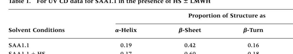
Circular dichroism (CD) measurements were done on a Jasco J-720 spectrophotometer (Jasco Inc., Easton, MD, USA). Measurements were made at 25C. Each sample was scanned 10 times and noise reduction applied to remove the high frequency, before calculating molar ellipticities. The voltage to photomultiplier was kept below 500 V to pre-vent distortion of the CD spectrum. The data were plotted as described previously (22,23). CD data were obtained from the SAA1.1 in 150 mM NaCl, 20 mM Tris-HCl, pH 8.4, 1 mM EDTA, and in the presence of 1 mM added calcium with or without HS (0.23 mg/ml).
Statistical Analysis
amyloid deposition, and dalteparin showed a simi-lar diminution of fibril deposition (Fig. 1e–h).
Quantification of the amyloid levels in the spleen indicated a significant reduction in mice treated with LMWHs (Fig. 1i). Dosing of mice with enoxaparin (0.01 mg/kg twice daily) or dalteparin (0.006 mg/kg/day) slowed the progression of amyloid deposition, resulting in a 4.5- and 4-fold decrease in amyloid detected in the spleen, respec-tively. Increasing the amount of LMWHs to 2 times the dose further reduced amyloid levels (Fig. 1i), whereas higher quantities did not further affect the amyloid present (data not shown). Treatment with LMWHs did not alter SAA levels as determined by SDS-PAGE and Western blot analysis, indicating that the effects were on the deposition of SAA into AA fibrils and not at the level of synthesis (Fig. 1j). Prophylactic treatment will be beneficial to many individuals at risk for amyloidoses; however, therapeutic treatment of individuals with estab-lished amyloid is an important consideration. To determine the effects of LMWHs on established amyloid, mice were injected with AEF and silver ni-trate and after 5 days LMWH therapy was initiated (Fig. 2). Whereas mice injected with AEF and silver probability value of less than 0.05 was considered
significant.
Results
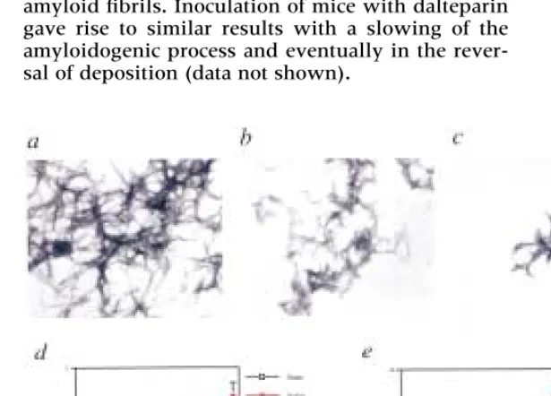
To test the hypothesis that heparin may interfere with amyloid fibrillogenesis, we injected animals with clinical doses of LMWHs. The LMWHs are cur-rently used in antithrombotic therapy in the treat-ment of unstable angina (24,25). The low molecular weight of these agents (4500–5000) and the clinical availability suggest that they may provide a novel treatment for amyloid diseases. Mice were injected with either enoxaparin (0.01 mg/mouse/twice daily) or dalteparin (0.006 mg/mouse/day) for 5 days prior to amyloid induction and continuing until the ani-mals were taken for amyloid analysis (Fig. 1). Mice were injected with AEF (20) and silver nitrate on day 5, and after 5 additional days spleens were collected and examined for amyloid load. Unin-jected mice were devoid of amyloid; in stark con-trast, mice injected with AEF and silver nitrate had significant amounts of splenic amyloid (Fig. 1a and 1b compared to 1c and 1d). Mice injected with enoxaparin, demonstrated a dramatic decrease in
nitrate followed by saline showed a continual in-crease in amyloid deposition, injection with enoxa-parin not only stopped the progression of deposi-tion, but also appeared to facilitate removal of the amyloid fibrils. Inoculation of mice with dalteparin gave rise to similar results with a slowing of the amyloidogenic process and eventually in the rever-sal of deposition (data not shown).
To demonstrate the ability of the LMWHs to in-hibit amyloidogenesis, purified SAA proteins were subjected to in vitrofibrillogenesis in the absence and presence of enoxaparin and dalteparin (Fig. 3). Incu-bation of fibrillogenic SAA1.1 (1 mg/ml) at 37C resulted in the formation of amyloid fibrils similar to those seen when fibrils are isolated from amyloidotic spleens [Fig. 3a, Axelrad et al. (20) and data not shown]. Incubation of SAA1.1 in the presence of a 10-fold molar excess of dalteparin (Fig. 3b) or enoxa-parin (Fig. 3c) limited fibril formation in vitro. Quan-tification of the fibril extension showed a dramatic decrease in fibrillogenesis in the presence of the LMWH molecules (Fig. 3d). In addition, fibrillogene-sis was inhibited when A peptides were utilized (Fig. 3e). As with the SAA proteins, enoxaparin and dalteparin were capable of blocking fibril formation of the A peptide in Alzheimer’s disease, indicating that the LMWHs are not specific for AA amyloid and may provide a novel treatment for AD. LMWHs were also capable of preventing the formation of -sheet structure as determined by circular dichroism analy-sis (Table 1). As shown in the table, HS interacts with specific protein to induce a shift in protein structure from random coil to -sheet, and in the presence of LMWH this shift was inhibited. This suggests that LWMH may prevent fibrillogenesis by not allowing for structural changes necessary for fibril interaction and in established amyloid may interfere with the as-sembly resulting in a shift back to a nonfibrillar form.
Discussion
We have here demonstrated that LMWHs are capa-ble of interacting with amyloidogenic proteins and inhibiting the fibrillogenic properties of the pro-teins and disrupting amyloid deposition in vivo. Furthermore, we have shown that LMWHs can slow
Fig. 2. Resolution of established amyloid in mice. LMWH resolved established amyloid in mice. Mice were treated with AEF and silver nitrate and after 5 days were subjected to sub-cutaneous injections of LMWH for the indicated times. Animals were killed on days 3, 5, 7, 9, and 11, and amyloid was quanti-fied and plotted as mean SEM (n8 for each time point). Mice were injected with AEF and silver nitrate followed by: saline (䊉䊉); enoxaparin at 0.01 mg per mouse twice a day (䉫䉫). On day 11, the LMWH-treated group was significantly different from the saline-treated group (p0.001).
sulfate, dextran sulfate, and pentosan polysulfate all enhanced the fibrillogenic capacity of the Apeptide significantly (33,34). On the other hand, Watson et al. (35) found that, using affinity co-electrophoresis, heparin bound with high affinity to the A peptide and to amylin and promoted fibril formation. Shuvaev and Siest (36) showed that LMWHs stimu-lated the formation of Afibrils, but that this activity was inhibited by high-affinity interactions with apolipoprotein E.
LMWHs have been implicated in altering the structural and synthetic properties of amyloidogenic proteins. HS proteoglycans are a component of the plaques associated with prion diseases and this in-teraction might have physiologic significance (37). HS can increase the concentration of prion proteins (PrP) in neuroblastoma cells; LMWHs appear to in-hibit the synthesis of PrPSc in scrapie infected cells. In addition, LMWHs were capable of converting PrPSc back to PrP as determined by the lack of in-fectivity of the molecules. Subsequently, data indi-cate that fragments of LMWHs (heparan disaccha-rides) were capable of blocking APP synthesis and inhibited the binding of heparin to APP (38). To help determine the role of LMWHs in amyloidogen-esis, we examined the effects of clinically available LMWHs on fibril formation both in vitro and in vivo using an animal model of AA amyloid.
In the current study, LMWHs did have a dra-matic effect on the fibrillogenic properties of the amyloidogenic proteins. CD analysis demonstrated that LMWH can interfere with the in vitro HS stim-ulated -sheet structure and this in turn inhibits the fibrillogenic capabilities of the molecules. This translated into an inhibition of amyloid deposition by preventing fibrillogenesis in vivoand by disrupt-ing the preformed fibrils allowdisrupt-ing for rapid degra-dation of amyloid fibrils. These observations con-tribute to a growing body of evidence implicating HS proteoglycans in the pathogenesis of amyloid diseases, including Alzheimer’s, which may provide a potential target for therapeutic intervention to disrupt common pathogenetic mechanisms.
or stop the progress of amyloidogenesis and acceler-ate the removal of established amyloid. Understand-ing the principals involved in the inhibition of fibrillogenesis may allow the development of deriva-tives that may limit the progression of amyloid diseases.
Studies have shown that HS proteoglycans in-teract with amyloidogenic proteins and facilitate fi-brillogenesis by altering the structural features of the molecules. Binding of fibrillogenic SAA1.1 to HS resulted in an increase in -sheet structure whereas the nonfibrillogenic SAA2.1 or SAA2.2 were not structurally altered by interaction with HS (22,26). Subsequent studies have shown that SAA can bind with high affinity and specificity to heparin and HS, indicating that this interaction may be im-portant in the amyloidogenicity of the proteins (27). In addition, high-affinity interactions between the Alzheimer’s -amyloid precursor protein (-APP) and HSPG have been detected and that this binding can be inhibited by heparin (28). Deletion mutagen-esis identified two heparin-binding domains in the APP protein, and synthetic peptides to candidate heparin-binding regions identified at least four hep-arin binding sites (29). The binding of HSPG to amylin (i.e., IAPP) enhanced fibril formation in a dose-dependent manner. Furthermore, HSPG inter-acts with amylin in a highly specific fashion and the association was abolished in the presence of heparin (30). We hypothesize that indeed the interaction be-tween amyloid proteins and HSPG may be impor-tant for fibrillogenesis.
There is some controversy over the role of hep-arin, derivatives of hephep-arin, and sulfated com-pounds in the amyloidogenesis. Initial studies by Kisilevsky et al. demonstrated that polyvinylsulfate and aliphatic polysulfonates could interfere with HSPG-induced A fibril formation and prevent the accumulation of AA amyloid in mice (16,31,32). Re-cently, one study showed that removal of the sulfate moieties from heparin resulted in a complete loss in the enhanced A fibrillogenesis (33). In addition, compounds such as chondroitin-4-sulfate, dermatin
Table 1. For UV CD data for SAA1.1 in the presence of HS ⫾LMWH
Proportion of Structure as
Solvent Conditions ␣-Helix -Sheet -Turn Remainder
SAA1.1 0.19 0.42 0.16 0.23
SAA1.1HS 0.17 0.60 0.18 0.05
SAA1.1HS LWMH 0.18 0.41 0.15 0.26
Acknowledgments
The authors wish to thank Mr. John Cranfill and Ms. Connie Gerardot for expert technical assistance. This work was supported by grants from the USPHS (NS31220 and AG12891) and the Stroke Program of the Sanders-Brown Center on Aging.
References
1. Selkoe DJ. (1997) Alzheimer’s disease: genotypes, phenotypes, and treatments. Science275:630–631.
2. Haass C, Selkoe DJ. (1993) Cellular processing of -amyloid precursor protein and the genesis of amyloid -peptide. Cell75:
1039–1042.
3. Yan SD, Zhu H, Golabek A, et al. (2000) Receptor-dependent cell stress and amyloid accumulation in systemic amyloidosis.
Nat. Med.6:643–651.
4. Selkoe DJ. (1999) Translating cell biology into therapeutic advances in Alzheimer’s disease. Nature399:A23–A31. 5. Kindy MS, deBeer FC. (2000) Amyloidosis. In: Massry SG,
Glassock RJ, eds. Textbook of nephrology. Philadelphia:Lippincott, Williams and Wilkins.
6. Kisilevsky R. (1993) Amyloidogenesis—A critical review. In: Kisilevsky R, Benson MD, Frangione B, Gauldie J, Muckle TJ, and Young ID, eds. Amyloid and amyloidosis. New York: Parthenon.
7. Botto M, Hawkins PN, Bickerstaff MC, Herbert J, et al. (1997) Amyloid deposition is delayed in mice with targeted deletion of the serum amyloid P component gene. Nat. Med.3:855–859. 8. Kindy MS, Rader DL. (1998) Reduction in amyloid A amyloid formation in apolipoprotein-E-deficient mice. Am. J. Pathol.152:
1387–1395.
9. Fraser PE, Nguyen JT, Chin DT, Kirschner DA. (1992) Effects of sulfate ions on Alzheimer-beta/A4 peptide assemblies— implications for amyloid fibril-proteoglycan interactions.
J. Neruochem.59:1531–1540.
10. Tennent GA, Lovat LB, Pepys MB. (1995) Serum amyloid P component prevents proteolysis of the amyloid fibrils of Alzheimer’s disease and systemic amyloidosis. Proc. Natl. Acad. Sci. U.S.A.92:4299–4303.
11. Wood SJ, Chan W, Wetzel R. (1996) Seeding of A beta fibril formation is inhibited by all three isotypes of apolipoprotein E.Biochemistry35:12623–12628.
12. Ma J, Yee A, Brewer HB, Das S, Potter H. (1994) Amyloid-associated proteins alpha 1-antichymotrypsin and apolipoprotein E promote assembly of Alzheimer beta-protein into filaments.
Nature372:92–94.
13. Bales KR, Verina T, Dodel RC, et al. (1997) Lack of apolipopro-tein E dramatically reduces amyloid beta-peptide deposition.
Nat. Genet.17:263–264.
14. Magnus JH, Stenstad T, Husby G, Kolset SO. (1992) Isolation and partial characterization of heparan sulphate proteoglycans from human hepatic amyloid. Biochem. J.288:225–231. 15. Lyon AW, Narindrasorasak S, Young ID, et al. (1991)
Co-deposition of basement membrane components during the in-duction of murine splenic AA amyloid. Lab. Invest.64:785–790. 16. Kisilevsky R, Lemieux LJ, Fraser PE, Kong X, Hultin PG,
Szarek WA. (1995) Arresting amyloidosis in vivo using small-molecule anionic sulphonates or sulphates: implications for Alzheimer’s disease. Nat. Med.1:143–148.
17.Physician’s desk reference,55th ed. (2001) Montvale, N.J.: Med-ical Economics; pp. 2613, 713.
18. Kindy MS, King AR, Yu J, Gerardot C, Whitley J, deBeer FC. (1998) Adenoviral expression of murine serum amyloid A pro-teins to study amyloid fibrillogenesis. Biochem. J.332:721–728. 19. Chiba T, Kogishi K, Wang J, et al. (1999) Mouse senile amyloid deposition is suppressed by adenovirus-mediated overexpression of amyloid-resistant apolipoprotein A-II. Am. J. Pathol.155:1319–1326.
20. Axelrad MA, Kisilevsky R, Willmer J, Chen SJ, Skinner M. (1982) Further characterization of amyloid-enhancing factor.
Lab. Invest.47:139–146.
21. Kindy MS, de Beer FC. (1999) A mouse model for serum amyloid A amyloidosis. Meth. Enzymol.309:701–716. 22. De Beer MC, de Beer FC, McCubbin WD, Kay CM, Kindy MS.
(1993) Structural prerequisites for amyloid A fibril formation.
J. Biol. Chem.268:20606–20612.
23. Provencher S, Glockner J. (1981) Estimation of globular pro-tein secondary structure from circular dichroism. Biochemistry
20:33–37.
24. Fragmin During Instability in Coronary Artery Disease (FRISC) Study Group. (1996) Low-molecular-weight heparin during instability in coronary artery disease. Lancet 347: 561–568 (1996).
25. Cohen M, Demers C, Gurfinkel EP, et al. (1997) A comparison of low-molecular-weight heparin with unfractionated heparin for unstable coronary artery disease. N. Engl. J. Med.337:447– 452.
26. McCubbin WD, Kay CM, Narindrasorasak S, Kisilevsky R. (1988) Circular-dichroism studies on two murine serum amy-loid A proteins. Biochem. J.256:775–783.
27. Ancsin JB, Kisilevsky R. (1999) The heparin/heparan sulfate-binding site on apo-serum amyloid A. implications for the therapeutic intervention of amyloidosis. J. Biol. Chem. 274:
7172–7181.
28. Narindrasorasak S, Lowery D, Gonzalez-De-Whitt P, Poorman RA, Greenberg B, Kisilevsky R. (1991) High affinity interac-tions between the Alzheimer’s beta-amyloid precursor pro-teins and the basement membrane form of heparan sulfate proteoglycan. J. Biol. Chem.266:12878–12883.
29. Clarris HJ, Cappai R, Heffernan D, Bryreuther K, Masters CL, Small DH. (1997) Identification of heparin-binding domains in the amyloid precursor protein of Alzheimer’s disease by deletion mutagenesis and peptide mapping. J. Neurochem.68:
1164–1172.
30. Castillo GM, Cummings JA, Yang W, et al. (1998) Sulfate content and specific glycosaminoglycan backbone of per-lecan are critical for perper-lecan’s enhancement of islet amyloid polypeptide (amylin) fibril formation. Diabetes 47: 612– 620.
31. Inoue S, Hultin PG, Szarek WA, Kisilevsky R. (1996) Effect of poly(vinylsulfonate) on murine AA amyloid: a high-resolution ultrastructural study. Lab. Invest. 74: 1081– 1090.
32. Leveugle B, Scanameo A, Ding W, Fillit H. (1994) Binding of heparan sulfate proteoglycan to -amyloid peptide: inhibition by potentially therapeutic polysulfated compounds. Neurore-port5:1389–1392.
33. Castillo GM, Lukito W, Wight TN, Snow AD. (1999) The sulfate moieties of glycosaminoglycans are critical for the enhancement of beta-amyloid protein fibril formation. J. Neurochem.72:1681– 1687.
34. McLaurin J, Franklin T, Zhang X, Deng J, Fraser PE. (1999) Interactions of Alzheimer amyloid-beta peptides with gly-cosaminoglycans effects on nucleation and growth. Eur. J. Biochem.266:1101–1110.
35. Watson DJ, Lander AD, Selkoe DJ. (1997) Heparin-binding properties of the amyloidogenic peptides Aand amylin. J. Biol. Chem.272:31617–31624.
36. Shuvaev VV, Siest G. (2000) Heparin specifically inhibits bind-ing of apolipoprotein E to amyloid -peptide. Neurosci. Lett.
280:131–134.
37. Gabizon R, Meiner Z, Halimi M, Ben-Sasson SA. (1993) Heparin-like molecules bind differentially to prion-proteins and changes their intracellular metabolic fate. J. Cell. Physiol.
157:319–325.