OncoTargets and Therapy
Dove
press
C a s e s e r i e s
open access to scientific and medical research
Open access Full Text article
successful treatment with apatinib for refractory
recurrent malignant gliomas: a case series
Honghong Zhang1,2
Fangfang Chen1,2
Zhiqiang Wang1,2
shaoxiong Wu1,2
1Department of radiation Oncology, sun Yat-sen University Cancer Center, 2state Key Laboratory of Oncology in southern China, Guangzhou, People’s republic of China
Abstract: Malignant glioma (MG) is a common and refractory primary tumor with a high recurrence rate. There is still a lack of effective therapy for recurrent MG (rMG). We present here two cases of refractory rMG treated using apatinib, which is a new highly selective inhibitor to VEGFR. Case 1, a 37-year-old female, was diagnosed with recurrent intracerebral high-grade glioma and failed to almost all treatments (including temozolomide, bevacizumab, nimotu-zumab, reradiation, etc) during her second relapse. Case 2, a 40-year-old male, was diagnosed with recurrent glioblastoma multiforme for the third time following multiple treatments includ-ing resurgery, temozolomide and radiation. These two patients were treated with oral apatinib (500 mg daily) during their most recent relapse and experienced rapid relief of central nervous system symptoms. Case 1 achieved near complete response evaluated by magnetic resonance imaging (MRI) after 6, 12 and 20 weeks medication and had an overall survival of 27 weeks. Case 2 achieved partial response evaluated by MRI after 4 and 12 weeks medication and had a progression-free survival of 12 months. The preliminary results of these two cases indicate that apatinib has outstanding efficacy for refractory rMG. It is worthwhile to develop a Phase II clinical trial to further evaluate the efficacy and toxicity of apatinib for rMG.
Keywords: apatinib, recurrent malignant glioma, targeted therapy, VEGFR
Introduction
Malignant glioma (MG) is one of the most common primary tumors in the central nervous system (CNS). The standard treatment for MG is surgery combined with postoperative radiotherapy (RT) and chemotherapy. The prognosis of MG is closely correlated with the pathological classification and World Health Organization (WHO) grade level. Unfortunately, intracerebral relapse is almost inevitable.1 At recurrence,
some tumors display new genetic mutations and can progress to a more aggressive state, which make treatments of recurrent gliomas more difficult.2 It was reported that
survival after relapse and retreatment of MG was usually in the range of 6–8 months,3
and the median time to the second progression was 14 weeks.4
Because growth of MG is dependent on new blood vessel formation, inhibitors to target tumor vasculation are considered promising therapeutic agents in these patients. Apatinib (Hengrui Pharmaceutical Co., Ltd, Shanghai, People’s Republic of China), a new-style small molecular tyrosine kinase inhibitor (TKI) to VEGFR, was approved for marketing in China in 2014 and admitted for the treatment of advanced gastric cancer patients who had failed after the second-line and above treatment.5,6 In
addition, several Phase II trials showed that apatinib had a significant improvement of survival for advanced nonsquamous non-small-cell lung cancer (NSCLC)7 or a
potential improvement of survival for metastatic breast cancer and hepatocellular Correspondence: shaoxiong Wu
Department of radiation Oncology, sun Yat-sen University Cancer Center, 651 Dong Feng road, east, Guangzhou 510060, People’s republic of China Tel +86 208 734 3374
Fax +86 208 734 3384 email wushx@sysucc.org.cn
Journal name: OncoTargets and Therapy Article Designation: Case Series Year: 2017
Volume: 10
Running head verso: Zhang et al
Running head recto: Apatinib for refractory rMGs DOI: http://dx.doi.org/10.2147/OTT.S119129
OncoTargets and Therapy downloaded from https://www.dovepress.com/ by 118.70.13.36 on 25-Aug-2020
For personal use only.
Number of times this article has been viewed
This article was published in the following Dove Press journal: OncoTargets and Therapy
Dovepress
Zhang et al
carcinoma.8–10 However, there have been no reports for
treat-ing recurrent MG (rMG) with this drug. Recently, we have used apatinib to treat two patients with refractory rMG and achieved exciting results, unexpectedly. The two cases are reported as follows.
Case presentation
Patient 1
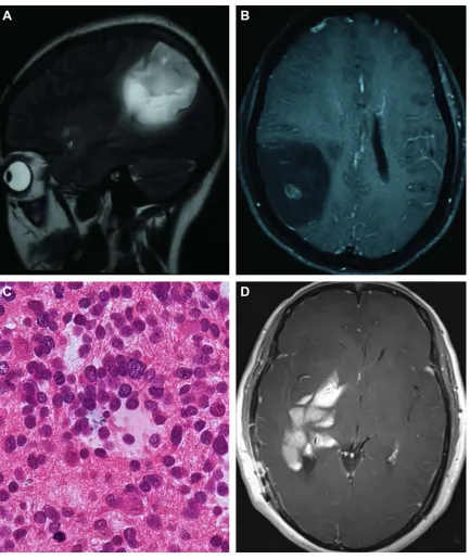
Patient 1, a 37-year-old female, was referred to an outside hospital due to headaches lasting 1 month without vomiting and blurred vision. Physical examination showed that her memory and numeracy had decreased. Magnetic resonance imaging (MRI) scan showed a space-occupying lesion in the right parietal lobe (Figure 1A and B). Surgical resection was performed on October 29, 2013. She was pathologically diagnosed with anaplastic astrocytoma (WHO grade III) after surgery (Figure 1C), and no antitumor therapy was executed. At 3 months postsurgery, the patient was referred to our hospital due to the emergence of headaches and vomiting. MRI showed an irregular mass in the anterior and inferior border of the original resected cavity, which extended from the right basal ganglia to the right temporal and frontal lobes, accompanied with obvious peripheral edema (Figure 1D). She was clinically diagnosed with rMG. Considering the extensive size and the poor location of the recurrent tumor, the patient was treated with intensity-modulated radiation therapy (IMRT) combined with concomitant temozolomide (TMZ) chemotherapy and bevacizumab-targeted therapy from February 19 to April 1, 2014. The prescribed dose of RT was 60 Gy in 28 fractions. TMZ at a dose of 75 mg/m2
daily and bevacizumab at a dose of 10 mg/kg every 2 weeks were given from the first to the last day of RT. Obvious regression of the recurrent lesion was observed on MRI scan at the end of treatment. After a 4-week break, she was treated using adjuvant chemotherapy with TMZ at a dose of 150–200 mg/m2 on days 1–5, repeated every 28 days. MRI
showed complete response (CR) after six cycles of TMZ adjuvant chemotherapy, and thus the TMZ regimen was continued. However, MRI after 12 cycles of TMZ showed several new lesions at the left cerebellar hemisphere and left parietal lobe in this patient. She was clinically diagnosed with ectopic rMG (Figure 2). Subsequently, the dose-dense chemotherapy of TMZ (150 mg/m2 on days 1–7 and 15–21)
and bevacizumab (300 mg on days 1 and 15) was instigated. After 1 month of this combined treatment, MRI showed sig-nificant progression of the lesions. Thereafter, local radiation using tomotherapy with 45 Gy in 15 fractions for all recur-rent lesions was used in this patient from July 1 to 25, 2015.
RT was concurrently combined with nimotuzumab-targeted therapy (200 mg weekly for 6 weeks) and EP regimen (etopo-side 100 mg/m2 on days 1–3, carboplatin AUC 5 on day 1,
repeated every 21 days for two cycles) chemotherapy. The patient complained about severe headaches and vomiting at 1 month after the combined treatment, which could not be relieved until mannitol (250 mL) and dexamethasone (10 mg) were administered daily. MRI showed continuous increase in the tumor size, and the tumor spread to the left frontal lobe (Figure 3A–D). We decided to try the new drug apatinib after consultation with a multidisciplinary team and extensive communication with the patient and obtained the informed consent from the patient and her family. Upon starting oral apatinib 500 mg daily on August 31, 2015, the patient’s headaches gradually decreased after 5 days of medication, and the dosage of dexamethasone was halved. When the headaches completely disappeared without any CNS symptoms after 8 days of medication, mannitol and dex-amethasone were stopped. After 6 weeks of medication, MRI showed all lesions to be near CR (Figure 3E–H). Apatinib was continued for 12 weeks, and the response remained the same. Thus, apatinib for maintenance was continued. MRI at 20 weeks medication still showed no disease progression. The patient remained without any CNS symptoms. However, after 24 weeks of medication, she suddenly suffered severe headaches and hypertension and subsequently fell into a coma in an outside hospital. Unfortunately, she died after 3 weeks. The cause of death could not be determined to be related to the drug or disease progression because the patient did not undergo computed tomography (CT) or MRI scan during her comatose condition. The overall survival from the start of apatinib for this patient was 27 weeks. During the treatment with apatinib, the patient experienced grade 1 hand–foot skin desquamation and grade 2 stomatitis, but these toxic reactions were relieved with symptomatic treatment.
Patient 2
Patient 2, a 40-year-old male, was referred to an outside hospital due to severe headaches and vomiting lasting 7 days. Physical examinations showed no obvious abnormality. MRI showed a lesion at the right parieto-occipital lobes (Figure 4A). Gross tumor resection was executed on December 26, 2014. The postoperative histopathology of the tumor was diagnosed as glioblastoma multiforme (WHO grade IV; Figure 4B). Immunohistochemistry examination revealed the following: MGMT (-), Ki-67 30% (+) and P53 (+). RT was rejected by the patient after surgery, and only TMZ (200 mg/m2 on days
1–5, repeated every 28 days) chemotherapy was executed.
OncoTargets and Therapy downloaded from https://www.dovepress.com/ by 118.70.13.36 on 25-Aug-2020
Dovepress apatinib for refractory rMGs
After six cycles of TMZ chemotherapy, the patient developed left limb weakness, dizziness and headaches. MRI showed tumor recurrence at the primary location (Figure 4C). Resec-tion was performed again on July 3, 2015, and the result of the postoperative histopathology remained the same. The patient was referred to our hospital as a result of severe headaches
again at 43 days postsurgery. MRI showed a recurring nodular enhanced lesion in the front edge of the operative cavity (Figure 4D). This was considered as the second tumor recurrence. IMRT of 60 Gy in 28 fractions combined with concomitant TMZ (75 mg/m2/day for 40 days) was executed
52 days after reoperation. The headaches gradually subsided Figure 1 Pre- and postoperative Mri and histopathological diagnosis for patient 1.
Notes: (A and B) Preoperative T2-weighted sagittal and T1 contrast-enhanced axial images showed a space occupying lesion with T2 heterogeneous hyperintensity and
central dotty enhancement in the right parietal lobe. (C) Postoperative histopathological section (HE staining, magnification ×400) showed features of anaplastic astrocytoma (WHO grade iii) with increased cellularity, nuclear atypia and mitotic activity. (D) T1 contrast-enhanced axial image at 3 months after operation showed an irregular mass
with gross enhancement in the right basal ganglia and temporal lobe.
Abbreviations: Mri, magnetic resonance imaging; He, hematoxylin–eosin; WHO, World Health Organization.
OncoTargets and Therapy downloaded from https://www.dovepress.com/ by 118.70.13.36 on 25-Aug-2020
Dovepress
Zhang et al
during the treatment. MRI after the combined therapy procedure showed that the nodular lesion shrunk and the edema also subsided. However, the patient’s headaches and left limb weakness reappeared at 3 weeks after the treatment. MRI showed that the previous lesion and periphery edema had obviously increased again (Figure 5A and B). Through consultation with a multidisciplinary team, the patient was clinically diagnosed with a third tumor progression. Beva-cizumab targeted therapy was recommended; however, the patient refused this recommendation due to financial reasons. He decided to accept apatinib therapy on November 3, 2015, and signed the informed consent. Thus, apatinib 500 mg daily was administered to this patient. The symptoms of headaches and limb weakness were gradually relieved after medication. MRI at 4 weeks of medication showed partial response of the recurrent lesion and periphery edema (Figure 5C and D). The patient had grade 3 hand–foot skin toxicity and grade 2 stomatitis during the medication. Therefore, the dosage of apatinib was halved (250 mg daily) in order for him to continue and the toxic reactions subsided to some extent after symptomatic treatment. MRI after 12 weeks of medication still revealed partial response of the lesion, and the half dosage of apatinib was continued in maintenance therapy. Thereafter, MRI was performed every 3 months and indicated no disease progression. The patient had a progression-free survival of
12 months until a new lesion in the contralateral parietal lobe was found. He is still alive with clear consciousness, although he has developed severe bilateral limb weakness.
Discussion
Until now, no standard treatment regimen has been proposed for rMG. Individual treatment regimen for rMG is usually recommended after the comprehensive considerations of multiple factors, including the general status of patients, location and size of recurrent tumor and the approaches and efficacy of previous treatment.
Reoperation may be the first choice for rMG. However, only ∼25% of patients are suitable for reoperation.11 The
value of reoperation is often limited by the infiltrative nature and the proliferation rate of the tumor itself, as well as a high risk for surgery-related complications. Generally, reoperation is suitable for young patients with high Karnofsky perfor-mance status (KPS), longer interval from the last surgery, singular and smaller tumor and good location. In this series, case 1 had multiple recurrent lesions, and case 2 had a very short interval from the second surgery. Therefore, they were not suitable for reoperation.
RT/re-RT is one of the traditional treatments for rMG, particularly suitable for those patients without previous RT or with intervals longer than 1 year from the first RT. In this Figure 2 Mri during the second relapse for patient 1.
Notes: T1 contrast-enhanced axial images showed multiple oval and patchy nonhomogeneous enhancement lesions in the left cerebellar hemisphere (A) and
parietal lobe (B).
Abbreviation: Mri, magnetic resonance imaging.
OncoTargets and Therapy downloaded from https://www.dovepress.com/ by 118.70.13.36 on 25-Aug-2020
Dovepress apatinib for refractory rMGs
series, case 1 had ectopic relapses at the opposite side of the primary tumor and had an interval .1 year from the first RT. Therefore, she was suitable for re-RT. Unfortunately, the outcome of re-RT in this patient was poor, and the recurrent lesions continued enlarging and multiplying after re-RT. Case 2 did not receive RT after the first surgery, and the tumor recurred again rapidly after reoperation for the first recurrent lesion, thus he received RT in conjunction with TMZ concomitant chemotherapy at the second recurrence. Despite the recurrent lesion being partially reduced at the end
of RT, it had progressed again in ,1 month after RT. This result indicated that the effect of RT combined with TMZ was poor in this patient.
Recently, a Phase II clinical trial reported that TMZ dose-dense regimen treated rMG with a certain efficacy.12
However, TMZ dose-dense regimen was not effective for case 1 in this report.
Molecular-targeted drugs have been the recent research hot spot for rMG. Bevacizumab, an anti-VEGF monoclonal antibody, can achieve favorable efficacy for the treatment
Figure 3 (Continued)
OncoTargets and Therapy downloaded from https://www.dovepress.com/ by 118.70.13.36 on 25-Aug-2020
Dovepress
Zhang et al
of recurrent glioblastoma in isolated use or in combination with chemotherapy with 6-month progression-free sur-vival of 43%–50%.13,14 Nimotuzumab, a type of humanized
anti-EGFR (epidermal growth factor receptor) monoclonal antibody, combined with RT has been reported to have better efficacy for rMG15 and newly diagnosed diffuse
intrinsic pontine glioma among children and adolescents.16
In this report, unfortunately, bevacizumab combined
with TMZ chemotherapy was ineffective for case 1 at the second recurrence. Whereafter, nimotuzumab com-bined with chemotherapy (etoposide and carboplatin) and synchronous re-RT also did not show any efficacy for this patient.
Apatinib as a small molecular TKI to VEGFR can highly selectively compete for the adenosine triphosphate (ATP) binding site of intracellular VEGFR-2 for blocking Figure 3 Mri comparison between pre- and post-apatinib for patient 1.
Notes: Before oral apatinib, FLair axial (A and B) and T1 contrast-enhanced axial images (C and D) showed multiple masses or nodular lesions with heterogeneous
intensity and nonhomogeneous enhancement accompanied with central necrosis and peripheral edema in the left cerebellar hemisphere, parieto-frontal lobes. after 6 weeks medication with apatinib, FLair axial (E and F) and T1 contrast-enhanced axial images (G and H) showed all the lesions and peripheral edema significantly reduced compared
with pre-apatinib Mri.
Abbreviations: FLAIR, fluid attenuation inversion recovery; MRI, magnetic resonance imaging.
OncoTargets and Therapy downloaded from https://www.dovepress.com/ by 118.70.13.36 on 25-Aug-2020
Dovepress apatinib for refractory rMGs
downstream signal transduction and inhibiting tumor angio-genesis. A multicenter randomized double-blind phase III clinical trial has confirmed the overall survival improve-ment of apatinib for patients with metastatic gastric cancer who failed after second-line or above chemotherapy (6.5 vs 4.7 months, P,0.016).6
Since MG consists of abundant and aberrant blood ves-sels, we tried apatinib for the treatment of the two cases with refractory rMG in this report and achieved apparent thera-peutic efficacy, especially for case 1 who did not respond to almost all drugs and modalities. Apatinib can quickly take effect and rapidly relieve the CNS symptoms of the patient. Figure 4 Pre- and postoperative MRI between the first and second operation and histopathological diagnosis for patient 2.
Notes: (A) T1 contrast-enhanced axial image showed an irregular occupying mass with nonhomogeneous enhancement accompanied with necrosis and peripheral edema at
the right parieto-occipital lobes before the first operation. (B) Histopathological section (HE staining, magnification ×200) after surgery showed the features of glioblastoma multiforme (WHO grade iV), with highly increased cellularity, marked nuclear atypia and mitotic activity, hemorrhage and pseudopalisading necrosis. (C) at 6 months after
the first operation, T1 contrast-enhanced axial image showed that a mass with nonhomogeneous enhancement reappeared at the primary location, accompanied with central necrosis and peripheral edema. (D) at 43 days after the second operation, T1 contrast-enhanced axial image showed a recurrent nodular enhanced lesion in the front edge
of the operative cavity accompanied with obvious peripheral edema.
Abbreviations: Mri, magnetic resonance imaging; He, hematoxylin–eosin; WHO, World Health Organization.
OncoTargets and Therapy downloaded from https://www.dovepress.com/ by 118.70.13.36 on 25-Aug-2020
Dovepress
Zhang et al
Why does apatinib have such a strong inhibitory effect on rMG? It may be due to its stronger interaction with intracel-lular VEGFR-2 compared with other TKIs (such as sorafenib, pazopanib and sunitinib).17 Results from animal experiments
showed that the value of half maximal inhibitory concentra-tion (IC50) to VEGFR-2 for apatinib was only 2 nM, while the values of IC50 for sorafenib, pazopanib and sunitinib
were 90, 30 and 10 nM, respectively,18 which indicated the
high specificity of VEGFR-2 for apatinib.
It was reported that hematological toxicities for apatinib mainly included leukopenia, neutropenia and thrombocy-topenia, and nonhematological toxicities included hyperten-sion, proteinuria, hand–foot skin reaction, fatigue, diarrhea, etc, most of which are grade 1 and 2.5 In this report, case 1 Figure 5 Mri comparison between pre- and post-apatinib for patient 2.
Notes: Before oral apatinib, FLair axial (A) and T1 contrast-enhanced axial images (B) showed an enlarged recurrent lesion accompanied with central necrosis and
obvious peripheral edema. at 4 weeks medication of apatinib, FLair axial (C) and T1 contrast-enhanced axial images (D) showed that the lesion and peripheral edema were
significantly reduced compared with pre-apatinib MRI.
Abbreviations: MRI, magnetic resonance imaging; FLAIR, fluid attenuated inversion recovery.
OncoTargets and Therapy downloaded from https://www.dovepress.com/ by 118.70.13.36 on 25-Aug-2020
OncoTargets and Therapy
Publish your work in this journal
Submit your manuscript here: http://www.dovepress.com/oncotargets-and-therapy-journal
OncoTargets and Therapy is an international, peer-reviewed, open access journal focusing on the pathological basis of all cancers, potential targets for therapy and treatment protocols employed to improve the management of cancer patients. The journal also focuses on the impact of management programs and new therapeutic agents and protocols on
patient perspectives such as quality of life, adherence and satisfaction. The manuscript management system is completely online and includes a very quick and fair peer-review system, which is all easy to use. Visit http://www.dovepress.com/testimonials.php to read real quotes from published authors.
Dovepress
Dove
press
apatinib for refractory rMGs
showed only mild hand–foot skin and oral mucosa reactions without other discomfort. Case 2 showed severe hand–foot syndrome in addition to mild stomatitis, but he was able to tolerate the symptoms after halving the dosage and symp-tomatic treatment.
Conclusion
According to the preliminary results on the two cases in this report, apatinib expresses outstanding efficacy for patients with rMG who did not respond to multiple treatments. In addition, it implies that apatinib is also effective in those patients who had no response to bevacizumab. These results also suggest that it is necessary to launch a Phase II clinical trial of apatinib to further evaluate its efficacy on rMG.
Disclosure
The authors report no conflicts of interest in this work.
References
1. Omuro A, DeAngelis LM. Glioblastoma and other malignant gliomas: a clinical review. JAMA. 2013;310(17):1842–1850.
2. Johnson BE, Mazor T, Hong C, et al. Mutational analysis reveals the origin and therapy-driven evolution of recurrent glioma. Science. 2014;343(6):189–193.
3. Nieder C, Grosu A, Molls M. A comparison of treatment results for recurrent malignant gliomas. Cancer Treat Rev. 2000;26(6):397–409. 4. Huncharek M, Muscat J. Treatment of recurrent high grade
astrocy-toma; results of a systematic review of 1,415 patients. Anticancer Res. 1997;18(2B):1303–1311.
5. Li J, Qin S, Xu J, et al. Apatinib for chemotherapy-refractory advanced metastatic gastric cancer: results from a randomized, placebo-controlled, parallel-arm, phase II trial. J Clin Oncol. 2013;31(26):3219–3225. 6. Li J, Qin S, Xu J, et al. Randomized, double-blind, placebo-controlled
phase III trial of apatinib in patients with chemotherapy-refractory advanced or metastatic adenocarcinoma of the stomach or gastroesopha-geal junction. J Clin Oncol. 2016;34(13):1448–1454.
7. Zhang L, Shi M, Huang C, et al. A phase II, multicenter, placebo-controlled trial of apatinib in patients with advanced nonsquamous non-small cell lung cancer (NSCLC) after two previous treatment regimens. J Clin Oncol. 2012;30(15_suppl):7548.
8. Hu X, Zhang J, Xu B, et al. Multicenter phase II study of apatinib, a novel VEGFR inhibitor in heavily pretreated patients with metastatic triple-negative breast cancer. Int J Cancer. 2014;135(8):1961–1969. 9. Hu X, Cao J, Hu W, et al. Multicenter phase II study of apatinib in
non-triple-negative metastatic breast cancer. BMC Cancer. 2014; 14(1):1–8.
10. Qin S. Apatinib in Chinese patients with advanced hepatocellular carcinoma: a phase II randomized, open-label trial. Asco Meeting
Abstracts. 2014;32(15_suppl):4019.
11. Dützmann S, Geßler F, Bink A, et al. Risk of ischemia in glioma surgery: comparison of first and repeat procedures. J Neurooncol. 2012;107(3):599–607.
12. Galldiks N, Berhorn T, Blau T, Dunkl V, Fink GR, Schroeter M. “One week on–one week off”: efficacy and side effects of dose-intensified temozolomide chemotherapy: experiences of a single center.
J Neurooncol. 2013;112(2):209–215.
13. Reardon DA, Desjardins A, Vredenburgh JJ, et al. Metronomic chemotherapy with daily, oral etoposide plus bevacizumab for recur-rent malignant glioma: a phase II study. Br J Cancer. 2009;101(12): 1986–1994.
14. Friedman HS, Prados MD, Wen PY, et al. Bevacizumab alone and in combination with irinotecan in recurrent glioblastoma. J Clin Oncol. 2009;27(28):4733–4740.
15. Yang QY, Guo CC, Chen ZP. Profile of nimotuzumab in the treatment of high-grade glioma. Onco Targets Ther. 2015;8:819–825.
16. Epelman S, Odone V, Gorender E, Medeiros RSS, Martins L. Phase II study of nimotuzumab and radiotherapy in children and adolescents with newly diagnosed diffuse intrinsic pontine gliomas (DIPG). J Clin
Oncol. 2015;33(15_suppl):10061.
17. Li J, Zhao X, Chen L, et al. Safety and pharmacokinetics of novel selective vascular endothelial growth factor receptor-2 inhibitor YN968D1 in patients with advanced malignancies. BMC Cancer. 2010;10(1):529–536.
18. Cowey CL, Sonpavde G, Hutson TE. New advancements and develop-ments in treatment of renal cell carcinoma: focus on pazopanib. Onco
Targets Ther. 2009;3(4):147–155.
OncoTargets and Therapy downloaded from https://www.dovepress.com/ by 118.70.13.36 on 25-Aug-2020