Regulation of expression of the
rat
CYP2B1
and
CYP2B2
genes
Siew Cheng Wong
A dissertation submitted to the Faculty of Life Sciences in
partial fulfilment of the requirements for the degree of
Doctor of Philosophy in the University of London
Department of Biochemistry and Molecular Biology
University College London
ProQuest Number: U642016
All rights reserved
INFORMATION TO ALL USERS
The quality of this reproduction is dependent upon the quality of the copy submitted.
In the unlikely event that the author did not send a complete manuscript and there are missing pages, these will be noted. Also, if material had to be removed,
a note will indicate the deletion.
uest.
ProQuest U642016
Published by ProQuest LLC(2015). Copyright of the Dissertation is held by the Author.
All rights reserved.
This work is protected against unauthorized copying under Title 17, United States Code. Microform Edition © ProQuest LLC.
ProQuest LLC
789 East Eisenhower Parkway P.O. Box 1346
Abstract
CYP2B1 and CYP2B2 proteins are highly induced in rat liver by
phénobarbital (PB). CYP2B1 promoter sequences from -179 to -347 bp and
-348 to -451 bp were observed in gel shift assays to bind liver nuclear
protein that was either more abundant or activated from PB-treated than
untreated rats.
The DNA-binding activity of the protein bound to the sequence
between -348 and -451 bp was enhanced when liver nuclear extracts from
both untreated and PB-treated rats were treated with ATP prior to gel shift
assays. While pre-treatment with either calf intestinal alkaline phosphatase
(CIP) or 2-aminopurine (2-AP), a general protein kinase inhibitor inhibited
complex formation. Thus, phosphorylation of this protein increases its
binding to DNA and dephosphorylation inhibits binding. When primary
hepatocytes or whole animals were treated with 2-aminopurine, it could
totally suppress both basal and PB-induced CYP2B mRNAs expression in
vitro but only partially suppress PB-induction of CYP2B mRNAs in vivo.
A PB-responsive element (PBRE) has been identified in the distal
region of the CYP2B2 promoter. The homologous region in the CYP2B1
promoter between -2142 and -2301 bp was cloned into a reporter gene
construct and was shown to confer PB-responsiveness to the luciferase
gene when transfected either into primary rat hepatocytes or directly into rat
liver tissues. A higher fold induction was observed with in vivo DNA
when the -348 to -451 bp sequence was included in the promoter region of
the reporter construct.
The CYP2B2 promoter, between -183 and -199 bp, also bound more
liver nuclear protein from PB-treated rats in gel shift assay. An octamer
consensus oligonucleotide competed for protein binding to this region. An
antibody which recognised the DNA-binding domain of Oct-1 and Oct-2
inhibited complex formation and an Oct-1 specific antibody supershifted the
A cknowledgements
This research degree could not have progressed smoothly and be fulfilled without the support of a number of people whom I would like to extend my sincere gratitude to.
My parents who have been extremely supportive. They have always understood my desire to pursue my studies further and given me lots of encouragement throughout the years. I am also immensely proud of my brother, Kok Meng who has taken full responsibility in caring for our parents all these while. I love them very much and would like them to know that I have put the best into what I am doing.
However, without the urge and encouragement from Kia Joo, I might not even have come to England in the first place. He has never failed to give me his full support and has helped me through difficult times. I also admire his passion and interest in research which has inspired me greatly.
My supervisor. Prof. Elizabeth Shephard had me given me a lot of help and guidance. I appreciated her patience and encouragement to me. I am also grateful to both her and Prof. Ian Phillips for the discussion sessions we had together and for their input of ideas into the research.
The lab would not be such an exciting and happy place to work in without my fellow colleagues. Dr Azara Janmohamed, Roongsiri Muangmoonchai, Penny Smirlis and Marie Bootman. I truly enjoyed their company and appreciated all their help, support, friendship and encouragement. Previous members of Lab 402 have also given me help and I would like to take this opportunity to thank them too. I also owe a great deal to Dr Mina Edwards who had assisted me with the perfusion and culturing work. She gave me a lot of encouragement along the way and I am grateful to her. I would like to thank Dr Parmjit Jat, my internal monitor for his time and his suggestions towards my research. The gene gun transfection experiment would not have been possible without the facilities offered by Prof. Jeremy Brocks and the assistance of Cristina Velloso.
Dr Shaun Thomas for his kind gift of the Oct1/2 antibody and Dr Peter Shaw for his gift of the pBLCAT(3.9kb) construct containing the CYP2B1
promoter sequence up to 3.9 kb.
Many thanks to my relatives and friends back in Singapore as well as in England for all their support. I would also like to extend my gratitude to Prof. Lee and Prof. Yap for their confidence in my abilities and support in my decision to pursue this degree. There were many other people, whose names have not been mentioned here, who had given me help in one way or another throughout the duration of this research and I am thankful to them.
Table of Contents
Abstract
... 2Acknowledgements
...5Table of Contents
...6List of Figures
...
11List of Tables
...
15Abbreviations
...
16Introduction
...201.1 Overview of xenobiotic metabolism... 21
1.2 Cytochrome P450-dependent mixed function oxygenase system 22 1.3 Discovery of cytochrome P450...27
1.4 The cytochrome P450 gene superfamily...29
1.5 Molecular mechanisms of CYP gene expression...33
1.5.1 Xenobiotic inducible CYPs... 33
1.5.1.1 Steroid inducible GYP genes...35
1.5.1.2 Peroxisome proliferator inducible GYP genes... 38
1.5.1.3 Polycyclic aromatic hydrocarbon inducible GYP genes 39 1.5.1.4 Phénobarbital (PB) inducible GYP genes...42
1.6 PB-responsive regulatory elements in PB-inducible GYP genes 47 1.6.1 Elements proximal to the transcription start site...47
1.6.2 Elements distal to the transcription start site... 49
1.7 CYP2B1 and CYP2B2... 53
1.8 CYP2B1 and CYP2B2 5’-flanking sequences... 54
1.9 Other regulatory elements...54
1.10 Regulation of CYP2B gene expression... 55
1.10.1 Tissue-specific expression... 55
1.10.2 Developmental-specific expression... 57
1.10.3 Gender-specific expression... 57
1.10.4 Strain-specific expression... 60
1.11 Factors affecting basal and/or PB-induced CYP2B gene expression...61
1.11.1 Cytokines...61
1.11.2 Insulin... 62
1.11.3 H aem... 63
1.12 Inhibitors of protein synthesis... 65
1.13 Protein kinases...67
1.13.1 Protein kinase A (PKA)... 67
1.13.2 Other protein kinases...69
1.15 Scope of this thesis...72
Materials and Methods
... 742.1 DNA cloning and sub-cloning...75
2.1.1 DNA digestion with restriction endonucleases...75
2.1.2 Agarose gel electrophoresis... 76
2.1.3 DNA purification from low melting point agarose...77
2.1.4 Dephosphorylation o f linearised plasmid DNA...79
2.1.5 Phosphorylation o f synthetic oligonucleotide 5’ ends... 81
2.1.6 Ligation reaction... 82
2.1.6.1 Ligation of DNA with sticky ends...82
2.1.6.2 Ligation of blunt-ended D N A ...82
2.1.7 Transformation o f bacterial cells...83
2.1.7.1 Preparation of fresh competent cells... 85
2.1.7.2 Preparation of frozen competent cells... 85
2.1.8 Transformation reaction... 86
2.1.9 Confirmation o f recombinants... 87
2.1.10 Preparation o f glycerol stocks... 87
2.2 Mini-scale preparation of plasmid DNA...89
2.3 Maxi-scale preparation of plasmid DNA (Maxi Qiagen® plasmid kit, Cat. No. 12162/3)...91
2.4 Annealing of single-stranded DNA sequences... 94
2.4.1 Calculation o f melting temperature...94
2.4.2 For T^ above 25 °C:-...95
2.4.3 For T^ below 25°C:-...95
2.5 Radioisotope-labelling of DNA... 96
2.5.1 DNA with a 5’-overhang... 96
2.5.2 DNA with blunt ends...97
2.5.3 Purification o f radiolabeled DNA probe... 98
2.5.3.1 Chroma Spin™ Column... 98
2.5.3.2 NucTrap® probe purification column... 98
2.5.4 Determination o f radioactive counts o f a radiolabeled probe 99 2.5.4.1 Calculation of percentage of incorporation... 100
2.6 Isolation and culturing of rat hepatocytes... 101
2.6.1 Coating o f tissue culture Permanox® plate...101
2.6.2 Isolation o f primary rat hepatocytes... 102
2.6.3 Culturing o f hepatocyte... 107
2.7 Chemical treatment of primary rat hepatocytes...109
2.7.1 2-aminopurine treatment... 109
2.7.2 PB treatment... 110 2.8 Nuclear protein extraction...I l l
2.8.1.1 Treatment of animals...113
2.8.1.2 Isolation of nuclei... 113
2.8.1.3 Preparation of nuclear extracts... 114
2.8.2 From primary rat hepatocyte culture (Rosette and Karin, 1995)... 117
2.9 Protein quantitation (Lowry, et al., 1951)...119
2.10 SDS-PAGE gel electrophoresis of protein...121
2.11 Staining of SDS-PAGE gel... 123
2.12 Gel retardation assay... 124
2.12.1 Competitive gel retardation assays...126
2.12.2 Use o f antibodies to identify DNA-binding proteins... 127
2.12.3 Chemical treatment o f nuclear protein... 127
2.13. DNase I footprinting (SureTrack Footprinting Kit from Pharmacia)... 128
2.13.1 Radiolabelling o f DNA...130
2.13.2 G + A reaction... 130
2.13.3 DNase I titration... 131
2.13.4 Binding and footprinting reaction...134
2.14 Partial nuclear protein purification using cellulose chromatography column... 135
2.14.1 Pre-cycling o f P11 phosphocellulose resins...135
2.14.2 Equilibrating column and fractionation o f nuclear proteins 136 2.15 Protein purification by magnetic DNA affinity... 138
2.15.1 Preparation o f DNA for coupling to Dynai® magnetic beads.... 138
2.15.1.1 Phosphorylation of Oligo 2 ...139
2.15.1.2 Annealing of tether DNA... 139
2.15.1.3 Preparation of DNA containing binding site of interest 140 2.15.1.4 Ligation of DNA to tether... 140
2.15.2 Preparation o f DNA-coated magnetic beads...140
2.15.2.1 Preparation of M-280 Dynabeads® for coupling... 141
2.15.2.2 Coupling of biotinylated DNA to pre-washed M-280 Dynabeads®...142
2.15.3 Binding, washing and elution conditions for protein purification... 142
2.15.4 Determination o f optimal elution conditions...144
2.15.5 Regeneration o f used DNA-coated magnetic beads... 144
2.16 Northern blot hybridisation...145
2.16.1 Isolation o f total R N A...145
2.16.2 Northern blotting... 146
2.16.2.3 Blotting of RNA...149
2.16.3 Radiolabelling by random priming...149
2.16.4 Calculation o f percentage incorporation, probe yield and specific activity o f the radiolabeled probe...150
2.16.5 Hybridisation o f RNA b lo t... 151
2.16.6 Stripping o f hybridised membrane...154
2.17 Transient Transfection of rat primary hepatocyte... 155
2.17.1 Transfection Using calcium phosphate (CaP04)...155
2.17.1.1 Preparation of CaPO^DNA precipitate... 156
2.17.1.2 Transfection... 156
2.17.2 Transfection using TransFast™ Transfection Reagent (Promega Corporation)... 157
2.17.3 Superfect™ Transfection Reagent (Qiagen Ltd)...159
2.17.4 Effectene™ Transfection Reagent (Qiagen Ltd)...160
2.18 In vivo gene transfer using the Helios™ gene gun system (Bio-Rad)...162
2.18.1 Cartridge Preparation...162
2.18.2 Precipitation o f DNA onto gold microcarrier... 163
2.18.3 Loading DNA-coated gold microcarrier into tubing using the tubing prep station... 164
2.18.4 Particle delivery using Helios™ gene gun... 165
2.19 Dual-Luciferase™ reporter assay system (Promega Corporation)... 168
2.19.1 Lysis o f cultured cells...169
2.19.2 Lysis of whole tissues...169
2.19.3 Dual-Luciferase Assay...170
Results and Discussion
... 171I Analysis o f the 5’-flanking sequence o f CYP2B1 and CYP2B2 genes using in vitro systems...172
3.1.1 Sub-cloning of the CYP2B1 proximal promoter region... 173
3.1.2 Multiple proteins including members of the C/EBP transcription factor family bind to the CYP2B1 proximal promoter sequence... 177
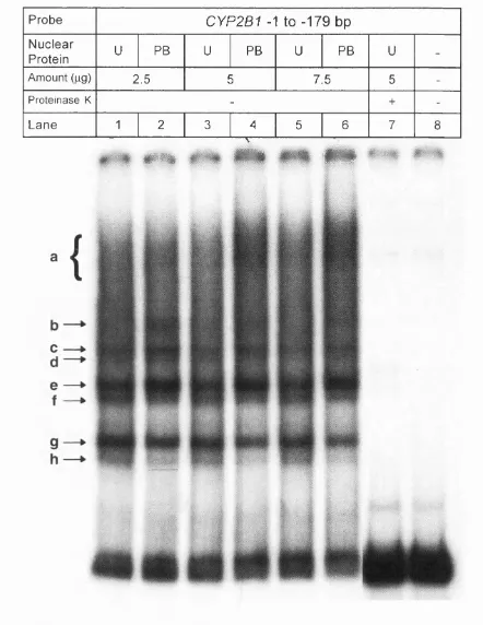
3.1.3 CYP2B1 promoter sequence between -179 and -348 bp binds more nuclear protein from rats treated with P B ... 186
3.1.4 CYP2B1 5’-flanking sequence between -348 and -451 bp also binds more nuclear protein from rats treated with PB ... 191
3.1.5 DNase I footprinting analysis of the CYP2B1 proximal promoter re gion ...200
efficient in binding nuclear protein from PB-treated rats as the
-348 to -451 bp sequence... 217 3.1.8 The CYP2B1 promoter regions from -179 to -348 bp and -348
to -451 bp could cross-compete each other in gel shift assay using
rat liver nuclear extracts... 220 3.1.9 Excessive homogenisation of liver tissue from untreated rats
can alter the abundance of nuclear protein binding to -348 and -451
bp 5’-flanking sequence of the CYP2B1 gene...227 3.1.10 Phosphorylation enhances and dephosphorylation inhibits
liver nuclear protein binding to the CYP2B1 5’-flanking sequence
rom -348 to -451 bp ...232 3.1.11 Nuclear protein binding to the CYP2B1 5'-flanking sequence
between -348 and -451 bp with rat liver protein appears to be lost in
primary rat hepatocyte cultures... 244 3.1.12 DNA affinity purification using Dynabeads® of liver nuclear
protein binding to the CYP2B1 5’-flanking sequence between -348
and -451 bp... 258 3.1.13 Oct-1 binds to the CYP2B2 promoter between
-183 and -199 bp... 267
II Effect o f 2-aminopurine on basal and PB-inducible expression of
CYP2B m RNAs... 275 3.2 2-Aminopurine down-regulates both basal and PB-inducible
expression of CYP2B genes... 276
III Analysis o f the 5’-flanking sequence o f CYP2B1 gene using transient transfection assays... 285
3.3.1 Preparation of luciferase reporter constructs... 286
3.3.1.1 Preparation o f luciferase reporter constructs containing various CYP2B1 proximal promoter regions... 286 3.3. f . 2 Heterologous promoter constructs... 289 3.3.1.3 Preparation o f luciferase reporter constructs containing proximal and distal 5’-flanking regions o f the CYP2B1 gene... 289 3.3.2 Determination of promoter and enhancer properties of CYP2B1 5'- flanking sequences using in vitro and in vivo transient transfection
syste m s...291
General Discussion
...308List of Figures
1.1 Catalytic cycle of cytochrome P450...26
1.2 Examples of diverse activities catalysed by cytochrome P450s...28
1.3 Diverse mechanisms in the regulation of CYP expression... 34
1.4 Simplified general model for the transcription activation of various
xenobiotic inducible CYP genes by their respective prototype inducers 37
1.5 Model for the induction of CYP1A1 transcription by polycyclic
aromatic hydrocarbon (PAH)...41
1.6 Pleiotropic effects of PB... 43
1.7 Structure diversity of PB and PB-like inducers... 44
1.8 Schematic representation of proposed PB regulatory elements in the
proximal and distal regions of CYP2B promoters...50
3.1.1 Agarose gel electrophoresis of restriction digestion of Clone 27
and subfragments of the first 450 bp promoter sequence of CYP2B1
cloned into pBluescript KSII... 175
3.1.2 Comparison of nucleotide sequences of the proximal promoter
region between CYP2B1 and CYP2B2 genes... 176
3.1.3 Sequence comparison of the CYP2B promoters and the C/EBP
consensus binding site... 178
3.1.4 Gel shift analysis of the CYP2B1 promoter sequence from
-1 to -179 bp... 180
3.1.5 Competitive gel shift analysis of the CYP2B1 promoter sequence
between -1 and -179 bp...181
3.1.6 DNase I footprint analysis of the template strand of CYP2B1
promoter between -1 and -179 bp...183
3.1.7 Supershift analysis of the CYP2B1 promoter sequence between
-1 and -179 bp...184
3.1.8 Gel shift analysis of the CYP2B1 promoter sequence between
-179 and -348 bp... 187
3.1.9 Competitive gel shift analysis of the CYP2B1 promoter sequence
3.1.10 Schematic diagram showing different subfragments that were
either able or not able to compete for protein(s) binding to the CYP2B1
promoter sequence between -179 and -348 bp... 190
3.1.11 Gel shift analysis of the CYP2B1 5’-flanking sequence between
-348 and -451 bp... 192
3.1.12 Competitive gel shift analysis of the CYP2B1 5’-flanking
sequence between -348 and -451 bp... 193
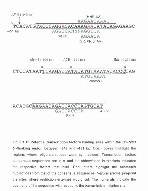
3.1.13 Potential transcription factors binding sites within the CYP2B1
5’-flanking region between -348 and -451 bp... 196
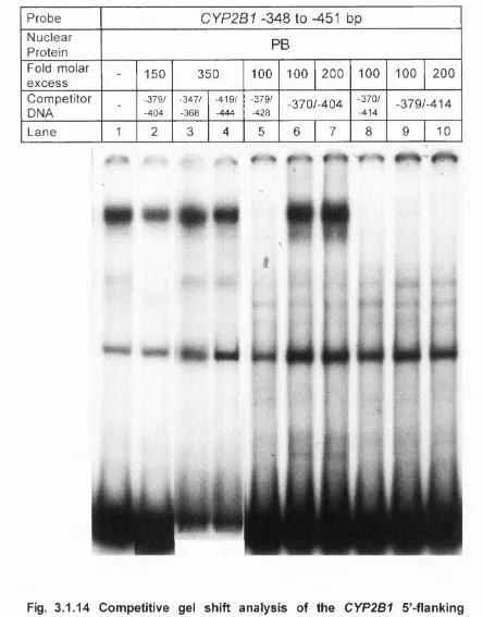
3.1.14 Competitive gel shift analysis of the CYP2B1 5’-flanking
sequence between -348 and -451 bp...197
3.1.15 Schematic diagram showing different subfragments that were
either able or not able to compete for protein(s) binding to the CYP2B1
promoter sequence between -348 and -451 bp...199
3.1.16 DNase I footprint analysis of the sense strand of the CYP2B1
promoter between -179 and -451 bp... 202
3.1.17 DNase I footprint analysis of the template strand of the CYP2B1
promoter between -179 and -451 bp... 203
3.1.18 DNase I footprint analysis of the sense strand of the CYP2B1
promoter between -348 and -451 bp... 205
3.1.19 DNase I footprint analysis of the EBNA-1 sequence... 206
3.1.20 DNase I footprint analysis of the template strand of CYP2B1
promoter between -1 and -179 bp...207
3.1.21 DNase I footprint analysis of the sense strand of the CYP2B1
promoter between -348 and -451 bp using varying concentrations of
Ca^^ and Mg^'"... 209
3.1.22 Sequence comparison of the CYP2B1 5’-fllanking sequence
with YY1 consensus binding sites... 212
3.1.23 Gel shift analysis of the YY1 consensus oligonucleotide
sequence...213
3.1.24 Competitive gel shift assay of the CYP2B1 5'-flanking sequence
with YY1 oligonucleotide sequence... 214
5’-flanking sequence between -348 and -451 bp as probes...218
3.1.26 Competitive gel shift analysis of the CYP2Bf promoter sequence
between -179 and -348 bp...221
3.1.27 Competitive gel shift analysis of the CYP2B' promoter between
-348 and -451 bp...222
3.1.28a Schematic diagram of a possible scenario whereby the CYP2B1
promoter from -179 to -348 bp and -348 to -451 bp could cross compete
with each other for nuclear protein binding in gel shift assays...225
3.1.28b Schematic diagram of a possible scenario whereby the CYP2B1
promoter from -179 to -348 bp and -348 to -451 bp could cross compete
with each other for nuclear protein binding in gel shift assays...226
3.1.29 Gel shift analysis of the CYP2B1 5’-flanking sequence between
-348 and -451 bp with different batches of liver nuclear extracts...229
3.1.30 The effects of different chemicals on liver nuclear protein binding
to the CYP2B1 5’-flanking sequence between -348 and -451 bp... 234
3.1.31 The effects of different chemicals on liver nuclear protein binding
to the CYP2B1 5’-flanking sequence between -348 and -451 bp... 236
3.1.32 The effects of protein kinase and phosphatase inhibitors on the
CYP2B1 promoter between -348 and -451 bp... 238
3.1.33 The effects of different salts on liver nuclear protein binding to
CYP2B1 5’-flanking sequence between -348 and -451 bp...241
3.1.34 The effects of chemicals on liver nuclear protein already bound
to the CYP2B1 5’-flanking sequence between -348 and -451 bp... 242
3.1.35 Gel shift analysis of the CYP2B1 5’-flanking sequence between
-348 and -451 bp with FAZA 967 cell lines... 246
3.1.36 Gel shift analysis of nuclear protein isolated from primary rat
hepatocytes on the CYP2B1 5'-flanking sequence between
-348 and -451 bp... 247
3.1.37 SDS-polyacrylamide gel electrophoresis of nuclear extracts
isolated using different methods... 248
3.1.38 Gel shift analysis of the CYP2B1 5'-flanking sequence between
3.1.39 Comparison of three methods of isolating nuclear protein from
primary rat hepatocytes...252
3.1.40 The effect of trolox on nuclear protein from primary rat hepatocytes
at various time-points upon seeding...253
3.1.41 Gel shift analysis of the CYP2B1 5’-flankirg sequence between
-348 and -451 bp with liver nuclear extract isolated using the Rosette
and Karin m ethod... 256
3.1.42 Schematic diagram of DNA affinity purification of liver nuclear
protein with Dynabeads... 259
3.1.43 Gel shift analysis of CYP2B1 5’-flanking sequence between
-348 and -451 bp with liver enriched nuclear extracts from
PB-treated rats... 261
3.1.44 Gel shift analysis of CYP2B1 5’-flanking sequence between
-348 and -451 bp with DNA affinity enriched liver ruclear extracts from
PB-treated rats... 263
3.1.45 Gel shift analysis of CYP2B1 5’-flanking sequence between
-348 and -451 bp with partially purified liver nuclear extracts from
PB-treated rats... 265
3.1.46 Sequence comparison of the CYP2B promoter regions between
-183 and -199 bp with the Octamer consensus binding site... 268
3.1.47 Gel shift analysis of the CYP2B2 promoter region between
-183 and -199 bp... 269
3.1.48 Antibody interference analysis using an octamer antibody... 271
3.1.49 Supershift analysis using an octamer antibody...272
3.2.1 The effect of 2-AP on CYP2B mRNA in primary rat hepatocytes 279
3.2.2 The effect of 2-AP on CYP2B mRNA in rat liver in vivo... 281
3.3.1 Schematic diagram of the pGL3 and pRL luciferase vector maps. ...287
3.3.2 Schematic diagram of reporter gene constructs for transient
transfection experiments...288
3.3.3 Transient luciferase activity of pGL3 reporter constructs... 292
3.3.4 Transient luciferase activity of pGL3 reporter constructs transfected
into either primary rat hepatocyte cultures or HeLa cell cultures...294
the pGL3 reporter constructs into primary rat hepatocyte cultures...296
3.3.6 Transient transfection of pGL3 constructs containing CYP2B1 proximal promoter sequences... 298
3.3.7 Transient transfection of the CYP2B1 promoter fusion constructs....301
3.3.8 Transient transfection of CYP2B1 promoter fusion constructs into rat livers in vivo... 305
3.3.9 Fold induction of luciferase activity upon PB treatment as a consequence of different CYP2B1 promoter constructs... 307
List of Tables
1.1 Examples of drug-metabolising enzymes involved in Phase I and Phase II reactions... 231.2 Diversity of nomenclature of some mammalian CYPs... 30
1.3 Overview of CYP families and enzymes functions in various species. ..32
1.4 Specific CYP genes, whose expression is increased, by distinct classes of inducers...36
2.1 Preparation of successive dilution of DNase 1...131
2.2 DNase I mock binding reaction mix... 131
Abbreviations
AGs anti-glucocorticoids
AhR Aryl hydrocarbon receptor
ALAS 5-aminolevulinate synthase
2-AP 2-aminopurine
AR Androgen receptor
Arnt AhR nuclear translocator protein
ATP Adenine triphosphate
ATPy-S Adenosine-5’-0-(3-thiotriphosphate)Li4
bHLH basic helix-loop-helix
BSA Bovine serum albumin
cAMP cyclic adenosine monophosphate
CAR Constitutively activated receptor
CAT Chloramphenicol acetyltransferase
C/EBP CCAAT/Enhancer binding protein
CD! 1-Cyclohexyl-3-(2-morpholinoethyl) carbodiimide metho-
p-toluene sulfonate
CIP Calf intestinal alkaline phosphatase
C0CI2 Cobalt chloride
CTF CCAAT-transcription factor
CYP Cytochrome P450
db-cAMP Dibutyryl cAMP
DDT 1,1,1-trichloro-2,2-bis(p-chlorophenyl)ethane
Dex Dexamethasone
DME Drug-metabolising enzymes
DMSO Dimethylsulphoxide
dNTP 2’ Deoxynucleoside 5’- triphosphate
DPBF Dystrophin promoter bending factor
DTT Dithiothreitol
EDNA Epstein-Barr virus nuclear antigen
EBSS Earles-balanced salt solution
EDTA Ethylenediaminetetra-acetic acid
EGTA Ethylene glycol-0,-0’-bis(2-amino-ethyl)-A/,A/,A/’,A/’-tetra acetic acid
EMSA Electrophoresis mobility shift assay
EtBr Ethidium bromide
FBS Foetal bovine serum
GAPDH Glyceraldehyde-3-phosphate dehydrogenase
GR Glucocorticoid receptor
GRE Glucocorticoid -responsive element
HAH Halogenated aromatic hydrocarbon
HEPES N-2-Hydroxyethylpiperazine-N’-2-ethanesulphonic acid
HNF Hepatocyte nuclear factor
hPAR human peroxisome proliferator activator receptor
hsp Heat-shock protein
IL Interleukin
IP Intra-peritoneal
IPTG Isopropyl p-D-Thiogalactopyranoside
1RS Insulin responsive sequence
LB Luria-Bertani
MAP Mitogen-activated protein kinase
MOPS 3-[N-Morpholino]propane-sulfonic acid
NADH Nicotinamide-adenine dinucleotide (reduced)
NADPH Nicotinamide-adenine dinucleotide phosphate (reduced)
NaF Sodium fluoride
NaNs Sodium azide
NE Negative element
NF1 Nuclear factor 1
NP-40 Nonidet-40
Cet Octamer
OK Okadaic acid
PAGE Polyacrylamide gel electrophoresis
PB Phénobarbital
PBRE PB responsive element
PBREM PB response enhancer module
PBRU PB response unit
PBS Phosphate buffered saline
pBS pBluescript
PCB Polychlorinated biphenyl
PCN Pregnenolone-16a-carbonitrile
PCR Polymerase chain reaction
PDE Phosphodiesterase
PE Positive element
PEPCK Phosphoenolpyruvate carboxykinase
PK Protein kinase
PLB Passive lysis buffer
PMSF Phenylmethylsulfonyl fluoride
PNK Polynucleotide kinase
PP Peroxisome proliferator
PP1 Protein phosphatase-1
PP2A Protein phosphatase-2A
PPAR Peroxisome proliferator activator receptor
PPRE Peroxisome proliferator response element
PR Prolactin receptor
psi pound per square inch
PXR Pregnane X receptor
RARE Retinoic acid response element
RP Random prime
RT Room temperature
RXR Retinoid X receptor
SAPK Stress-activated protein kinase
SDS Sodium dodecyl sulphate
(Sp)-cAMPS Phosphorothioate stereoisomer of cAMP
SRC-1 Steroid receptor coactivator-1
SRE Serum response element
SRF Serum response factor
STAT Signal transducer and activator
SV40 Simian virus 40
TAE T ris-acetate-EDTA
TBE Tris-borate-EDTA
TCP Ternary complex formation
TCPOBOP 1,4-bis-[2-(3,5-dichloropyridyloxy)]benzene
TE Tris-EDTA
TEMED A/,A/,A/’,A/’-tetramethylethylenediamine
TESS Transcription element search software
TK Thymidine kinase
Tm Melting temperature
TNF Tumour necrosis factor
Tris Trizma® Base (Tris[hydroxymethyl]amino-methane)
VDR Vitamin-D receptor
X-gal 5-Bromo-4-Chloro-3-indolyl-p-D-Thiogalactopyranoside
XRE Xenobiotic-responsive element
Chapter One
1.1 Overview of xenobiotic metaboiism
Each living organism, from microorganisms, plants to animals are
under constant exposure to many different kinds of foreign compounds.
These are both naturally occurring and synthetic chemicals. Most of the
xenobiotics, being lipophilic in nature, are easily absorbed by an organism.
The very property, i.e. lipophilicity, which facilitates the absorption of these
chemicals also makes them difficult to eliminate. If they are continually
absorbed and not excreted from the organism fast enough, they would
accumulate to eventually overwhelm and kill the organism. To prevent these
chemicals from accumulating to toxic levels, organisms have evolved
defense mechanisms to eliminate these chemicals. There are two main
detoxification systems: one is a non-catalytic multidrug resistance system,
where harmful compounds bind to P-glycoprotein and are then transported
out of the cell (Endicott and Ling, 1989); the other is a metabolic process
involving several enzyme systems. These enzymes are generally known as
drug-metabolising enzymes and they eliminate foreign compounds
basically by making them more water-soluble. The latter system is the major
detoxification mechanism utilised by an organism and is the most complex
one too.
Drug-metabolising enzymes are generally divided into two broad
categories, ‘Phase I’ and ‘Phase 11’ (Williams, 1971). Phase I
(functionalisation) reactions involve hydrolysis, reduction and oxidation. They
expose or introduce a functional group, such as a hydroxyl, to the parent
usually small and further Phase II (conjugation) reactions, whereby a
conjugate such as glutathione is added to the functional group, are usually
required to generate a product relatively hydrophilic that is readily excreted
(Testa and Jenner, 1976, Nebert, 1994). Unfortunately, the detoxication
process can sometimes produce intermediate or final products that are
more toxic and/or carcinogenic than the parent compound. Exahiples of
some drug-metabolising enzymes involve in Phase I or II reactions are
listed in Table 1.1.
1.2 Cytochrome P450~dependent mixed function oxygenase
system
Among the Phase I enzymes, cytochrome P450 is one of the m ost
versatile biological catalysts because of the number of xenobiotics it
detoxifies and activates to reactive intermediates (Guengerich, 1987,
Waterman and Johnson, 1991). Cytochrome P450 is widely distributed in
nature and has been found to be present in virtually all mammalian tissues
examined, with the greatest abundance in the liver. With the exception of
some soluble bacterial proteins, all known cytochrome P450s are
membrane-bound and located predominantly in the endoplasmic reticulum.
However, some cytochrome P450s have also been found in mitochondria
(Hollis, 1990).
Cytochrome P450 is a monooxygenase and catalyses the
Phase I enzymes
Cytochrome P450
Flavin-containing monooxygenase Aldehyde dehydrogenase
Epoxide hydrolase
NADPH-cytochrome P450 reductase Monoamine oxidase
Carboxylesterase
Phase II enzymes
UDP glucuronosyltransferase Glutathione-S-transferase Acetyltransferase
Methyltransferase P-glucuronidase Sulfotransferase
Table 1.1 Examples of drug-metabolising enzymes involved in Phase I and
concomitant reduction of the other oxygen atom to water as represented in
the following equation, where RH refers to the
substrate:-RH + O
2+ NAD(P)H + H*
Cytochrome P45^RQH + HgO + NAD(P)*
Cytochrome P450 cannot carry out the above reaction alone but requires
other components in the cytochrome P450-dependent mixed function
oxygenase system (Lu and Coon, 1968). The mammalian microsom al
system is made up of cytochrome P450, NADPH-cytochrome P450
reductase and phospholipid. NADPH is the reducing co-factor that donates
electrons to mammalian cytochrome P450 for oxidative reaction to occur,
while NADH is the electron donor in bacterial systems. However, NAD(P)H
is a two-electron donor and cytochrome P450 accepts one electron at a
time. Cytochrome P450, therefore cannot interact directly with the co-factor.
Instead, it receives the electrons via an accessory enzyme. In microsom al
systems, the accessory enzyme is NADPH-cytochrome P450 reductase.
This is a flavoprotein and possesses two flavin prosthetic groups. This
enables it to accept the two electrons donated by NAD(P)H simultaneously
and transfer one electron to each of two different cytochrome P450s.
Because NADPH-cytochrome P450 reductase transfers electrons much
faster than cytochrome P450 can use them, one NADPH-cytochrome P450
reductase provides electrons for more than one cytochrome P450. This may
account for the low ratio of NADPH-cytochrome P450 reductase to
NADPH-cytochrome P450 reductase to every 10 to 20 molecules of NADPH-cytochrome
P450) (Shephard, etal., 1983, Parkinson, 1996, Josephy, etal., 1997).
Another constituent of the cytochrome P450-dependent system is
phospholipid. It is not required for soluble forms of cytochrome P450 but is
particularly important for membrane-bound forms because it facilitates
interactions between NADPH-cytochrome P450 reductase and cytochrome
P450 in the endoplasmic reticulum. However, the actions by which
phospholipid facilitates this interaction is not very clear (Parkinson, 1996,
Josephy, etal., 1997).
Hepatic microsomes also contain another haemoprotein, cytochrome
bg. Although NADPH-cytochrome P450 reductase is absolutely required for
cytochrome P450 activity, cytochrome bg can transfer the second of the two
electrons required by cytochrome P450. Cytochrome P450 catalysed
turnover of some substrates can be increased synergistically by electron
transfer from cytochrome bg. This is not always simply due to an increase in
the rate of catalysis by cytochrome P450, cytochrome bg can also increase
the apparent affinity with which certain cytochrome P450s bind their
substrates. Some cytochrome P450s, including CYP3A, CYP2E1 and
CYP2C9 have been demonstrated to require cytochrome bg for maximal
catalytic activities in reconstituted monooxygenase systems (Shet, at a/.,
1995, Yamazaki, at a/., 1996, Shimada and Yamazaki, 1998). A more
detailed description of the oxygenation of a substrate by cytochrome P450 is
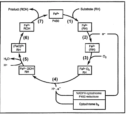
presented in Fig. 1.1.
Substrate (RH) Product (RON)
P450
H+
H + , e
-NADPH'Cytochrome P450 reductase
Cytochrome bg
Fig. 1.1 Catalytic cycle of cytochrome P450. (1) binding of substrate (RH),
(2) reduction of ferric, substrate-bound enzyme to the ferrous form, (3)
binding of oxygen, (4), (5) and (6) addition of second electron, released
water and oxidized substrate and (7) releasing of oxidized substrate (ROM),
Apart from hydroxylation reaction, cytochrome P450 also catalyses a
range of other reactions such as N-, 0-, S-dealkylation, sulfoxidation,
deamination and N-oxide reduction as shown in Fig. 1.2.
1.3 Discovery of cytochrome P450
In 1958, Garfinkel and Klingenberg while studying microsomal haem
proteins by optical spectroscopy, observed a strong absorption band at 450
nm (Soret band) when pig and rat liver microsomes were treated with a
reducing agent in the presence of carbon monoxide (Garfinkel, 1958,
Klingenberg, 1958). This was a unique characteristic because many other
haemoproteins that form a complex with carbon monoxide absorb light
maximally at ~420 nm. The protein was later purified by Omura and Sato,
who confirmed that the unique absorption spectra was indeed due to a new
class of haem-binding protein and named it cytochrome P450, based on its
atypical absorption maximum at 450 nm (Omura and Sato, 1964). With time,
this terminology was found to be unsuitable since cytochrome P450 acts as
an oxygenase rather than just an electron carrier. The term ‘haem-thiolate
protein’ was suggested in replacement of ‘cytochrome’, however it did not
gain favour and the name cytochrome P450 is still universally recognised
R — ^ R — Ch^OH
Aliphatic Oxidation
OH
Aromatic Hydroxylation
R -N H ^C H 3
R —S - CH3
(R-.MH-CH2OHI
N-Dealkylation
[R --O -C H2OHÎ*
O-Dealkylation
. [ R - S - C H2OH3.
S-Dealkylation
R — CH—CH3
; ^
NH2
OH R — C—CH3
► R - N H2 + HCHO
A - O H + HCHO
R - S H + HCHO
0
I
Rf — S — R)
R — C— CH3 + N H j
NH2
Oxidative Deamination
H'^r OH
,Ri- S - R 2 J - > ë - R ; +
Sulfoxide Formation
] * !J 0 I (CH3)3N H 4
-(CHsJsN - OhI (CH3)3N + —0 ” + H +
N-Oxidation
OH R^l — NH — R2 R^ — N — R2
N-Hydroxylation
Fig. 1.2 Examples of diverse activities catalysed by cytochrome P450s.
1.4 The cytochrome P450 gene superfamily
When cDNA and cloning techniques were introduced in the 1980s,
more new forms of cytochrome P450 were isolated. Each laboratory involved
in cytochrome P450 isolation began developing its own nomenclature
system according to electrophoretic mobility, substrate specificity or
maximal absorption wavelength making the situation very complex. There
were cases where an enzyme was designated several different names. The
fact that cytochrome P450s have a broad substrate specificity and catalyse
different reactions make the classical method of naming an enzyme
according to its function very difficult.
When amino acid sequence data was derived from DNA sequences,
it made possible a naming system based on the amino acid sequence
similarities. Table 1.2 lists a few examples of cytochrome P450s to illustrate
the diversity of the previous nomenclature and how the new classification
helped to overcome this complexity. Since it was first recommended in 1987
(Nebert, et al., 1987), there have been a few revisions (Nebert, at al., 1989,
Nebert, etal., 1991, Nelson, etal., 1993, Nelson, et al., 1996). A cytochrome
P450 gene is named by the italicised root symbol ‘CVP (‘Cyp’ for mouse
and Drosophila) to denote Cytochrome P450, followed by an Arabic number
for the family, a letter for the subfamily and another Arabic number for the
individual gene, i.e. CYP2B1 ÇCyp2bT in mouse). A pseudogene will have
a ‘P (‘ps’ in mouse and Drosophila) after the gene number. The non
italicised form and all capital letters should be used for mRNA, cDNA and
Trivial name
Rat
(name according to the laboratories of)
Gene Symbol Ryan Guengerich Waxman Rabbit Mouse Human CYP1A1 c pNF-B p-NF-B LM6 Pi450 Pi
CYP1A2 d pNF/ISF-G ISF-G LM4 P3 4 5O Pi
CYP2A1 a UT-F 3 - -
-CYP2B1 b PB-B PB-4 LM2 -
-CYP2B2 e PB-D PB-5 LM2 -
-CYP2C6 k PB-C PB-1 - -
-CYP2C11 h UT-A 2c - P450 16a
-CYP2C12 i UT-I 2d - P450 15P
-CYP2D1 - UT-H - - - dbi
CYP2E1 j - - LM3a - j
CYP3A1 P - - LM3 - P450nf
CYP4A1 - PB/PCN-E PB-2a - -
-Table 1.2 Diversity of nomenclature of some mammalian CYPs. (adapted
from (Paine, 1991, Soucek and Gut, 1992) and references therein for
1996). Members within the same family are defined as usually having >40%
amino acid sequence identity and mammalian sequences within the sam e
subfamily are always >55% identical. Although these definitions were made
arbitrarily, they turned out to be very useful despite a few exceptions
(reviewed in (Nelson, etal., 1993, Nelson, 1998)).
By 1996, 481 CYP genes were identified in 85 eukaryote and 20
prokaryote species, the number is still increasing (Nelson, at a!., 1996). But
how did CYP evolve to become such a superfamily of proteins? This
superfamily is ancient and believed to have begun with only a few genes
coding for CYP forms that were engaged in the metabolism of endogenous
substrates important for cellular functions (Nebert, 1991, Soucek and Gut,
1992). The increase in the number of CYP genes, according to the
evolutionary tree, arose during the past 400 million years. And ‘animal-plant
war-fare' is believed to be the driving force for the recent burst in new CYP
genes, particularly in the CYP2 family (Nebert and Gonzalez, 1987, Gonzalez
and Nebert, 1990). New genes encoding for new forms of CYP appear
through increased frequency of gene duplications and conversions as the
animal continues to encounter new types of foreign compounds, including
drugs and pesticides of the present days.
The diversity of genes has evolved mainly in the CYP families 1 to 4.
Hence, it is not surprising to find these four families more important in
xenobiotic metabolism than the other CYP families which are involved
mainly in the metabolism of endogenous substrates such as steroids, fatty
acids and hormones (Table 1.3). Apparently, most of the CYPs involved in
Gene families
Occurrence and functions
CYP1 Vertebrates; dioxin-induclbie; metabolism of polycyclic
hydrocarbon, halogenated and heterocyclic hydrocarbon, and aromatic amines
CYP2 Vertebrates and invertebrates; metabolism of drugs
and environmental chemicals
CYP3 Vertebrates; metabolism of drugs and environmental
chemicals
CYP4 Vertebrates, fatty acid hydroxylases; invertebrates,
unknown function(s)
CYP5 Vertebrates; thromboxane synthase
CYP6 Insects; metabolism of plant products and pesticides
CYP7A Vertebrates; cholesterol 7a-hydroxylase
CYP7B Vertebrates; unknown function(s)
CYP8 Vertebrates; prostacyclin synthase
CYP9 Insects
CYP10 Molluscs (mitochondrial enzyme)
CYP11 Vertebrates; cholesterol side-chain cleavage, steroid
1 1p-hydroxylase, and aldosterone synthase
(mitochondrial enzyme)
CYP12 Insects (mitochondrial enzyme)
CYP13 Nematodes
CYP14 Nematodes
CYP15 Insects
CYP16 Nematodes
CYP17 Vertebrates; steroid 17a-hydroxylase
CYP18 Insects
CYP19 Vertebrates; aromatization of androgens
CYP21 Vertebrates; steroid 21-hydroxylase
CYP24 Vertebrates; steroid 24-hydroxylase (mitochondrial enzyme)
CYP27 Vertebrates; steroid 27-hydroxylase (mitochondrial enzyme)
CYP51 Animals, filamentous fungi, yeast and plants; sterol biosynthesis
CYP52 Yeast; alkane hydroxylase
CYP53 to CYP62 Fungi
CYP71 to CYP92 Plants
CYP73 Plants, cinnamic acid hydroxylase
CYP101 to CYP118 Bacteria
Table 1.3 Overview of CVP familles and enzymes functions in various
in xenobiotic metabolism exhibit broad and overlapping substrate
specificities allowing them to deal with a wide range of foreign compounds.
1.5 Molecular mechanisms of CYP gene expression
The regulation of CYP gene expression is complex and governed by
several different mechanisms. CYP expression can be tissue, strain and
sex-specific and/or regulated at the level of development. Certain families of
CYP, particularly those involved in xenobiotic metabolism, can also be
induced in response to many foreign compounds (reviewed in (Bernhardt, et
a/., 1995; Gonzalez, etal., 1989). And more than one mechanism is usually
involved in the regulation of any particular CYP.
Although the most common means of regulating CYP expression is
at the level of transcription, some forms of CYP have been found to be
regulated via post-transcriptional mechanisms, at the level of mRNA (Song,
etal., 1987) or protein (Eliasson, etal., 1990) stabilisation as shown in Fig.
1.3 (reviewed in (Gonzalez, 1989, Okey, 1990)).
1.5.1 Xenobiotic inducible CYPs
It was recognised more than 20 years ago that many xenobiotics can
induce their own metabolism and the metabolism of other compounds of
similar structure (Conney, 1967). This happens because they induce the
expression of one or more CYPs that are responsible for their metabolism.
Gene mRNA
transcription Processing stabilisation Translation
i
i
1
HHHHf
AAAAAAAEnzyme stabilisation
1A1 2012 3A6 1A2 1A1 2H1
1A2 2D9 4A1 2B1 2H2
2B1 2E1 11A1 2B2 3A1/2
2B2 2H1 11B1 2012 3A5
2C7 2H2 17 2E1 11A1
2011 3A1/2 21A1
2E1 2E1
3A1/2 3A6
Fig. 1.3 Diverse mechanisms in the regulation of CYP expression.
(reproduced from (Porter and Coon, 1991) and references therein for the
induced only in response to the xenobiotics that they metabolised.
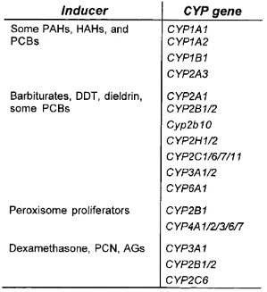
Mammalian CYP inducers can be categorised into four distinct classes as
shown in Table 1.4. Although each class of inducer is capable of inducing a
number of CYP genes, a particular subfamily of CYP genes, most efficient in
metabolising the inducers, are predominantly induced.
For instant, polycyclic aromatic hydrocarbons (PAHs) such as
benzopyrenes and anthrenes induce CYPs belonging to the CYP1A
subfamily (Whitlock, etal., 1996), chlorinated pesticides such as DDT and
drugs like PB greatly induce proteins of the CYP2B subfamily (Waxman and
Azaroff, 1992). While peroxisome proliferators and glucocorticoids such as
dexamethasone predominantly induce members of the CYP4A (Reddy and
Mannaerts, 1994) and CYP3A (Okey, 1990) subfamily respectively. The
increase in the amount of these four families of CYPs is achieved mainly by
transcriptional activation and a simplified model of the m olecular
mechanisms known so far is presented in Fig. 1.4 (Dogra, at a!., 1998).
1.5.1.1 Steroid inducible CYP genes
Glucocorticoids, such as dexamethasone, activate the transcription of
numerous genes in the liver, such as the rat CYP3A1 gene, via the classical
glucocorticoid receptor-dependent mechanism. Members of the CYP3A
subfamily are the most induced by these molecules. However, the
involvement of the glucocorticoid receptor in the transcriptional activation of
CYP3A genes is controversial (Fig. 1.4). In humans, CYP3A5 induction
Inducer
CYP gene
Some PAHs, HAHs, and RGBs
CYP1A1 CYP1A2 CYP1B1 CYP2A3
Barbiturates, DDT, dieldrin, some RGBs
CYP2A1 CYP2B1/2 Cyp2b10 CYP2H1/2 CYP2C1/6/7/11 CYP3A1/2 CYP6A1
Peroxisome proliferators CYP2B1
CYP4A1/2/3/6/7
Dexamethasone, PGN, AGs CYP3A1
CYP2B1/2 CYP2C6
Table 1.4 Specific CVP genes, whose expression is increased, by distinct
classes of inducers. PAHs, polycyclic aromatic hydrocarbons; HAHs,
halogenated aromatic hydrocarbons; RGBs, polychlorinated biphenyls; DDT,
1,1,1 -trichloro-2,2-bis(p-chlorophenyl)ethane; PON,
pregnenolone-16a-carbonitrile; AGs, anti-glucocorticoids, (reproduced and modified from
Nucleus PAH
I
Arnt / )hsp90 AhR AmtXRE PPARa RXRa PP
T RXRa
GYP4A
PPRE
PB PPARa
0 ^ 6
J CARandrostane
RXRa
♦
CYP2B
NF1
PBRE
DEX
f
ORE GR7
Fig. 1.4 Simplified general model for the transcription activation of various
xenobiotic inducible CYP genes by their respective prototype inducers.
PAH, polycyclic aromatic hydrocarbon; PP, peroxisome proliferator; PB,
phénobarbital; Dex, dexamethasone; AhR, aryihydrocarbon receptor; hsp90,
heat-shock protein 90; PPARa, peroxisome proliferator-activated receptor;
CAR, constitutively activated receptor; GR, glucocorticoid receptor; Arnt, AhR
nuclear translocator protein; RXRa, retinoid X receptor; XRE,
xenobiotic-responsive element; PPRE, peroxisome proliferator response element;
GRE, glucocorticoid-responsive element, (adapted and modified from
rat, CYP3A23 induction is not mediated through the classical mechanism.
Instead, Schuetz et al (1998) (Schuetz, et a!., 1998) and Lehmann et ai
(1998) (Lehmann, et al., 1998) have recently reported the activation of a
novel orphan receptor, PXR (pregnane X receptor) by glucocorticoids,
pregnanes and other compounds that induce CYP3A. These compounds
also activate another nuclear receptor termed hPAR (human peroxisome
proliferator activator receptor) which apparently regulates only human and
not mouse CYP3A expression (Bertilsson, et al., 1998). It is possible that
PXR and hPAR represent orthologous proteins from different species.
In rats, PXR heterodimerises with RXR (retinoid X receptor) and binds
to a dexamethasone-responsive element in the promoter sequence of the
CYP3A23 gene and is able to activate reporter gene transcription
(Quattrochi, etal., 1998).
1.5.1.2 Peroxisome proliferator inducible CYP genes
Peroxisome proliferators comprise a variety of structurally dissim ilar
compounds including hypolipidemic drugs, industrial solvents and
herbicides. As the name implies, they induce peroxisome proliferation and
also lead to an increase in the oxidation of fatty acids through peroxisomal
P-oxidation and microsomal co-oxidation pathways (Lock, et al., 1989,
Johnson, etal., 1996). The microsomal co-oxidation reaction is catalysed by
the CYP4A subfamily.
Specific members of the CYP4A subfamily are induced by
1993, Roman, et al., 1993). The rat CYP4A1 and rabbit CYP4A6 are the most
highly induced in the liver (Johnson, at a!., 1996). A member of the nuclear
receptor superfamily known as peroxisome proliferator activator receptor
(PPARa) is involved in the transcriptional activation of CYP4A genes
(Issemann and Green, 1990). However, its ability to activate transcription
requires the binding of ligand. A number of exogenous peroxisome
proliferators (Devchand, at al., 1996) as well as fatty acids (Keller, at al.,
1993) have been observed to bind and activate PPARa. Because of the
structural diversity of exogenous peroxisome proliferators, the idea of fatty
acids being the true ligand for PPARa is more favourable. There is a
possibility that peroxisome proliferators may lead to an accumulation of
endogenous fatty acids which then activate PPARa resulting in
transcriptional activation (Dogra, at al., 1998). The general molecular
mechanism for the activation of CYP4A gene expression is shown in Fig.
1.4. A peroxisome proliferator responsive element (PPRE) has been
identified in the promoter of genes responsive to this class of inducers.
Apparently, PPARa has to heterodimerise with RXRa in order to bind to the
PPRE and inducers activate transcription by enhancing the dimérisation
between PPARa and RXRa (Palmer, at ai, 1994).
1.5.1.3 Polycyclic aromatic hydrocarbon inducible CYP genes
Unlike chemicals that induce CVP4AorCYP2B subfamily members, high
similar structures, i.e. uniformly planar and aromatic, which include dioxins,
3-methylcholanthrene and benzo(a)pyrene. Of all the xenobiotic inducible
CYP subfamilies, the molecular mechanism regulating CYP1A1 gene
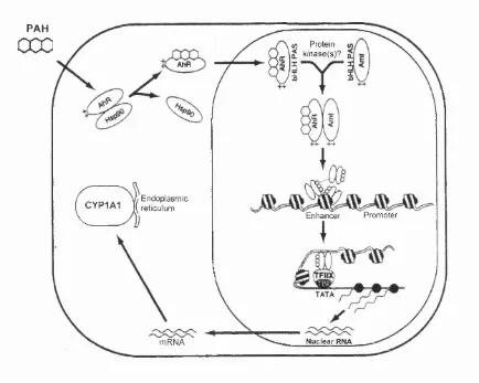
expression is the most well-characterised to-date.
The inducer on entering the cell interacts with a protein known as
aryihydrocarbon receptor (AhR) and dissociates the AhR from heat-shock
protein 90 (hsp90) in the cytosol. The liganded AhR then translocates into
the nucleus and heterodimerises with a nuclear protein called AhR nuclear
translocator protein (Arnt) through the Helix-Loop-Helix (bHLH) and the PAS
domains. The heterodimer then binds to specific DNA sequences known as
xenobiotic-responsive elements (XRE) and activates transcription. Protein
phosphorylation may be required for heterodimerisation (Chen and Tukey,
1996), XRE-binding (Pongratz, etal., 1991) and/or transcriptional activation
(Li and Dougherty, 1997) to occur. The chromatin structure of the enhancer
(XRE) and the promoter region of the CYP1A1 gene assumes a
nucleosomal configuration in the uninduced state. Upon induction, the
AhR/Arnt heterodimer binding to the XRE can disrupt a nucleosome. It then
recruits general transcription factors, somehow disrupts the nucleosomal
structure at the promoter region, and finally stabilises the binding of the
general transcription factors to the promoter (reviewed (Whitlock, et a/.,
1996)). Fig. 1.5 shows the molecular mechanism in more detail than Fig.
PAH
000
V) Protein^ kinase(s)? AhR
-H-Endoplasmic reticulum
CYP1A1 I
Promoter Enhancer
N uclear RNA m RN A
Fig. 1.5 Model for the induction of CYP1A1 transcription by polycyclic
1.5.1.4 Phénobarbital (PB) inducible CYP genes
PB, an anti-epileptic drug, causes pleiotropic effects which include
induction of numerous drug-metabolising enzymes and various cellular
processes (Fig. 1.6 and (Honkakoski and Negishi, 19G8b)). A wide variety of
structurally dissimilar compounds such as pesticides, chlorinated
biphenyls, organic solvents and drugs also induce a similar set of enzymes
as PB and are generally known as ‘PB-like’ inducers (Fig. 1.7).
The mechanism whereby PB induces gene expression was thought
to be highly conserved when it was found to induce genes in both
prokaryotes (bacteria) and eukaryotes (birds and mammals). However, this
does not appear to be the case. PB regulation of certain genes such as
CYP2B1, CYP2B2 and CYP102 is blocked by cycloheximide signifying that
ongoing protein synthesis is required for their increased transcription (Bhat,
etal., 1987, Waxman and Azaroff, 1992). For CYP3A, CYP2H1 and CYP2H2
genes, cycloheximide is observed to synergise with PB leading to
‘superinduction’ (Burger, at a/., 1990, Hamilton, at a/., 1992). The
phenomenon seen in the latter case is deduced to be due to the loss of a
labile repressor protein (Dogra, at a/., 1993). Furthermore, the inductive
response of different proteins to PB can differ enormously (e.g. 50- to 100-
fold with CYP2B1 and CYP2B2 and 2- to 4-fold with CYP2A1 and CYP2C6). It
therefore seems that PB might induce CYPs by more than one mechanism.
So far, the molecular mechanism of gene activation by steroids,
peroxisome proliferators and polycyclic aromatic hydrocarbon has been
Cell Growth
Induction of Genes
. . .
.
\
f
CYP450 reductase
Liver h y p e rtro p h y ^ \
/
^poxkis hydrolase
Tumour Promotion-*-
cyp450
s-2A,2B,2C,3A
"
UDP-glucuronyltransferase
_
^ /
\
Glutathione S-transferases
Cell Communication
/
\
Haem Synthesis & Metabolism Hepatic ER Proliferation
Fig. 1.6 Pleiotropic effects o f PB. (adapted from (Honkakoski and Negishi,
CH3CH2 o
NH
Phénobarbital (PB)
9 ^
C H —CHCH3
Isosafrole
% o
NH.
ÇCI2
Cl
■Cl
Allylisopropylacetamide (AIA)
Chlordane
Fig. 1.7. Structure diversity of PB and PB-iike inducers, (adapted
regulatory elements. Thus, the idea of a receptor involved in PB mediated
transcriptional activation is highly probable.
Recently, Honkakoski and co-workes (1998) (Honkakoski, et al.,
1998b) have identified an orphan nuclear receptor known as constitutively
activated receptor (CAR) th a t is involved in PB induction o f the m ouse
Cyp2b10 and the human CYP2B6. CAR acivates Cyp2b10 and CYP2B6
gene transcription by binding to nuclear receptor binding sites, i.e. NR1
and/or NR2 (see section 1.6.2 for more detail) within the PB-responsive
enhancer module (PBREM, generally knov\n as PB-responsive element
(PBRE)) as a heterodimer with RXRa. CAR la s previously been shown to
dimerise with RXR and bind to a subset of retinoic acid response elements
(RAREs) (Baes, et al., 1994). However, CAR appears to function differently
from the conventional nuclear receptor pathway. Unlike classical nuclear
receptors which are activated by their cognate ligands, CAR is a constitutive
transcriptional activator (Choi, et al., 1997). Recently, androstane
metabolites have been identified, in the mouse, as ligands for an isoform of
CAR called CARp. Instead of activating the receptor, these metabolites
inhibit the constitutive activity of CARp (Forman, et al., 1998). These
androstane ligands are found to be examples of naturally occurring inverse
agonists that can reverse the transcriptional activation by nuclear receptors
(Klein, etal., 1996).
For the activation of classical receptors, ligand binding induces a
transcriptional co-activators like steroid receptor coactivator-1 (SRC-1) for
transcriptional activation (Onate, et al., 1995). On the other hand, CAR has
been shown to act in an opposite manner and is speculated to adopt an
active conformation in the absence of ligand. The binding of ligand to CAR
has been shown to directly dissociate the interaction between CAR and
SRC-1 by shifting the receptor to an inactive conformation (Forman, at a!.,
1998). However, the micromolar concentration of androstenol needed to
inhibit CAR in in vitro studies is higher than that reported in the circulation of
adult man (Gower and Ruparelia, 1993). Hence, it is still not entirely clear as
to how CAR is repressed in the liver.
Sueyoshi ef a/ (1999) (Sueyoshi, etal., 1999) demonstrated that the
cotransfection of CAR with a reporter construct containing either the mouse
or human PBREM sequence into HepG2 cells gave high levels of luciferase
activity. The endogenous CYP2B6 mRNA, normally not expressed in HepG2
cells, were also detected upon transfection with CAR. Both the luciferase
activity and the endogenous CYP2B6 mRNA expression were suppressed
when CAR transfected cells were treated with androstenol. The subsequent
addition of PB to androstenol-treated CAR-transfected cells overcomes the
suppressive effect of androstenol and was observed to induce luciferase
activity and endogenous CYP2B6 mRNA expression. Sueyoshi et al (1999)
(Sueyoshi, etal., 1999) believe the transfection results suggested that PB
induces CAR probably by displacing androstenol. However, there is no
evidence at present indicating that CAR does bind PB. Furthermore,
CYP2BS can also be induced by many other structurally dissimilar ‘PB-like’
clear. It is not known if PB and PB-like inducers act like peroxisome
proliferators, leading to the accumulation of an endogenous substrate which
then derepressed CAR resulting in transcriptional activation. An indication
that this might be the case had been proposed by Shaw et al (1993) (Shaw,
at a/., 1993) when they observed that the antiprogestin-antiglucocorticoid, RU486 could block the increased expression of reporter constructs
containing either the CYP2B1 or CYP2B2 5’-flanking sequences by PB. They proposed that PB acts indirectly to cause the accumulation of an
endogenous steroid, which is the direct inducer of CYP2B genes.
1.6 PB-responsive regulatory elements In PB-lnduclble CYP
genes
1.6.1 Elements proximal to the transcription start site
An element known as the ‘Barbie Box’ located within the promoter
region of the CYP102 and CYP106 genes in Bacillus megaterlum w as initially identified and reported to be important for PB induction (He and
Fulco, 1991, Liang, etal., 1995, Liang and Fulco, 1995). However recently, this sequence is found to be responsive to PB only in the more PB-inducible
CYP102 and not the less PB-inducible CYP106 gene (Shaw, et al., 1998). But the ‘Barbie Box’ sequence is important in the negative regulation of both
CYP102 and CYP106 genes in uninduced bacteria (Shaw, et al., 1998). Liang ef a/ (1995) (Liang, etal., 1995) has reported the presence of a Barbie-box like DNA sequence within many mammalian PB-inducible CYP