Bosn J Basic Med Sci 2014; 14 (2): 81-88
Abstract
Purpose of the study: Can minimally invasive intramedullary osteosynthesis of distal radius fractures provide better therapeutic results than multidirectional locking plates. Retrospective study of patients operated for distal radius fractures, were treated with intramedullary X-screw (XSCR) fi xation and with the multidirectional angle-stable plate system (APTUS). Th e evaluation at -year follow-up included functional status of the wrist and hand, and radiographic fi ndings. In the XSCR group, the functional outcomes of the treated extremity did not achieve values comparable with those of the uninjured side in any of the parameters measured. Th e radiographic fi ndings did not meet the requirements of successful healing due to failure to restore an anatomical volar tilt in . cases. In the APTUS group, comparable values of the injured and the uninjured side were achieved in radial deviation, ulnar deviation, pronation, supination and grip strength. Th e radiographic criteria of successful healing were met by all fractures treated by locking plate osteosynthesis. Implant migration associated with secondary displacement of bone fragments was recorded in . of the XSCR patients and only in . of the APTUS patients. Th e overall evaluation show that intramedullary osteosynthesis does not produce better treatment outcomes compared with plate osteosynthesis in indicated types of fractures. © Association of Basic Medical Sciences of FB&H. All rights reserved
KEY WORDS: distal radius fracture, locking plate osteosynthesis, intramedullary osteosynthesis, complications
osteosynthesis in comparison with volar plating real
benefi t in the treatment of distal radius fractures?
Martin Vlček1*, Edib Jaganjac1, Jan Pech1, David Jonáš1, Radek Kebrle2
1Teaching Hospital Motol, 1st Clinic of Orthopaedic Surgery, V Úvalu 84, 150 06 Prague 5, Czech Republic. 2Institut of Hand and Plastic
Surgery Vysoké nad Jizerou, Dr. Farského 267, 512 11 Vysoké nad Jizerou, Czech Republic.
INTRODUCTION
Fractures of the distal radius are complex injuries with varied prognosis depending on the type of fracture and method of treatment [].Th ere is still no clinical evidence suggesting a superior modality for their management []. The current therapy most frequently involves open reduc-tion and plate osteosynthesis []. Angle-stable fixareduc-tion provides sufficient stability of the fracture to allow early start of rehabilitation soon after surgery; consequently, the total treatment time is shorter and good functional outcomes are achieved even in unstable grossly com-minuted intra-articular fractures [, ]. Application of a volar plate with angle-stable fixation has been used suc-cessfully in a number of cohort studies but needs to be ex-amined in stringent trials to determine if there is any
ben-efit when compared with other treatment modalities []. Intramedullary osteosynthesis is used for fragment fixa-tion less frequently and good radiographic and funcfixa-tional outcomes have been published []. However, its range of indications limited to the management of extra-articular and simple intra-articular fractures is a disadvantage of the method []. Only a few clinical evaluations are available. Lerch et al. [] in an isolated study on a small patient group report comparable results for plate and intramedullary os-teosyntheses (Targon DR, Aesculap Implant Systems, Cen-ter Valley, PA, U.S.A.) and relate the excellent functional scores for nailing to the minimally invasive procedure. Ilyas et al. [] have reported that using the intramedullary nail (Micronail, Wright Medical Technologies, Arlington, TN, U.S.A.) in the treatment of displaced distal radius fractures can result in good functional outcome, but is associated with a high incidence of complications, i.e., screw penetra-tion into the distal radioulnar joint, and transient super-fi cial radial sensory neuritis, . No studies evaluating the results of X-screw (Zimmer, Inc., Warsaw, IN, U.S.A.) in-tramedullary osteosynthesis have been published so far. Since for surgical stabilization to all types of dis-tal radius fractures multidirectional angle-stable plates
* Corresponding author: Martin Vlček,
Teaching Hospital Motol, 1st Clinic of Orthopaedic Surgery, Department of Traumathology, V Úvalu 84, 150 06 Prague 5, Czech Republic Phone.: + 420 224 438 601
Fax: + 420 224 438 623 e-mail: dr.martinvlcek@gmail.com
Bosn J Basic Med Sci 2014; 14 (2): 82-88applied from volar approach are used in our prac-tise. In we have also started to use X-screw im-plant allowing intramedullary osteosynthesis. In a ret-rospective study, we decided to elucidate whether this minimally invasive implant can provide comparably good therapeutic results as multidirectional locking plates.
MATERIALS AND METHODS
Th erapeutic approaches at our department
Patients with non-displaced fractures of the distal radius referred to the Trauma Center at the Teaching Hospital in Motol are indicated for conservative treatment. Th ose with displaced fractures receive urgent treatment at the in-patient and emergency department of the center, involving reduction of the fracture under local anesthesia ( ml Mesocaine injected into the fracture site) and immobilization of the ex-tremity with a dorsal and a volar plaster splint. Based on plain postero-anterior and lateral radiographs taken after reduction, the patients are indicated for further treatment. Patients who failed to achieve anatomical reduction that meets the criteria for successful treatment [] are then indicated for surgical treatment either by intramedullary or plate osteosynthesis.
Patient groups
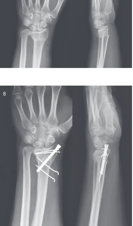
Between February and November , a total of fractures of the distal radius were treated at our de-partment, and of this number were fractures surgi-cally solved. Of the patients treated, came to the final examination and evaluation of outcomes at a follow-up of months although all patients operated on by the fi rst author in the period mentioned above had been invited. All patients were adults with an uninjured contralateral ex-tremity, who underwent surgery within days of injury. Th e indication criteria for treatment of distal radius fractures by intramedullary X-screw osteosynthesis included an absence of osteopenia on plain X-ray and the patient’s preference for a minimally invasive procedure. Th e remaining patients were treated by open reduction and internal fi xation (ORIF) using volar locking plates with variable-angle screw insertion. Indi-cations for surgery were based on antero-posterior and lateral radiographs; when a fracture was more complicated, CT ex-amination was carried out. Fracture types were assessed ac-cording to the AO classifi cation system []. Patients with pseudoarthrosis, pathological fractures, refractures, open fractures, multiple factures of the ipsilateral extremity and fractures associated with multiple injuries were excluded. The XSCR group of patients with distal radius fractures treated by intramedullary X-screw osteosynthesis (Zimmer, Inc., Warsaw, Indiana, U.S.A.) comprised patients with a mean age of . years (range, to ; SD, .) (Figure ).
FIGURE 1. A type A3 fracture of the distal radius. Postero-anterior and lateral radiographs of the fracture (patient B.N.) A) after injury; B) at 3 months after intramedullary X-screw fi xation.
A
Th e mean injury-surgery interval was days (range, to ; SD, .). Four patients had distal radius fractures without joint involvement and had fractures with intra-articular injury. Th e fractures were due to falling after a stumble while walking (. ) or were sustained in sports activities (. ). The APTUS group included patients with a distal ra-dius fracture treated by the multidirectional angle-stable plate Aptus Radius (Medartis, Basel, Switzerland) (Fig-ure ). Th e mean age of the group was . years (range, to ; SD, .). The mean injury-surgery interval was . days (range to ; SD, .). Nine fractures were without and with joint involvement. Th ey were due to falling af-ter a stumble (. ) or were sustained in association with sports activities (. ) or in motor car accidents (. ). Both the XSCR and APTUS groups were matched as for gender, injured body side and AO fracture type, but diff ered
Bosn J Basic Med Sci 2014; 14 (2): 83-88
signifi cantly in patient age. Patient characteristics are shown in Table . Lower number of fractures treated by intramed-ullary method compared to group of plate osteosynthesis was given a narrower indication extent of X-screw implant.
Operative techniques
All patients were operated on with intravenous infusion of a second generation cephalosporin antibiotic before a tour-niquet was applied. Th e X-screw is a form of intramedullary osteosynthesis of the distal radius based on combination of a special screw with threaded Kirschner wires. A -mm incision was made on the radial side over the radial styloid process. After reduction, the fracture was temporarily fi xed with a Kirschner wire. A cannulated screw was inserted over the wire and tightened to further compress the fragments and hold the reduction. Once the screw was anchored, no
further correction of fracture position was possible. Osteo-synthesis was completed with an adequate number of wires inserted by aimer device from small incisions. The wrist was immobilized with a dorsal plaster splint for weeks in type A3 fractures and for weeks in type C fractures. Type A2 fractures were not immobilized. Supervised rehabilita-tion of the hand and wrist started at post-operative weeks. Th e multidirectional angle-stable plates Aptus radius were applied from the flexor capri radialis (FCR) approach as follows: access to the radius was gained between the ra-dial vascular bundle and the FCR tendon. The pronator quadratus muscle was dissected from the anterior surface of the radius, shifted medially in a fl ap and secured with a hook. Fluoroscopy-guided reduction of the fracture was performed and fragments were temporarily fixed with Kirschner wires. Subsequently, the plate itself was applied.
FIGURE 2. A type C1 fracture of the distal radius. Postero-anterior and lateral radiographs of the fracture (patient V.H.) A) after injury; B) at 3 months after Aptus radius locking plate fi xation.
Patient characteristics APTUS X-SCREW p value
(n = 50 ) (n = 18)
Age at the time of injury, mean (range) years 48.5 (22-77) 61.0 (40-75) <0.05
Gender Men 15 2 0.2
Women 35 16
Injured side
Right 25 6
0.28
Left 25 12
Dominant 27 6
0.27
Non-dominant 23 12
AO fracture type
A2 2 2
0.54
A3 7 2
C1 14 4
C2 10 6
C3 17 4
Injury-to-surgery interval, mean (range) days 6.1 (0-22) 5.6 (2-10) 0.68
TABLE 1. Patient characteristics.
Bosn J Basic Med Sci 2014; 14 (2): 84-88The extremity was immobilized in a brace. In less serious cases, supervised rehabilitation was started at weeks and, in more complex fractures, at weeks post-operatively.
Follow-up examination
Clinical fi ndings were evaluated immediately after surgery and then at , , and weeks and , and months of fol-low-up. Radiographs were obtained within the visits at and weeks and , and months. Th e clinical outcome was evaluated using international scores at one-year follow-up. Th e range of wrist motion (volar fl exion, dorsal fl exion,
ra-dial deviation, ulnar deviation, supination and pronation) was measured with a goniometer and grip strength was assessed with a hand dynamometer (Vigorimeter, Martin, Tuttlingen, Germany). Th e values were compared with those obtained for the uninjured side. We were aware of diff erent muscle strength between the dominant and non-dominant hand but, because of a signifi cant similarity of this characteristic in both groups (p=.), this was not taken into consideration. Antero-posterior and lateral radiographs were examined for radial height, radial inclination, ulnar variance, volar tilt and articular surface step-off. The findings were com-pared with the standard anatomy of the distal radius to avoid X-ray exposure in the contralateral wrist. The val-ues of radial height, radial inclination, ulnar variance and volar tilt in lateral projection described by Ruedi et al. [] were taken as the standard anatomical parameters. In all patients the results were evaluated using the scoring systems of Gartland and Werley [], Castaing [] and the
Disability of the Arm, Shoulder and Hand (DASH) question-naire [].
Statistical analysis
The data obtained were statistically evaluated (t -test and chi-square -test) at the . level of signifi-cance using Statistica (StatSoft, Incorporation, Tul-sa, Oklahoma, U.S.A.) and SPSS Statistics (SPSS Incorporation, Chicago, Illinois, U.S.A.) software programs.
RESULTS
Functional outcomes
In the XCSR group, in comparison with the contra-lateral wrist, the following values were achieved: vo-lar flexion, .; dorsal flexion, .; radial devia-tion, .; ulnar deviadevia-tion, .; pronadevia-tion, .; supination, . and grip strength, .. These values ware not comparable with those of the uninjured side. In the APTUS group, the values achieved were as follows: vo-lar fl exion, .; dorsal fl exion, .; radial deviation, .; ulnar deviation, .; pronation, .; supination, . and grip strength, .. Th ese values were comparable with those of the uninjured side in radial deviation, ulnae deviation, pro-nation et supipro-nation. Th e results are summarized in Table .
Radiographic fi ndings
Radiographs obtained at a one-year follow-up showed complete bony union in all fractures of both groups. In
Group Side
Volar fl exion (degrees)
Dorsal fl exion (degrees)
Radial deviation (degrees)
Ulnar deviation (degrees)
Pronation (degrees)
Supination (degrees)
Grip strength (kg) Mean ± S.D. Mean ± S.D. Mean ± S.D. Mean ± S.D. Mean ± S.D. Mean ± S.D. Mean ± S.D.
APTUS
Injured side 53.40 ± 10.42 60.00 ± 9.24 25.60 ± 9.07 37.40 ± 10.06 88.60 ± 4.52 84.80 ± 10.54 20.48 ± 7.59 Uninjured side 63.00 ± 9.94 66.60 ± 9.60 26.60 ± 7.17 40.00 ± 9.03 89.40 ± 3.13 87.20 ± 7.29 23.52 ± 6.30 p value <0.05 <0.05 0.54 * 0.17 * 0.3 * 0.18 * <0.05
XSCR
Injured side 52.22 ± 8.08 55.56 ± 8.56 20.00 ± 0.00 21.11 ± 11.32 83.33 ± 10.85 78.89 ± 14.10 12.22 ± 6.25 Uninjured side 63.33 ± 4.85 67.78 ± 4.28 23.33 ± 4.85 34.44 ± 5.11 90.00 ± 0.00 88.89 ± 3.23 16.56 ± 1.82 p value <0.05 <0.05 <0.05 <0.05 <0.05 <0.05 <0.05
Group Extremity
Radial height (mm)
Radial inclination (degrees)
Volar tilt
(degrees) Ulnar variance (mm)
Articular surface step-off (mm)
Mean ± S.D. Mean ± S.D. SD Mean ± S.D. Mean ± S.D. Mean ± S.D.
APTUS
Injured side 11.86 ± 1.76 25.00 ± 2.25 2.25 9.12 ± 5.28 -0.68 ± 2.25 0.18 ± 0.43 Anatomical standard 12.00 ± 0.00 23.00 ± 0.00 0.00 12.00 ± 0.00 0.00 ± 0.00 0.00 ± 0.00 p value 0.58 * <0.05 <0.05 <0.05 <0.05 0.18 *
XSCR
Injured side 10.67 ± 1.19 21.44 ± 2.96 2.96 9.11 ± 14.10 2.89 ± 3.41 0.56 ± 0.98 Anatomical standard 12.00 ± 0.00 23.00 ± 0.00 0.00 12.00 ± 0.00 0.00 ± 0.00 0.00 ± 0.00 p value <0.05 <0.05 0.39 * <0.05 <0.05 <0.05
TABLE 2. Functional ranges of motion of the wrist joint and grip strength.
TABLE 3. Radiographic fi ndings.
* Parameters with corresponding values of both extremities
Bosn J Basic Med Sci 2014; 14 (2): 85-88
the XSCR group, only the volar tilt was fully restored. Th e other parameters were significantly different from the anatomical standards. The radiographic findings did not meet the criteria for successful treatment [] in four pa-tients (.), of whom one had an insufficient radial height and three had no satisfactory radial inclination.
In the APTUS group, all fractures were completely healed but only the radial height was comparable with the anatomi-cal standard; the other parameters were signifi cantly diff er-ent (p<.) (Table ). However, the criteria of a successful outcome [], were fulfilled in all fractures in this group.
Scores
Th e XSCR group showed . excellent and good results on the Castaing scoring system and . on the Gartland-Werley score. The average DASH score was . points. In the APTUS group, excellent and good results were achieved in . and . of the patients assessed us-ing the Castaus-ing and Gartland-Werley scores, respec-tively. The average DASH score was . points. The results are presented in Table and Figures and .
Complications
Secondary fragment displace-ment during healing was found in six patients of the XSCR group (.). It occurred in osteopenic patients with grossly comminuted fractures (types C2 and C3). But in the end, these patients found the functional state of their wrists and hand satisfactory and underwent only hardware removal. Partial migration of Kirschner wires was recorded in four patients (.) in this group, but did not result in loss of correction. In the APTUS group, secondary fragment displacement was recorded in two osteopenic patients (.) with grossly commi-nuted fractures (types A3 and C3). Temporary irritation of the sensi-tive branch of the radial nerve in two patients (.) of the XSCR group resolved spontaneously without any therapy. Temporary irritation of the median nerve was recorded in one APTUS patient (. ) who had paresthesia of the distal phalanx of the third finger. It subsided spontaneously within months after surgery with no need to indicate electromyogra-phy examination or any therapy. No tendon injury was found in the XSCR group. One APTUS patient (.) suffered a rupture of the
Scoring system
Castaing (points)
Gartland Werley (points)
DASH (points) Mean ± S.D. Mean ± S.D. Mean ± S.D. APTUS 5.26 ± 4.22 5.86 ± 6.24 10.05 ± 7.71 XSCR 6.22 ± 5.82 4.44 ± 3.73 12.12 ± 8.48 p value 0.45* 0.37* 0.35*
TABLE 4. Scoring systems.
* Parameters with corresponding values of both extremities
FIGURE 4. Functional outcome evaluation using the Gartland-Werley score.
Bosn J Basic Med Sci 2014; 14 (2): 86-88thumb extensor tendon caused by the tip of a screw project-ing through the dorsal cortex of the radius. Th is was managed by hardware removal and transfer of the extensor indicis pro-prius tendon onto the distal part of extensor pollicis tendon. Complex regional pain syndrome developed in two XSCR (.) and three APTUS (.) patients. Treat-ment with intranasal calcitonin application and in-creased calcium intake resulted in subsidence of clinical and radiographic findings in months. The com-plications and their management are shown in Table .
DISCUSSION
Restoration of the normal anatomy as a prerequisite for good wrist and hand function is currently taken as an imperative in the management of distal radius fractures. However, in unstable fractures refractory to conservative treatment there is still no clear evidence suggesting which method of osteo-synthesis should be indicated for each of the fracture types. Th e functional outcomes of intramedullary osteosynthesis of the distal radius generally have very good scores, chiefl y due to minimal invasiveness of the methods used []. With the intramedullary nail Targon DR, the resulting values for the range of wrist motion in all directions and for grip strength reached to of those measured on the uninjured side []. However, our XSCR patients achieved the level only in pronation. Th e least satisfactory range of motion (ROM) was in ulnar deviation (. of the healthy wrist value). Fail-ure in direct relation with restoring ulnar deviation has to be considered radiographic results and inability to restore radial length (mean ulnar variance of . mm). Diffi culties in ana-tomical restoration of normal volar tilt ( degrees) with the use of nailing techniques have been published. In a study com-prising patients, Gradl et al. [] reported achieving only a mean volar tilt of . degrees. Th e mean value of . degrees
for volar tilt in our XSCR patients can be regarded as a very good result testifying for good stability of the X-screw im-plant in terms of radiographic parameter. On the other hand, radial height and radial inclination were not fully restored in these patients. However, problems with maintaining volar tilt while achieving good restoration of the other radiographic parameters have also been described for the Micronail []. Th e Castaing scores showed excellent and good results in . , and the Gartland-Werley scores were excellent and good in . of the XSCR patients. Worse results with the Castaing scoring system were due to relative lengthening of the ulna. In the XSCR group, the most serious complication included loss of correction in . of the patients. Th is value is ex-tremely high compared to the . of the patients treated by plate osteosynthesis. Secondary displacement in the XSCR group was always associated with grossly comminuted fractures (A3, C3 and C2). Th is result should be taken into consideration when indicating complex fractures of the dis-tal radius for intramedullary osteosynthesis. Kirschner wire migration did not prove a serious complication because it occurred after complete fracture union and was not associ-ated with secondary fragment displacement. Paresthesia in the region innervated by a sensitive branch of the radial nerve resolved spontaneously. In the future this can be avoided by a careful operative technique involving good intra-operative visualization of the nerve. Th e absence of any injury to ten-dons is a great advantage of the intramedullary technique. Our experience with intramedullary osteosynthesis, as well as conclusions of other authors [, ] have shown that the major advantage of this technique is its good functional outcome. A full range of wrist motion at post-operative months with use of a Micronail® implant has been described. Th is advantage is in contrasted to high complication rate and
a narrow indication range regarding to AO fracture types []. In the APTUS group, the range of motion achieved in radial and ulnar deviation and in pronation and supination was comparable with that of the uninjured side; however, the val-ues for both volar and dorsal fl exion were markedly lower. A restricted ROM of only to of that at the uninjured wrist has repeatedly been reported at -year follow-up in patients treated by the volar locking plate system [, ]. However, the hand/wrist functional status recorded at year need not be fi nal and may further improve. A previous study has shown that patients achieve most of their improvement in wrist motion range and grip strength by months, but they may continue to improve up to around months []. The functional outcomes assessed using the Gartland-Werley scores were excellent and good in . of the AP-TUS patients. Th is is in agreement with the results reported by Osada et al. [] in displaced intra-articular fractures treated with a volar locking plate system. Good functional
Complication Group Number of
patients aff ected Treatment
Loss of correction APTUS 2 No treatment
XSCR 6 Hardware removal
Implant migration, no loss of correction
APTUS 0
-XCSR 4 Hardware removal
Nerve irritation
APTUS 1 (median nerve) Spontaneous resolution
XSCR 2 (radial nerve) Spontaneous resolution
Tendon rupture APTUS
1 (extensor tendon)
Hardware removal, tendon transfer
XSCR 0
-Complex regional pain syndrome
APTUS 3 Medications
(calci-tonin and calcium)
XSCR 2 Medications
(calci-tonin and calcium)
Bosn J Basic Med Sci 2014; 14 (2): 87-88
outcomes in the treatment of distal radius fractures using plate osteosynthesis are achieved in spite of a high incidence of complications []. Based on the criteria for success-ful treatment [], our results are excellent and are similar to those described by other authors for the treatment of distal radius fractures with volar locking plates [, ]. In one APTUS patient, injury to the extensor pollicis longus tendon was caused by a screw penetrating through the dorsal corticalis. It was managed by implant removal and repair of the rupture with tendon transfer. Th is damage can be avoid-ed by a careful operative technique with use of fl uoroscopic guidance during screw insertion to prevent penetration of a screw through the dorsal corticalis of the distal radius. The loss of correction was the most serious complication. Two APTUS patients (. ), women with osteopenia aged and years, experienced secondary displacement of type A3 and type C3 fractures, respectively. Revision sur-gery was not indicated; no patient wanted to have the im-plant removed. Koenig et al. performed a study to evaluate whether early internal fi xation or non-operative treatment is preferred for displaced, potentially unstable distal radial fractures with initial adequate reduction. They found that internal fi xation with a volar plate provided a higher prob-ability of painless union for potentially unstable distal radius fractures. However, patients older than years, who had lower risk of symptomatic malunion seemed to prefer non-operative treatment []. Our results of the APTUS group confirm this statement. Because due to poor bone quality even angle-stable plate does not suffi ciently guarantees good stability for all bone fragments. On the other hand, the risk of fracture re-displacement in a plaster cast is higher in os-teoporotic bone compared to healthy bone. Clayton et al. [] identifi ed a high correlation between bone mineral density and the severity of distal radius fractures. Th ey found that the probability of early instability was in osteopenic patients and only in those with normal bone mineral density. But other authors reported on high stability of lock-ing plate osteosynthesis for osteoporotic bone fractures []. Th e evaluation of our groups based on the scoring systems for the assessment of wrist functional state, both objective and subjective, as well as clinical and radiographic find-ings showed that the results achieved with either locking plates or intramedullary osteosynthesis were comparable and generally good. In terms of objective evaluation, the pa-tients with locking plate osteosynthesis had a better ROM, i.e., the range was closer to that of the contralateral wrist, than the patients with intramedullary osteosynthesis. Ra-diographs demonstrated good restoration of radial height in the APTUS group and maintenance of adequate volar tilt in the XSCR group. Regarding complications, there was a high incidence of secondary loss of correction and
im-plant migration in the XSCR group. Overall assessment it must be taken into consideration that the APTUS group included a signifi cantly higher number of younger patients.
CONCLUSION
The overall evaluation based on the conventional scor-ing systems show that intramedullary osteosynthesis does not produce better treatment outcomes compared with plate osteosynthesis in indicated types of fractures. Our study showed that the minimally invasive applica-tion of intramedullary osteosynthesis in treatment of the distal radius fractures is only a relative advantage over sur-gical stabilisation using multidirectional locking plates. In intramedullary techniques good anatomical reduc-tion and its maintenance while the implant is being in-serted, is often difficult to achieve from through small incision without a direct visualisation of the fracture. Intramedullary techniques are also associated with frequent secondary displacement of bone fragments and implant mi-gration.
DECLARATION OF INTEREST
Th e authors declare no confl ict of interest.
REFERENCES
[] Osada D, Kamei S, Masuzaki K, Takai M, Kameda M, Tamai K. Prospective study of distal radius fractures treated with a volar locking plate system. J Hand Surg Am. ; :-.
[] Chen NC, Jupiter JB. Management of distal radial fractures. J Bone Joint Surg Am. ; : -.
[] Wong KK, Chan KW, Kwok TK, Mak KH. Volar fi xation of dorsal-ly displaced distal radial fracture using locking compression plate. J Orthop Surg (Hong Kong) ; :-.
[] Leung F, Tu YK, Chew WY, Chow SP. Comparison of external and percutaneous pin fi xation with plate fi xation for intra-articular distal radial fractures. A randomized study. J Bone Joint Surg Am. ; :-.
[] Lattmann T, Meier C, Dietrich M, Forberger J, Platz A. Results of volar locking plate osteosynthesis for distal radial fractures. J Trau-ma. ; :-.
[] Gradl G, Wendt M, Gierer P, Beck M, Mittlmeier T. Fixation of distal radial fractures with the Targon DR nail. Oper Orthop Trau-matol. ; :-.
[] Tan V, Bratchenko W, Nourbakhsh A, Capo J. Comparative Analy-sis of Intramedullary Nail Fixation Versus Casting for Treatment of Distal Radius Fractures. J Hand Surg Am. ; :-. [] Lerch S, Sextro HG, Wilken F, Wittenberg CE. Clinical and
radio-logical results after distal radius fracture: intramedullary locking nail versus volar locking plate osteosynthesis. Z Orthop Unfall. ; :-.
[] Ilyas AM, Th oder JJ. Intramedullary fi xation of displaced distal radi-us fractures: a preliminary report. J Hand Surg Am. ; :-.
Bosn J Basic Med Sci 2014; 14 (2): 88-88 [] Müller ME, Nazarian S, Koch P. Classifi cation AO des fractures: lesos longs. Springer-Verlag, Berlin Heidelberg New York;. [] Rüedi TP, Buckley RE, Moran CG. AO Principles of Fracture
Man-agement. Th ieme Medical Publishers, New York; .
[] Gartland JJ, Werley CW. Evaluation of healed Colles' fractures. J Bone Joint Surg Am. ; :-.
[] Castaing J. Les fractures recentes de l'extremite inferieure du radius chez l'adulte. Rev Chir Orthop. ; :-.
[] Hudak PL, Amadio PC, Bombardier C. Development of an upper extremity outcome measure: the DASH (disabilities of the arm, shoulder and hand). [corrected]. Th e Upper Extremity Collabora-tive Group (UECG). Am J Ind Med. ; :-. Erratum in: Am J Ind Med. ; :.
[] Brooks KR, Capo JT, Warburton M, Tan V. Internal fi xation of distal radius fractures with novel intramedullary implants. Clin Or-thop Relat Res. ; :-.
[] Geerts RW, Toonen HG, van Unen JM, van Vugt R, Werre AJ. A new technique in the treatment of distal radius fractures: the Mi-cronail®. Acta Orthop Traumatol Turc. ; :-.
[] Chung KC, Watt AJ, Kotsis SV, Margaliot Z, Haase SC, Kim HM. Treatment of unstable distal radial fractures with the volar locking plating system. J Bone Joint Surg Am. ; :-. [] Mehling I, Meier M, Schlör U, Krimmer H. Multidirectional
pal-mar fi xed-angle plate fi xation for unstable distal radius fracture.
Handchir. Mikrochir Plast Chir. ; :-.
[] MacDermid JC, Richards RS, Roth JH. Distal radius fracture: a pro-spective outcome study of patients. J Hand Th er. ; :-.
[] Rozental TD, Blazar PE. Functional outcome and complications after volar plating for dorsally displaced, unstable fractures of the distal radius. J Hand Surg Am. ; :-.
[] Murakami K, Abe Y, Takahashi K. Surgical treatment of unstable distal radius fractures with volar locking plates. J Orthop Sci. ;:-.
[] Orbay JL, Fernandez DL. Volar fi xed-angle plate fi xation for un-stable distal radius fractures in the elderly patient. J Hand Surg Am. ; :-.
[] Koenig KM, Davis GC, Grove MR, Tosteson AN, Koval KJ. Is early internal fi xation preferred to cast treatment for well-reduced un-stable distal radial fractures? J Bone Joint Surg Am. ; : -.
[] Clayton RA, Gaston MS, Ralston SH, Court-Brown CM, McQueen MM. Association between decreased bone mineral density and se-verity of distal radial fractures. J Bone Joint Surg Am. ; :-.