The
Aspergillus fumigatus
Mucin MsbA Regulates the Cell Wall
Integrity Pathway and Controls Recognition of the Fungus by
the Immune System
Isabella Luísa da Silva Gurgel,
aKarina Talita de Oliveira Santana Jorge,
aNathália Luísa Sousa de Oliveira Malacco,
aJéssica Amanda Marques Souza,
aMarina Campos Rocha,
bMarina Faria Fernandes,
aFlávia Rayssa Braga Martins,
aIran Malavazi,
bMauro Martins Teixeira,
cFrederico Marianetti Soriani
aaCentro de Pesquisa e Desenvolvimento de Fármacos, Instituto de Ciências Biológicas, Departamento de Genética, Ecologia e Evolução, Universidade Federal de Minas
Gerais, Belo Horizonte, Brazil
bDepartamento de Genética e Evolução, Centro de Ciências Biológicas e da Saúde, Universidade Federal de São Carlos, São Carlos, São Paulo, Brazil
cCentro de Pesquisa e Desenvolvimento de Fármacos, Departamento de Bioquímica e Imunologia, Instituto de Ciências Biológicas, Universidade Federal de Minas
Gerais, Belo Horizonte, Brazil
ABSTRACT
Aspergillus fumigatus
is a filamentous fungus which causes invasive
pul-monary aspergillosis in immunocompromised individuals. In fungi, cell signaling and
cell wall plasticity are crucial for maintaining physiologic processes. In this context,
Msb2 is an important signaling mucin responsible for activation of a variety of
mitogen-activated protein kinase (MAPK)-dependent signaling pathways that
regu-late cell growth in several organisms, such as the cell wall integrity (CWI) pathway.
Here, we aimed to characterize the MSB2 homologue in
A. fumigatus
. Our results
showed that MsbA plays a role in the vegetative and reproductive development of
the fungus, in stress adaptation, and in resistance to antifungal drugs by modulating
the CWI pathway gene expression. Importantly, cell wall composition is also
respon-sible for activation of diverse receptors of the host immune system, thus leading to
a proper immune response. In a model of acute
Aspergillus
pulmonary infection,
re-sults demonstrate that the Δ
msbA
mutant strain induced less inflammation with
di-minished cell influx into the lungs and lower cytokine production, culminating in
in-creased lethality rate. These results characterize for the first time the role of the
signaling mucin MsbA in the pathogen
A. fumigatus
, as a core sensor for cell wall
morphogenesis and an important regulator of virulence.
IMPORTANCE
Aspergillus fumigatus
is an opportunistic fungus with great medical
importance. During infection,
Aspergillus
grows, forming hyphae that colonize the
lung tissue and invade and spread over the mammal host, resulting in high
mortal-ity rates. The knowledge of the mechanisms responsible for regulation of fungal
growth and virulence comprises an important point to better understand fungal
physiology and host-pathogen interactions. Msb2 is a mucin that acts as a sensor
and an upstream regulator of the MAPK pathway responsible for fungal
develop-ment in
Candida albicans
and
Aspergillus nidulans
. Here, we show the role of the
sig-naling mucin MsbA in the pathogen
A. fumigatus
, as a core sensor for cell wall
mor-phogenesis, fungal growth, and virulence. Moreover, we show that cell wall
composition, controlled by MsbA, is detrimental for fungal recognition and clearance
by immune cells. Our findings are important for the understanding of how fungal
sensors modulate cell physiology.
KEYWORDS
Aspergillus fumigatus
, cell wall integrity, immune response,
msb2
, mucin,
virulence
CitationGurgel ILDS, Jorge KTDOS, Malacco NLSDO, Souza JAM, Rocha MC, Fernandes MF, Martins FRB, Malavazi I, Teixeira MM, Soriani FM.
2019. TheAspergillus fumigatusmucin MsbA
regulates the cell wall integrity pathway and controls recognition of the fungus by the immune system. mSphere 4:e00350-19.
https://doi.org/10.1128/mSphere.00350-19.
EditorAaron P. Mitchell, Carnegie Mellon University
Copyright© 2019 Gurgel et al. This is an open-access article distributed under the terms of theCreative Commons Attribution 4.0 International license.
Address correspondence to Frederico Marianetti Soriani, fredsori@icb.ufmg.br.
Received15 May 2019
Accepted25 May 2019
Published
RESEARCH ARTICLE
Host-Microbe Biology
19 June 2019
on September 8, 2020 by guest
http://msphere.asm.org/
F
ungal cell survival is dependent on the organization, composition, and function of
the cell wall in which synthesis and remodeling are highly regulated. The cell wall
integrity (CWI) pathway is responsible for the maintenance of a rigid but dynamic cell
wall, which is important for hyphal growth, adaptation to environmental challenges,
and host invasion and colonization (1). The CWI functionality relies on the activation of
external sensors and the downstream activation of the mitogen-activated protein
kinase (MAPK) cascade, which in turn phosphorylates targeted transcription factors that
ultimately induce the expression of different proteins involved in cell wall
reinforce-ment (1–3). In addition, external sensors, such as mucins, are able to sense the activity
of O-mannosyltransferases and the protein glycosylation status via its extracellular
domain. As a result, the cleaved cytoplasmic domain can mobilize Cdc42, which is
essential for MAPK activation and cell wall rearrangement (4, 5).
Signaling mucins are transmembrane proteins that play an important role in cell
wall plasticity after O-glycosylation. Members of this class are integral-membrane
glycophosphatidylinositol (GPI)-anchored proteins with a cytoplasmic domain, which
interfaces with signaling transduction machinery (5). The signaling mucin Msb2
acti-vates the CEK1-MAPK pathway in
Candida albicans
, KSS1 in
Saccharomyces cerevisiae
,
Fmk1 in
Fusarium oxysporum
, and the cell wall integrity pathway in
Aspergillus nidulans
.
Msb2 signaling has also been extensively characterized as an external sensor for cell
growth (4, 6–12).
Msb2 has also been recognized as a virulence determinant in pathogenic fungi, such
as
C. albicans
. Indeed, Szafranski-Schneider et al. (13) demonstrated that upon release
to the outer cell space, the glycosylated extracellular domain of Msb2 protects the
fungal cells from antimicrobial peptides such as histatin and cathelicidins produced by
the mucosa and immune cells of the host such as neutrophils and macrophages
(13–16). Also, Msb2 is responsible for activation of the MAPK Fmk1 in the soilborne
fungus
F. oxysporum
, regulating invasive growth and virulence (7). Additionally, in the
rice blast fungus
Magnaporthe oryzae
, Msb2 is responsible for appressorium formation,
penetration, and invasive growth through activation of the Pmk1 MAPK pathway and
Ras2 GTPase (10).
In the present study, we have identified and characterized an
S. cerevisiae MSB2
homologue, in the pathogenic filamentous fungus
A. fumigatus
, named here
msbA
. We
show that
msbA
influences vegetative and reproductive growth, reflecting into the
cell-cell adhesion properties and biofilm formation. These phenotypes are related to a
modulation of the expression of the CWI pathway genes that are thought to control cell
wall composition. MsbA is shown to be crucial for survival in an immunocompetent
model of
A. fumigatus
lung infection. Moreover, an
msbA
mutant modulates activation
of the immune system affecting cell influx into the airways and production of
inflam-matory mediators. Altogether, we demonstrate for the first time that the
A. fumigatus
signaling mucin, MsbA, represents a core sensor for cell wall morphogenesis and an
important regulator of virulence.
RESULTS
Identification of the Msb2 homologue in
A. fumigatus
and construction of the
⌬
msbA
mutant strain.
In order to identify the putative
MSB2
orthologue in
A.
fumigatus
, we performed a BLASTp search using
S. cerevisiae MSB2
and
A. nidulans MSBA
as queries. Our search revealed a putative orthologue in
A. fumigatus
, Afu4g04070,
named here
msbA
to be consistent with
A. nidulans
nomenclature. The
A. fumigatus
msbA
gene is a 2,706-nucleotide open reading frame, located in the short arm of
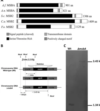
chromosome 4, with a predicted 901-amino-acid protein sequence. Comparisons of
msbA
with homologue sequences from other fungal species denote a high identity
of 75.6% (E value 7.7e
⫺
61) with
A. nidulans MSBA
but 36.1% (E value 4.3e
⫺
5) protein
identity with
S. cerevisiae MSB2,
36.4% (E value 1.4e
⫺
4) with
C. albicans MSB2,
and
34.5% (E value 1.9e
⫺
17) with
F. oxysporum MSB2
. Moreover,
in silico
analysis
demon-strated that
A. fumigatus
MsbA protein contains a single transmembrane region as well
as serine/threonine-rich regions, both common features in other MSB2 mucin proteins
on September 8, 2020 by guest
http://msphere.asm.org/
previously described (Fig. 1A). In addition, it has been described that mucins undergo
posttranslational modifications, especially glycosylation. We performed a prediction of
O-GlcNAcylated sites and observed that
A. fumigatus
MsbA presents a similar
distribu-tion pattern of these sites as reported for other fungal mucins (data not shown).
To determine the role of
msbA
in
A. fumigatus
, a deletion mutant strain was
constructed by replacing the genomic sequence of
msbA
with the
pyrG
marker in the
⌬
akuB
Ku80strain (wild type) (17). The double crossover of the deletion cassette occurred
in approximately 100 transformants, which were confirmed by Southern blotting
(Fig. 1B). After genomic DNA restriction by NcoI, we were able to identify a 1.34-kb
fragment in the wild-type strain and a fragment of approximately 3.45 kb in the Δ
msbA
mutant strain (Fig. 1C). We also constructed the complemented strain by reconstitution
of the
msbA
gene. The complementation of the
msbA
gene in the
⌬
msbA
mutant
background was confirmed by PCR (see Fig. S1A in the supplemental material).
msbA
is involved in the maintenance of vegetative growth and adhesion ability
of
A. fumigatus
.
In
A. nidulans, msbA
regulates filamentous growth and the CWI
FIG 1 Domain architecture prediction and knockout strain construction. (A)A. fumigatusMsbA shares common features of signaling mucins withA. nidulansMSBA,S. cerevisiaeMSB2,C. albicansMSB2, andF. oxysporumMSB2. All of these mucin proteins contain a cleaved signal peptide (SignalP) and one transmembrane domain (TMHMM) close to the C terminus. The large extracellular part is Ser/Thr rich (ProtParam; Color Protein Sequence). Right after the transmembrane region, the short cytoplasmic tail contains a positively charged motif (RR-RKKR--HRR inA. fumigatus; RR-RKKR--HRR inA. nidulans; RRR in
S. cerevisiae; RK-RK inC. albicans; and RR-KRKK---HRR inF. oxysporum). (B) ThemsbAgene was replaced, through homologous recombination, by the auxotrophic markerpyrG.(C) Southern blotting of wild-type strain presenting a fragment of 1.34 kb and the knockout strain (⌬msbA) presenting a fragment of 3.45 kb.
Aspergillus fumigatusMsbA Regulates CWI and Virulence
on September 8, 2020 by guest
http://msphere.asm.org/
pathway (11); thus, we tried to understand the role played by
msbA
in the maintenance
of cell wall integrity in
A. fumigatus
. Initially, we investigated the role of
msbA
in
vegetative growth of
A. fumigatus
. Colonies lacking
msbA
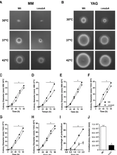
had impaired radial growth
at 30°C, 37°C, and 42°C on both minimal medium (MM) and complete medium (YAG)
(Fig. 2A and B, respectively). The effect on radial growth was more evident at 37°C, the
optimal temperature of growth, with a reduction of about 30% in colony diameter of
the
⌬
msbA
mutant. The alteration of growth phenotype was quantified and is shown
in Fig. 2C to H. The complemented strain reverted the defective growth phenotype of
the mutant strain (Fig. S1B to E). Furthermore, conidium formation was also affected by
deletion of
msbA,
which is evidenced by the decrease in the number of conidia
produced by the
⌬
msbA
strain compared to the wild-type strain (Fig. 2J). We
hypoth-esized that the morphotypic alteration in the conidiophores of the Δ
msbA
strain could
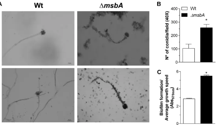
explain the decrease of conidiation. However, microscopic analyses of the aerial
structures of the null mutant showed no evident morphologic differences between
strains (Fig. 3A).
We also observed that
msbA
deletion leads to a significant delay in germ tube
formation in liquid medium. Only 20% of mutant strain conidia emitted germ tubes
compared to approximately 65% of the wild-type conidia after 2 h of incubation
(Fig. 2I). These results indicate that vegetative growth is controlled, at least in part, by
msbA
and that,
in A. fumigatus
, conidiation is affected by this mucin, independently of
morphotypic alterations of conidiophores.
From the beginning of the initial phenotypic analyses, we observed that Δ
msbA
conidia massively adhered to elongated hyphae, which was not evident for the
wild-type hyphae, suggesting an alteration in conidial adhesion properties. Therefore,
we assessed the adherence ability of
A. fumigatus
mutant strain conidia.
⌬
msbA
conidium adhesion was increased about 2.5 times compared to the wild-type strain
(Fig. 3B). Adhesion is the initial phase of biofilm formation. Consistently, biofilm
formation was significantly higher in the
⌬
msbA
strain than the wild type after 24 h of
incubation at 37°C (Fig. 3C).
msbA
plays a role in maintaining the cell wall integrity.
Cell adhesion and biofilm
formation are intimately related to cell wall properties. We subsequently evaluated the
sensitivity of
⌬
msbA
to a variety of cell wall stressors. The
⌬
msbA
mutant strain
presented increased sensitivity to chitin binding and chitin synthesis inhibitor agents,
such as Congo red (CR), calcofluor white (CFW), and nikkomycin Z (Fig. 4A and B). It has
also been demonstrated that Msb2 is able to sense osmolarity changes in the
high-osmolarity glycerol (HOG) pathway in
S. cerevisiae
(18). Likewise, MsbA also seems to
play a role in osmosensing in our system, as the mutant strain presented higher
sensitivity to the osmotic stressor agent NaCl (Fig. 4A). The complemented strain
reverted these phenotypes (Fig. S1F).
Moreover, we assessed the sensitivity of the mutant strain in the presence of
different antifungal drugs. An Etest assay demonstrated that the
⌬
msbA
strain shows
higher MICs of voriconazole and itraconazole (inhibitors of ergosterol biosynthesis) and
caspofungin (

-1,3-glucan synthase inhibitor) (Fig. 4C and D).
In order to explore the evidence regarding the function of
msbA
in cell plasticity, we
investigated the cell wall ultrastructure in the mutant strain by transmission electron
microscopy (TEM). Interestingly, the
⌬
msbA
hypha cell walls were significantly thicker
than the wild type under control conditions. In contrast, while wild-type cells showed
an increase in cell wall thickness after CFW challenge, the mutant strain had a decrease
in cell wall thickness compared to control condition (Fig. 5A). Comparing the two
strains exposed to CFW, we observed that the mutant strain decreased about 50% in
thickness (Fig. 5B). Taken together, these results suggest that
msbA
plays an important
role in cell wall organization and plasticity that affects the fungal response to cell wall
damage.
A. fumigatus msbA
modulates the expression of the CWI pathway genes.
All the
phenotypes mentioned above suggest an important role of
msbA
in the maintenance
on September 8, 2020 by guest
http://msphere.asm.org/
of the integrity of
A. fumigatus
cell wall, which may be reflected in the perturbed
downstream signaling cascade due to abnormal sensing of cell wall disruption. To
address this question, we evaluated the expression profile of genes related to cell wall
biosynthesis and remodeling, such as the kinases
pkcA
and
mpkA
and the CWI
tran-FIG 2 The ΔmsbAstrain has reduced vegetative growth and conidiation. The mutant strain has a diminished radial growth at 30°C, 37°C, and 42°C compared to wild type, in both MM and YAG. (A) MM for 48 h; (B) YAG for 48 h; (C to E) MM at 30°C, 37°C, and 42°C, respectively; (F to H) YAG at 30°C, 37°C, and 42°C, respectively. Quantification was performed by colony diameter measurement every 24 h during 96 h (C to H). Asterisk represents statistical difference (P⬍0.05) from wild type under the same growth condition. (I) Percentage of polarized growth after HU release. Samples of germlings were analyzed at 0 h, 0.5 h, 1 h, 1.5 h, and 2 h after release of HU blockade. Asterisk represents statistical difference (P⬍0.05) from wild type under the same growth condition. (J) Number of conidia/colony diameter after growth in MM at 37°C for 72 h. Asterisk represents statistical difference (P⬍0.05) from wild type.
Aspergillus fumigatusMsbA Regulates CWI and Virulence
on September 8, 2020 by guest
http://msphere.asm.org/
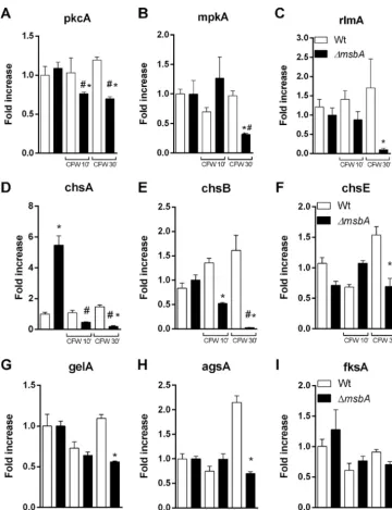
scription factor
rlmA
. Our results demonstrate that upon CFW exposition, the CWI
signaling genes were downregulated in the mutant strain, while we were able to
identify a slight upregulation of these genes in the wild-type strain (Fig. 6A to C).
We also examined the transcriptional levels of the CWI target genes such as chitin
synthases (
chsA, chsB,
and
chsE
),
␣
-1,3-glucan synthase (
agsA
),

-1,3-glucan synthase
(
fksA
), and 1-3-

-glucanosyltransferase (
gelA
). Likewise, we observed lower expression
levels of the evaluated genes after 30 min of CFW stimuli in the mutant strain, except
for
fksA
. Curiously, the mutant strain expressed about five times more
chsA
mRNA than
the wild type under the control condition (Fig. 6D to I). This finding is consistent with
the cell wall alterations observed in the mutant and may help to explain phenotypes
such as those described in Fig. 3. Our results demonstrate that the mucin MsbA
regulates the CWI pathway under stress conditions, acting as a stress sensor.
msbA
contributes to downmodulating inflammatory responses after
A.
fumiga-tus
infection.
Then, we evaluated whether
msbA
deletion affected
A. fumigatus
viru-lence after infection with a single inoculum of
A. fumigatus
in C57BL/6
immunocom-petent mice. During the infection in mice, the expression of
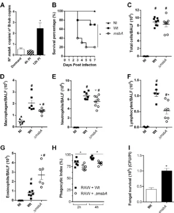
msbA
was analyzed from
fungal cells retrieved from the lungs 4 h and 12 h postinfection. Results show that, at
the initial steps of fungal infection, expression of
msbA
was unaltered compared to the
basal levels of dormant conidia. In contrast, 12 h postinfection,
msbA
was highly
expressed, suggesting that it plays an important role in the vegetative growth of the
fungus upon contact with host cells (Fig. 7A).
Survival rates of animals infected with the wild-type and knockout strains, after 7
days, showed that mice infected with the
⌬
msbA
strain were highly susceptible to the
infection. Indeed, 80% of the mice infected with
A. fumigatus
⌬
msbA
succumbed to
infection at day 5 compared to only 30% of the ones infected with the wild-type fungus
(Fig. 7B). To understand the underlying mechanism of this difference in mortality, we
analyzed the cellular inflammatory profile in the airways. The results demonstrate the
same amounts of total cells infiltrated into the alveoli as assessed from bronchoalveolar
lavage fluid (BALF) of mouse groups infected with both strains 24 h postinfection
FIG 3 The ΔmsbAstrain shows enhanced adhesion properties and biofilm formation. (A) Optical microscopy of conidiophores from wild-type and ΔmsbAstrains. On the right side are shown two independent experiments on wild-type conidiophores. On the left side are shown two independent experiments on ΔmsbAconidiophores. The coverslips were stained using lactophenol blue solution and analyzed by optical microscopy using⫻40 magnifi-cation. Bars, 50m. Experiments were performed in triplicate. (B) Adhesion properties of conidia were analyzed after 4 h of growth in RPMI at 37°C. Adhered conidium number was determined in at least 6 microscope fields and expressed as total percent conidia in each assay. (C) Biofilm formation was measured indirectly by absorbance at 570 nm. Absorbance values were normalized by growth rate of each strain in MM. Values are shown as mean⫾ SEM. Asterisks represent statistical difference (P⬍0.05) from wild type under the same growth condition.on September 8, 2020 by guest
http://msphere.asm.org/
(Fig. 7C). However, the profile of recruitment of cells was altered. The mutant strain
infection induced the migration of a decreased number of macrophages, lymphocytes,
and neutrophils (Fig. 7D to F) and of a higher number of eosinophils (Fig. 7G) recruited
to the site of infection. In the lung tissue, there was a greater accumulation of
eosinophils and neutrophils in mice infected with the
⌬
msbA
strain. The recruitment of
macrophages presented no differences in the lung tissue between the two groups (see
Fig. S2 in the supplemental material). We hypothesized whether these alterations in the
FIG 4 The⌬msbAstrain has altered sensitivity to cell wall stressors and antifungal agents. (A) Tenfold dilution dropout growth in solid MM plates supplemented with different cell wall-perturbing agents: Congo red (CR), calcofluor white (CFW), and NaCl. (B) Conidia (1⫻104of each strain) were incubated in liquid MM at 37°C for 48 h in the presence of the indicated concentrations of nikkomycin Z. (C) Antifungal susceptibility using Etest gradient strips for voriconazole, itraconazole, and caspofungin. (D) MICs of antifungal drugs analyzed in panel C.FIG 5 The⌬msbAstrain has altered cell wall thickness. (A) Transmission electronic microscopy images of hyphae from wild-type and⌬msbAstrains grown in liquid MM with or without CFW (100g/ml). Bars, 500 nm. (B) Cell wall thickness was quantified under each condition. Values are shown as mean⫾SEM from 50 different sections. Asterisk represents statistical difference (P⬍0.05) compared with wild type under the same growth condition. Number sign represents statistical difference (P⬍0.05) compared within the group (wild type⫻wild type/⌬msbAstrain⫻ ⌬msbAstrain) under control condition.
Aspergillus fumigatusMsbA Regulates CWI and Virulence
on September 8, 2020 by guest
http://msphere.asm.org/
immune system modulation would be the result of an alteration in the capacity of
recognition of the fungus and activation of a proper response by phagocytes. In this
way,
in vitro
phagocytic and clearance abilities of macrophages were accessed. After a
2-h assay, the wild-type strain was highly recognized and phagocytosed while the
⌬
msbA
strain showed a lower phagocytic index. The same pattern was observed after
a 4-h assay; approximately 15% fewer conidia of the mutant strain were phagocytosed
compared to the wild-type counterpart (Fig. 7H). Moreover, fungal clearance was
accessed via CFU counting, after 6 h of incubation. It showed a significant increase in
⌬
msbA
conidium survival (Fig. 7I), meaning that macrophages were not able to clear
FIG 6 The⌬msbAstrain has decreased levels of expression of cell wall integrity pathway genes. Strains were grown in YG medium for 24 h at 37°C and treated with CFW (100g/ml) for 10 and 30 min. The control group was not treated with CFW. Fold increase in each strain represents the normalized mRNA abundance relative to the wild-type strain under the control condition. Values are shown as mean⫾SEM. Asterisk represents statistical difference (P⬍0.05) from wild type under the same growth condition. Number sign represents statistical difference (P⬍0.05) compared within the group (wild type⫻wild type/⌬msbAstrain⫻ ⌬msbAstrain) under the control condition.on September 8, 2020 by guest
http://msphere.asm.org/
⌬
msbA
conidia properly. Thus, these results demonstrate that alterations of the cell wall
remodeling mechanisms orchestrated by the stress sensor, MsbA, in
A. fumigatus
led to
an altered recognition of conidia by phagocytes. Furthermore, beside recognition, the
mutant strain is capable of evading killing machinery during phagocytosis and
surviv-ing more than the wild-type strain.
FIG 7 MsbA contributes to downmodulating the inflammatory response afterA. fumigatusinfection. (A)msbA
mRNA expression during infection.A. fumigatuscells were harvested from lungs of infected animals after 4 h and 12 h (PI, postinfection). mRNA was extracted for qRT-PCR assay. Data are presented as mean⫾SEM (n⫽3 to 4 mice per group).*, significantly different (P⬍0.05). (B) Lethality of mice infected with wild-type or⌬msbAstrain. Mice were infected intranasally with 40l of suspension containing 1⫻108conidia of wild-type or⌬msbAstrain, and mortality was monitored for 7 days. NI, noninfected. (C to G) Inflammatory infiltrate was analyzed in BALF of infected animals. Infection with⌬msbAstrain altered inflammatory cells recruitment into airways. Intranasally infected mice had BALFs harvested at day 1 postinfection for inflammatory cell infiltrate determination. Total cell (C), macrophage (D), neutrophil (E), lymphocyte (F), and eosinophil (G) absolute counts in BALF. Data are presented as mean⫾SEM (n⫽5 to 8 mice per group).*, significantly different (P⬍0.05) compared to wild-type-infected group. #, significantly different (P⬍0.05) compared to noninfected group. (H)In vitrophagocytosis was performed with RAW 264.7 immortalized macrophages challenged with wild-type or ⌬msbA strain for 2 h and 4 h for phagocytosis. The relationship between the number of macrophages containing conidia inside and the total number of macrophages was used to calculate phagocytic index. One hundred macrophages were counted for each coverslip. (I) Fungal survival was quantified as CFU/phagocytic index (PI).⌬msbAconidium number after 6 h of phagocytosis was higher.*, significantly different (P⬍0.05) compared to wild type.
Aspergillus fumigatusMsbA Regulates CWI and Virulence
on September 8, 2020 by guest
http://msphere.asm.org/
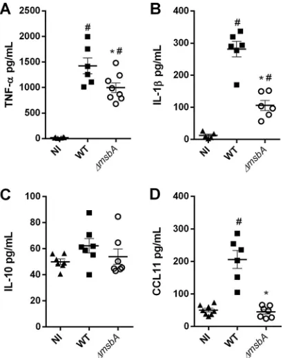
The inflammatory response is a complex process that involves inflammatory signals
such as cytokine and chemokine production (19). Therefore, we evaluated the role of
the
⌬
msbA
mutant in the stimulation of immune system and the production of key
mediators in BALF after
A. fumigatus
infection. Our data demonstrate that mice infected
with the
⌬
msbA
strain showed a reduction of important inflammatory mediators like
TNF-
␣
and IL-1

, resulting in almost a 30% reduction of both cytokines, after 24 h of
infection (Fig. 8A and B). The levels of IL-10 remained unaltered in the airways of
infected animals compared to the noninfected mice (Fig. 8C). Curiously, despite the
higher levels of eosinophils quantified at the infection site, CCL11/eotaxin1, one of the
main chemokines responsible for recruitment of these cells, was detected at a lower
concentration in BALF of
⌬
msbA
strain-infected animals (Fig. 8D). Taken together, these
results demonstrate that, during infection, MsbA is an important signaling protein in
A.
fumigatus
and takes part in fungal physiological processes that culminate in the
recognition of the pathogen during lung infection in mammalian hosts. This evidence
is especially important to guide cell infiltration into the airways and for the production
of inflammatory mediators. The
msbA
gene is expressed at higher levels during the
course of infection, which results in alterations that occur upon the loss of function of
this mucin, modulating the overall innate immune response to affect the outcome of
infection.
DISCUSSION
In this study, we characterized the mucin-like protein MsbA in the human pathogen
A. fumigatus
. Here, we show that MsbA controls fungal development, as a result of the
modulation of the cell wall integrity pathway, which culminates in the regulation of cell
adhesion and biofilm formation and responses to cell wall stressors and to antifungal
drugs. Moreover, MsbA deletion compromises the
A. fumigatus
recognition by
phago-cytes.
MsbA is involved in the vegetative growth of many yeasts and filamentous fungi. In
FIG 8 The⌬msbAstrain stimulates fewer inflammatory mediators in a model ofA. fumigatusacute lung infection. Mice were infected with wild-type or ⌬msbA strains, and inflammatory mediators were analyzed by ELISA in BALFs at day 1 postinfection: TNF-␣(A), IL-1(B), IL-10 (C), and CCL11/eotaxin (D).*, significantly different (P⬍0.05) from wild-type-infected group. #, significantly different (P⬍0.05) from noninfected (NI) group.
on September 8, 2020 by guest
http://msphere.asm.org/
C. albicans
, Msb2 regulates the yeast filamentous growth by controlling Cek1 MAPK
phosphorylation (8). Additionally, Brown et al. (11) demonstrated that in
A. nidulans
,
MsbA regulates not only vegetative growth but also the conidiation process. Our data
corroborate these results in
A. fumigatus
and highlight that
msbA
is required for the
normal hyphal growth and conidiation of the fungus.
The fungal cell wall has a crucial role in pathogenic fungi as it is responsible for the
pathogen survival, adaptation, and signaling under stressful conditions during infection
(1, 20). It has been shown that Msb2 homologues in
C. albicans
,
A. nidulans
, and
F.
oxysporum
regulate the response to cell wall stressors such as in the presence of Congo
red, calcofluor white, and caspofungin (7, 8, 11). Brown et al. (11) showed that MsbA
modulates the cell wall integrity (CWI) pathway, interfering with the production of
chitin in the cell wall. The CWI pathway is the main signaling pathway controlling the
synthesis of cell wall components in response to environmental stresses in
S. cerevisiae
(21). Here, we show that deletion of MsbA interferes in the expression of the
A.
fumigatus
CWI pathway. Consequently, CWI target genes, such as the chitin synthase
genes, were also misregulated, thus resulting in changes in the fungal cell wall
morphometry. Collectively, our results corroborate literature described for different
fungal species, showing that MsbA is similarly responsible for controlling the response
of
A. fumigatus
in the presence of cell wall stress.
Changes in the cell wall composition can be responsible for altered recognition of
the pathogen by host cells. This process can change the course of the inflammatory
response, affecting the secretion of cytokines and chemokines and cell influx. Indeed,
our results demonstrate an impairment of inflammatory cell migration into the airways
after infection with the Δ
msbA
strain. These results were in accordance with the
sensitivity assays and the expression levels of CWI genes. Along with the diminished
influx of macrophages, lymphocytes, and neutrophils into the airways, there were
reduced levels of TNF-
␣
and IL-1

in mice infected with the
⌬
msbA
strain. This evidence
indicates that the mucin MsbA is involved in the regulation of CWI in
A. fumigatus
,
controlling the composition of fungal cell wall components (pathogen-associated
molecular patterns [PAMPs]).
After infection,
A. fumigatus
cells are recognized by the immune system, triggering
a series of events that promote clearance of the fungus from infected host tissues. This
recognition involves primarily activation of pattern recognition receptors (PRRs) by the
cell wall PAMPs (19). Alveolar macrophages are crucial components of the host defense
against
A. fumigatus
infections not only because they promote the phagocytosis and
killing of these fungal conidia but also because they initiate a proinflammatory
re-sponse that recruits other leukocytes to the infection site, such as neutrophils (19,
22–24).
A number of factors, including inflammatory mediators such as cytokines and
chemokines, mediate leukocyte influx into the site of infection. TNF-
␣
is mainly secreted
by alveolar macrophages upon stimulation, increasing the phagocytic activity of
mac-rophages and neutrophil fungicidal activities against hyphae (25, 26). Besides TNF-
␣
,
alveolar macrophages secret other proinflammatory mediators, such as IL-1

and IL-1
␣
(26). Werner et al. (26) showed a correlation between a decreased production of these
cytokines and nonresponsive macrophages in
A. fumigatus
.
Altogether, our results show an important role of the
A. fumigatus
mucin MsbA in
the fungal growth, maintenance, and plasticity of the cell wall. MsbA also contributes
to fungal cell wall properties such as adhesion and biofilm formation and resistance to
antifungals. Proper plasticity of the cell wall is crucial for fungal survival to phagocytosis
and for the adequate innate immune response during mammalian pulmonary infection.
Overall, our results show that the inflammatory profile triggered by infection with the
A. fumigatus
strain lacking mucin MsbA affects not only the host cell recruitment but
also the production of inflammatory mediators. Additional studies are required to
elucidate how this protein connects with the CWI pathway and the mechanisms
whereby MsbA modulates the host response to
A. fumigatus
.
Aspergillus fumigatusMsbA Regulates CWI and Virulence
on September 8, 2020 by guest
http://msphere.asm.org/
MATERIALS AND METHODS
Mutant construction.Construction of the deletion cassette byin vivorecombination inS. cerevisiae
was performed as previously described by Colot et al. (27). Briefly, fragments 5=untranslated region (UTR) (1,505 bp) and 3=UTR (1,532 bp) of themsbAgene (AFUA_4G04070) from the⌬akuBku80strain (17) were
amplified by PCR, as well as the genepyrGfrom PCDA21. All fragments contained flanking sequences. The three fragments plus BamHI-EcoRI-cut pRS426 were transformed in S. cerevisiae(28). Positive transformant genomic DNA was transformed inEscherichia colichemically competent DH5ɑcells, aiming at amplification of the deletion cassette. The constructed deletion cassette was transformed in the wild-typeAspergillus fumigatus(⌬akuBku80) through double homologous recombination, mediated by the
polyethylene glycol-mediated protoplast technique (29). The Southern blot technique was utilized to confirm mutant construction.
To complement the ΔmsbAstrain, the cassette containing themsbAgene plus the two 1.5-kb flanking regions was PCR amplified using the genomic DNA from the⌬akuBku80strain as a template (see Table S1
in the supplemental material). Protoplasts from the ΔmsbAstrain were transformed with the 5,736-bp PCR product and plated onto medium containing 200g/ml of CR and 100g/ml of CFW. Two revertants, which were able to grow under these conditions, were further analyzed by PCR, with the primer sets msbA FWD and msbAREV (Fig. S1A). The complemented strains were also tested for complementing phenotypes, and they yielded the same results (Fig. S1B to F). These strains were named ΔmsbA::msbA1 and ΔmsbA::msbA2.
Culture conditions.A. fumigatusstrains were grown in minimal medium (MM), complete medium (YAG), YG, or RPMI 2⫻agar, 2% (wt/vol) (30), at 28°C, 30°C, 37°C, or 42°C. When necessary, YAG TOP agar (1% agar) was utilized. Conidia were harvested and collected from agar medium after 48 h or 72 h. Next, conidia were diluted and counted in a Neubauer chamber. Conidiation (microcultivation) assay was carried out as described by Lin and Momany (31) with adaptations. Briefly, coverslips were placed on top of potato dextrose agar, on which conidia of both strains were previously deposited, and cultivated at 28°C for 48 h/72 h. For conidiophore structure observation, coverslips with aerial hyphae and conidio-phores attached were stained using lactophenol blue solution, mounted on coverslips, and observed microscopically using⫻40 magnification.
Biofilm formation and adhesion assay.A. fumigatusbiofilm was produced and quantified accord-ing to the method of Mowat et al. (32) and Gravelat et al. (33), with adaptations. Briefly, 2⫻104conidia of each strain were inoculated in 200 ml MM into a flat-bottom 96-well polystyrene plate. The plate was incubated at 37°C for 24 h. After incubation, MM was removed from wells, and cells were washed four times with 1⫻PBS. For biofilm quantification, 150l of 0.5% (wt/vol) crystal violet was added to each well for 5 min, at environmental temperature. Mycelia were washed with sterile Milli-Q water. Remaining crystal violet was eluted with 200l 100% (vol/vol) ethanol per well. Ethanol solution was transferred to a new 96-well plate. Absorbance was determined at 570 nm.
The adhesion assay was performed according to the work of Shopova et al. (34), with adaptations. Briefly, conidium adhesion to the polystyrene plate surface was quantified through visual counting using inverted light microscopy. Conidia of each strain (5⫻105) were diluted in 5 ml of RPMI. After 4 h of incubation at 37°C, RPMI was washed with 10 ml of sterile 1⫻PBS. The number of adhered conidia was determined in at least 6 optical fields and expressed as total percentage of conidia in each assay.
Polarization.Conidia of each strain (1⫻105) were incubated at 37°C for 5 h in 5 ml of YG containing 50 mM hydroxyurea (HU), followed by washing with distilled water and then incubation in YG at 37°C. Samples were taken at 0 h, 0.5 h, 1 h, 1.5 h, and 2 h after HU blockage release, and germ tubes were quantified by direct counting of at least 100 cells per plate.
Susceptibility assay for antifungal, cell wall, and osmotic stressor agents.The antifungal Etest assay was conducted according to the method of Rocha et al. (3) with adaptations. Briefly, 1⫻105 conidia in 100l 1⫻PBS were mixed into 10 ml RPMI TOP agar, placed over 15 ml RPMI agar in 90-mm plates, and left to dry. Etest strips were applied on the top of the plates, incubated at 37°C for 36 h, and photographed. MICs were determined as the minimum drug concentration of the inhibitory halo intercepting the test strip. To evaluate growth under cell wall and osmotic stress, concentrations of Congo red (200g/ml), calcofluor white (50g/ml), and sodium chloride (400 mM) were used. Five-microliter drops with 10⫻dilutions of both strains were plated in MM plus the stressor agent and grown for 30 h at 37°C. To evaluate sensitivity to nikkomycin Z, 1⫻104conidia were cultivated in MM in a 24-well plate containing serial dilutions of the drug (50 mM, 100 mM, and 150 mM) at 37°C for 48 h and photographed.
TEM analysis.Wild-type and ΔmsbAstrains (1⫻107conidia) were grown in liquid MM for 24 h at 37°C prior to exposure to CFW (100g/ml) for 2 h. Cells were processed essentially as described previously with modifications (35). Briefly, mycelium was fixed in 0.1 M sodium phosphate buffer (pH 7.4)-2.5% (vol/vol) glutaraldehyde for 24 h at 4°C. Samples were encapsulated in 2% (wt/vol) agar and subjected to fixation (1% OsO4), contrast (1% uranyl acetate), ethanol dehydration, and a two-step infiltration process with propylene oxide-EMbed 812 (Electron Microscopy Sciences) of 16 h and 3 h at room temperature. Additional infiltration was provided under vacuum at room temperature before embedding in BEEM capsules (Electron Microscopy Sciences) and polymerization at 60°C for 72 h. Semithin (0.5-mm) survey sections were stained with toluidine blue to identify the areas of greatest cell density. Ultrathin sections (60 nm) were prepared and stained with uranyl acetate (1%) and lead citrate (2%). Transmission electron microscopy (TEM) images were obtained using a Tecnai G2-12-SpiritBiotwin FEI electron microscope at an acceleration voltage of 120 kV (Center of Microscopy from UFMG, Brazil) using a charge-coupled device (CCD) camera. Cell wall thicknesses of 50 sections of different germlings were measured using magnification of⫻26,500 and ImageJ software analysis (13).
on September 8, 2020 by guest
http://msphere.asm.org/
RNA extraction and real-time qRT-PCR procedures.Mycelia were disrupted by grinding in liquid nitrogen with a pestle and mortar. Total RNA was extracted using TRIzol (ThermoFisher Scientific) according to the manufacturer’s protocol. Samples were treated with DNase I (Invitrogen). RNA concen-tration and quality were assessed with a nanophotometer (NanoDrop; ThermoFisher Scientific). A total of 2g of DNase-treated total RNA from eachA. fumigatusstrain was reverse transcribed using the SuperScript III reverse transcriptase kit (Invitrogen) and oligo(dT) primers. Real-time RT-PCR was con-ducted using Power Sybr green PCR master mix (Applied Biosystems). Primers used are listed in Table S1 in the supplemental material. RT-PCR was performed in duplicate in a 7500 Fast real-time PCR system (Applied Biosystems) according to the manufacturer’s instructions. Nontemplate controls (NTC) were used to confirm elimination of contaminating DNA in every run. After completing PCR, melt curve analysis was performed to confirm the absence of nonspecific amplification products. The results were normalized using -tubulin copy number, calculated in reference to a standard curve with known amounts ofA. fumigatusgenomic DNA (36).
Fungicidal clearance capacity by phagocytic cells.In vitrophagocytosis was performed according to the method of Bom et al. (35) with adaptations. Immortalized macrophages (1⫻105; RAW 264.7 strain) were cultured in 1 ml of Dulbecco’s minimal essential medium (DMEM), supplemented with 2.5% (vol/vol) fetal bovine serum (FBS) in a 24-well plate containing 13-mm round coverslips. Cells were maintained at 37°C in a 5% CO2atmosphere. After incubation, wells were washed 3 times with DMEM with no antibiotics. One milliliter of DMEM plus 2% (vol/vol) FBS plus 1⫻106conidia of both strains (1:10) were added to each well. Samples were incubated for 2 h and 4 h. Each well was washed 4 times with 1⫻PBS at 37°C. The 13-mm round coverslips were stained with the Panoptic kit (Laborclin) and submitted to phagocytosis counting. For fungicidal clearance evaluation, 1⫻106RAW macrophages were incubated in DMEM-2.5% (vol/vol) FBS in a 24-well plate. Wells were washed prior to incubation with DMEM⫹2% (vol/vol) FBS plus 1⫻107conidia of both strains (1:10) in each well. Samples were incubated at 37°C in a 5% CO2atmosphere for 6 h. Wells were washed for cell lysis with sterile water, and the remaining conidia were plated in YAG medium to quantify the CFU.
Ethics statement.Animal experiments were approved by the Institution Ethics Committee (Comis-são de Ética no Uso de Animais, CEUA/UFMG, protocol number 187/2018), according to Brazilian national guidelines on animal work (Conselho Nacional de Controle de Experimentação Animal [CONCEA]).
Animal infection andin vivoassays.In this study, we used male and female 8- to 12-week-old C57BL/6J specific-pathogen-free (SPF) mice. Prior to infection, mice were anesthetized by inhaling up to 3% isoflurane (Biochimico, Brazil) with oxygen. Mice were intranasally infected with 1⫻108conidia of theA. fumigatuswild-type or⌬msbAstrain in 40l of sterile PBS. After 24 h of incubation, infected mice were euthanized with a solution of 180 mg/kg of body weight of ketamine and 24 mg/kg of xylazine. Subsequently, bronchoalveolar lavage fluid (BALF) was harvested by washing the lungs twice with 2 ml of PBS. Fluid was centrifuged, and cell pellets were used for total and differential leukocytes counts. Supernatants were used for cytokine and chemokine quantification (37). Prior to removal and freezing of the right lobes of the lungs for myeloperoxidase (MPO),N-acetylglucosaminidase (NAG), and eosinophil peroxidase (EPO) analysis, lungs were perfused with 5 ml of 1⫻PBS (38–40). Additionally, lungs were harvested for the fungal burden analysis.
Cytokine and chemokine measurement.DuoSet ELISA kits (R&D) were used to quantify cytokine and chemokine levels in BALF, according to the manufacturer’s instructions.
Statistical analysis.Experiments were performed at least twice. Data are presented as the mean⫾ SEM. Statistical differences were analyzed with one-way analysis of variance (ANOVA), followed by Holm-Sidak posttest. Normal distribution was evaluated by the D’Agostino-Person test. Nonparametric data were analyzed with the Kruskal-Wallis test. Mann-Whitney test (nonparametric) orttest (parametric) was used to compare two groups. Survival analysis was performed using log rank test. Statistical significance was set asP⬍0.05. Graphs and analysis were performed using GraphPad Prism 6.0 software (GraphPad Software Inc., San Diego, CA, USA).
SUPPLEMENTAL MATERIAL
Supplemental material for this article may be found at
https://doi.org/10.1128/
mSphere.00350-19
.
FIG S1
, TIF file, 2 MB.
FIG S2
, TIF file, 2.2 MB.
TABLE S1
, PDF file, 0.03 MB.
ACKNOWLEDGMENTS
This work was supported by Conselho Nacional de Desenvolvimento Científico e
Tecnológico 474528-2012-0 and 483184-2011-0 and Fundação de Amparo à Pesquisa do
Estado de Minas Gerais APQ- 01756-10, APQ-02198-14, and APQ-03950-17. It was financed
in part by the Coordenação de Aperfeiçoamento de Pessoal de Nível Superior-Brasil
(CAPES), finance code 001, and Instituto Nacional de Ciência e Tecnologia (INCT) em
Dengue e Interação Microrganismo-Hospedeiro. The funders had no role in study design,
data collection and analysis, decision to publish, or preparation of the manuscript.
We thank Ilma Marçal and Rosemeire Oliveira for technical support.
Aspergillus fumigatusMsbA Regulates CWI and Virulenceon September 8, 2020 by guest
http://msphere.asm.org/
REFERENCES
1. Latgé J-P. 2010. Tasting the fungal cell wall. Cell Microbiol 12:863– 872. https://doi.org/10.1111/j.1462-5822.2010.01474.x.
2. Rocha MC, Godoy KF, de Castro PA, Hori JI, Bom VLP, Brown NA, Cunha AF, Goldman GH, Malavazi I. 2015. The Aspergillus fumigatus pkcA G579R mutant is defective in the activation of the cell wall integrity pathway but is dispensable for virulence in a neutropenic mouse infection model. PLoS One 10:e0135195.https://doi.org/10.1371/journal.pone.0135195.
3. Rocha MC, Fabri J, de Godoy KF, de Castro PA, Hori JI, da Cunha AF, Arentshorst M, Ram AFJ, van den Hondel C, Goldman GH, Malavazi I. 2016. Aspergillus fumigatus MADS-box transcription factor rlmA is re-quired for regulation of the cell wall integrity and virulence. G3 (Bethesda) 6:2983–3002.https://doi.org/10.1534/g3.116.031112. 4. Cullen PJ, Sabbagh W, Jr, Graham E, Irick MM, van Olden EK, Neal C,
Delrow J, Bardwell L, Sprague GF, Jr. 2004. A signaling mucin at the head of the Cdc42- and MAPK-dependent filamentous growth pathway in yeast. Genes Dev 18:1695–1708.https://doi.org/10.1101/gad.1178604. 5. Cullen PJ. 2007. Signaling mucins: the new kids on the MAPK block. Crit
Rev Eukaryot Gene Expr 17:241–257. https://doi.org/10.1615/CritRev EukarGeneExpr.v17.i3.50.
6. Lanver D, Mendoza-Mendoza A, Brachmann A, Kahmann R. 2010. Sho1 and Msb2-related proteins regulate appressorium development in the smut fungusUstilago maydis. Plant Cell 22:2085–2101.https://doi.org/ 10.1105/tpc.109.073734.
7. Perez-Nadales E, Di Pietro A. 2011. The membrane mucin Msb2 regulates invasive growth and plant infection in Fusarium oxysporum. Plant Cell 23:1171–1185.https://doi.org/10.1105/tpc.110.075093.
8. Román E, Cottier F, Ernst JF, Pla J. 2009. Msb2 signaling mucin controls activation of Cek1 mitogen-activated protein kinase in Candida albicans. Eukaryot Cell 8:1235–1249.https://doi.org/10.1128/EC.00081-09. 9. Leroch M, Mueller N, Hinsenkamp I, Hahn M. 2015. The signalling mucin
Msb2 regulates surface sensing and host penetration via BMP1 MAP kinase signalling in Botrytis cinerea. Mol Plant Pathol 16:787–798. https://doi.org/10.1111/mpp.12234.
10. Wang G, Li G, Zhang S, Jiang C, Qin J, Xu J. 2015. Activation of the signalling mucin MoMsb2 and its functional relationship with Cbp1 in Magnaporthe oryzae. Environ Microbiol 17:2969 –2981.https://doi.org/ 10.1111/1462-2920.12847.
11. Brown NA, dos Reis TF, Goinski AB, Savoldi M, Menino J, Almeida MT, Rodrigues F, Goldman GH. 2014. The Aspergillus nidulans signalling mucin MsbA regulates starvation responses, adhesion and affects cellu-lase secretion in response to environmental cues. Mol Microbiol 94: 1103–1120.https://doi.org/10.1111/mmi.12820.
12. Roberts RL, Fink GR. 1994. Elements of a single MAP kinase cascade in Saccharomyces cerevisiae mediate two developmental programs in the same cell type: mating and invasive growth. Genes Dev 8:2974 –2985. https://doi.org/10.1101/gad.8.24.2974.
13. Szafranski-Schneider E, Swidergall M, Cottier F, Tielker D, Román E, Pla J, Ernst JF. 2012. Msb2 shedding protects Candida albicans against anti-microbial peptides. PLoS Pathog 8:e1002501.https://doi.org/10.1371/ journal.ppat.1002501.
14. Durr M, Peschel A. 2002. Chemokines meet defensins: the merging concepts of chemoattractants and antimicrobial peptides in host de-fense. Infect Immun 70:6515– 6517. https://doi.org/10.1128/iai.70.12 .6515-6517.2002.
15. Peschel A, Sahl H. 2006. The co-evolution of host cationic antimicrobial peptides and microbial resistance. Nat Rev Microbiol 4:529 –536.https:// doi.org/10.1038/nrmicro1441.
16. Oudhoff MJ, Blaauboer ME, Nazmi K, Scheres N, Bolscher JGM, Veerman ECI. 2010. The role of salivary histatin and the human cathelicidin LL-37 in wound healing and innate immunity. Biol Chem 391:541–548.https:// doi.org/10.1515/BC.2010.057.
17. Da Silva Ferreira ME, Kress MRVZ, Savoldi M, Goldman MHS, Härtl A, Heinekamp T, Brakhage AA, Goldman GH. 2006. The akuB(KU80) mutant deficient for nonhomologous end joining is a powerful tool for analyzing pathogenicity in Aspergillus fumigatus. Eukaryot Cell 5:207–211.https:// doi.org/10.1128/EC.5.1.207-211.2006.
18. Tatebayashi K, Tanaka K, Yang H-Y, Yamamoto K, Matsushita Y, Tomida T, Imai M, Saito H. 2007. Transmembrane mucins Hkr1 and Msb2 are putative osmosensors in the SHO1 branch of yeast HOG pathway. EMBO J 26:3521–3533.https://doi.org/10.1038/sj.emboj.7601796.
19. Dagenais TRT, Keller NP. 2009. Pathogenesis of Aspergillus fumigatus in
invasive aspergillosis. Clin Microbiol Rev 22:447– 465.https://doi.org/10 .1128/CMR.00055-08.
20. Latgé J-P, Beauvais A, Chamilos G. 2017. The cell wall of the human fungal pathogenAspergillus fumigatus: biosynthesis, organization, im-mune response, and virulence. Annu Rev Microbiol 71:99 –116.https:// doi.org/10.1146/annurev-micro-030117-020406.
21. Levin DE. 2011. Regulation of cell wall biogenesis in Saccharomyces cerevisiae: the cell wall integrity signaling pathway. Genetics 189: 1145–1175.https://doi.org/10.1534/genetics.111.128264.
22. Shaffner A, Douglas H, Braude A. 1982. Selective protection against conidia by mononuclear and against mycelia by polymorphonuclear phagocytes in resistance to Aspergillus. J Clin Invest 69:617– 631.https:// doi.org/10.1172/JCI110489.
23. Duong M, Ouellet N, Simard M, Bergeron Y, Olivier M, Bergeron MG. 1998. Kinetic study of host defense and inflammatory response to Aspergillus fumigatus in steroid-induced immunosuppressed mice. J Infect Dis 178:1472–1482.https://doi.org/10.1086/314425.
24. Stephens-Romero SD, Mednick AJ, Feldmesser M. 2005. The pathogen-esis of fatal outcome in murine pulmonary aspergillosis depends on the neutrophil depletion strategy. Infect Immun 73:114 –125. https://doi .org/10.1128/IAI.73.1.114-125.2005.
25. Balloy V, Chignard M. 2009. The innate immune response to Aspergillus fumigatus. Microbes Infect 11:919 –927.https://doi.org/10.1016/j.micinf .2009.07.002.
26. Werner JL, Metz AE, Horn D, Schoeb TR, Hewitt MM, Schwiebert LM, Faro-Trindade I, Brown GD, Steele C. 2009. Requisite role for the dectin-1 beta-glucan receptor in pulmonary defense against Asper-gillus fumigatus. J Immunol 182:4938 – 4946.https://doi.org/10.4049/ jimmunol.0804250.
27. Colot HV, Park G, Turner GE, Ringelberg C, Crew CM, Litvinkova L, Weiss RL, Borkovich KA, Dunlap JC. 2006. A high-throughput gene knockout procedure for Neurospora reveals functions for multiple transcription factors. Proc Natl Acad Sci U S A 103:10352–10357.https://doi.org/10 .1073/pnas.0601456103.
28. Soriani FM, Malavazi I, Savoldi M, Espeso E, Dinamarco TM, Bernardes LAS, Ferreira MES, Goldman MHS, Goldman GH. 2010. Identification of possible targets of theAspergillus fumigatus CRZ1 homologue, CrzA. BMC Microbiol 10:12.https://doi.org/10.1186/1471-2180-10-12. 29. Osmani SA, May GS, Morris NR. 1987. Regulation of the mRNA levels of
nimA, a gene required for the G2-M transition in Aspergillus nidulans. J Cell Biol 104:1495–1504.https://doi.org/10.1083/jcb.104.6.1495. 30. Soriani FM, Malavazi I, Da Silva Ferreira ME, Savoldi M, Von Zeska Kress
MR, De Souza Goldman MH, Loss O, Bignell E, Goldman GH. 2008. Functional characterization of the Aspergillus fumigatus CRZ1 homo-logue, CrzA. Mol Microbiol 67:1274 –1291.https://doi.org/10.1111/j.1365 -2958.2008.06122.x.
31. Lin X, Momany M. 2003. The Aspergillus nidulans swoC1 mutant shows defects in growth and development. Genetics 165:543–554.
32. Mowat E, Butcher J, Lang S, Williams C, Ramage G. 2007. Development of a simple model for studying the effects of antifungal agents on multicellular communities of Aspergillus fumigatus. J Med Microbiol 56:1205–1212.https://doi.org/10.1099/jmm.0.47247-0.
33. Gravelat FN, Ejzykowicz DE, Chiang LY, Chabot JC, Urb M, Macdonald KD, Al-Bader N, Filler SG, Sheppard DC. 2010. Aspergillus fumigatus MedA governs adherence, host cell interactions and virulence. Cell Microbiol 12:473– 488.https://doi.org/10.1111/j.1462-5822.2009.01408.x. 34. Shopova I, Bruns S, Thywissen A, Kniemeyer O, Brakhage AA, Hillmann F.
2013. Extrinsic extracellular DNA leads to biofilm formation and colocalizes with matrix polysaccharides in the human pathogenic fungus Aspergillus fumigatus. Front Microbiol 4:141.https://doi.org/10.3389/fmicb.2013.00141. 35. Bom VLP, de Castro PA, Winkelströter LK, Marine M, Hori JI, Ramalho
LNZ, dos Reis TF, Goldman MHS, Brown NA, Rajendran R, Ramage G, Walker LA, Munro CA, Rocha MC, Malavazi I, Hagiwara D, Goldman GH. 2015. The Aspergillus fumigatus sitA phosphatase homologue is impor-tant for adhesion, cell wall integrity, biofilm formation, and virulence. Eukaryot Cell 14:728 –744.https://doi.org/10.1128/EC.00008-15. 36. Semighini CP, Marins M, Goldman MHS, Goldman GH. 2002. Quantitative
analysis of the relative transcript levels of ABC transporter Atr genes in Aspergillus nidulans by real-time reverse transcription-PCR assay. Appl Environ Microbiol 68:1351–1357.https://doi.org/10.1128/aem.68.3.1351 -1357.2002.
on September 8, 2020 by guest
http://msphere.asm.org/
37. Russo RC, Guabiraba R, Garcia CC, Barcelos LS, Roffê E, Souza ALS, Amaral FA, Cisalpino D, Cassali GD, Doni A, Bertini R, Teixeira MM. 2009. Role of the chemokine receptor CXCR2 in bleomycinduced pulmonary in-flammation and fibrosis. Am J Respir Cell Mol Biol 40:410 – 421.https:// doi.org/10.1165/rcmb.2007-0364OC.
38. Reiner RG, Tanner AR, Keyhani AH, Wright R. 1981. A comparative study of lysosomal enzyme activity in monocytes and Kupffer cells isolated simulta-neously in a rat model of liver injury. Clin Exp Immunol 43:376–380.
39. Strath M, Warren DJ, Sanderson CJ. 1985. Detection of eosinophils using an eosinophil peroxidase assay. Its use as an assay for eosinophil differ-entiation factors. J Immunol Methods 83:209 –215. https://doi.org/10 .1016/0022-1759(85)90242-X.
40. Huang J, Milton A, Arnold RD, Huang H, Smith F, Panizzi JR, Panizzi P. 2016. Methods for measuring myeloperoxidase activity toward assessing inhibitor efficacy in living systems. J Leukoc Biol 99:541–548.https://doi .org/10.1189/jlb.3RU0615-256R.
Aspergillus fumigatusMsbA Regulates CWI and Virulence