Activation of the Extracytoplasmic Function
Factor
P
by

-Lactams in
Bacillus thuringiensis
Requires the Site-2
Protease RasP
Theresa D. Ho,aKelsie M. Nauta,aUte Müh,a Craig D. Ellermeiera,b
aDepartment of Microbiology and Immunology, Carver College of Medicine, University of Iowa, Iowa City, Iowa, USA bGraduate Program in Genetics, University of Iowa, Iowa City, Iowa, USA
ABSTRACT Bacteria can utilize alternativefactors to regulate sets of genes in
re-sponse to changes in the environment. The largest and most diverse group of alter-nativefactors are the extracytoplasmic function (ECF)factors.Pis an ECF
fac-tor found in Bacillus anthracis, Bacillus cereus, and Bacillus thuringiensis. Previous work showed thatPis induced by ampicillin, a-lactam antibiotic, and required for
resistance to ampicillin. However, it was not known how activation of P is
con-trolled or what other antibiotics may activateP. Here, we report that activation of
Pis specific to a subset of-lactams and thatPis required for resistance to these
-lactams. We demonstrate that activation ofPis controlled by the proteolytic
de-struction of the anti-factor RsiP and that degradation of RsiP requires multiple pro-teases. Upon exposure to -lactams, the extracellular domain of RsiP is cleaved by an unknown protease, which we predict cleaves at site-1. Following cleavage by the unknown protease, the N terminus of RsiP is further degraded by the site-2 intramembrane protease RasP. Our data indicate that RasP cleavage of RsiP is not the rate-limiting step in P activation. This proteolytic cascade leads to
acti-vation of P, which induces resistance to -lactams likely via increased
expres-sion of-lactamases.
IMPORTANCE The discovery of antibiotics to treat bacterial infections has had a
dra-matic and positive impact on human health. However, shortly after the introduction of a new antibiotic, bacteria often develop resistance. The bacterial cell envelope is essential for cell viability and is the target of many of the most commonly used anti-biotics, including -lactam antibiotics. Resistance to -lactams is often dependent upon -lactamases. In B. cereus, B. thuringiensis, and some B. anthracis strains, the expression of some -lactamases is inducible. This inducible -lactamase expression is controlled by activation of an alternative factor called P. Here, we show that
-lactam antibiotics inducePactivation by degradation of the anti-factor RsiP.
KEYWORDS cell envelope, extracellular signaling, gene expression, sigma factors,
signal transduction, stress response
T
he bacterial cell envelope is essential for cell viability and is the target of many of the most commonly used antibiotics, including-lactams like penicillins, penems, and cephalosporins. These are broad-spectrum antibiotics that target peptidoglycan (PG) biosynthesis by inhibiting the transpeptidase activity of penicillin-binding proteins (PBPs). This results in decreased and/or altered cross-linking of peptidoglycan, which leads to cell envelope damage and subsequent cell lysis and death (1, 2).Members of theBacillus cereusgroup, includingBacillus thuringiensis andBacillus cereusand some strains ofBacillus anthracis, are highly resistant to-lactam antibiotics (3–6). This resistance is due in part to expression of at least two-lactamases (3, 5). The
CitationHo TD, Nauta KM, Müh U, Ellermeier CD. 2019. Activation of the extracytoplasmic function σ factor σPby β-lactams inBacillus
thuringiensisrequires the site-2 protease RasP. mSphere 4:e00511-19.https://doi.org/10.1128/ mSphere.00511-19.
EditorSarah E. F. D’Orazio, University of Kentucky
Copyright© 2019 Ho et al. This is an open-access article distributed under the terms of theCreative Commons Attribution 4.0 International license.
Address correspondence to Craig D. Ellermeier, [email protected].
Received17 July 2019
Accepted18 July 2019
Published7 August 2019
on September 8, 2020 by guest
http://msphere.asm.org/
expression of these-lactamases is induced by ampicillin and is dependent upon the alternative factor P. Pbelongs to the extracytoplasmic function (ECF) family of
alternativefactors (5).
Bacteria often utilize alternativefactors to regulate subsets of genes required for survival under specific environmental conditions or for stress responses. ECFfactors are the largest and most diverse group of alternativefactors and represent the “third pillar” of bacterial signal transduction (7, 8). ECFfactors belong to the70family, but
unlike the “housekeeping”factor,70, ECFfactors contain only region 2 and region
4.2 of 70, which recognize and bind to the ⫺10 and ⫺35 regions of promoter
sequences, respectively (8, 9). In addition, unlike70, ECFfactors are generally held
inactive by anti-factors until bacteria encounter an inducing signal (10, 11). Upon induction, ECF factors are released from their cognate anti- factors to promote transcription of specific stress response genes.
The ECF factors have been subdivided into more than 40 distinct groups, with ECF01 being the best studied (reviewed in references 7, 11, and 12).Pbelongs to the
ECF01 family, which includes members likeEandWfromEscherichia coliandBacillus
subtilis, respectively. The activities of the ECF01 family are inhibited by their cognate transmembrane anti-factors (8, 13). To activate ECF01 factors, the anti- factors must be destroyed via a proteolytic cascade (14, 15). For example, theE. coli anti- factor RseA is degraded in response to outer membrane stress, leading toEactivation
(16, 17). DegS, a serine protease, cleaves the anti- factor RseA at site-1 (14, 18, 19). After site-1 cleavage, the conserved site-2 protease, RseP, cleaves RseA within the membrane, leading to increasedEactivity (14, 20, 21). Similarly, theWanti-factor,
RsiW, fromB. subtilisis proteolytically degraded by site-1 and site-2 proteases. In the case of RsiW, the site-1 protease is PrsW, a metalloprotease unrelated to DegS. PrsW cleaves RsiW in response to antimicrobial peptides, vancomycin, and pH change (22–24). RsiW is further processed by the conserved site-2 protease RasP, a homolog of RseP (15).
The closely related ECF30 family memberVfromB. subtilisis activated by lysozyme
(25–29). Activation ofVdiffers fromEandWactivation in thatVis not controlled
by a dedicated site-1 protease but instead utilizes signal peptidases (30, 31). Signal peptidases are essential proteases which are required to cleave substrates secreted from the general secretion or twin arginine secretion systems (32–34). The anti-factor RsiV binds to lysozyme, which allows signal peptidase to cleave RsiV at site-1 (30, 31). This allows the site-2 protease RasP to cleave RsiV, leading toVactivation (35).
Previous studies found thatPis induced by ampicillin (Amp) and that its activity is
required for resistance to ampicillin (5). The activity ofPis inhibited by the
transmem-brane anti-factor RsiP (5, 6). However, whetherPis activated specifically by ampicillin
or more generally by cell wall stress is not known. In B. subtilis, activation of Vis
specific to lysozyme (26, 27), while activation ofW,X, andMis in response to more
general cell envelope stress (9, 36, 37). Here, we show thatPis activated by a specific
subset of -lactams and that this activation occurs via regulated intramembrane proteolysis of the anti-factor RsiP.
RESULTS
A subset of-lactams inducesPactivation.Previously, Koehler and colleagues demonstrated that ampicillin induces expression of the-lactamase encoded bybla1 (hd73_3490) in aP-dependent manner inB. thuringiensisandB. cereus(5). Activation
of some ECFfactors is highly specific to an inducing signal, while others are activated by more general cell envelope stress. Thus, we sought to determine the specificity of
Pactivation usingB. thuringiensisas a model system.
Like many ECF factor systems, Pis required for its own transcription (5). To
monitorPactivation, we fused thePpromoter (P
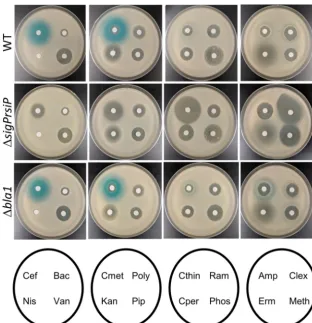
sigP) to thelacZreporter gene and integrated this construct into the genome ofB. thuringiensis(THE2549thrC::PsigP-lacZ). We tested several classes of-lactams and cell wall-targeting antibiotics for their ability to induce expression of PsigP-lacZ. We observed wide zones of PsigP-lacZ induction
on September 8, 2020 by guest
http://msphere.asm.org/
around cefoxitin and cefmetazole (Fig. 1). We detected fainter zones of induction in the areas around cephalothin and cephalexin (Fig. 1). Very faint zones of induction were present in the cells around ampicillin and methicillin (Fig. 1). Interestingly, we did not observe this induction surrounding the -lactams cefoperazone and piperacillin or antibiotics that target other steps in cell wall biosynthesis, including ramoplanin, phosphomycin, nisin, bacitracin, and vancomycin (Fig. 1). We also tested compounds that do not target peptidoglycan biosynthesis, including kanamycin, polymyxin B, and erythromycin (Erm), and saw no induction of PsigP-lacZ(Fig. 1).
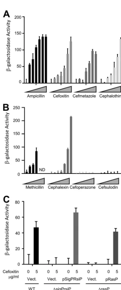
To quantify the levels of -lactam induction, we tested eight-lactams for their ability to activate the PsigP-lacZfusions using a-galactosidase assay. Mid-log cells were incubated in the presence of various concentrations of ampicillin, cefoxitin, cefmeta-zole, cephalothin, methicillin, cephalexin, cefoperazone, and cefsulodin for 1 h at 37°C. We observed dose-dependent induction with a subset of these-lactams (Fig. 2A and B). Interestingly, ampicillin, methicillin, and cephalexin showed low levels of PsigP-lacZ induction when spotted onto a lawn of cells (Fig. 1) but strongly induced PsigP-lacZin liquid assays (Fig. 2A and B), a point we will return to later. In contrast, neither cefoperazone nor cefsulodin was able to induce on the plates or in liquid (Fig. 1 and 2B). This confirms our observation that a subset of-lactams inducesPactivation.
FIG 1 Expression ofsigPis specifically induced by-lactams. All the strains contained PsigP-lacZin either a wild-type (THE2549), a ΔsigP-rsiP(EBT232), or a Δbla1(EBT215) background. Mid-log cells were washed and diluted 1:100 in molten LB agar containing X-Gal (100g/ml) and poured into empty 100-mm petri dishes. Filter disks containing cefoxitin (Cef) (1l of 5-mg/ml cefoxitin), bacitracin (Bac) (1l of 50-mg/ml bacitracin), nisin (Nis) (3l of 100-mg/ml nisin), vancomycin (Van) (1l of 10-mg/ml vancomycin), cefmetazole (Cmet) (1l of 5-mg/ml cefmetazole), polymyxin B (Poly) (1l of 50-mg/ml polymyxin B), kanamycin (Kan) (1l of 10-mg/ml kanamycin), piperacillin (Pip) (1l of 5-mg/ml piperacillin), cephalo-thin (Ccephalo-thin) (1l of 50-mg/ml cephalothin), ramoplanin (Ram) (1l of 25-mg/ml ramoplanin), cefopera-zone (Cper) (1l of 50 mg/ml cefoperazone), phosphomycin (Phos) (1l of 100-mg/ml phosphomycin), Amp (2l of 200-mg/ml ampicillin), cephalexin (Clex) (1l of 50-mg/ml cephalexin), Erm (1l of 5-mg/ml erythromycin), and methicillin (Meth) (2l of 100-mg/ml methicillin) were then placed on the top agar and incubated for 16 h at 30°C.
on September 8, 2020 by guest
http://msphere.asm.org/
We found that deletion of thesigP-rsiPgenes blocked expression of PsigP-lacZin the presence of-lactams (Fig. 1 and 2C), demonstrating thatPis required for induction
of PsigP-lacZin response to-lactams. When we introduced a low-copy-number plasmid containing PsigP-sigP⫹-rsiP⫹ into the ΔsigP-rsiP mutant (ΔsigP-rsiP/pSigPRsiP), we re-FIG 2 Expression of PsigP-lacZ is dose dependent and dependent upon P and RasP. (A) B.
thuringiensiswith transcriptional fusion PsigP-lacZ(THE2549) was grown overnight at 30°C, subcul-tured in LB, and grown to an OD600of⬃0.8 before being incubated with various concentrations of -lactams (0, 0.0625, 0.125, 0.25 0.5, 1, and 2g/ml) for 1 h. Cells were collected and resuspended in Z buffer. (B)B. thuringiensiswith transcriptional fusion PsigP-lacZ(THE2549) was grown overnight at 30°C, subcultured in LB, and grown to an OD600of⬃0.8 before being incubated with various concentrations of-lactams (0, 0.0625, 0.125, 0.25 0.5, 1, and 2g/ml) for 1 h. Cells were collected and resuspended in Z buffer. (C) All strains contain PsigP-lacZand the genotype and plasmid noted: wild type/Vect. (EBT169),sigP/Vect. (EBT251), ΔsigP-rsiP/pSigPRsiP (EBT238), ΔrasP/Vect. (EBT175), and rasP/pRasP (EBT176). Strains were grown to mid-log phase and then treated with 5g/ml cefoxitin or untreated (0) and incubated for 1 h.-Galactosidase activity was calculated as described in Materials and Methods. These experiments were done in triplicate, and standard deviations are represented by error bars.
on September 8, 2020 by guest
http://msphere.asm.org/
stored the induction of PsigP-lacZin response to cefoxitin (Fig. 2C). Taken together, these data suggest that a subset of-lactam antibiotics activatesP.
P and Bla1 are involved in resistance to some-lactams.To determine the impact ofPon resistance to-lactams, we measured the MICs of several-lactams for
wild-type and ΔsigP-rsiPmutant strains. We found that the wild type was greater than 100-fold more resistant to ampicillin, methicillin, and cephalothin than was the Δ sigP-rsiPmutant (Table 1). The wild type was 16- to 50-fold more resistant to cefmetazole, cefoxitin, and cephalexin than the mutant (Table 1). There was little or no difference in resistance to piperacillin, cefoperazone, and cefsulodin, which also failed to activateP
(Table 1 and Fig. 1). We also demonstrate that complementing the⌬sigP-rsiPmutant with a plasmid carrying PsigP-sigP⫹-rsiP⫹restored resistance to ampicillin and cefoxitin (Table 2). For reasons that remain unclear, strains containing plasmids, including empty vector, have slight increases in-lactam resistance. However, this does not impact the observation that the presence of PsigP-sigP⫹-rsiP⫹restored resistance to ampicillin and cefoxitin.
SincePwas shown to control expression ofhd73_3490(referred to here asbla1),
which encodes a-lactamase, we sought to determine if this gene played a role in resistance to -lactams. We made a deletion of bla1 and determined the MIC of ampicillin and cefoxitin for this strain. Thebla1mutant was 8- to 16-fold more sensitive to ampicillin and ⬃5-fold more sensitive to methicillin but no more sensitive to cefoxitin than the wild type (Table 2). This contrasts with the sigPmutant, which is greater than 1,000-fold more sensitive to ampicillin, 600-fold more sensitive to meth-icillin, and ⬃25-fold more sensitive to cefoxitin than the wild type (Table 2). This suggests that Bla1 plays a more important role in resistance to ampicillin and methi-cillin than to cefoxitin. Furthermore, our data suggest that while Bla1 contributes to
-lactam resistance, additional P-regulated genes must also contribute to-lactam
resistance.
When we tested various-lactams for induction of PsigP-lacZon 5-bromo-4-chloro-3-indolyl--D-galactopyranoside (X-Gal) plates, we did not consistently observe a strong zone of induction surrounding ampicillin and methicillin (Fig. 1). We hypothesized that this weak induction zone was due to the wild type efficiently producing-lactamases which degraded the inducer (ampicillin and methicillin). Thus, we were unable to TABLE 1⌬sigP-rsiPmutant is more sensitive to-lactams than wild type
Drug
MIC (g/ml) for strain (meanⴞSD):
Fold difference WT ⌬sigP-rsiPmutant
Ampicillin 6,000⫾0 1.67⫾0.5 3,592
Cefoxitin 200⫾0 20⫾0 10
Methicillin 666⫾115 1⫾0 666
Piperacillin 5⫾0 1.25⫾0 4
Cephalothin 88⫾25 0.25⫾0 350
Cephalexin 200⫾0 4⫾0 50
Cefmetazole 44⫾13 2.8⫾1.1 16
Cefoperazone 5⫾2 4⫾0 1.25
Cefsulodin 400⫾0 400⫾0 1
TABLE 2RasP is required for resistance to-lactamsa
Genotype Vector
MIC (g/ml) of drug (meanⴞSD):
Ampicillin Cefoxitin Methicillin
WT Empty 8,000⫾0 200⫾0 666.7⫾115
⌬sigP-rsiP Empty 2⫾0 20⫾0 1⫾0
⌬sigP-rsiP pSigP 6,666⫾3,011 100⫾0 ND
⌬rasP Empty 6.7⫾2.1 20⫾0 ND
⌬rasP pRasP 6,333⫾1,966 133⫾57.7 ND
⌬bla1 Empty 400⫾0 200⫾0 125⫾50
aAbbreviations: WT, wild type; ND, not determined.
on September 8, 2020 by guest
http://msphere.asm.org/
observe the increased production of -galactosidase. To test this hypothesis, we determined the effect of a Δbla1mutant onPactivation. We found that in the Δbla1
mutant, ampicillin and methicillin produced more distinct zones of induction (Fig. 1). However, all other induction zones of the Δbla1mutant were similar to the wild type. Thus, in the absence of Bla1, which degrades ampicillin and methicillin, we detected greater induction of PsigP-lacZexpression. Taken together, these observations suggest that the weak ampicillin induction of PsigP-lacZon plates is in part due to the efficient degradation of the inducer by-lactamases.
RsiP is degraded in response to cefoxitin in a dose-dependent manner. The anti- factors of other ECF01 family members are degraded, which leads to the activation of their cognatefactors (7, 14, 15). We sought to determine if-lactams activatePby inducing degradation of RsiP. To investigate this, we constructed a strain
with an anhydrotetracycline (ATc)-inducible copy of green fluorescent protein (GFP) fused to the N terminus of RsiP (GFP-RsiP). The inducible promoter allows us to uncouple expression of RsiP from induction ofP. The GFP-RsiP fusion allows us to
follow the fate of the cytoplasmic portion of RsiP. Expression of GFP-RsiP complements an rsiPnull mutation (see Fig. S1 in the supplemental material) and localizes to the membrane (Fig. S2). We then induced the synthesis of GFP-RsiP in exponential-phase cells and monitored its processing before and after treatment with cefoxitin. We chose to utilize cefoxitin for these experiments because cefoxitin inducesPactivation over
a wide concentration range and the⌬sigP-rsiPmutant strain grows at most of these concentrations (Fig. 2A and Table 1). Cell pellets were then lysed by sonication, and Western blot analyses were performed using anti-RsiP antisera against the extracellular portion of RsiP or anti-GFP antisera, which detect GFP fused to the intracellular portion of RsiP.
When cells producing GFP-RsiP were grown in the absence of cefoxitin, we detected full-length GFP-RsiP at the expected size of⬃60 kDa using anti-RsiP antisera. This band was absent in the empty-vector control (Fig. 3A). When cells were incubated with cefoxitin (5g/ml) for various times, we found that the level of full-length GFP-RsiP decreased over time (Fig. 3A and Fig. S3A). We observed loss of GFP-RsiP by 30 min to 1 h after exposure to cefoxitin (Fig. 3A and Fig. S3A). This suggests that GFP-RsiP is likely degraded in the presence of cefoxitin.
We also tested the effect of cefoxitin concentration on GFP-RsiP levels by incubating cells with a range of cefoxitin concentrations (0 to 500g/ml) for 1 h. We found that increasing concentrations of cefoxitin resulted in a greater decrease of full-length GFP-RsiP (Fig. 3B and Fig. S3B). We obtained comparable results when we performed blotting assays for the N-terminal domain using anti-GFP antisera (Fig. S4). These data suggest that activation of Poccurs via loss of RsiP in a cefoxitin dose-dependent
manner.
RasP is necessary forPactivation.BothEandWare activated by regulated
intramembrane proteolysis of their cognate anti-factors. Proteolysis of these anti- factors requires multiple proteases, including the highly conserved site-2 proteases RseP and RasP, respectively (14, 15). We hypothesize that activation of P requires
multiple proteases, including the conserved site-2 protease RasP to degrade RsiP. To test this, we used BLAST to identify a putative membrane-embedded metalloprotease, HD73_4103, which is 76% similar and 60% identical to B. subtilis RasP and is here referred to as RasP (Fig. S5) (38–43). To determine if RasP was required forPactivation,
we generated a strain containing a deletion ofrasPand the PsigP-lacZreporter. In the absence of RasP, we did not detect increased expression of PsigP-lacZ reporter in response to cefoxitin (Fig. 2C). In MIC experiments, we found that, similarly to the ΔsigP-rsiP mutant, the ΔrasP mutant was more sensitive to ampicillin and cefoxitin (Table 2). We found that both resistance to-lactams and induction of PsigP-lacZcould be complemented when a plasmid expressing rasP⫹was introduced into the ΔrasP mutant (Fig. 2C and Table 2). These data suggest that RasP is required forPactivation.
on September 8, 2020 by guest
http://msphere.asm.org/
RasP is required for degradation of RsiP. To determine if RasP is required for degradation of RsiP, we expressed the GFP-RsiP fusion in both the wild type and a
⌬rasPmutant. We treated cells with 5g/ml cefoxitin for various lengths of time from 0 to 180 min (Fig. 4 and Fig. S6). In the wild type, we observed loss of full-length RsiP over time (Fig. 4 and Fig. S6). In contrast, we observed loss of full-length GFP-RsiP and the accumulation of a smaller⬃35-kDa band in the ΔrasPmutant (Fig. 4 and Fig. S6). This suggests that RasP is required for complete degradation of RsiP. Since a truncated product accumulates in the⌬rasPmutant, RasP is likely required for site-2 cleavage and an unidentified protease is required for cleavage at site-1.
FIG 3 RsiP levels decrease in the presence of cefoxitin.B. thuringiensisexpressing tetracycline-inducible
gfp-rsiP(EBT360) or empty vector (EV; EBT169) was subcultured 1:50 into LB supplemented with ATc (50 ng/ml). At mid-log phase, cells were incubated with 5g/ml of cefoxitin for various times (0, 15, 30, 60, 120, or 180 min) (A) or increasing concentrations of cefoxitin (0, 0.05, 0.5, 5, 50, or 500g/ml) for 1 h (B). The immunoblot was probed with antisera against RsiP (␣-RsiP76 –275). Streptavidin IR680LT was used to detect HD73_4231 (PycA homolog), which served as a loading control (62, 63). The color blot showing both anti-RsiP and streptavidin on a single gel is shown in Fig. S3. Numbers at right indicate molecular masses in kilodaltons.
FIG 4 RsiP degradation is dependent upon the site-2 protease RasP.B. thuringiensiswild type (EBT360) or⌬rasP
(EBT366) containing a tetracycline-inducible copy ofgfp-rsiPwas subcultured 1:50 into LB supplemented with ATc (50 ng/ml). At mid-log phase, cultures were incubated with cefoxitin (5g/ml) for the time indicated at 37°C. The immunoblot was probed with anti-GFP antisera. EV is wild type with pAH9 (EBT169), and GFP is wild type with pAH13 (UM20). Streptavidin IR680LT was used to detect HD73_4231 (PycA homolog), which served as a loading control (62, 63). The color blot showing both anti-GFP and streptavidin on a single gel is shown in Fig. S6. Numbers at right are molecular masses in kilodaltons.
on September 8, 2020 by guest
http://msphere.asm.org/
Mutations inrsiPresult in constitutivesigPexpression.To further characterize thePsignal transduction system, we isolated mutants which resulted in constitutive
expression of PsigP-lacZ. We selected for mutants with increased resistance to cefoxitin by plating cultures of the wild-type PsigP-lacZstrain (THE2549) on LB-cefoxitin (200g/ ml) agar. At this concentration of cefoxitin, wild-typeB. thuringiensisfails to grow. These strains were tested for PsigP-lacZexpression in the absence of cefoxitin by streaking on LB–X-Gal. We isolated 8 independent mutants with increased resistance to cefoxitin that have constitutive PsigP-lacZexpression. We hypothesized that these strains har-bored mutations inrsiP. We PCR amplified and sequenced thesigPandrsiPgenes from the constitutive mutants. The 8 constitutive mutants contained mutations in different regions of thersiP gene that resulted in C-terminal truncations of RsiP (Fig. S7). We selected fourrsiPmutants for further study. We found that each mutant strain showed increased PsigP-lacZ expression even in the absence of -lactams (Fig. 5). When a wild-type copy ofrsiP(pSigPRsiP) was introduced to each of these mutants, PsigP-lacZ expression was no longer constitutive but was induced in the presence of cefoxitin (Fig. S8). This indicates that the rsiP mutations were responsible for the increased PsigP-lacZexpression.
In theVandWsystems, RasP cleaves the anti-factors RsiW and RsiV within the
transmembrane domain to activate the cognatefactors (15, 35). The RsiP transmem-brane is predicted to be residues 54 to 71 based on TMHMM (44). Two of the four RsiP truncations produce proteins with the transmembrane domain intact, while the re-maining RsiP truncations lack the transmembrane domain. Since RasP is known to cleave proteins within the transmembrane domain, we hypothesized that those trun-cations which still contain a transmembrane domain would require RasP in order to activate P. To test this, we introduced the ⌬rasP mutation into each of the rsiP
mutants. In the absence of RasP, strains containing truncations which have a trans-membrane domain (RsiP1–220and RsiP1– 80) (Fig. 4 and Fig. S7) no longer constitutively
activateP(Fig. 5). However, the strains with thersiPtruncation lacking the
transmem-brane domain (RsiP1–16and RsiP1– 61) constitutively activatePeven in the absence of
RasP (RsiP1–16and RsiP1– 61) (Fig. 4 and Fig. S5). Thus, RasP is required forPactivation
when the transmembrane domain of RsiP is intact, consistent with the role of RasP as a site-2 protease.
FIG 5 Truncations of RsiP lead to constitutive Pactivation. To determine if RasP was required for ampicillin-inducible PsigP-lacZexpression, we assayed-galactosidase activity ofB. thuringiensiswith transcriptional fusion PsigP-lacZand differentrsiPtruncation mutants (WT, THE2549; RsiP1–220, THE2602; RsiP1– 80, THE2628; RsiP1– 61, THE2637; RsiP1–16, THE2642) and a ΔrasPdeletion (WT, EBT140; RsiP1–220, EBT116; RsiP1– 80, EBT148; RsiP1– 61, EBT133; RsiP1–16, THE2605). Cells were grown overnight at 30°C, subcultured in LB, and grown to an OD600of⬃0.8 before being incubated with cefoxitin (5g/ml) for 1 h. The experiment was performed in triplicate, and standard deviations are represented by error bars.
on September 8, 2020 by guest
http://msphere.asm.org/
RasP cleaves within the transmembrane domain of RsiP and is not the regu-lated step inPactivation.In the case ofWandV, the rate-limiting step infactor
activation is site-1 cleavage (15, 35). Since the identity of the site-1 protease is not currently known, we sought to determine if RasP cleavage of RsiP is a rate-limiting step in P activation. To test this, we constructed truncations of GFP-RsiP that lack the
extracellular portion of RsiP. One truncation includes the transmembrane domain (gfp-rsiP1–72), and one truncation lacks the transmembrane domain (gfp-rsiP1–53). We
expressed the truncated GFP-RsiP proteins in wild-type and ⌬rasPbackgrounds and exposed these strains to cefoxitin (5g/ml). In wild-type strains, we found that both GFP-RsiP1–72and GFP-RsiP1–53were degraded (Fig. 6 and Fig. S9). However, in the ΔrasP
mutant GFP-RsiP1–72accumulated, while GFP-RsiP1–53was degraded (Fig. 6 and Fig. S9).
These data indicate that GFP-RsiP1–72requires RasP for degradation while GFP-RsiP1–53
does not. One possible interpretation is that GFP-RsiP1–72is not produced or localized
properly to the membrane. Thus, we confirmed that GFP-RsiP1–72 localizes to the
membrane by fluorescence microscopy (Fig. S2). This suggests that the RasP cleavage site of RsiP occurs within the transmembrane domain between amino acids 53 and 72. The presence or absence of cefoxitin had no effect on the degradation (Fig. 6 and Fig. S9). Since GFP-RsiP1–72 is constitutively degraded, we conclude that GFP-RsiP1–72
mimics the site-1 cleavage product and that RasP activity is not induced by cefoxitin. This suggests that RasP cleavage of RsiP is not the regulated step inPactivation and
that site-1 cleavage is the step that is controlled by the presence of-lactams.
DISCUSSION
Many ECF factors are induced in response to extracytoplasmic stressors and initiate transcription of a subset of genes to modulate the cell’s response to these stresses. ECFfactors can respond to signals such as misfolded periplasmic protein, antimicrobial peptides, or lysozyme. The ECF factors encoded in highly related organisms can vary widely. For example, B. subtilisencodes 7 ECF factors, whileB. thuringiensis encodes 15 predicted ECF factors. The only ECF factor that these organisms share is M (45). Thus, there is a variability in how bacteria utilize ECF
factors to respond to stress. Ross et al. demonstrated that the novel ECFfactorPis
induced in the presence of ampicillin and initiates transcription of -lactamases (5). Here, we demonstrated thatPresponds specifically to a subset of-lactams, while
other-lactams and cell wall-targeting antibiotics fail to inducePactivation. We also
showed thatPconfers various degrees of resistance to these-lactam antibiotics. We
found thatPwas not required for resistance to other cell wall antibiotics, including
FIG 6 Truncation of RsiP results in constitutive degradation in a RasP-dependent manner.B. thuringiensis
containing a tetracycline-inducible copy ofgfp-rsiP,gfp-rsiP1–72(rsiPwithout the extracellular domain), or
gfp-rsiP1–53(rsiPwithout the transmembrane and extracellular domains) was constructed in either the wild type (rasP⫹) or a⌬rasPmutant strain (GFP-RsiP wild type [rasP⫹], EBT360; GFP-RsiP⌬rasP, EBT366; GFP-RsiP1–53wild type [rasP⫹], EBT518; GFP-RsiP1–53⌬rasP, EBT510; GFP-RsiP1–72wild type [rasP⫹], EBT516; GFP-RsiP1–72⌬rasP, EBT533). Strains were subcultured 1:50 into LB supplemented with ATc (100 ng/ml), grown to mid-log phase, and then incubated for 2 h without (⫺) or with (⫹) cefoxitin treatment (5g/ml) at 37°C. The immunoblot was probed with anti-GFP antisera. Streptavidin IR680LT was used to detect HD73_4231 (PycA homolog), which served as a loading control (62, 63). The color blot showing both anti-GFP and streptavidin on a single gel is shown in Fig. S9. Numbers at right are molecular masses in kilodaltons.
on September 8, 2020 by guest
http://msphere.asm.org/
vancomycin, nisin, and bacitracin, suggesting specificity in resistance to-lactams and not a general cell envelope stress response.
For ECFfactors to be activated, their cognate anti-factors must be inactivated. This can be accomplished via various mechanisms, including a conformational change of the anti- factor; partner switching, where an anti-anti-factor frees thefactor from the anti-factor; or proteolytic destruction of the anti-factor (9, 11). The anti- factors RseA in E. coliand RsiW and RsiV in B. subtilis are degraded sequentially by regulated intramembrane proteolysis. Each of these anti-factors requires a different family of proteases to cleave the anti-factor at site-1 (14, 22, 30, 46, 47), while site-2 cleavage is carried out by the conserved site-2 protease (14, 15, 35). We hypothesize thatPis activated in a similar manner. Our data indicate thatPis released from RsiP
by proteolytic degradation when-lactams are present. We found that RasP is required for activation ofP. We also observe that an RsiP degradation product approximately
the size of our predicted RasP substrate accumulates in a⌬rasPmutant. This indicates that RasP is required for degradation of RsiP. Our data also suggest, similarly to other anti- factors, that site-2 cleavage of RsiP is not the rate-limiting step, since the C-terminal RsiP truncations are constitutively degraded and lead to constitutive P
activation in the absence of-lactams. Thus, we hypothesize that RasP is required for site-2 cleavage of RsiP and that an as-yet-unidentified protease is required to initiate degradation of RsiP by cleaving RsiP at site-1. We hypothesize that, like other ECF factors activated by regulated intramembrane proteolysis, site-1 cleavage of RsiP is likely the rate-limiting step inPactivation.
Our data suggest that a subset of-lactams inducePactivation. We found that, in
addition to ampicillin,Pis activated by cefoxitin, cefmetazole, cephalothin, cephalexin,
and methicillin but not by piperacillin, cefoperazone, cefsulodin, or antibiotics that target other steps in peptidoglycan biosynthesis. This raises the question of what the signal is for Pactivation. The -lactams could be sensed directly or indirectly. For
example, RsiV directly senses lysozyme and degradation of RsiV is rapid (31). In contrast, activation ofEis indirect and due to buildup of products that occur when the outer
membrane is damaged (31, 48). Our data suggest that RsiP degradation is a relatively slow process. One possible interpretation of this is that-lactam-induced peptidogly-can (PG) damage must accumulate to induce RsiP degradation. We hypothesize that the -lactams that we tested have different affinities for penicillin-binding proteins (PBPs) and that this affinity may explain why some-lactams inducePwhile others do
not. In other organisms, including Streptococcus pneumoniae, B. subtilis, and E. coli,
-lactams can differentially target PBPs (49–51). This raises the possibility that activa-tion ofPcould be the result of inhibition of specific PBPs. Unfortunately, at this time
we do not know which PBPs are targeted by the different-lactams inB. thuringiensis. Thus, the precise mechanism and signal responsible for Pactivation remain to be
clearly defined.
MATERIALS AND METHODS
Media and growth conditions. All B. thuringiensisstrains are isogenic derivatives of AW43, a derivative ofBacillus thuringiensissubsp.kurstakistrain HD73 (52). All strains and genotypes can be found in Table 3. AllB. thuringiensisstrains were grown in or on LB medium at 30°C unless otherwise specified. Cultures ofB. thuringiensiswere grown with agitation in a roller drum. Strains containing episomal plasmids were grown in LB containing chloramphenicol (Cam; 10g/ml) or erythromycin (Erm; 10g/ ml).E. colistrains were grown at 37°C using LB-ampicillin (Amp; 100g/ml) or LB-Cam (10g/ml) medium. To screen for threonine auxotrophy,B. thuringiensisstrains were patched on minimal medium plates without or with threonine (50g/ml) (53, 54). The -galactosidase chromogenic indicator 5-bromo-4-chloro-3-indolyl--D-galactopyranoside (X-Gal) was used at a concentration of 100g/ml. Anhydrotetracycline (ATc; Sigma) was used at a concentration of 100 ng/ml.
Strain and plasmid construction.All plasmids are listed in Table 4, which includes information relevant to plasmid assembly. Plasmids were constructed by isothermal assembly (55). Regions of plasmids constructed using PCR were verified by DNA sequencing. The oligonucleotide primers used in this work were synthesized by Integrated DNA Technologies (Coralville, IA) and are listed in Table S1 in the supplemental material. All plasmids were propagated using OmniMax 2-T1R as the cloning host and passaged through the nonmethylatingE. colistrain INV110 before being transformed into aB. thurin-giensisrecipient strain.
on September 8, 2020 by guest
http://msphere.asm.org/
To construct deletion mutants, we cloned DNA 1 kb upstream and 1 kb downstream of the site of desired deletion using primers listed in Table S1 onto the temperature-sensitive pMAD plasmid (eryth-romycin resistant) between the BglII and EcoRI sites (56).
Complementation constructs were constructed in pAH9, which is anE. coli–Gram-positive bacterial shuttle vector with a pE194 origin of replication (57). Chromosomal DNA including the promoter sequence was cloned for PsigP-sigP⫹-rsiP⫹and cloned into pAH9 digested with EcoRI and HindIII, while
rasPwas cloned downstream of the PsarApromoter fromStaphylococcus aureusby digesting with EcoRI and KpnI. InB. thuringiensis, PsarAhas moderate constitutive expression.
To generate strains containing thesigPpromoter fused to thelacZreporter integrated into the chromosome, we constructed a number of intermediate vectors. To switch the antibiotic resistance of the temperature-sensitive pMAD vector, we constructed pTHE946, which contains theE. coliorigin (ColE1 ori) of replication, an Erm resistance gene (for selection in Gram-positive bacteria), an Amp resistance gene (for selection inE. colistrains), and the temperature-sensitive origin (pE194 ori) from pMAD (7.3-kb StuI and BamHI fragment) as well as the conjugation origin of transfer and the Cam resistance gene from pRPF185 (SmaI and BamHI fragment). ThethrC(primers 2917 and 2918) andthrB(primers 2919 and 2920) genes were cloned into the ScaI- and SalI-digested pTHE946 plasmid (lacking Ermrand Amprgenes) to generate a vector (pTHE948) which can integrate into thethrCoperon. A promoterlesslacZfragment (primers 2922 and 2923) was added between thethrCandthrBgenes of pTHE948 (XhoI and SbfI) to generate pTHE950. This plasmid (XhoI and NotI digested) was used to clone thesigPpromoter (primers TE2929 and 2930) to generate the PsigP-lacZpromoter fusion (pTHE949).
TABLE 3Strains
Species and strain Description Reference or source
B. thuringiensis
AW43 B. thuringiensissubsp.kurstakiHD73 cured of both pAW63 and pHT73, Nalr
52
THE2549 AW43thrC::PsigP-lacZ This study
EBT140 AW43thrC::PsigP-lacZΔrasP This study
EBT232 AW43thrC::PsigP-lacZΔsigP-rsiP This study
EBT215 AW43thrC::PsigP-lacZΔbla1 This study
EBT360 AW43thrC::PsigP-lacZ/pAH9 Ptet-gfp-rsiP This study EBT366 AW43thrC::PsigP-lacZΔrasP/pAH9 Ptet-gfp-rsiP This study EBT510 AW43thrC::PsigP-lacZΔrasP/pAH9 Ptet-gfp-rsiP1–53 This study EBT516 AW43thrC::PsigP-lacZ/pAH9 Ptet-gfp-rsiP1–72 This study EBT518 AW43thrC::PsigP-lacZ/pAH9 Ptet-gfp-rsiP1–53 This study
EBT533 AW43thrC::PsigP-lacZΔrasP/pAH9 Ptet-gfp-rsiP1–72 This study
EBT175 AW43thrC::PsigP-lacZΔrasP/pAH9 This study
EBT176 AW43thrC::PsigP-lacZΔrasP/pAH9rasP This study
EBT238 AW43thrC::PsigP-lacZΔsigP-rsiP/pAH9 PsigP-sigP-rsiP This study
EBT251 AW43thrC::PsigP-lacZΔsigP-rsiP/pAH9 This study
THE2642 AW43thrC::PsigP-lacZ rsiP1–16 This study
THE2637 AW43thrC::PsigP-lacZ rsiP1–61 This study
THE2628 AW43thrC::PsigP-lacZ rsiP1–80 This study
THE2602 AW43thrC::PsigP-lacZ rsiP1–220 This study
THE2605 AW43thrC::PsigP-lacZΔrasP rsiP1–16 This study
EBT133 AW43thrC::PsigP-lacZΔrasP rsiP1–61 This study
EBT148 AW43thrC::PsigP-lacZΔrasP rsiP1–80 This study
EBT116 AW43thrC::PsigP-lacZΔrasP rsiP1–220 This study
EBT567 AW43thrC::PsigP-lacZ rsiP1–16/pAH9 PsigP-sigP-rsiP This study EBT566 AW43thrC::PsigP-lacZ rsiP1–61/pAH9 PsigP-sigP-rsiP This study EBT565 AW43thrC::PsigP-lacZ rsiP1–80/pAH9 PsigP-sigP-rsiP This study
EBT564 AW43thrC::PsigP-lacZ rsiP1–220/pAH9 PsigP-sigP-rsiP This study EBT168 AW43thrC::PsigP-lacZ/pAH9 PsigP-sigP-rsiP This study
EBT169 AW43thrC::PsigP-lacZpAH9 This study
EBT563 AW43thrC::PsigP-lacZ rsiP1–16/pAH9 This study
EBT562 AW43thrC::PsigP-lacZ rsiP1–61/pAH9 This study
EBT561 AW43thrC::PsigP-lacZ rsiP1–80/pAH9 This study
EBT560 AW43thrC::PsigP-lacZ rsiP1–220/pAH9 This study
UM20 AW43/pAH13 This study
EBT587 AW43thrC::PsigP-lacZ rsiP1–80/pAH9 Ptet-gfp-rsiP This study E. coli
OmniMax 2-T1R F={proAB⫹lacIqlacZΔM15 Tn10(Tetr) Δ(ccdAB)}mcrA
Δ(mrr-hsdRMS-mcrBC)80(lacZ)ΔM15 Δ(lacZYA-argF)U169 endA1 recA1 supE44 thi-1 gyrA96 relA1 tonA panD
Invitrogen
INV110 endA1 rpsL thr leu thi lacY galK galT ara tomA tsx dam dcm supE44Δ(lac-proAB) [F=traD36 proAB lacIqZΔM15]
Invitrogen
on September 8, 2020 by guest
http://msphere.asm.org/
B. thuringiensisDNA transformation.Plasmids were introduced intoB. thuringiensisby electropo-ration (58, 59). Briefly, recipient cells were grown to late log phase at 37°C. For each transformation, cells (1.5 ml) were pelleted by centrifugation (9,000⫻g) and washed twice in room-temperature sterile water. After careful removal of all residual water, 100l of sterile 40% polyethylene glycol (PEG) 6000 (Sigma) was used to gently resuspend cells. Approximately 2 to 10l of unmethylated DNA (⬎50 ng/l) was added to cells and transferred to an 0.4-cm-gap electroporation cuvette (Bio-Rad). Cells were exposed to 2.5 kV for 4 to 6 ms. LB was immediately added, and cells were incubated at 30°C for 1 to 2 h prior to plating on selective media.
Construction of deletions or promoter-lacZfusions inB. thuringiensis.To generate unmarked mutants andthrC::PsigP-lacZstrains, we used plasmid vectors containing the temperature-sensitive origin of replication (pE194 ori) from the pMAD plasmid (56). At permissive temperatures (30°C), pMAD replicates episomally as a plasmid. At nonpermissive temperatures (42°C), pMAD must integrate into the chromosome via homologous recombination; otherwise, the plasmid will be lost to segregation and the strain will become sensitive to erythromycin. Plasmids were transformed into aB. thuringiensisrecipient strain and selected for on LB-Erm agar at 30°C. To select for the integration of the deletion plasmid into the recipient strain genome, plasmid-containing bacteria were grown at 42°C on LB-Erm plates. The plasmid-integrated strain was then struck on LB agar at 30°C twice. Individual colonies were patched on LB and LB-Erm agar to identify the Erm-sensitive bacteria which had lost the deletion plasmid by segregation. To verify each deletion, genomic DNA was isolated from each strain candidate and PCR was used to verify the deletion. Integration of the PsigP-lacZfusion into thethrCoperon results in threonine auxotrophy and can be identified by lack of growth on minimal medium plates without threonine.
Zones of inhibition and zones of induction.To determine the zones of inhibition and induction by various antibiotics, we first washed mid-logarithmically grown cells in fresh LB. Washed cells were diluted 1:100 in molten LB agar containing X-Gal (100g/ml) and poured into empty 100-mm petri dishes. Sterile cellulose disks (8 mm) were saturated with different antibiotics and allowed to dry for longer than 10 min. After each antibiotic disk was placed on the solidified agar, plates were incubated at 30°C overnight before observation.
-Galactosidase assays. To quantify expression from the sigP promoter, we measured the -galactosidase activity of cells containing a PsigP-lacZpromoter fusion. Overnight cultures were diluted 1:50 in fresh LB medium and incubated for 3 h at 30°C. One milliliter of each subculture was pelleted (9,000⫻g), washed (in LB broth), and resuspended in 1 ml LB broth lacking or including specified antibiotics. After 1 h of incubation at 37°C, 1 ml of each sample was pelleted and resuspended in 200l of Z buffer. Cells were permeabilized by mixing with 16l chloroform and 16l 2% Sarkosyl (26, 60). Permeabilized cells (100l) were mixed with 10 mg/mlortho-nitrophenyl--galactoside (ONPG; Research Products International; 50l), and optical density at 405 nm (OD405) was measured over time using an Infinite M200 Pro plate reader (Tecan).-Galactosidase activity units (mol of ONP formed min⫺1)⫻ 103/(OD
600⫻ml of cell suspension) were calculated as previously described (61). Experiments were performed in triplicate with the mean and standard deviation being shown.
MIC assay.To determine the MICs of various antibiotics, we diluted overnight cultures of bacteria (washed in LB) 1:1,000 in medium containing 2-fold dilutions of each antibiotic. All MIC experiments were performed in round-bottom 96-well plates. Each experiment was performed in triplicate, and the plates were allowed to incubate for 24 h at 37°C before observation of growth or no growth.
Immunoblot analysis.Samples were electrophoresed on a 15% SDS-polyacrylamide gel, and proteins were then blotted onto a nitrocellulose membrane (GE Healthcare, Amersham). Nitrocellulose was blocked with 5% bovine serum albumin (BSA), and proteins were detected with either 1:10,000 anti-GFP or 1:5,000 anti-RsiP76 –275antiserum. Streptavidin IR680LT (1:10,000) was used to detect two biotin-containing proteins, PycA (HD73_4231) and AccB (HD73_4487), which served as loading controls (62, 63). To detect primary antibodies, the blots were incubated with 1:10,000 goat anti-rabbit IR800CW (Li-Cor) and imaged on an Odyssey CLx scanner (Li-Cor) or an Azure Sapphire imager (Azure Biosystems). All immunoblot assays were performed a minimum of three times with a representative example being shown.
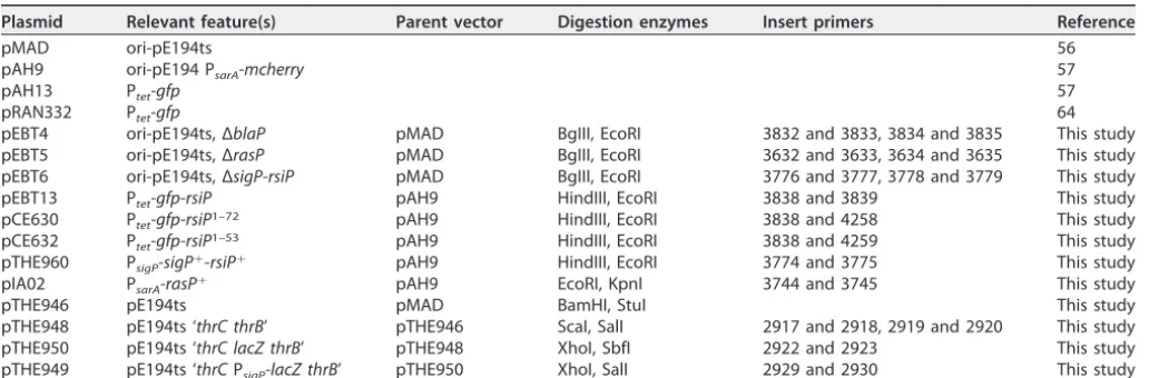
TABLE 4Plasmids
Plasmid Relevant feature(s) Parent vector Digestion enzymes Insert primers Reference
pMAD ori-pE194ts 56
pAH9 ori-pE194 PsarA-mcherry 57
pAH13 Ptet-gfp 57
pRAN332 Ptet-gfp 64
pEBT4 ori-pE194ts, ΔblaP pMAD BgIII, EcoRI 3832 and 3833, 3834 and 3835 This study pEBT5 ori-pE194ts, ΔrasP pMAD BgIII, EcoRI 3632 and 3633, 3634 and 3635 This study pEBT6 ori-pE194ts, ΔsigP-rsiP pMAD BgIII, EcoRI 3776 and 3777, 3778 and 3779 This study
pEBT13 Ptet-gfp-rsiP pAH9 HindIII, EcoRI 3838 and 3839 This study
pCE630 Ptet-gfp-rsiP1–72 pAH9 HindIII, EcoRI 3838 and 4258 This study pCE632 Ptet-gfp-rsiP1–53 pAH9 HindIII, EcoRI 3838 and 4259 This study pTHE960 PsigP-sigP⫹-rsiP⫹ pAH9 HindIII, EcoRI 3774 and 3775 This study
pIA02 PsarA-rasP⫹ pAH9 EcoRI, KpnI 3744 and 3745 This study
pTHE946 pE194ts pMAD BamHI, StuI This study
pTHE948 pE194ts ‘thrC thrB’ pTHE946 ScaI, SalI 2917 and 2918, 2919 and 2920 This study pTHE950 pE194ts ‘thrC lacZ thrB’ pTHE948 XhoI, SbfI 2922 and 2923 This study pTHE949 pE194ts ‘thrCPsigP-lacZ thrB’ pTHE950 XhoI, SalI 2929 and 2930 This study
on September 8, 2020 by guest
http://msphere.asm.org/
SUPPLEMENTAL MATERIAL
Supplemental material for this article may be found at https://doi.org/10.1128/ mSphere.00511-19.
FIG S1, TIF file, 1.9 MB.
FIG S2, TIF file, 1.4 MB.
FIG S3, TIF file, 1.5 MB.
FIG S4, TIF file, 1.8 MB.
FIG S5, TIF file, 1.3 MB.
FIG S6, TIF file, 2.7 MB.
FIG S7, TIF file, 1.5 MB.
FIG S8, TIF file, 1.8 MB.
FIG S9, TIF file, 1.6 MB.
TABLE S1, PDF file, 0.1 MB.
ACKNOWLEDGMENTS
This work was supported by the Department of Microbiology and Immunology at the University of Iowa and by NIH grant R21AI146769 to C.D.E.
We thank Theresa Koehler for strains and advice. We also thank Leyla Slamti for protocols and members of the Ellermeier and Weiss labs for helpful comments.
REFERENCES
1. Waxman DJ, Strominger JL. 1983. Penicillin-binding proteins and the mechanism of action of beta-lactam antibiotics. Annu Rev Biochem 52:825– 869.https://doi.org/10.1146/annurev.bi.52.070183.004141. 2. Tomasz A. 1979. The mechanism of the irreversible antimicrobial effects
of penicillins: how the beta-lactam antibiotics kill and lyse bacteria. Annu Rev Microbiol 33:113–137.https://doi.org/10.1146/annurev.mi.33 .100179.000553.
3. Chen Y, Succi J, Tenover FC, Koehler TM. 2003.-Lactamase genes of the penicillin-susceptible Bacillus anthracisSterne strain. J Bacteriol 185: 823– 830.https://doi.org/10.1128/JB.185.3.823-830.2003.
4. Chen Y, Tenover FC, Koehler TM. 2004.-Lactamase gene expression in a penicillin-resistantBacillus anthracisstrain. Antimicrob Agents Chemother 48:4873– 4877.https://doi.org/10.1128/AAC.48.12.4873-4877.2004. 5. Ross CL, Thomason KS, Koehler TM. 2009. An extracytoplasmic function
sigma factor controls beta-lactamase gene expression inBacillus anthra-cisand otherBacillus cereusgroup species. J Bacteriol 191:6683– 6693.
https://doi.org/10.1128/JB.00691-09.
6. Gargis AS, McLaughlin HP, Conley AB, Lascols C, Michel PA, Gee JE, Marston CK, Kolton CB, Rodriguez-R LM, Hoffmaster AR, Weigel LM, Sue D. 2018. Analysis of whole-genome sequences for the prediction of penicillin resistance and-lactamase activity inBacillus anthracis. mSys-tems 3:e00154-18.https://doi.org/10.1128/mSystems.00154-18. 7. Staron´ A, Sofia HJ, Dietrich S, Ulrich LE, Liesegang H, Mascher T. 2009.
The third pillar of bacterial signal transduction: classification of the extracytoplasmic function (ECF) sigma factor protein family. Mol Micro-biol 74:557–581.https://doi.org/10.1111/j.1365-2958.2009.06870.x. 8. Helmann JD. 2002. The extracytoplasmic function (ECF) sigma factors.
Adv Microb Physiol 46:47–110.https://doi.org/10.1016/S0065-2911(02) 46002-X.
9. Helmann JD. 2016.Bacillus subtilisextracytoplasmic function (ECF) sigma factors and defense of the cell envelope. Curr Opin Microbiol 30: 122–132.https://doi.org/10.1016/j.mib.2016.02.002.
10. Helmann JD. 1999. Anti-sigma factors. Curr Opin Microbiol 2:135–141.
https://doi.org/10.1016/S1369-5274(99)80024-1.
11. Ho TD, Ellermeier CD. 2012. Extra cytoplasmic functionfactor activa-tion. Curr Opin Microbiol 15:182–188.https://doi.org/10.1016/j.mib.2012 .01.001.
12. Sineva E, Savkina M, Ades SE. 2017. Themes and variations in gene regulation by extracytoplasmic function (ECF) sigma factors. Curr Opin Microbiol 36:128 –137.https://doi.org/10.1016/j.mib.2017.05.004. 13. Yoshimura M, Asai K, Sadaie Y, Yoshikawa H. 2004. Interaction ofBacillus
subtilis extracytoplasmic function (ECF) sigma factors with the N-terminal regions of their potential anti-sigma factors. Microbiology 150:591–599.https://doi.org/10.1099/mic.0.26712-0.
14. Alba BM, Leeds JA, Onufryk C, Lu CZ, Gross CA. 2002. DegS and YaeL
participate sequentially in the cleavage of RseA to activate the sigma(E)-dependent extracytoplasmic stress response. Genes Dev 16:2156 –2168.
https://doi.org/10.1101/gad.1008902.
15. Schöbel S, Zellmeier S, Schumann W, Wiegert T. 2004. TheBacillus subtilis
sigmaW anti-sigma factor RsiW is degraded by intramembrane proteol-ysis through YluC. Mol Microbiol 52:1091–1105.https://doi.org/10.1111/ j.1365-2958.2004.04031.x.
16. Lima S, Guo MS, Chaba R, Gross CA, Sauer RT. 2013. Dual molecular signals mediate the bacterial response to outer-membrane stress. Sci-ence 340:837– 841.https://doi.org/10.1126/science.1235358.
17. Ades SE, Connolly LE, Alba BM, Gross CA. 1999. The Escherichia coli
E-dependent extracytoplasmic stress response is controlled by the regulated proteolysis of an anti- factor. Genes Dev 13:2449 –2461.
https://doi.org/10.1101/gad.13.18.2449.
18. Walsh NP, Alba BM, Bose B, Gross CA, Sauer RT. 2003. OMP peptide signals initiate the envelope-stress response by activating DegS pro-tease via relief of inhibition mediated by its PDZ domain. Cell 113:61–71.
https://doi.org/10.1016/s0092-8674(03)00203-4.
19. Wilken C, Kitzing K, Kurzbauer R, Ehrmann M, Clausen T. 2004. Crystal structure of the DegS stress sensor: how a PDZ domain recognizes misfolded protein and activates a protease. Cell 117:483– 494.https:// doi.org/10.1016/s0092-8674(04)00454-4.
20. Kanehara K, Ito K, Akiyama Y. 2003. YaeL proteolysis of RseA is controlled by the PDZ domain of YaeL and a Gln-rich region of RseA. EMBO J 22:6389 – 6398.https://doi.org/10.1093/emboj/cdg602.
21. Akiyama Y, Kanehara K, Ito K. 2004. RseP (YaeL), anEscherichia coliRIP protease, cleaves transmembrane sequences. EMBO J 23:4434 – 4442.
https://doi.org/10.1038/sj.emboj.7600449.
22. Ellermeier CD, Losick R. 2006. Evidence for a novel protease governing regulated intramembrane proteolysis and resistance to antimicrobial peptides inBacillus subtilis. Genes Dev 20:1911–1922.https://doi.org/10 .1101/gad.1440606.
23. Butcher BG, Helmann JD. 2006. Identification ofBacillus subtilis sigma-dependent genes that provide intrinsic resistance to antimicrobial com-pounds produced by bacilli. Mol Microbiol 60:765–782.https://doi.org/ 10.1111/j.1365-2958.2006.05131.x.
24. Pietiäinen M, Gardemeister M, Mecklin M, Leskelä S, Sarvas M, Kontinen VP. 2005. Cationic antimicrobial peptides elicit a complex stress response in
Bacillus subtilisthat involves ECF-type sigma factors and two-component signal transduction systems. Microbiology 151:1577–1592.https://doi.org/ 10.1099/mic.0.27761-0.
25. Benachour A, Muller C, Dabrowski-Coton M, Le Breton Y, Giard J-C, Rincé A, Auffray Y, Hartke A. 2005. TheEnterococcus faecalisSigV protein is an extracytoplasmic function sigma factor contributing to survival
on September 8, 2020 by guest
http://msphere.asm.org/
ing heat, acid, and ethanol treatments. J Bacteriol 187:1022–1035.
https://doi.org/10.1128/JB.187.3.1022-1035.2005.
26. Ho TD, Hastie JL, Intile PJ, Ellermeier CD. 2011. The Bacillus subtilis
extracytoplasmic function sigma factor sigma(V) is induced by lysozyme and provides resistance to lysozyme. J Bacteriol 193:6215– 6222.https:// doi.org/10.1128/JB.05467-11.
27. Guariglia-Oropeza V, Helmann JD. 2011. Bacillus subtilis (V) confers lysozyme resistance by activation of two cell wall modification path-ways, peptidoglycan O-acetylation and D-alanylation of teichoic acids. J Bacteriol 193:6223– 6232.https://doi.org/10.1128/JB.06023-11. 28. Ho TD, Williams KB, Chen Y, Helm RF, Popham DL, Ellermeier CD. 2014.
Clostridium difficileextracytoplasmic functionfactorVregulates ly-sozyme resistance and is necessary for pathogenesis in the hamster model of infection. Infect Immun 82:2345–2355.https://doi.org/10.1128/ IAI.01483-13.
29. Ho TD, Ellermeier CD. 2019. Activation of the extracytoplasmic function factor V by lysozyme. Mol Microbiol https://doi.org/10.1111/mmi .14348.
30. Hastie JL, Williams KB, Sepúlveda C, Houtman JC, Forest KT, Ellermeier CD. 2014. Evidence of a bacterial receptor for lysozyme: binding of lysozyme to the anti-factor RsiV controls activation of the ECFfactor V. PLoS Genet 10:e1004643. https://doi.org/10.1371/journal.pgen .1004643.
31. Hastie JL, Williams KB, Bohr LL, Houtman JC, Gakhar L, Ellermeier CD. 2016. The anti-sigma factor RsiV is a bacterial receptor for lysozyme: co-crystal structure determination and demonstration that binding of lysozyme to RsiV is required forVactivation. PLoS Genet 12:e1006287. https://doi.org/10.1371/journal.pgen.1006287.
32. Paetzel M, Karla A, Strynadka NCJ, Dalbey RE. 2002. Signal peptidases. Chem Rev 102:4549 – 4580.https://doi.org/10.1021/cr010166y. 33. Tuteja R. 2005. Type I signal peptidase: an overview. Arch Biochem
Biophys 441:107–111.https://doi.org/10.1016/j.abb.2005.07.013. 34. van Roosmalen ML, Geukens N, Jongbloed JDH, Tjalsma H, Dubois J-Y,
Bron S, van Dijl JM, Anné J. 2004. Type I signal peptidases of Gram-positive bacteria. Biochim Biophys Acta 1694:279 –297.https://doi.org/ 10.1016/j.bbamcr.2004.05.006.
35. Hastie JL, Williams KB, Ellermeier CD. 2013. The activity ofV, an extra-cytoplasmic function factor ofBacillus subtilis, is controlled by a regulated proteolysis of the anti-factor RsiV. J Bacteriol 195:3135–3144.
https://doi.org/10.1128/JB.00292-13.
36. Kingston AW, Liao X, Helmann JD. 2013. Contributions of the(W),(M) and(X) regulons to the lantibiotic resistome ofBacillus subtilis. Mol Microbiol 90:502–518.https://doi.org/10.1111/mmi.12380.
37. Jervis AJ, Thackray PD, Houston CW, Horsburgh MJ, Moir A. 2007. SigM-responsive genes ofBacillus subtilisand their promoters. J Bacteriol 189:4534 – 4538.https://doi.org/10.1128/JB.00130-07.
38. Schneider JS, Glickman MS. 2013. Function of site-2 proteases in bacteria and bacterial pathogens. Biochim Biophys Acta 1828:2808 –2814.
https://doi.org/10.1016/j.bbamem.2013.04.019.
39. Kinch LN, Ginalski K, Grishin NV. 2006. Site-2 protease regulated in-tramembrane proteolysis: sequence homologs suggest an ancient signaling cascade. Protein Sci 15:84 –93. https://doi.org/10.1110/ps .051766506.
40. Kroos L, Akiyama Y. 2013. Biochemical and structural insights into intramembrane metalloprotease mechanisms. Biochim Biophys Acta 1828:2873–2885.https://doi.org/10.1016/j.bbamem.2013.03.032. 41. Parrell D, Zhang Y, Olenic S, Kroos L. 2017.Bacillus subtilis
intramem-brane protease RasP activity inEscherichia coliandin vitro. J Bacteriol 199:e00381-17.https://doi.org/10.1128/JB.00381-17.
42. Altschul SF, Gish W, Miller W, Myers EW, Lipman DJ. 1990. Basic local alignment search tool. J Mol Biol 215:403– 410.https://doi.org/10.1016/ S0022-2836(05)80360-2.
43. Altschul SF, Madden TL, Schäffer AA, Zhang J, Zhang Z, Miller W, Lipman DJ. 1997. Gapped BLAST and PSI-BLAST: a new generation of protein database search programs. Nucleic Acids Res 25:3389 –3402.https://doi .org/10.1093/nar/25.17.3389.
44. Krogh A, Larsson B, von Heijne G, Sonnhammer EL. 2001. Predicting transmembrane protein topology with a hidden Markov model: appli-cation to complete genomes. J Mol Biol 305:567–580.https://doi.org/10 .1006/jmbi.2000.4315.
45. Schmidt TR, Scott EJ, Dyer DW. 2011. Whole-genome phylogenies of the family Bacillaceae and expansion of the sigma factor gene family in the
Bacillus cereusspecies-group. BMC Genomics 12:430.https://doi.org/10 .1186/1471-2164-12-430.
46. Heinrich J, Wiegert T. 2006. YpdC determines site-1 degradation in regulated intramembrane proteolysis of the RsiW anti-sigma factor of
Bacillus subtilis. Mol Microbiol 62:566 –579. https://doi.org/10.1111/j .1365-2958.2006.05391.x.
47. Castro AN, Lewerke LT, Hastie JL, Ellermeier CD. 2018. Signal peptidase is necessary and sufficient for site-1 cleavage of RsiV inBacillus subtilisin response to lysozyme. J Bacteriol 200:e00663-17. https://doi.org/10 .1128/JB.00663-17.
48. Chaba R, Alba BM, Guo MS, Sohn J, Ahuja N, Sauer RT, Gross CA. 2011. Signal integration by DegS and RseB governs the sigmaE-mediated envelope stress response inEscherichia coli. Proc Natl Acad Sci U S A 108:2106.https://doi.org/10.1073/pnas.1019277108.
49. Kocaoglu O, Tsui HCT, Winkler ME, Carlson EE. 2015. Profiling of-lactam selectivity for penicillin-binding proteins inStreptococcus pneumoniae
D39. Antimicrob Agents Chemother 59:3548 –3555.https://doi.org/10 .1128/AAC.05142-14.
50. Kocaoglu O, Carlson EE. 2015. Profiling of-lactam selectivity for penicillin-binding proteins inEscherichia colistrain DC2. Antimicrob Agents Che-mother 59:2785–2790.https://doi.org/10.1128/AAC.04552-14.
51. Kocaoglu O, Calvo RA, Sham LT, Cozy LM, Lanning BR, Francis S, Winkler ME, Kearns DB, Carlson EE. 2012. Selective penicillin-binding protein imaging probes reveal substructure in bacterial cell division. ACS Chem Biol 7:1746 –1753.https://doi.org/10.1021/cb300329r.
52. Wilcks A, Jayaswal N, Lereclus D, Andrupl L. 1995. Characterization of plasmid pAW63, a second self-transmissible plasmid inBacillus thurin-giensissubsp.kurstakiHD73. Plasmid 73:1263–1270.https://doi.org/10 .1099/00221287-144-5-1263.
53. Hoffmaster AR, Koehler TM. 1999. Autogenous regulation of theBacillus anthracis pagoperon. J Bacteriol 181:4485– 4492.
54. Koehler TM. 2009.Bacillus anthracisphysiology and genetics. Mol As-pects Med 30:386 –396.https://doi.org/10.1016/j.mam.2009.07.004. 55. Gibson DG, Young L, Chuang R, Venter JC, Hutchison CA, Smith HO.
2009. Enzymatic assembly of DNA molecules up to several hundred kilobases. Nat Methods 6:343–345.https://doi.org/10.1038/nmeth.1318. 56. Arnaud M, Chastanet A, Débarbouillé M. 2004. New vector for efficient allelic replacement in naturally nontransformable, low-GC-content, gram-positive bacteria. Appl Environ Microbiol 70:6887– 6891.https:// doi.org/10.1128/AEM.70.11.6887-6891.2004.
57. Malone CL, Boles BR, Lauderdale KJ, Thoendel M, Kavanaugh JS, Horswill AR. 2009. Fluorescent reporters forStaphylococcus aureus. J Microbiol Methods 77:251–260.https://doi.org/10.1016/j.mimet.2009.02.011. 58. Bone EJ, Ellar DJ. 1989. Transformation ofBacillus thuringiensisby
elec-troporation. FEMS Microbiol Lett 58:171–177.https://doi.org/10.1016/ 0378-1097(89)90033-5.
59. Lereclus D, Arantes O, Chaufaux J, Lecadet M-M. 1989. Transformation and expression of a cloned delta-endotoxin gene inBacillus thuringien-sis. FEMS Microbiol Lett 60:211–217.https://doi.org/10.1111/j.1574-6968 .1989.tb03448.x.
60. Griffith KL, Wolf RE. 2002. Measuring beta-galactosidase activity in bacteria: cell growth, permeabilization, and enzyme assays in 96-well arrays. Biochem Biophys Res Commun 290:397– 402.https://doi.org/10 .1006/bbrc.2001.6152.
61. Slauch JM, Silhavy TJ. 1991. Cis-acting ompF mutations that result in OmpR-dependent constitutive expression. J Bacteriol 173:4039 – 4048.
https://doi.org/10.1128/jb.173.13.4039-4048.1991.
62. Marini P, Li SJ, Gardiol D, Cronan JE, De Mendoza D. 1995. The genes encoding the biotin carboxyl carrier protein and biotin carboxylase subunits of Bacillus subtilis acetyl coenzyme A carboxylase, the first enzyme of fatty acid synthesis. J Bacteriol 177:7003–7006.https://doi .org/10.1128/jb.177.23.7003-7006.1995.
63. Karp PD, Billington R, Caspi R, Fulcher CA, Latendresse M, Kothari A, Keseler IM, Krummenacker M, Midford PE, Ong Q, Ong WK, Paley SM, Subhraveti P. 2017. The BioCyc collection of microbial genomes and metabolic pathways. Brief Bioinform 2017:bbx085. https://doi.org/10 .1093/bib/bbx085.
64. Ransom EM, Ellermeier CD, Weiss DS. 2015. Use of mCherry red fluores-cent protein for studies of protein localization and gene expression in
Clostridium difficile. Appl Environ Microbiol 81:1652–1660. https://doi .org/10.1128/AEM.03446-14.