Research and Reports in Tropical Medicine
Dovepress
R E V I E W
open access to scientific and medical research
Open Access Full Text Article
Mansonellosis: current perspectives
Thuy-Huong Ta-Tang1
James L Crainey2
Rory J Post3,4
Sergio LB Luz2
José M Rubio1
1Malaria and Emerging Parasitic Diseases Laboratory, National Microbiology Center, Instituto de Salud Carlos III, Majadahonda, Spain; 2Laboratory of Infectious Disease Ecology in the Amazon, Oswaldo Cruz Foundation, Instituto Leônidas e Maria Deane, Manaus, Brazil; 3School of Natural Sciences and Psychology, John Moores University, Liverpool, 4Department of Disease Control, Faculty of Infectious Tropical Diseases, London School of Hygiene and Tropical Medicine, London, UK
Abstract: Mansonellosis is a filarial disease caused by three species of filarial (nematode) parasites (Mansonella perstans, Mansonella streptocerca, and Mansonella ozzardi) that use humans as their main definitive hosts. These parasites are transmitted from person to person by bloodsucking females from two families of flies (Diptera). Biting midges (Ceratopogonidae) transmit all three species of Mansonella, but blackflies (Simuliidae) are also known to play a role in the transmission of M. ozzardi in parts of Latin America. M. perstans and M. streptocerca are endemic in western, eastern, and central Africa, and M. perstans is also present in the neotropical region from equatorial Brazil to the Caribbean coast. M. ozzardi has a patchy distribution in Latin America and the Caribbean. Mansonellosis infections are thought to have little pathogenicity and to be almost always asymptomatic, but occasionally causing itching, joint pains, enlarged lymph glands, and vague abdominal symptoms. In Brazil, M. ozzardi infections are also associated with corneal lesions. Diagnosis is usually performed by detecting microfilariae in peripheral blood or skin without any periodicity. There is no standard treatment at present for mansonellosis. The combination therapy of diethylcarbamazine plus mebendazole for M. perstans microfilaremia is presently one of the most widely used, but the use of ivermectin has also been proven to be very effective against microfilariae. Recently, doxycycline has shown excellent efficacy and safety when used as an antimicrobial against endosymbiotic Wolbachia bacteria harbored by some strains of M. perstans and M. ozzardi. Diethylcarbamazine and ivermectin have been used effectively to treat M. streptocerca infection. There are at present no estimates of the disease burden caused by mansonellosis, and thus its importance to many global health professionals and policy makers is presently limited to how it can interfere with diagnostic tools used in modern filarial disease control and elimination programs aimed at other species of filariae.
Keywords: mansonellosis, filariasis, M. perstans, M. ozzardi, M. streptocerca, neglected disease
Introduction
Mansonellosis is a filarial disease caused by various species of the genus Mansonella (Nematoda; Filarioidea; Onchocercidae) that use humans as their primary definitive hosts.1 By most modern definitions, there are just three filarial species considered
responsible for causing human mansonellosis: Mansonella perstans, Mansonella
streptocerca, and Mansonella ozzardi.1–5 Other Mansonella spp., like the chimpanzee
parasite Mansonella rodhaini, occasionally infect humans, and some can use humans as definitive hosts,6–9 producing patent infections with circulating microfilariae.
How-ever, they are rare infections in humans in comparison with other primates, and by convention they are not considered to cause mansonellosis.1–5
As M. ozzardi was originally the only parasite of the Mansonella genus known to infect humans,10 the term “mansonellosis” and its synonym “mansonelliasis” were,
until the mid-1980s, applied only to infections caused by this one species.11–15 The
Correspondence: José M Rubio Carretera Majadahonda–Pozuelo km 2.5, Majadahonda, Madrid 28220, Spain Tel +34 918 223 420
Fax +34 915 097 034 Email [email protected]
Journal name: Research and Reports in Tropical Medicine Article Designation: REVIEW
Year: 2018 Volume: 9
Running head verso: Ta-Tang et al Running head recto: Mansonellosis update DOI: http://dx.doi.org/10.2147/RRTM.S125750
Research and Reports in Tropical Medicine downloaded from https://www.dovepress.com/ by 118.70.13.36 on 27-Aug-2020
For personal use only.
Dovepress
Ta-Tang et al
modern use of the term1–5 began only after Orihel and
Eber-hard revised the genus Mansonella and added M. perstans and M. streptocerca.16,17 Prior to this, a number of systems
of nomenclature were used to describe Mansonella parasite infections,18–24 eg, mansonellosis was usually restricted to
infections caused by M. ozzardi19,20 and the terms
“per-stans filariasis” and “streptocerciasis” were often used to describe infections with M. perstans and M. streptocerca, respectively.18,21–24
Among the known human filarial infections, mansonellosis is probably the most prevalent23,25 but nevertheless the least
studied, and can be considered the most neglected filariasis. Cer-tainly, it is more prevalent and more neglected than other filarial diseases like lymphatic filariasis, onchocerciasis, and loiasis. It can even probably be considered one of the most neglected of all the tropical diseases, as it is subject to more neglect than schistosomiasis, taeniasis, echinococcosis, or rabies. 1–5
The three species of Mansonella parasite which cause mansonellosis in humans vary in biological, clinical, and epidemiological characteristics (Table 1).1–5,18,23–25 The life
cycles for all three species are similar, involving develop-ment in two hosts: an insect and a primate.4,15,25–27 Humans
are the only known natural vertebrate host for M. ozzardi, but other primates can serve as natural hosts for M. perstans and M. streptocerca.2,28 All of the known insect vectors of
mansonellosis are from the Diptera taxonomic grouping. The known vectors of M. perstans and M. streptocerca are from the biting-midge genus Culicoides. M. ozzardi, however, is not exclusively transmitted by Culicoides.15,26,27,29 A range of
non-Culicoides Ceratopogonidae biting midges and black-flies from the genus Simulium are known to be capable of transmitting M. ozzardi (Table 1).15,26,27,29
Females of all known vectors of mansonellosis consume blood meals in order for their eggs to mature.26,27 Microfilariae
are ingested by the vector when a female vector takes a blood meal from an infected host. The microfilariae penetrate the insect’s gut and go through several maturation stages in the thoracic flight muscles over 6–12 days before migrating to the head and proboscis, from where they can be transferred to a primate when the vector takes its next blood meal.4,26,27
During the blood meal, the third-stage (infective) filarial larvae actively break out of the mouthparts and penetrate into skin via the bite wound before migrating and maturing into adult worms. Males and females mate, and female worms begin to produce unsheathed microfilariae that circulate in the blood (M. perstans and M. ozzardi) or diffuse into the skin (M. streptocerca and M. ozzardi) of the primate host.4,26,27
Practical diagnostic guides often describe all three species
as having aperiodic microfilariae that circulate in peripheral blood or skin throughout the day and night,30 although reports
of cryptic periodicity for both M. perstans and M. ozzardi exist.4,31,32
Most of what is known of mansonellosis and the geo-graphic distribution of Mansonella parasites is based on the morphological identification of microfilariae recovered from blood or skin-snip samples.23 With adult filarial
para-sites, almost impossible to recover, even accounts of “new species” of Mansonella or Mansonella-like parasites have often been based solely on the morphological characteristics of microfilariae.33–36 The species-diagnostic characters of
microfilariae are sometimes difficult to see, and can vary greatly with sample preservation, mounting, and staining procedures,4–38 and hence microfilariae are easily
misidenti-fied.39,40 This has resulted in a number of questionable “new”
Mansonella species being described,33–36 some controversial
filarial parasite-distribution maps (generated by Mansonella parasites being confused with other filarial parasites and vice versa),39,40 inappropriate clinical treatments,39 and even
dubious clinical symptoms being attributed to mansonellosis infections.41 For example, there has only ever been one very
tentative report of M. perstans encountered in the cerebrospi-nal fluid of patients. The report concerned patients from the Zambezi valley region of Zimbabwe,41 and has subsequently
been attributed to a zoonotic filariasis,7,42,43 but there are a
number of published papers where M. perstans is described as occasionally causing severe neurological symptoms.15,44–47
Following the development of filarial parasite-diagnostic polymerase chain reaction (PCR) assays, many ambiguities concerning species’ distribution and “new species” have been resolved, and most modern filarial parasite research, including mansonellosis research, now suffers from far fewer parasite-identification uncertainties than used to be the case.48–53 Almost all of these assays use mitochondrial or
rDNA target sequences, and were developed from molecu-lar systematic studies (see “Diagnosis” section), which used DNA sequences from these regions to constructed molecular filarial parasite phylogenies.48–57 Although these
mitochondrial and rDNA phylogenetic studies and more recent multilocus sequence typing challenged traditional morphology-based phylogenies of the species within the family Onchocercidae,58 none has hitherto challenged the
morphology-based genus Mansonella proposed by Eberhard and Orihel in 1984, from which the modern use of the term “mansonellosis” has arisen.16,17,50,54–60
Later molecular phylogenies investigated the origins of the filarial endosymbiont Wolbachia, which infects many
Research and Reports in Tropical Medicine downloaded from https://www.dovepress.com/ by 118.70.13.36 on 27-Aug-2020
Dovepress Mansonellosis update
(though not all) filarial parasites.61–64 Data from these studies
suggested that filarial parasites had acquired Wolbachia endo-symbionts several times independently during the evolution of Onchocercidae.62–64 These Wolbachia endosymbionts have
important roles in the pathology of onchocerciasis and lym-phatic filariasis, and are also necessary for the reproduction and development of filarial parasites that cause these diseases. This has made them an important target for therapeutics.65
Table 1 Main characteristics of Mansonella perstans, Mansonella ozzardi, and Mansonella streptocerca
M. perstans M. streptocerca M. ozzardi
Microfilaria tail Blunt, rounded tail, body Nuclei extend to tip of tail
Hooked shape
Body nuclei extend to tip of tail
Long, thin, pointed tail
Body nuclei do not extend to tip of tail
Sheath in microfilariae Unsheathed Unsheathed Unsheathed
Microfilariae: periodicity and localization
Aperiodic microfilariae
Blood
Aperiodic microfilariae
Skin of the upper trunk and shoulder girdle
Non-periodic microfilariae
Blood and skin
Microfilaria: length ×
diameter
200×4–5 mm 180–240×3–5 mm 163–203×3–5 mm
Adults measurements ♂ 3.5–4.5 cm, ♀ 5–8 cm ♂ 1.3–1.8 cm, ♀ 2.7 cm ♂ 2.4–2.8 cm, ♀ 3.2–8.1 cm
Adults localization Serous body cavities, mainly peritoneal, but may also appear subcutaneously
Subcutaneous tissues of the dermis
Thoracic cavity and mesenteries of the peritoneal cavity
Vector
Africa Culicoides spp. (biting midges) Culicoides spp.(biting midges)
Central America • Simulium sanguineum (Panama)
South America Culicoides spp. (biting midges) Simuliidae
• Simulium exiguum (Argentina);
S. amazonicum (Venezuela, Colombia, and Brazilian Amazon); S. argentiscutum
(Colombia and Brazilian Amazon);
S. oyapockense (Colombia, Venezuela, and Brazil); S. sanguineum (Colombia);
S. guyanensis and S. sanchezi (Venezuela);
S. minusculum (Guyana)
Ceratopogonidae
• Culicoides insinuatus (Colombia);
C. guttatus (Guyana, Suriname and Venezuela); C. paraensis (French Guiana, Argentina and Bolivia); C. debilipalpis
(Bolivia and Argentina); C. lahillei
(Argentina)
Caribbean • C. furens (Haiti and St Vincent);
C. barbosai (Haiti); C. paraensis? (St Vincent); C. phlebotomus (Trinidad);
Leptoconops bequaerti (Haiti)
Geographical distribution
Central and west Africa, South America
Central and west Africa, western Uganda
Latin America and Caribbean islands
Reported clinical symptoms
Often asymptomatic Itching, pruritus, joint pains, enlarged lymph glands, Calabar swelling, neurological manifestations
Often asymptomatic Dermatitis, pruritus, rash, papular skin, inguinal adenopathy, occasional dizziness
Often asymptomatic
Moderate fever, headache, articular pain, rash, sensation of coldness in legs, foot edema and face edema, keratitis, or corneal opacity
Treatment Diethylcarbamazine + mebendazole: 200 mg/12 hours + 100–200 mg/day × 21 days
Mebendazole: 100 mg/12 hours × 30 days
Doxycycline: 200 mg/day × 6 weeks
Diethylcarbamazine: 6 mg/kg/day × 12 days
Ivermectin: 150 µg/kg, single dose; not active against the adult worm
Ivermectin: 200 µg/kg, single dose Not active against the adult worm
Research and Reports in Tropical Medicine downloaded from https://www.dovepress.com/ by 118.70.13.36 on 27-Aug-2020
Dovepress
Ta-Tang et al
The Wolbachia bacteria that infect M. ozzardi and M. perstans appear to share a common origin, but are genetically very distinct (belonging to the F superclade of Wolbachia) from the Wolbachia known to cause disease pathologies.64 It is
presently not clear if what is known about other filarial
Wolba-chia can be safely extrapolated to the Mansonella WolbaWolba-chia
endosymbionts and if they have a hitherto-uncharacterized role in mansonellosis pathology or indeed what their role in
Mansonella-parasite development and reproduction is.65,66
However, in one clinical trial in Mali, antibiotic treatment with doxycycline was seen to provide sustained clearance of M. perstans parasitemias 12 months following treatment, suggesting that Wolbachia may have an important role in the reproduction and survival of adult worms.67
Wolbachia from M. perstans cannot be easily detected,
and some M. perstans parasites may not be infected.66–69
It has been proposed that Mansonella parasites may have acquired their F-clade Wolbachia (the only Wolbachia superclade found in both arthropod and filarial worm hosts) more recently than other filariae, and it may not be an essential symbiont for some Mansonella spp. and strains.66
Consistent with this is the notion that M. perstans may be a species complex, and a potential new species or subspecies of M. perstans was recently described from rDNA ITS1
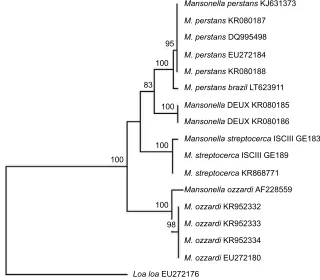
sequences. This potential “new species” was recovered from human blood samples taken in Gabon, and has provisionally been called Mansonella sp. “Deux”.60 A phylogenetic tree,
constructed with Treecon software70 by the neighbor-joining
method, shows that Mansonella sp. Deux forms a sister clade to standard forms of M. perstans from Brazil and Africa (Figure 1). However, further studies should be performed to characterize this new species and evaluate its clinical significance in humans.60
Epidemiology and distribution
Mansonella parasites are among the most common cause
of blood parasitemias and are encountered widely across Africa and Latin America.3,4,18,23,25,71 M. perstans is
consid-ered the most common of the mansonellosis parasites and is endemic in a large portion of sub-Saharan Africa, as well as a northern part of the Amazon rainforest stretching from equatorial Brazil to the Caribbean coast of South America, with molecular analysis suggesting that the parasite arrived in South America relatively recently and most likely as a consequence of the slave trade (Figure 2).18,23,25,50 From the
mid-1920s until the early 1970s, M. perstans was frequently recorded as occurring in Papua New Guinea, but there is very little primary-source data to support this and no recent survey
Figure 1 Neighbor-joining phylogenetic tree.
Notes: Tree compares the ITS1 partial sequences of human Mansonella spp. present in the GenBank (accession numbers are indicated in parentheses or with an internal code when not submitted to the GenBank). A sequence of L. loa (EU272176) was used as the out-group. Significant bootstrap values are indicated.
Mansonella perstans KJ631373
M. perstans KR080187
M. perstans DQ995498 95
100
83
100
100 100
100
98
M. perstans EU272184
M. perstans KR080188
M. perstans brazil LT623911
Mansonella DEUX KR080185
Mansonella DEUX KR080186
Mansonella streptocerca ISCIII GE183
M. streptocerca ISCIII GE189
M. streptocerca KR868771
Mansonella ozzardi AF228559
M. ozzardi KR952332
M. ozzardi KR952333
M. ozzardi KR952334
M. ozzardi EU272180
Loa loa EU272176
Research and Reports in Tropical Medicine downloaded from https://www.dovepress.com/ by 118.70.13.36 on 27-Aug-2020
Dovepress Mansonellosis update
Figure 2 Distribution of human Mansonella based on published reports before and after the year 2000.
Notes: 1, Before; 2, after.
Abbreviations: Mp, Mansonella perstans; Ms, Mansonella streptocerca; Mo, Mansonella ozzardi.
Research and Reports in Tropical Medicine downloaded from https://www.dovepress.com/ by 118.70.13.36 on 27-Aug-2020
Dovepress
Ta-Tang et al
data that indicate its occurrence anywhere in the Eastern hemisphere outside continental Africa.71 Overall, more than
100 million people may be infected by M. perstans, and it is estimated that 600 million people live at high risk of contract-ing an infection in Africa alone.23
A strong epidemiological link between banana plantations and M. perstans prevalence and parasitic loads was recorded more than 100 years ago, before its Culicoides vectors had been incriminated in its transmission (Table 1).4,32,72,73 Sharp
(the man who incriminated Culicoides in the transmission of M. perstans) was aware of this epidemiological link and presumably also that C. austeni uses banana-tree stumps and litter to support its larval development (because this had been published in the original description of the C. austeni). It is not clear to what extent, if any, the banana-plantation epi-demiological observation led to elucidation of the life cycle of M. perstans.32 Certainly, the epidemiological M. perstans
studies that have been carried out since then have not provided much more significant understanding of the parasites’ biology and transmission dynamics. However, they have shown that in endemic areas, prevalence of M. perstans infection is higher among older age-groups and that men are at more risk of infection than women.23–25 Recently, geographic
information-system data obtained from remote satellite imaging have been used in conjunction with M. perstans prevalence from school surveys to show M. perstans distribution varies with diurnal temperature range, vegetation, and cattle density.74
Epidemiological studies that have tried to link M. perstans parasitemias with clinical symptoms have thus far failed to detect any significant correlations.23,24
M. streptocerca seems to have a distribution limited to
continental Africa, where it occurs in the tropical rainfor-est areas of central and wrainfor-est Africa, as well as in Uganda (Figure 2).4,18,25 Perhaps because the skin-snip biopsies
required for its epidemiological monitoring are more invasive and painful than the blood sampling required for M. perstans surveys, or perhaps because blood sampling is simply more commonly done for other epidemiological studies, there have been fewer epidemiological surveys investigating
M. streptocerca epidemiology.4,25 As with M. perstans and M.
ozzardi and many other filarial infections, prevalence rates of M. streptocerca are highest in the oldest age-groups studied
and higher among men than women in endemic areas.21,25,75
M. streptocerca is transmitted by various species of Culi-coides and some of the same vector species that transmit M. perstans, although there are too few data available to
evaluate properly whether its distribution is influenced by the same ecological drivers as M. perstans (Table 1).15,77,78 The few
epidemiological studies that have tried to link patent M.
strep-tocerca infections with clinical symptoms have encountered
similar rates of skin disease among those infected and those uninfected in their study areas.75,76
M. ozzardi has a patchy geographic distribution across
Latin America. It has been recorded from southern Mexico to northwestern Argentina, but has not been reported in Chile, Uruguay, or Paraguay.3,4,18,25,29 The parasite occurs on
several Caribbean Islands, and like M. perstans has even been recorded in Papua New Guinea, although recent reports of its occurrence in the region are notable by their absence (Figure 2).4,71 In the Caribbean region, the parasite is
trans-mitted by a diverse range of biting midges from the family Ceratopogonidae, whereas in Central and South America it is known only to be transmitted by biting midges from the genus
Culicoides and blackflies from the genus Simulium.3,4,15,29,79–90
Because of these vectorial differences, it was once thought that M. ozzardi from the Caribbean islands and those from continental South America might be different species (Table 1).85,91 Artificial-infection experiments and the recent
publication of DNA sequences, however, strongly indicate that parasites from the two regions are morphologically, bio-logically, and genetically near identical, and thus that they are most parsimoniously regarded as just one species.2,17,28,48,50,58
Even though the various populations of M. ozzardi are best considered as belonging to the same species, it is not safe to assume, for example, that the epidemiology of M. ozzardi in the Brazilian Amazon region is more or less identical to its epidemiology in the Caribbean, because of the great diversity of vectors involved.3,4,15,29,79–90 In the Amazon region, although
it is transmitted by a range of Simulium and Culicoides spe-cies, the parasite appears to be most commonly transmitted by Simulium vectors from the amazonicum species group and to have a distribution that follows the riverine breeding sites of these Simulium vectors.29,79 In Trinidad, by way of
contrast, the only known vector is Culicoides phlebotomus (which uses sandy-beach breeding sites), and the parasite thus has a coastal distribution.15,88
However, it is important to note that vector distribution is not the only important driver of M. ozzardi distribution. For example, repeated M. ozzardi surveys in the Brazilian Amazo-nian state of Rondônia have found the area completely devoid of the parasite, despite the fact that the region shares a similar ecology and blackfly fauna to the neighboring Amazonas state. It has been proposed that low levels of human immigration may play an important role in the parasite’s failure to establish in the region.91 However, data from tourists indicate that M.
ozzardi infections are easily acquired, and as Rondônia is
Research and Reports in Tropical Medicine downloaded from https://www.dovepress.com/ by 118.70.13.36 on 27-Aug-2020
Dovepress Mansonellosis update
surrounded by highly endemic areas, migration levels would have to be very low indeed for this to be the most important factor limiting the parasite’s establishment in the region.19,91–95
Furthermore, since the first discovery of M. ozzardi, surveys have shown that very small isolated foci (in some cases, the size of a single village) on numerous islands can sustain themselves over prolonged periods, suggesting that even low levels of migration could supply a sufficient parasite reservoir to allow for the establishment of a new focus if the other epidemiological criteria for establishment were met.96
In Cameroon, it has been proposed that some areas of low onchocerciasis endemicity are explained by “zooprophylaxis”, whereby exposure to simuliid bites from insects infected with cattle parasite Onchocerca ochengi provide something akin to natural vaccination.97,98 In light of these observations, the
recent discovery of highly prevalent zoonotic Mansonella parasites in anthropophilic simuliid vectors of the region may thus been seen as an alternative explanation as to why
M. ozzardi has failed to establish in Rondônia.94,98,99
In line with epidemiological studies of other mansonel-losis, research in the Brazilian Amazon has identified outdoor workers, males, and older community members as being at elevated risk of developing M. ozzardi parasitemias.101,102
Importantly too, M. ozzardi parasitemias from this region have been shown to be positively correlated with joint pain, leg chills, headaches, and corneal lesions.103,104
Pathology and symptomatology of
mansonellosis
Although mansonellosis has very broad global distribution and is almost certainly the most common form of filarial parasite infection, little is known about its pathology or symptomatology, with most health experts and policy mak-ers viewing the parasites as largely innocuous and only rarely causing mild clinical symptoms.4,5,18,25 However, as
mentioned, recent studies investigating M. ozzardi infections in the Brazilian Amazon have found significant correlations between M. ozzardi infections and certain clinical symptoms, and other similar epidemiological studies investigating M.
ozzardi in other regions and M. perstans and M. streptocerca
have repeatedly failed to correlate clinical symptoms robustly with patent mansonellosis infections.101–107
This of course does not mean that mansonellosis is neces-sarily always completely asymptomatic outside the Brazilian Amazon region. Most of the epidemiological studies used to investigate symptomatology have had very small sample sizes and used diagnostic techniques of low sensitivity (ie, light microscopy-based diagnosis of thick blood smears), and
thus could not be expected to have had sufficient statistical power to detect subtle or complex correlations.24,75,76,101–109
Recent observations suggest that mansonellosis infections can influence the human immune system’s response and this can influence the development of secondary infections, like malaria.110 As mansonellosis infections are chronic and occur
in many tropical areas where a great number of other infec-tious diseases also exist, it is possible that the way manson-ellosis influences other pathogenic infections could actually account for a very substantial proportion of its pathogenicity and its total disease burden.4,5,18,23,25 At present, however,
although there have been many accounts of mansonellosis parasites co-occurring with other parasites, there have been very few accounts about how mansonellosis influences the pathogenicity of other infectious diseases.96,110–114
Most symptoms ascribed to M. perstans infections in modern scientific literature are based on symptoms that have been recorded in case study reports.23,25,44,115,116 As most
of these reports are based on the treatment of tourists and expatriate Europeans and North Americans returning home from endemic areas, and not on people who have lived all their lives in endemic areas, it is not clear whether the symp-toms reported from these studies can be used to compile a clinical picture that represents all or even most infections caused by mansonellosis.23,25,44,116,117 Based on these reports,
M. perstans can be considered to have little pathogenicity and
almost always to be asymptomatic, but it can occasionally cause itching, joint pains, enlarged lymph glands, and vague abdominal symptoms.5,23,25,116,117
High-level eosinophilia are regularly observed in some but not all patients with M. perstans infections, probably as a consequence of the body’s reaction against the adult worm, rather than against microfilariae.118 M. perstans is also
reported to be able to induce dermatological symptoms like the Calabar swellings of loiasis, fever, headaches, and pain in bursae and/or joint synovia or in serous cavities.23,25,44,118
There has also been ocular pathology ascribed to M. perstans in which yellowish nodules develop in the bulbar conjunctiva; it has been reported that this pathology can on occasion pro-voke edema of the eyelids and proptosis.7,118 This pathology,
which has been described as a rare clinical manifestation of
M. perstans infections caused by adult M. perstans
migrat-ing to the eye, is commonly referred to as “bulge” or “bung” eye disease.7,118 Whether, however, this pathology is caused
by M. perstans or a zoonotic parasite from the Mansonella genus is not presently clear.7,118 Certainly, it is not
uncom-mon for zoonotic filarial infections to have ocular clinical manifestations of this type.7,8
Research and Reports in Tropical Medicine downloaded from https://www.dovepress.com/ by 118.70.13.36 on 27-Aug-2020
Dovepress
Ta-Tang et al
In addition to showing that M. ozzardi infections are associated with joint pain, leg chills, headaches, and corneal lesions, epidemiological studies have also added support to the notion that M. ozzardi infections are mostly asymptom-atic.101–105 Case reports from tourist-acquired infections have
also suggested that M. ozzardi infections can cause fatigue, respiratory problems, dermal swellings, and itching; however, evidence that such symptoms occur regularly in endemic settings is still lacking.19,95
There have been very few studies of M. streptocerca infections. From what data are available, however, it is clear that infections with this parasite are very often (if not exclusively) asymptomatic.4,5,21,22,25,75 However, it has been
proposed that infections cause various types of dermatologi-cal symptoms in the shoulders and thorax, where the parasite is mostly encountered.4,5,21,22,25,75 Hyperpigmented macules
have been attributed to infections occurring in the Demo-cratic Republic of the Congo, but have not been reported everywhere the parasite is found, including Uganda, and thus some authors have cautioned against their attribution to
M. streptocerca infections.21,22,75 Chronic papular dermatitis
has been encountered in most M. streptocerca-endemic areas and has been reported to be cleared from patients successfully treated for M. streptocerca infections.75 However, in endemic
areas where epidemiological studies have been performed, chronic papular dermatitis is no more common in those patients who have detectable M. streptocerca in their skin snips than in those that do not, and thus at present there is no statistically robust evidence to support the parasites causing any symptoms at all.4,5,21,22,25,75
Diagnosis
Clinical diagnosis
As indicated in the previous section, very few clinical symptoms have been robustly linked to mansonellosis in general,4,5,25 and at present there is robust evidence to support
the notion that only M. ozzardi infections cause any clini-cal symptoms at all.103,104 Therefore, it is near impossible to
make a reliable clinical diagnosis of mansonellosis, even for M. ozzardi-caused mansonellosis. This is because most of the symptoms that have statistically robust associations with mansonellosis, such as joint pain, are so aspecific, dif-ficult to score, and caused by numerous other pathogens that occur in mansonellosis endemic region as to be of almost no utility at all.101–103 Even the potential of corneal lesions for
clinical diagnosis is limited, because although they are quite a perceptible physical symptom, they are quite aspecific, occurring in both infected and uninfected individuals from
M. ozzardi-endemic areas. Furthermore, corneal lesions
are not manifest in everyone who is infected or even occur everywhere that M. ozzardi occurs (eg, there are no reports of M. ozzardi causing corneal lesions anywhere in the Carib-bean area).103,104 In summary, it is presently impossible to use
the clinical presentation of mansonellosis to make a reliable diagnosis of the disease.
Parasitological diagnosis
Traditional parasitological diagnosis, which is based on direct observation of a causative parasite, is still the most common way mansonellosis infections are diagnosed. It is performed by the detection and identification of sheathless
Mansonella microfilariae in the skin or blood at any time of
day or night.4,5,18,25,30 Detection of skin-dwelling microfilariae
is usually done by examination of ~1.5 mg (~2 mm diam-eter) skin-biopsy (skin-snip) samples, taken with a Walser or Holth corneoscleral punch.4,5,18,25,30,119 The skin-snip test
requires that a skin biopsy be taken from areas of presumed optimal microfilariae density, and this varies depending on geographic location. Blood samples used for mansonellosis diagnosis are often taken from peripheral blood samples by way of finger pricks, although venous blood samples can also be used for this purpose.4,5,18,25,30,119
When microfilariae are present in high numbers in the patient’s blood, as is often the case in endemic areas, they may be easily found and identified in thick or thin blood smears stained with Giemsa or hematoxylin.5,25,30,119 Skin
snips are usually incubated in water or saline, and the emer-gent microfilariae can be stained on microscope slides, but in fresh unstained wet preparations of skin snips, the way in which the microfilariae move can also help with their iden-tification. The main diagnostic microfilariae characteristics can be found in Table 1.25,29,30,38,119 Morphologically, the most
important features to identify microfilariae in blood smears stained with Giemsa are size and shape of the tail, presence or absence of a sheath, and the arrangement of terminal nuclei (in the tail).18,25,30,38,119 All Mansonella microfilariae
are unsheathed, and for diagnostic purposes are treated as aperiodic.5,18,25,30,38,120
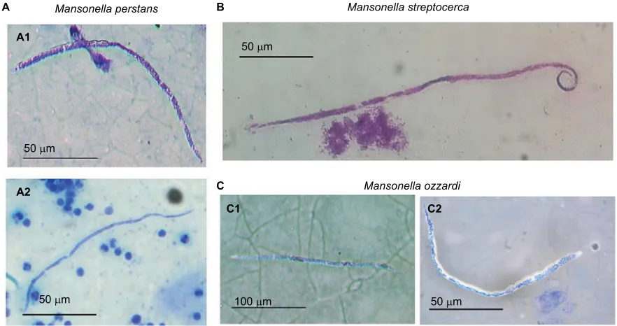
The microfilariae of M. perstans are reported to be 200×4–5 μm, to have blunt rounded tails, and to have nuclei extending to the tips of their tails (Figure 3). M. perstans is easily distinguishable from other blood-dwelling micro-filariae, which have overlapping distributions (Loa loa or
Wuchereria bancrofti), by their being smaller, their lack of
an enveloping sheath, and their tail features (their terminal nuclei are bigger than the other microfilariae).5,18,25,30,119
Research and Reports in Tropical Medicine downloaded from https://www.dovepress.com/ by 118.70.13.36 on 27-Aug-2020
Dovepress Mansonellosis update
M. streptocerca microfilariae are smaller and thinner
(180–240×3–5 μm) than Onchocerca volvulus, and have hook-shaped tails with nuclei that extend to the end (no nuclei are seen in the pointed tail region of O. volvulus microfilariae) (Figure 3).5,18,25,30,119 In wet mounts, live M. streptocerca
microfi-lariae are usually less motile than those of O. volvulus.5,119 With
these morphological descriptions, the two filariae should be eas-ily distinguished. In the past, however, in some countries, such as Uganda, M. streptocerca and O. volvulus have been confused, despite the clear morphological differences between them.30,39,75
The microfilariae of M. ozzardi are reported to be 163– 203×3–5 μm, with long thin pointed tails and body nuclei that do not extend to the tips of their tails.2–5,30,38 As they can locate
in both the blood and skin, from a public health perspective it is most important that the microfilariae can be differentiated from W. bancrofti and O. volvulus.2–5,25,30,38,119–122 The
microfi-lariae of M. ozzardi can be easily distinguished from those of
W. bancrofti by virtue of being smaller and having no sheath
(Figure 3).5,18,25,30,120 Living microfilariae of M. ozzardi can be
obtained from skin snips and are not easily differentiated from
O. volvulus by movement.2–5,25,30,38,119–122 Their morphology is
similar to the pathogenic O. volvulus, and it is especially impor-tant to distinguish these two species where they are sympatric, eg, in Amazonia onchocerciasis focus. Although morphologi-cal characters can be used to discriminate these two species,38
difficulties have led to false reports of new onchocerciasis foci
and continue to pose a problem for onchocerciasis epidemio-logical monitoring in the Amazonia focus, which is now the last Latin America onchocerciasis focus where transmission is considered to be ongoing.30,38,40,99,123,124
Inmunodiagnosis
Immunological methods involve detection of either antibody or antigen. When a filarial parasite immunodiagnostic assay is detecting antibodies, enhanced specificity is often achieved by assessment of IgG4 rather than total IgG, as IgG4 antibod-ies are significantly elevated in microfilaria-positive individu-als.125 Although there is presently no effective antigen- or
antibody-detecting immunological assay for diagnosis or epidemiological monitoring of mansonellosis infections, both kinds of tools have been developed for onchocerciasis and lymphatic filariasis disease-control programs, and have been tested for cross-reactivity with mansonellosis sera.4,25,51,126–128
The reliability (specificity and sensitivity) of these immuno-diagnostic tools for lymphatic filariasis and onchocerciasis has not been fully characterized for all of the tests, and the best-understood and most reliable tests are not necessarily the most convenient, and thus whether they should be employed to assist with mansonellosis research and diagnostics is questionable.4,51,124,126–128 However, two recently published M.
perstans studies used generic immunological filariasis assays
to support light-microscopy-based diagnoses.116
Figure 3 Comparison of the three agents causing mansonellosis illustrating the morphological characteristic of each microfilariae.
Notes: (A) M. perstans microfilariae seen in Giemsa-stained thick blood smear with blunt rounded tail and nuclei extending to the tip of the tail: M. perstans parasite from São Gabriel da Cachoeira (Amazonas state, Brazil) (A1); African M. perstans (A2). (B) M. streptocerca microfilariae from a skin-snip fixed and Giemsa-stained showing the
characteristic hooked tail. (C) M. ozzardi microfilariae from São Gabriel da Cachoeira (Amazonas state, Brazil) stained with Giemsa (C1, C2).
Mansonella perstans
A
A1
A2
C1 C2
B
C
50 µm
50 µm 100 µm 50 µm
50 µm
Mansonella streptocerca
Mansonella ozzardi
Research and Reports in Tropical Medicine downloaded from https://www.dovepress.com/ by 118.70.13.36 on 27-Aug-2020
Dovepress
Ta-Tang et al
Molecular diagnosis
Although DNA-based diagnostic tools can be used to detect and identify all of the known human filarial parasites, their most important utility to public health is their ability to differ-entiate skin infections of M. streptocerca and M. ozzardi from
O. volvulus skin infections and M. perstans parasitemias from L. loa and W. bancrofti parasitemias.51,126 DNA-based
tech-niques for filarial parasite detection and identification have been proven to be both sensitive and specific, and have now begun to replace light-based microscopy in epidemiological surveys of mansonellosis.4,49,51,52,114,129–133 Molecular diagnosis
may be used to detect microfilariae in both peripheral blood and skin biopsies, and adult worms in other tissues.4,51,52,114,129
PCR-based amplification of species-specific target sequences allows increasing diagnostic sensitivity compared with microscopic methods and reliable differentiation of samples taken from individuals living in coendemic areas.51,52,114,127
In 2010, Tang et al53 developed a popular nested PCR that
could detect any form and life stage of filariae in the human host or vector. This assay uses universal filariae PCR primers to amplify a variable portion of filarial parasite ribosomal ITS1 DNA, and allows for the subsequent identification of species based on the size of the amplified fragment. This technique is used in conjunction with gel electrophoresis and/ or Sanger sequencing, and allows for the characterization of previously unknown species (the M. perstans variant Deux was found in this way).49,53,114,129 Recently, this rDNA
ITS1-based method was turned into a single-step diagnostic by adapting it for real-time PCR.113,131 This new assay allows for
the identification of filarial parasite without the need for gel electrophoresis or Sanger sequencing, but does require more expensive reagents and infrastructure than standard PCR assays, and does not directly allow for the characterization of novel or unexpected filarial species.113,131 Other methods
(such as PCR–restriction-fragment-length polymorphism [RFLP]) that do not allow for the characterization of novel or unexpected filarial species, but require less infrastructure to support them and can differentiate a broad range of filarial species using universal primers with a combination of PCR and RFLP, have also been reported.132,133 Despite requiring
less complex infrastructural support than alternative methods, PCR-RFLP assays still require access to PCR reagents and a PCR machine, which can be a limiting factor for filarial parasite epidemiological studies.
Last year, the first DNA-detecting loop-mediated iso-thermal amplification (LAMP) filarial parasite assays were reported.134,135 Although these assays have not yet been
rigorously tested in the field or developed for the detection
of Mansonella parasites, there is no theoretical reason they could not be adapted for mansonellosis research.134,135
Requir-ing almost no infrastructural support, LAMP assays are a particularly appealing diagnostics option for research stud-ies that need to be done in resource-limited settings.51,134,135
For this reason, these new LAMP assays can be seen as a welcome development for filarial parasite research in gen-eral and an interesting new tool of research to develop for mansonellosis.51,134,135
Treatment
Among the three types of human mansonellosis, the one caused by M. perstans is usually regarded as the most dif-ficult to treat.4,18,23,25,116 Treatment studies have provided
conflicting results, and there are at present no international guidelines on best treatment.18,23,25,67,116,136–140 Few studies have
recruited sufficient patients to provide statistically robust results to support their conclusions, and many are based on the treatment of one or two European or North American expatriates or tourists returning from endemic areas, who may not respond to treatments in the same way as residents of endemic areas.4,23,25,116 The existence of genetically distinct
strains of M. perstans (like Deux or strains that lack
Wolba-chia) complicates the formulation of treatment guidelines
further, as it also possible that the efficacy of therapeutics varies between these strains.60,66
In one of the more robust M. perstans mansonellosis studies hitherto, conducted in the south of Chad, symptom-atic subjects with evidence of M. perstans microfilariae in the peripheral blood were divided in groups and tested with the antiparasitic drugs diethylcarbamazine (DEC), mebenda-zole, ivermectin, praziquantel, thiabendamebenda-zole, and DEC plus mebendazole.137 DEC plus mebendazole proved to be the most
effective antiparasitic treatment to reduce microfilaremia, and it is now one of the most used. Thiabendazole was second, with a single treatment significantly reducing microfilariae in blood. Ivermectin and praziquantel did not prove to be useful in the treatment of M. perstans infections. Mebendazole alone was seen to be a good alternative treatment and appeared to be more active than DEC alone in eliminating the infection.137
In another study carried out in Uganda, ivermectin alone, albendazole alone, and the two drugs in combination were assessed in three groups infected with M. perstans.138 In
this study, in concordance with the aforementioned study, microfilarial levels were unaffected by ivermectin or alben-dazole treatment when these drugs were used alone. Van den Enden et al136 reached the same conclusion about albendazole
treatment after finding that treatment had not decreased
Research and Reports in Tropical Medicine downloaded from https://www.dovepress.com/ by 118.70.13.36 on 27-Aug-2020
Dovepress Mansonellosis update
microfilarial counts after a median follow-up period of 45 days. However, it has been found that when these drugs (iver-mectin + albendazole) are used in combination, microfilarial loads decrease slightly after treatment.136,139
In contrast to conventional anthelmintic treatments, doxycycline has proved to be excellent, effective, and safe in the treatment of M. perstans infections.4,67,116,139,140 However,
the course of treatment over 6 weeks, which is necessary for this type of therapy, probably makes it impractical for con-trol programs, although the fact that it appears to be curative makes it a very desirable therapeutic for travel medicine.4,65,67
However, doxycycline treatment may not work on all M.
perstans infections, as there is evidence of subspecies genetic
diversity among M. perstans parasites and it is presently unclear whether all M. perstans strains harbor the Wolbachia endosymbiont.60,66 This treatment’s safety profile and all
hitherto-published results on its efficacy, however, suggest that doxycycline treatment should be the first-choice treat-ment for perstans mansonellosis wherever it can be feasibly administered.7,67,115,116
The use of ivermectin for treating M. ozzardi has proven to be very effective against microfilariae in a number of stud-ies.3,23,25,29,139–143 A single dose of ivermectin given to infected
adults was seen to reduce microfilariae densities and provide both short- and long-term reductions in M. ozzardi micro-filaraemia.29,142,143 Adverse events, however, have been seen
in some patients with M. ozzardi treated with ivermectin.143
Two elderly patients from Argentina developed serious adverse events that resembled a Mazzotti reaction following ivermectin administration, although the patients recovered without sequelae after 2–3 days.143 DEC has little or no effect
on microfilariae of M. ozzardi.3
Because of its effectiveness in the treatment of M. ozzardi, ivermectin has been used as a mansonellosis control tool in the Brazilian Amazon region.139,140 The recent discovery of M.
per-stans in the northern regions of the Brazilian Amazon, however,
suggests that this approach will not be effective everywhere.50
As M. ozzardi are also known to harbor the endosymbiotic
Wolbachia, it maybe that mansonellosis in this region can be
tackled with a single anti-Wolbachia treatment regime using an antibacterial agent.3,4,144 Although the six-week treatment
course required for doxycycline-based therapies makes them impractical for filarial parasite-control programs, new
anti-Wolbachia therapeutics with far shorter treatment courses
(of 3–7 days) are in the pipeline and could make appealing treatment options throughout the whole Amazon region and beyond.145–148 However, it is important that before such a
measure is employed, efforts are made to determine whether
this treatment interrupts transmission (like ivermectin treat-ment does). This is important to establish, as anti-Wolbachia treatments are not thought to kill Mansonella microfilariae directly and may thus not interrupt mansonellosis transmis-sion as effectively as some of the alternative options.67,115,117
DEC and ivermectin have been used with good effect to treat M. streptocerca infection.4,18,25,39,149 Fischer et al found
that a single dose of 150 μg/kg body weight of ivermectin suppresses microfilaria for a year or more, with 46% of individuals displaying no detectable microfilaria on skin biopsy 1 year after treatment.149 The most common treatment
for M. streptocerca infections is DEC given at 6 mg/kg/day orally in three doses for 12 days.139,140 At present, it is unclear
whether M. streptocerca harbors a Wolbachia endosymbiont like M. ozzardi and M. perstans.4,66 If it does, the possibility of
using an anti-Wolbachia therapeutic like doxycycline for the treatment of M. streptocerca should also be investigated.4,139,140
Global burden
The high prevalence of asymptomatic cases and the lack of a universally agreed clinical profile for any of the three forms of mansonellosis infection has left many researchers in the field reluctant to even describe mansonellosis as a disease, with some authors conspicuously avoiding the word “disease” and some preferring to use the term “condition”.1–5,18,25 This
prevailing perspective of mansonellosis as a benign parasitic infection with little or no direct effect on patients’ well-being has left mansonellosis research trapped in a vicious circle of neglect. Mansonellosis research is unable to generate robust clinical data exactly because of the present paucity of robust clinical data makes funding mansonellosis research difficult for research councils to justify.
In addition to this, the symptoms currently directly attributed to mansonellosis are difficult to utilize in the modern metric systems used to assess disease burden and in the allocation of research funding. From the data presently available, it is impossible to assign any mortalities directly to mansonellosis infection and even difficult to assign the disability-adjusted life-years or years lost to disability, which have been used so effectively in the advocacy of attracting research funding to the 13 core neglected tropical diseases.150–152 Recent surveys of the global burden of disease
commissioned by a major tropical disease research fund have omitted mansonellosis from their analyses, which has surely resulted in the disease receiving a neglect that may be inappropriate.151–154
Even without a recognized disease burden, however, it is still clear that mansonellosis has an important role in
Research and Reports in Tropical Medicine downloaded from https://www.dovepress.com/ by 118.70.13.36 on 27-Aug-2020
Dovepress
Ta-Tang et al
global health by indirectly interfering with the effectiveness of control programs targeting more serious diseases.4,29,53 As
mentioned earlier, the similarity between the microfilariae of
O. volvulus with M. streptocerca (in Africa) and M. ozzardi
(in Latin America) encountered in skin snips has led to false reports of onchocerciasis foci and misallocation of precious onchocerciasis-control resources.39,40,120 Although we are
unaware of published accounts of M. perstans in skin being confused with O. volvulus, the recent molecular detection of
M. perstans DNA in skin snips on the island of Bioko
(Equato-rial Guinea) suggests that it could potentially have happened without being detected.113 Similarly, mansonellosis parasites
have been shown to interfere with some onchocerciasis immu-nodiagnostic assays and are suspected of interfering with the reliability of others.51,120,124 In relation to lymphatic filariasis
control, false-positive results with the Binax Now filariasis immunochromatographic test have recently been recorded in several independent studies carried out in central Africa. It is presently argued that the observed cross-reactivity is entirely attributable to L. loa infections, but a role for M. perstans in cross-reactivity has not yet been definitively ruled out.128,151,152
Beyond mansonellosis’s direct and indirect (by raising morbidity in coinfection cases) disease burden and beyond even its role in interfering with the diagnostics used in other neglected tropical disease-control planning, it could also be negatively affecting global health by negatively impacting the efficacy of vaccine programmes.154–156 It has been shown,
for example, that certain lymphatic filariasis infections can affect the efficacy of some vaccines (including tuberculosis and HIV vaccines), and thus it is quite possible that mansonel-losis infections could have a similar effect.156 For this reason,
some authors have discussed the possibility of auxiliary “deworming” programs to accompany vaccine programs as a way of potentially improving their efficacy.154–156 Given the
importance of vaccines as tools for infectious disease con-trol and the extremely high prevalence of mansonellosis in infectious disease hot spots, the influence mansonellosis has on the efficacy of vaccine programs may in fact be the most important impact mansonellosis has on global health.154–156
Disclosure
The authors report no conflicts of interest in this work.
References
1. Downes BL, Jacobsen KH. A systematic review of the epidemiology of mansonelliasis. Afr J Infect Dis. 2010;4(1):7–14.
2. Bain O, Mutafchiev Y, Junker K, et al. Review of the genus
Man-sonella Faust, 1929 sensu lato (Nematoda: Onchocercidae), with
descriptions of a new subgenus and a new subspecies. Zootaxa. 2015;3918(2):151–193.
3. Lima NF, Aybar CA, Juri MJ, Ferreira MU. Mansonella ozzardi: a neglected New World filarial nematode. Pathog Glob Health. 2016;110(3):97–107.
4. Medeiros JF, Crainey JL, Pessoa FA, Luz SL. Mansonelliasis. In: Mar-condes CB, editor. Arthropod Borne Diseases. Heidelberg: Springer; 2017:405–426.
5. Nutman TB. Filarial infections. In: Cohen J, Powderly WG, Opal SM, editors. Infectious Diseases. Amsterdam: Elsevier; 2016:1046–1052. 6. Richard-Lenoble D, Kombila M, Bain O, Chandenier J, Mariotte O.
Filariasis in Gabon: human infections with Microfilaria rodhaini. Am
J Trop Med Hyg. 1988;39(1):91–92.
7. Orihel TC, Eberhard ML. Zoonotic filariasis. Clin Microbiol Rev. 1998;11(2):366–381.
8. Otranto D, Eberhard ML. Zoonotic helminths affecting the human eye. Parasit Vectors. 2011;4:41.
9. Sucharit S. Mansonella infecting man in Thailand. J Med Assoc Thai. 1988;71(10):587–588.
10. Raccurt CP. Mansonella ozzardi and its vectors in the New World: an update with emphasis on the current situation in Haiti. J Helminthol. Epub 2017 Oct 25.
11. Nelson GS. Filarial infections as zoonoses. J Helminthol. 1965; 39(2):229–250.
12. Marinkelle CJ, German E. Mansonelliasis in the Comisaria del Vaupes of Colombia. Trop Geogr Med 1970;22(1):101–111.
13. Neafie R, Meyers W, Connor DJ. Mansonelliasis. In: Binford C, Con-nor D, editors. Pathology of Tropical and Extraordinary Diseases. Washington: Armed Forces Institute; 1976:390.
14. Shelley AJ, Dias AP, Moraes MA. Simulium species of the amazonicum group as vectors of Mansonella ozzardi in the Brazilian Amazon. Trans
R Soc Trop Med Hyg. 1980;74(6):784–788.
15. Linley JR, Hoch AL, Pinheiro FP. Biting midges (Diptera: Cerato-pogonidae) and human health. J Med Entomol. 1983:20(4):347–364. 16. Eberhard ML, Orihel TC. The genus Mansonella (syn. Tetrapetalo-nema): a new classification. Ann Parasitol Hum Comp. 1984;59(5): 483–496.
17. Orihel TC, Eberhard ML. Mansonella ozzardi: a redescription with com-ments on its taxonomic relationships. Am J Trop Med Hyg. 1982;31(6): 1142–1147.
18. Muller R. Worms and Human Disease. 2nd ed. Wallingford, UK: CABI International; 2002.
19. Nutman TB, Nash TE, Ottesen EA. Ivermectin in the successful treatment of a patient with Mansonella ozzardi infection. J Infect Dis. 1987;156(4):662–665.
20. Kozek WJ, D’Alessandro A, Silva J, Navarette SN. Filariasis in Colom-bia: prevalence of mansonellosis in the teenage and adult population of the Colombian bank of the Amazon, Comisaria del Amazonas. Am
J Trop Med Hyg. 1982;31(6):1131–1136.
21. Meyers WM, Connor DH, Harman LE, Fleshman K, Moris R, Neafie RC. Human streptocerciasis: a clinico-pathologic study of 40 Africans (Zairians) including identification of the adult filaria. Am J Trop Med
Hyg. 1972;21(5):528–545.
22. Duke BO. A case of streptocerciasis in a European. Ann Trop Med
Parasitol. 1957;51(4):364–367.
23. Simonsen PE, Onapa AW, Asio SM. Mansonella perstans filariasis in Africa. Acta Trop. 2011;120 Suppl 1:S109–S120.
24. Bassene H, Sambou M, Fenollar F, et al. High prevalence of
Man-sonella perstans filariasis in rural Senegal. Am J Trop Med Hyg.
2015;93(3):601–606.
25. Simonsen PE, Fischer PU, Hoerauf A, Weil GJ. The filariasis. In: Farrar J, Hotez PJ, Junghanss T, Kang G, Lalloo D, White NJ, editors.
Man-son’s Tropical Diseases. 23rd ed. London: Saunders; 2014:737–765.
26. Lane RP, Crosskey RW. Medical Insects and Arachnids. Heidelberg: Springer Netherlands; 1993.
27. Crosskey RW. The Natural History of Blackflies. Chichester, UK: Wiley; 1990.
28. Orihel TC, Lowrie RC Jr, Eberhard ML, et al. Susceptibility of labora-tory primates to infection with Mansonella ozzardi from man. Am J
Trop Med Hyg. 1981;30(4):790–794.
Research and Reports in Tropical Medicine downloaded from https://www.dovepress.com/ by 118.70.13.36 on 27-Aug-2020
Dovepress Mansonellosis update
29. Shelley AJ, Coscaron S. Simuliid blackflies (Diptera: Simuliidae) and ceratopogonid midges (Diptera: Ceratopogonidae) as vectors of
Man-sonella ozzardi (Nematoda: Onchocercidae) in northern Argentina. Mem Inst Oswaldo Cruz. 2001;96(4):451–458.
30. World Health Organization. Bench Aids for the Diagnosis of Filarial
Infections. Geneva: WHO; 1997.
31. Pichon G. Crypto-periodicity in Mansonella ozzardi. Trans R Soc Trop
Med Hyg. 1983;77(3):331–333.
32. Sharp NA. Filaria perstans: its development in Culicoides austeni.
Trans R Soc Trop Med Hyg. 1928:21:371–396.
33. Manson P. On certain new species of nematode haematozoa occurring in America. Br Med J. 1897:2:1837–1838.
34. Godoy GA, Orihel TC, Volcan GS. Microfilaria bolivarensis: a new species of filaria from man in Venezuela. Am J Trop Med Hyg. 1980;29(4):545–547.
35. Adami YL, Moraes MA, Lanfredi RM, Maia-Herzog M. An atypical microfilaria in blood samples from inhabitants of Brazilian Amazon.
Parasitol Res. 2008;104(1):95–99.
36. Arrospide N, Adami YL, Gutierrez S, Vargas J. [Morphological charac-terization of atypical and ozzardi microfilariae of Mansonella gender].
Rev Peru Med Exp Salud Publica. 2012;29(1):161–163. Spanish.
37. Medeiros, Costa FA, Martins M. The importance of thick blood film method for sympatric filarial diagnosis in Amazon basin. Acta Amazon. 2010;40(4):779–780.
38. Post RJ, Adams Z, Shelley AJ, Maia-Herzog M, Dias AP, Coscarón S. The morphological discrimination of microfilariae of Onchocerca
vol-vulus from Mansonella ozzardi. Parasitology. 2003;127(Pt 1):21–27.
39. Fischer P, Bamuhiiga J, Büttner DW. Treatment of human
Manson-ella streptocerca infection with ivermectin. Trop Med Int Health.
1997;2(2):191–199.
40. Moraes MA, Almeida MM, Lovelace JK, Chaves GM. [Mansonella ozzardi among Ticuna Indians of the state of Amazonas, Brazil]. Bol
Oficina Sanit Panam. 1978;85(1):16–25. Portuguese.
41. Dukes DC, Gelfand M, Gadd KG, Clarke VD, Goldsmid JM. Cerebral filariasis caused by Acanthocheilonema perstans. Cent Afr J Med. 1968;14(2):21–27.
42. Holmes GK, Gelfand M, Boyt W, Mackenzie P. A study to investigate the pathogenicity of a parasite resembling Acanthocheilonema
per-stans. Trans R Soc Trop Med Hyg. 1969;63(4):479–484.
43. Orihel TC, Esslinger JH. Meningonema peruzzii gen. et sp. n. (Nema-toda: Filarioidea) from the central nervous system of African monkeys.
J Parasitol. 1973;59(3):437–441.
44. Rubin RH, Austen KF, Goetzl EJ. Studies of immediate hypersensitivity in a patient with Acanthocheilonema perstans filarial infection. J Infect
Dis. 1975;131 Suppl:S98–S103.
45. Sondergaard J. Filariasis caused by Acanthocheilonema perstans. Arch
Dermatol 1972;106(4):547–548.
46. Berkowitz AL, Raibagkar P, Pritt BS, Mateen FJ. Neurologic manifesta-tions of the neglected tropical diseases. J Neurol Sci. 2015;349(1–2): 20–32.
47. Fux CA, Chappuis B, Holzer B, et al. Mansonella perstans causing symptomatic hypereosinophilia in a missionary family. Travel Med
Infect Dis. 2006;4(5):275–280.
48. Marcos LA, Arrospide N, Recuenco S, Cabezas C, Weil GJ, Fischer PU. Genetic characterization of atypical Mansonella (Mansonella)
ozzardi microfilariae in human blood samples from northeastern Peru. Am J Trop Med Hyg. 2012;87(3):491–494.
49. Ta-Tang TH, Luz SL, Merino FJ, et al. Atypical Mansonella ozzardi microfilariae from an endemic area of Brazilian Amazonia. Am J Trop
Med Hyg. 2016;95(3):629–632.
50. da Silva LB, Crainey JL, da Silva TR, et al. Molecular verification of New World Mansonella perstans Parasitemias. Emerg Infect Dis. 2017;23(3): 545–547.
51. Alhassan A, Li Z, Poole CB, Carlow CK. Expanding the MDx toolbox for filarial diagnosis and surveillance. Trends Parasitol. 2015;31(8):391–400.
52. Ricciardi A, Ndao M. Diagnosis of parasitic infections: what’s going on? J Biomol Screen. 2015;20(1):6–21.
53. Tang TH, Lopez-Velez R, Lanza M, Shelley AJ, Rubio JM, Luz SL. Nested PCR to detect and distinguish the sympatric filarial species
Onchocerca volvulus, Mansonella ozzardi and Mansonella perstans in
the Amazon region. Mem Inst Oswaldo Cruz. 2010;105(6):823–828. 54. Morales-Hojas R, Cheke RA, Post RJ. A preliminary analysis of the
population genetics and molecular phylogenetics of Onchocerca
volvulus (Nematoda: Filarioidea) using nuclear ribosomal second
internal transcribed spacer sequences. Mem Inst Oswaldo Cruz. 2007;102(7):879–882.
55. Morales-Hojas R, Cheke RA, Post RJ. Molecular systematics of five
Onchocerca species (Nematoda: Filarioidea) including the human
parasite, O. volvulus, suggest sympatric speciation. J Helminthol. 2006;80(3):281–290.
56. Morales-Hojas R, Post RJ, Shelley AJ, Maia-Herzog M, Coscarón S, Cheke RA. Characterisation of nuclear ribosomal DNA sequences from
Onchocerca volvulus and Mansonella ozzardi (Nematoda: Filarioidea)
and development of a PCR-based method for their detection in skin biopsies. Int J Parasitol. 2001;31(2):169–177.
57. Xie H, Bain O, Williams SA. Molecular phylogenetic studies on filarial parasites based on 5S ribosomal spacer sequences. Parasite. 1994;1(2):141–151.
58. Lefoulon E, Bain O, Bourret J, et al. Shaking the tree: multi-locus sequence typing usurps current onchocercid (filarial nematode) phy-logeny. PLoS Negl Trop Dis. 2015;9(11):e0004233.
59. Morales-Hojas R. Molecular systematics of filarial parasites, with an emphasis on groups of medical and veterinary importance, and its relevance for epidemiology. Infect Genet Evol. 2009;9(5):748–759. 60. Mourembou G, Fenollar F, Lekana-Douki JB, et al. Mansonella,
including a potential new species, as common parasites in children in Gabon. PLoS Negl Trop Dis. 2015;9(10):e0004155.
61. McGarry HF, Pfarr K, Egerton G, et al. Evidence against Wolbachia symbiosis in Loa loa. Filaria J. 2003;2(1):9.
62. Ferri E, Bain O, Barbuto M, et al. New insights into the evolution of
Wolbachia infections in filarial nematodes inferred from a large range
of screened species. PLoS One. 2011;6(6):e20843.
63. Casiraghi M, Bain O, Guerrero R, et al. Mapping the presence of
Wol-bachia pipientis on the phylogeny of filarial nematodes: evidence for
symbiont loss during evolution. Int J Parasitol. 2004;34(2):191–203. 64. Nikoh N, Hosokawa T, Moriyama M, Oshima K, Hattori M, Fukatsu T. Evolutionary origin of insect-Wolbachia nutritional mutualism. Proc
Natl Acad Sci U S A. 2014;111(28):10257–10262.
65. Taylor MJ, Hoerauf A, Bockarie M. Lymphatic filariasis and oncho-cerciasis. Lancet. 2010;376(9747):1175–1185.
66. Gehringer C, Kreidenweiss A, Flamen A, Antony JS, Grobusch MP, Bélard S. Molecular evidence of Wolbachia endosymbiosis in Mansonella perstans in Gabon, central Africa. J Infect Dis. 2014;210(10):1633–1638.
67. Coulibaly YI, Dembele B, Diallo AA, et al. A randomized trial of doxycycline for Mansonella perstans infection. N Engl J Med. 2009;361(15):1448–1458.
68. Büttner DW, Wanji S, Bazzocchi C, Bain O, Fischer P. Obligatory symbiotic Wolbachia endobacteria are absent from Loa loa. Filaria J 2003;2(1):10.
69. Grobusch MP, Kombila M, Autenrieth I, Mehlhorn H, Kremsner PG. No evidence of Wolbachia endosymbiosis with Loa loa and
Manson-ella perstans. Parasitol Res. 2003;90(5):405–408.
70. Van de Peer Y, De Wachter R. Treecon for Windows: a software package for the construction and drawing of evolutionary trees for the Microsoft Windows environment. Comput Appl Biosci. 1994;10(5):569–570. 71. Crainey JL, da Silva TR, Luz SL. Historic accounts of Mansonella
para-sitaemias in the South Pacific and their relevance to lymphatic filariasis elimination efforts today. Asian Pac J Trop Med. 2016;9(3):205–210. 72. Christy C. The distribution of sleeping sickness, Filaria perstans, etc.
in East Equatorial Africa. Rep R Soc Sleep Sickn Comm. 1903;2:2–8.
Research and Reports in Tropical Medicine downloaded from https://www.dovepress.com/ by 118.70.13.36 on 27-Aug-2020