for antigen processing
by
Richard James Glynne
A thesis submitted for the degree of Doctor of Philosophy, May 1994
Imperial Cancer Research Fund
Lincoln's Inn Fields
London WC2A 3PX
and
Department of Genetics and Biometry
All rights reserved
INFORMATION TO ALL USERS
The quality of this reproduction is dependent upon the quality of the copy submitted.
In the unlikely event that the author did not send a complete manuscript and there are missing pages, these will be noted. Also, if material had to be removed,
a note will indicate the deletion.
uest.
ProQuest 10044384
Published by ProQuest LLC(2016). Copyright of the Dissertation is held by the Author.
All rights reserved.
This work is protected against unauthorized copying under Title 17, United States Code. Microform Edition © ProQuest LLC.
ProQuest LLC
789 East Eisenhower Parkway P.O. Box 1346
ACKNOWLEDGEMENTS...2
TABLE OF CONTENTS...3
TABLE OF FIGURES...8
ABSTRACT... 12
C hapter 1: INTRODUCTION... 14
1. A h is to ry of im m u n o g e n etic s a n d th e m ajo r histocom patibility com plex... 14
2. M HC-encoded proteins present antigens to T c e lls... 17
3. Processing of antigen for presentation th ro u g h MHC class I and class II... 17
4. Elution of peptides from class I m olecules... 19
5. Processing of proteins for presentation through MHC class 1 ...21
6. M u tan t cell lines u n ab le to process an tig en for presentation through class 1...22
7. Assembly of class I, p2m and p e p tid e ...24
8. T ow ards a genetic explanation of the defect in the m u tan t cell lines - cloning of ATP-binding cassette (ABC) transporter genes in the M H C ...26
9. The function of the TAP gene pro d u cts defined by transfection...28
10. The function of the TAPs: biochemical assay s... 31
11. The d eg rad atio n m achinery for the p ro d u ctio n of p e p tid e s to be p resen te d th ro u g h class I - cytosolic proteolysis and the p ro teaso m e... 35
11a. Characterisation of the proteasom e... 35
11c. Mechanism of proteolysis by the proteasom e...39
l i d . Ubiquitin and the 26S com plex...43
l ie . Regulation of the proteasom e...51
I lf. Structure of the proteasom e...56
l lg . Prim ary sequences of proteasom e su b u n its...61
12. T h eL M P s...69
13. Is there a role for the class I molecule in the processing of its ow n antigens?...72
14. Sum m ary of antigen processing for p resen tatio n through class I ... 75
C hapter 2: MATERIALS AND M ETHODS...80
1. S olutions...80
2. DNA preparation and m anipulation...82
Labelling of DNA fragm ents using random hexam er p rim ing...82
Screening of cDNA lib raries...82
H ybridisation of filters...83
Southern b lo ttin g ...84
Plasmid m in ip rep s...84
Plasmid m axipreps...85
Sequencing of double stranded plasm id D N A ...87
Shotgun sequencing...88
M13 v e cto r...88
In sert...88
L igation...89
Preparation of JM109 bacteria... 89
Single strand M13 m inipreps for sequencing...90
Solid phase sequencing of PGR p ro d u c ts...91
Subcloning of plasm id inserts...92
Vector p rep a ratio n ... 92
Insert prep aratio n ...92
L igations...93
Subcloning of PCR p ro d u cts...93
Preparation of electrocompetent b acteria...94
Single strand conformational polym orphism (SSCP)...95
3. RNA preparation and m an ip u latio n ...96
RNA p rep a ratio n s...96
N orthern blotting...96
Preparation of gel for northern blo ttin g ...96
Blotting of the RNA onto nitrocellulose and hybridisation...97
4. P rep aratio n of antisera, im m u n o p récip itatio n s and w estern b lo ttin g ...97
Production of BNFl antiserum ...97
ELISA assay ...98
Im m unoprécipitation...99
Proteolysis assay... 100
Pulse chase an aly sis... 100
R eprecipitation... 101
W estern blotting of im m unoprecipitates... 102
W estern blotting of cell lysates... 102
D e n a tu r in g p o l y a c r y la m id e p r o t e i n g e l electrophoresis... 103
C hapter 3: CLONING OF LMP7, A PROTEASOME-RELATED GENE IN THE CLASS H REGION OF THE M H C ... 104
INTRODUCTION... 104
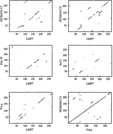
Cloning and sequence analysis of L M P7... 105
P o ten tial active site resid u es w ith in the LMP7 sequence... I l l Cloning and sequence analysis of L M P2... 116
MRNA expression of the LM Ps... 117
R e la tio n s h ip b e tw e e n th e LM Ps a n d o th e r proteasom e cDNA sequences... 119
Evolution of the T A P i LM P gene cluster and the LMP-related genes... 120
CONCLUSIONS... 122
ACKNOWLEDGEMENTS... 123
C h a p te r 4: A N INVESTIGATION OF THE LMP GENE PRODUCTS... 124
SUMMARY... 124
INTRODUCTION... 124
RESULTS and DISCUSSION... 126
Production and specificity of the BNFl antiserum ... 126
LMP7 is a component of the proteasom e com plex... 132
LMP2 is a component of the proteasom e... 134
The LMPs are synthesised w ith an N -term inal leader sequence... 138
Assembly of the LMPs into the proteasom e... 145
The m ature forms of the LMPs are upreg u lated by interferon-y... 152
LM P7-containing proteasom es are proteolytically active... 158
CONCLUSIONS... 160
C hapter 5: UNUSUAL GENOMIC ORGANISATION OF THE
LMP7 LOCUS... 163
SUMMARY... 163
INTRODUCTION... 164
RESULTS and DISCUSSION... 168
Isolation of two different LMP7 transcripts... 168
M essenger RNA expression levels of the alternative splice p ro d u cts... 174
A coding polym orphism w ith in the first exon of LMP7b... 177
Three types of transcripts from the opposite strand to LMP7 and T A PI... 180
RING9 RNA is upregulated by interferon-y... 186
A com parison of the hum an and m ouse genom ic DNA sequences at the TAPI / LMP7 locus... 188
CONCLUSIONS... 196
C hapter 6: CONCLUDING COMMENTS... 199
Do the LMPs have a role in antigen processing?... 199
Future p ro sp ects...206
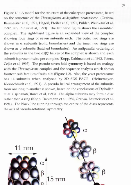
TABLE OF FIGURES Figure 1.1
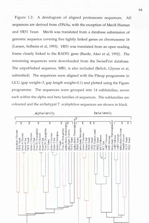
A m odel for the structure of the eukaryotic p ro teaso m e...59 Figure 1.2
A dendogram of aligned proteasom e sequences... 64 Figure 1.3a
A sim ilarity plot of the alpha-type proteasom e sequences... 67 Figure 1.3b
A similarity plot of the beta-type proteasom e sequences...68 Figure 1.4
Potential processes involved in the processing of antigen for
class I p resen tatio n ...76 Figure 3.1
The genomic position of LMP2 and LMP7 in relation to the
TAP g e n e s...107 Figure 3.2
The nucleotide sequence of the LMP7 cDNA...109 Figure 3.3
A lignm ent of LMP7 w ith proteasom e sequences...110 Figure 3.4
LMP7 com pared to proteasom e sequences by diagon p lo ts...113 Figure 3.5
Com parison of LMP7 w ith protease active sites...114 Figure 3.6
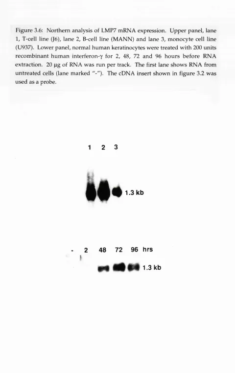
N orthern analysis of LMP7 mRNA ex p ressio n...118 Figure 4.1
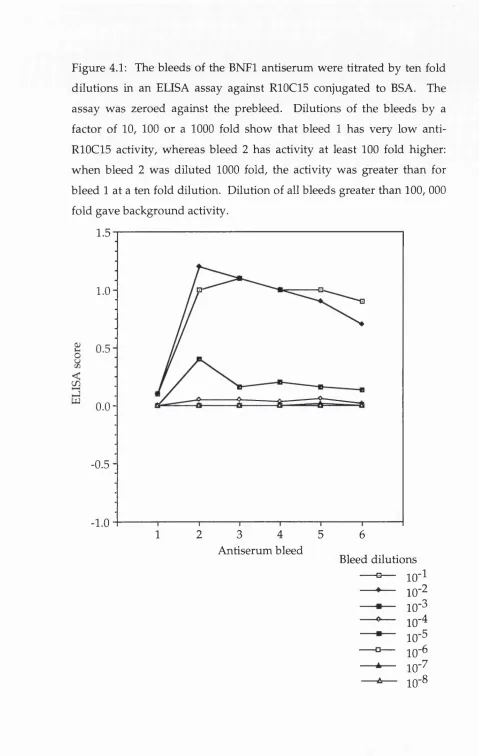
A ctivity of the anti-LM P7 serum assessed in an ELISA
Figure 4.2
Specificity of the anti-LMP7 serum determ ined by w estern
b lo t... 129 Figure 4.3
Specificity of th e anti-LM P7 se ru m d e te rm in e d b y
im m unoprécipitation... 129 Figure 4.4
Im m unoprécipitation of proteins by the BNFl antiserum
can be inhibited by R10C15 p e p tid e ... 131 Figure 4.5
Im m unoprécipitations w ith the BNFl antiserum resem ble
those of an anti-proteasome seru m ... 133 Figure 4.6
W estern analysis of proteasome p recipitates... 135 Figure 4.7
The anti-LM P2 serum specifically recognises a 22 kD
p ro te in ...136 Figure 4.8
Specificity of th e anti-LM P2 se ru m d e te rm in e d b y
im m unoprécipitation... 137 Figure 4.9
Precipitation of LMP7 after dissociation of the pro teaso m e... 139 Figure 4.10
LMP7 is processed from a 28 kD p recu rso r... 141
Figure 4.11
Precipitation of LMP2 after dissociation of the p ro teaso m e... 143 Figure 4.12
Schematic of the reprecipitation experim ent of figure 4.14...146 Figure 4.14
The m atu re form s of the LMPs are assem bled into the p ro tea so m e a p p ro x im ately co in cid en tally w ith th e ir
processing ... 147 Figure 4.15
The LMPs are upregulated by interferon-y... 153 Figure 4.16
Surface expression of class I parallels expression of the
LM Ps... :... 155 Figure 4.17
The LMP7-containing complex has proteolytic activity... 159 Figure 5.1a
Genomic organisation of LM P7... 169 Figure 5.1b
Genomic sequence of the alternative first exons of LMP7... 170 Figure 5.1c
H om ology betw een the interferon response elem ent of the
GBP prom oter and genomic sequence 5' of the LMP7b gene... 171 Figure 5.2
A com parison of the derived protein sequences of LMP7a,
LMP7b and MC13 by diagon analysis... 173 Figure 5.3
N orthern analysis of LMP7a and LM P7b... 175 Figure 5.4
Schematic representation of the RING9 splice p a tte rn s ... 181 Figure 5.5
Sequences of the RING9 clones and the exons of T A P I,
Figure 5.6
N orthern analysis of RING9 expression ... 187 Figure 5.7
A com parison of h u m an and m ouse RING9 genom ic
sequence by diagon analysis... 191 Figure 5.8
A sim ilarity plot of the RING9 genomic sequence of hum an
and m o u s e ... 192 Figure 5.9
A lignm ents of conserved, non-coding regions from the
hum an and m ouse RING9 genomic sequence... 193 Figure 6.1
ABSTRACT
The aim of the w ork described in this thesis w as to clone n ew genes from the class II region of the h u m an m ajor histocom patibility complex (MHC) and to investigate their function.
The first section describes the cloning of LMP2 and LMP7, tw o genes w ith a proposed role in antigen processing. Both genes have hom ology to com ponents of the proteasom e, a 700 kD cytoplasm ic complex, containing 10-20 subunits. The proteasom e has well defined protease activities, suggesting that LMP2 and LMP7 m ay function in the degradation of cytoplasmically synthesised proteins to form antigenic peptides. This hypothesis is strengthened by the fact that the LMPs are closely linked to, and coordinately regulated with, another pair of genes, called TAPI and TAP2. Studies w ith m utant cell lines have show n th at the TAP gene products function to m ediate the transport of antigenic peptides into the endoplasmic reticulum.
Using antisera raised against LMP2 and LMP7,1 show ed that both proteins are synthesised as precursors. These precursors are cleaved at the N term inus to form m ature proteins which are incorporated into the proteasom e. The expression of both LMP2 and LMP7 proteins w as u p regulated by interferon-y, a cytokine w hich also u p -reg u lates M HC proteins at the cell surface.
w ere isolated from cDNA libraries. In addition, three splice variants transcribed from the opposite strand to T A P I and LMP7 w ere cloned. These did n o t contain an obvious open reading frame. A com parison of this locus w ith that of the mouse show ed that only one of the alternative
CHAPTER 1: INTROD UCTIO N
1. A h isto ry of im m u n o g en etics an d th e m ajo r h isto co m p atib ility complex
The m ajor histocom patibility complex, or MHC, is possibly the best characterised region of the hum an genome. The phenom enon of histocom patibility came from w ork on the house mouse. E. E. Tyzzer, in 1916, show ed that tum ours from one strain of inbred m ouse w ere unable to grow w hen grafted onto other strains. W hen unrelated strains w ere crossed, first generation mice were susceptible to tum ours grafted from a p arent. H ow ever, a variable num ber of the second generation w ere susceptible to such grafted tum ours. It w as concluded that the trait of susceptibility to a grafted tu m o u r w as polygenic (Little and Tyzzer, 1916).
The field of blood group antigens w as linked to tum our rejection by P. A. Gorer. There w as a correlation betw een the agglutination of erythrocytes from a range of m ouse strains by a rabbit anti-mouse serum and the rejection of a tum our grafted onto the sam e m ouse strains. It seem ed th at a blood group antigen, called antigen II, and the tu m o u r rejection antigen m ay be related and it w as concluded th at "antigen II m u st be p resen t in the tissues of the host, otherw ise the tu m o u r w ill regress" (Gorer, 1937).
same patient was rapidly rejected. A similar process occurred when grafting betw een mice strains was used as an experimental system: sensitisation of graft rejection was donor specific and was independent of where the second graft was transplanted (Medawar, 1946).
To separate the loci involved in tum our rejection, G. D. Snell em barked on a program m e of back-crossing progeny from a cross between mice of different tumour rejection phenotypes. At each cross the animals were selected for the ability to reject a given tumour. The result was a series of congenic lines that differed in some of the resistance loci but not all. Use of the congenic lines showed that histocompatibility antigens could be divided into major antigens, which produced a strong rejection response, and minor, which produced only a weak effect, often overcome by the tum our (reviewed in Snell, 1981 and Bailey, 1975). One set of crosses showed that a tumour resistance phenotype was genetically linked to a fused tail mutation present in one of the parents (Snell, 1948). The absence of antigen II from these mice led to the suggestion that antigen II and the tum our resistance phenotype were also linked (Gorer, Lyman et al, 1948). This locus was named histocompatibility-2, or H-2.
A second type of lym phocyte antigen w as defined using techniques other than serology and grafting. Mixing lymphocytes from unrelated individuals induced the cells to transform and proliferate. This became the basis of the mixed lymphocyte reaction or MLR (Bach and Amos, 1967). Typing of lymphocytes using MLR or serology gave results which were broadly consistent. However, rare recombination events were able to distinguish the MLR locus from the serology locus (Yunis and Amos, 1971).
A further typing assay was devised in the guinea pig and the mouse which relied on the response of the animal to injections of simple polypeptides. The response to such challenges wasjfcontrolled by what became known as the immune response gene, or Ir. Again there was a strong correlation between the response to the polypeptides and the conventional serological typing. More detailed typing of mouse strains recombinant over the H-2 region showed that the Ir locus was linked to, but distinct from, H-2 (McDevitt, Deak et al, 1972).
2. M H C-encoded proteins present antigens to T cells
M atching of MHC haplotypes is now routinely u sed in organ transplant procedures. However, a more physiological role for the MHC w as detailed by Zinkem agel and Doherty. Sensitised m ouse cytotoxic T cells (CTL) w ere able to lyse targ et cells w h en infected b y the lymphocytic choriomeningitis virus. This lysis only occurred if the CTL and the target cells shared alleles at class I of the MHC. The theory that CTL recognised viral proteins in the context of particular MHC-encoded proteins becam e know n as MHC restriction (Zinkem agel and Doherty, 1974, Blanden, D oherty et al, 1975). A sim ilar phenom enon w as seen w hen hum an cells were used (McMichael, Ting et al, 1977).
Two subclasses of T cells could be defined on the basis of the differential expression of cell surface m arkers. O n the Ly m arking system, T cells differentiated from Ly 123"*' to be either Ly 1+ or Ly 23‘*‘. O nly those T cells which were Ly 23''" (CDS'*", CTL) w ere able to develop cytolytic activity. This cytolytic activity could be am plified by the presence of Ly I"*" helper T cells (Th). Both CTL and Th are restricted th ro u g h allelic p ro d u cts of the MHC. H ow ever, CTL are restricted through MHC class I and Th through MHC class II (Cantor and Boyse, 1975).
3. Processing of an tig en for p resen tatio n th ro u g h M H C class I an d class II
restricted antigen processing (Ziegler and Unanue, 1982). The catabolism a n d p re se n ta tio n of th is a n tig en to Th w ere in h ib ite d b y th e lysosom otropic agents chloroquine and am m onia, although uptake and ingestion w ere unaffected. These d ata suggested th at p resen tatio n th ro u g h class II required degradation of the antigen in an acidified intracellular compartm ent.
CTL lysis of infected cells th ro u g h class I w as th o u g h t to be
[d
6
initiated by the recognition of viralfencoded surface g ly co p ro teir^ ex t to MHC class I in the cell membrane. However, experim ents using defined reassortant influenza A viruses show ed th at a subset of anti-viral CTL w ere specific for non-m em brane proteins such as viral polym erase and nucleoprotein (Bennmk, Yewdell et al, 1982, Townsend and Skehel, 1982, Kees and Krammer, 1984). These results w ere confirmed by transfection of the individual viral nucleoprotein gene into L cells. The transfected cells are targets for CTL lysis despite the lack of detectable nucleoprotein at the cell surface (Townsend, 1984). Furtherm ore, transfection only of fragm ents of nucleoprotein could allow CTL lysis (Townsend, Gotch et al, 1985). Subsequently, synthetic peptides w ere also used to sensitise cells to CTL lysis and to define the epitopes w ithin an antigenic protein (Townsend, Rothbard et al, 1986).
cytotoxic T cells w ere used w ith either a B cell lym phom a line or class II transfected L cells as the presenting cells.
Several differences betw een the class I and class II presentation p ath w ay s w ere described. O nly the class II restricted clones could recognise targ et cells treated w ith non-infectious virions. C lass I restricted clones required àe novo protein synthesis in the target cell for cytotoxicity w hereas inhibition of protein synthesis h ad no effect on the presentation of HA to class II restricted clones. Conversely, only the class II p ath w ay of p resen tatio n w as sensitive to chloroquine. Finally, expression of HA from a recombinant vaccinia virus sensitised the target cells to lysis by the class I restricted T cell clones b u t not those restricted th ro u g h class II. The conclusion from these experim ents w as th at presentation through class I occurred through an "endogenous" pathw ay necessitating cytoplasm ic p ro tein synthesis. Class II p resen tatio n occurred through an "exogenous" pathw ay - able to utilise only protein added externally and degrading this protein in a chloroquine-sensitive step (Morrison, Lukacher et al, 1986, Braciale, M orrison et al, 1987). The w ork described in this thesis is likely to have m ore bearing on the mechanics of processing antigen for presentation through MHC class I. The p ro d u c tio n an d p resen tatio n of cytosolic p ro tein s by class I molecules is, therefore, described in more detail below.
4. E lution of p eptides from class I m olecules
sequence (Bjorkman, Saper et al, 1987). In the light of the above data (Townsend, Gotch et al, 1985, Townsend, R othbard et al, 1986) this w as interpreted as being due to epitopes bound in one of tw o conformations: either -2 0 am ino acid (aa) peptides b o u n d as alpha-helices or 8-9 aa peptides bound in an extended conformation.
To stu d y the n atu rally processed p ep tid es in m ore detail, a preparation of the total cellular peptide pool w as m ade from influenza infected tu m o u r cells. This procedure relies u p o n the fact th at m ost p ep tid es are soluble in trifluoroacetic acid (TFA) w hile m ost higher m olecular w eig h t m aterial can be rem oved by d é n a tu ra tio n an d precipitation. The acid soluble m aterial w as fractionated by HPLC and the fractions assayed for antigenic peptides by specific CTL. Influenza specific CTL restricted through two different class I molecules recognised
s
different peptides from flu nucleoprotein (R o t^ h k e , Falk et al, 1990). These peptides tu rn ed out to be sim ilar to the epitopes defined using synthetic peptides: an analysis of the synthetic p ep tid es by HPLC show ed th at lower m olecular w eight by-products of the synthesis w ere recognised by the CTL m ore effectively than the m ain product. For both class I molecules, one of these by-products corresponded to the peptide p resen t in the acid soluble extracts. Both epitopes w ere nonam ers, consistent w ith Bjorkman's "extra" electron density as peptides b o u n d in an extended conformation. A more refined class I crystal structure, of the B27 molecule, confirmed the presence of nonam er peptides in the a l / o 2 groove (M adden, Gorga et al, 1991).
ch ro m a to g ra p h y (HPLC) an d the m ore a b u n d a n t p e p tid e s w ere sequenced. This ap p ro ach revealed th at the p e p tid es b o u n d to a particular class I molecule are of a specific length (8 or 9 aa) and fit into consensus sequences. These consensus sequences consist of one or tw o w ell conserved "anchor" positions: the carboxy-term inal resid u e is hydrophobic (for example D^, and HLA-A2, Falk, Rotzschke et al, 1991) or basic (for example, HLA-B27, Jardetzky, Lane et al, 1991); other anchor positions occur at different positions dependent on the class I molecule (for example, a preference for arginine at position 2 in HLA- B27, b u t for asparagine at position 5 in H-2-D^).
5. Processing of proteins for presentation through M HC class I
genes. It w as proposed that antigenic peptides could be p roduced by translation of short genetic regions, rather th an by degradation of the intact protein (Boon and van Pel, 1989).
6. Mutant cell lines unable to process antigen for presentation through class 1
The use of m utant cell lines deficient in class 1 surface expression has allowed the genes involved in the processing of antigen through class 1 to be characterised. The hum an B cell line LCL721 w as m utated w ith
y-irradiation and selected w ith antibody and com plem ent for loss of the class 1 allele B8. The resultant cell line, LCL721.45 (.45), h ad lost the entire B8-containing MHC haplotype. LCL721.45 w as further m utated and selected w ith antibodies against class 1 A2, p ro d u cin g the line LCL721.134 (.134). The cell line LCL721.174 (.174) was produced by the selection of m utagenised LCL721.45 w ith an anti-class 11 DR and DQ antibody.
m apping of the deletion of .174 suggested that there w as a gene or genes w ithin the class II region that affected class I expression (DeMars, Chang et al, 1984, DeMars, R udersdorf et al, 1985).
This interpretation was supported w hen .174 w as fused to a T cell line GEM to create XI, w hich expressed high levels of A2 and B5. A derivative of T l, called T2, w hich had lost bo th copies of the GEM derived chrom osom e 6, had a phenotype sim ilar to .174. This suggests that the trans-acting factor responsible for class I expression is encoded on chrom osome 6, and is presum ably w ithin the hom ozygous deletion in the class II region of .174. Preclearing of .45 lysates w ith anti-p2m serum led to an 80% reduction in the subsequent precipitation of class I heavy chain. In contrast, less than 20% of class I in the .174 line w as associated w ith p2m by the same analysis. A pulse chase experim ent show ed that im m unoprecipitated class I w as Endoglycosidase H (Endo H) resistant after 60 m in for .45. In .174, Endo H sensitivity w as seen after 3 hours chase. Thus class I molecules in .174 neither assem ble w ith P2m nor reach the m edial Golgi (Salter and Gresswell, 1986).
BJAB-B95.8.6 by m utagenesis and selection for loss of the class I allele Bw6 w ith a monoclonal antibody and com plem ent (Ziegler, M uller et al, 1985?KeUy, Powis et al, 1991).
7. A ssem bly of class I, p2m and p eptide
RMA-S and .174 w ere used to examine the biochem istry of class I assembly w ith peptide and p2m, and for their ability to present antigen to CTL. RMA-S w as resistan t to lysis by CTL specific for influenza nucleoprotein (NP) after infection w ith the virus, w hereas RMA w as efficiently recognised. H ow ever, treatm ent of the tw o cell lines w ith a synthetic p ep tid e N P epitope led to recognition of RMA-S by CTL at levels sim ilar to RMA. This was accompanied by an increase in surface expression of H-2-D^, associated w ith |32m, in RMA-S. The restoration of surface expression was peptide specific and there w as a correlation betw een those peptides able to inhibit CTL recognition of RMA-S treated w ith the N P epitope and those p ep tid es able to induce surface expression. A dditionally, the same peptides w ere able to prom ote the assembly of w ith P2m, to prom ote the folding of as assayed by a conformation sensitive antibody, and to prom ote Endo H resistance of glycosylation. Treatment of RMA-S w ith brefeldin A, to prevent egress of class 1 from the endoplasm ic reticulum (ER), prevented the p ep tid e- induced surface expression of D^. p2m association of the heavy chain w as not inhibited, im plying that this process is induced by peptide in the ER (Townsend, Ohlen et al, 1989).
folding of class I molecules could also be driven by the addition of high concentrations of P2m to the lysate, w ithout the addition of peptides. In contrast to the effect of added peptides which w ere specific for different class I m olecules, excess P2m w as able to stabilise both and
(Townsend, Elliottet al, 1990; Elliott, Cerondolo et al, 1991). ^
Experim ents on the cell line .174 gave sim ilar results to RMA-S. .174 w as unable to present antigen from infecting viruses, bu t w as able to present synthetic peptide epitopes w hen added exogenously. In fact, .174 could present exogenous peptide better than the w ild type cell line 721. Surface class I expression w as also in d u ced by p e p tid e e p ito p e (Cerundolo, Alexander et al, 1990). Similarly, the derivative of .174, T2 (Salter an d C ressw ell, 1986), w as unable to p resen t en d o g en o u sly synthesised antigen b u t could present added peptide epitope (Hosken and Bevan, 1990).
Class I molecules can be stabilised at the cell surface of RMA-S by a drop in tem perature. The class I is associated w ith P2m b u t is unable to present endogenous antigens. H ow ever, RMA-S w as able to p resent exogenous peptide epitope better at 26° C than the parent cell RMA w as at 37° C. The addition of peptides to RMA-S at 26° C was able to stabilise class I w hen the cell line w as shifted to 37° C (Ljunggren, Stam et al, 1990). Direct binding of peptide to the surface of RMA-S cultured at 26° C w as dem onstrated using a radio-labelled peptide epitope (Schumacher, Heem els et al, 1990).
The data sum m arised in this section is consistent w ith a defect in the supply of peptide to the ER for class I assembly in these m utant lines.
the ER, probably by association w ith the chaperonin p88 (Degen and Williams, 1991 and Jackson, Cohen-Doyle et al, 1994). How ever, addition
« j t
of exogenous peptide allows exit to the cell surface. This m ay occur by stabilising "empty" class I molecules w hich have "leaked" to the cell surface from the ER w ithout peptide. These em pty class I m olecules w ould be unstable and be degraded so th at steady state levels of class I w ould be low w ithout the addition of peptide. Stabilisation of class I by a drop in tem perature or addition of peptide w ould then allow significant am ounts of class I to accumulate at the cell surface (Ljunggren, Stam et al, 1990). Presum ably, the increased ability of the class I molecules of RMA- S to p resen t exogenous peptide in this experim ent reflected a lack of endogenous peptide in the class I groove.
In the case of .174 and T2, the gene or genes responsible for this defect could be m apped to the .174 deletion in the class II region of the MHC. Fusion of RMA-S (H-2^ haplotype) w ith a w ild-type cell line (H- 2^ haplotype) restored the w ild-type phenotype. H ow ever, w hen the fusion clones w ere selected for loss of the H-2^ haplotype derived from the w ild-type cell line, the resultant cell populations h ad the RMA-S p h en o ty p e of low surface H-2^. This experim ent suggested th at the RMA-S defect w as linked to the H-2 locus on chrom osome 17 (Hosken and Bevan, 1992).
8. T ow ards a genetic explanation of the defect in the m u tan t cell lines - cloning of A TF-binding cassette (ABC) transporter genes in the M HC
and mouse. The circumstances by which these were cloned are described below.
Antisera raised between congenic mouse strains were used in immunoprécipitation studies. A group of low molecular weight proteins (LMPs) were precipitated by such sera (Monaco and McDevitt, 1982, Monaco and McDevitt, 1984, Monaco and McDevitt, 1986, this work is further discussed in section 12). Recombinations within the H-2 region mapped the LMP genes to between Pb and Ob, the murine equivalents of
DPB and D O B in the hum an MHC (Hanson and Trowsdale, 1991). Cosmids over this region had previously been isolated (Steinmetz, Stephan et al, 1986) and were used to screen cDNA libraries for new genes. Two genes, called H A M l and HAM2 (Histocompatibility antigen modifier) were isolated but did not code for the LMPs (Monaco, Cho et al, 1990). However, they did have homology to the ATP-binding cassette family of transporters. This family of genes includes members known to have a role in the transport of molecules across cell membranes. In particular, the oligopeptide permease operon of bacteria transports small peptides (Hiles, Gallagher et al, 1987).
A locus in the rat, called dm for class I modification, had been described that altered the properties of the rat class I molecule RTl.A^. Mapping of the dm locus placed it in the class II region of the rat MHC, outside the RTl.A class I locus (Livingstone, Powis et al, 1989). The class I allelic gene product RTl.A^ differs in its reactivity to a monoclonal antibody JY3/84 depending on its association with different alleles at the
w as dom inant over slow processing The speed of processing of another class I allelic product, RT1.AC, w as not affected by the d m locus (Livingstone, Powis et al, 1989, Powis, H ow ard et al, 1991). The d m locus w as fu rth er m ap p ed by typing a panel of FI hybrid rats from MHC recom binant strains (Livingstone, Powis et al, 1991). Cosm ids over this region detected tw o clones m tp l and m tp2 (Deverson, C ow et al, 1990). These w ere highly hom ologous to H A M l and HAM2 and p ro m p ted speculation that they w ere responsible for the dm phenotype.
Finally tw o groups cloned hom ologous genes w ithin the hum an MHC by screening cDNA libraries w ith cosmids that m apped across the region defined by the .174 deletion (Blanck and Strom inger, 1988), in p articular close to CpG islands (Bird, 1987). Two genes w ere cloned, nam ed RING4 and R IN G ll by one group (Trowsdale, H anson et al, 1990, Powis, Mockridge et al, 1991) and PSFl cmd PSF2 (Peptide Supply Factor) by the other (Spies, Bresnahan et al, 1990, Colonna, Bresnahan et al, 1992). These genes have now been renam ed TAPI and TAP2.
9. The function of the TAP gene products defined b y transfection
The mtp genes in the rat m apped w ithin the region defined for the
An antiserum raised against TAPI specifically precipitated two pcLyv^ophcusof -80 kD. One of these showed altered mobility on SDS-PAGE consistent with polymorphisms in the TAP2 gene product. Thus, the two
TAP genes encode that associate with each other, possibly as a heterodimeric complex (Kelly, Powis et al, 1991). This would fit with a model in which the TAPs function in a similar way to other members of the ABC family - a dimer of TAPI and TAP2 would form a complex similar in structure to the membrane proteins encoded in the opp operon. The same serum, when used for electron m icrography of frozen cell sections, show ed that TAPI was located in the ER and cis Golgi mem brane - consistent w ith a role in peptide transport into the ER (Kleijmeer, Kelly et al, 1992). Direct evidence to support a role of the TAPs in antigen processing came from transfection experiments.
Anti-TAPl precipitations from the mutant cell line BM36.1 showed that the upper band of the doublet was fainter and of a higher molecular weight than that of the parent cell line BM28.7. Sequencing of TAP2 cDNA from BM36.1 revealed a frame-shift m utation that resulted in replacement of the C-terminal 52 amino-acids of the ABC domain and an extension of the open reading frame by 51 amino acids, consistent with the immunoprécipitation result. To show that the TAP2 m utation was causative for the BM36.1 phenotype, w ild-type TAP2 cDNA was transfected into BM36.1 and caused an increase in surface class I to 50% that of the parental line BM28.7. Presentation of intracellular antigen was restored to that of the parent line (Kelly, Powis et al, 1991).
(Spies and DeMars, 1991, Spies, Cerundolo et al, 1992). The defect in RMA-S could be restored by transfection of a HAM2 cDNA, leading to increased class I assembly, surface expression and presentation of antigen (Attaya, Jameson et al, 1992). Transfection of the rat HAM2 homologue, mtp2, had a similar effect on RMA-S (Powis, Townsend et al, 1991).
As the mtp genes m apped to the dm locus in the rat, it was reasoned that the cim phenotype might be caused by different mtp alleles. The mtp genes from a dominant haplotype were transfected into a cell line with a dm ^ haplotype. Transfection of m tpl cDNA had no effect on the phenotype of the recipient cell line. However, transfection of the mtp2 allele transferred to the transfected cell line a dm^-like phenotype. RT1.A^+ was now expressed, as determined by serology or CTL, and was rapidly processed. Sequencing of mtp2 from several haplotypes w ith defined cim phenotypes revealed 29 amino acid differences between the alleles, of which 25 were correlated with cim phenotype. These were predominantly in the membrane spanning region. Peptides recovered by acid elution from RTl.A^ in either cim"^ or d m ^ backgrounds I were separated by HPLC. RT1.A^+ had peptides eluting from HPLC as a broad peak. RTl.A^', on the other hand, had a profile showing more hydrophobic, late-eluting peptides. The peptide-elution phenotype of RTl.A^" could be shifted by transfection of mtp2 from the dominant drn ^
haplotype (Powis, Deverson et al, 1992). Thus, the m tpl allele had a direct affect on the peptides that were presented in the RTl.A^ molecule, consistent w ith the m tp gene products forming a peptide pum p to provide the substrates for class I presentation.
m ouse have a phenotype sim ilar to the m u tan t cell lines described in section 8 - they are able to present exogenously added p ep tid e to CTL, b u t not endogenously synthesised proteins, and have low class I surface expression. A n analysis of the T cells from various organs show ed th at C D 4/C D 8 double positive cells are present at w ild type levels, as are CD4 single positives. How ever, CD8 positive CTL T cells are not found in the spleen, blood or lym ph nodes of the m u ta n t mice. This is consistent w ith a role for class I in the positive selection of CD8 positive cells in the thym us (van Kaer, Ashton-Rickardt et al, 1992).
10. The function of the TAPs: biochem ical assays
Reconstitution of m u tan t cell lines using the TAP genes and an effect of the rat TAP alleles on the peptides eluted from class I both suggest a role for the TAP complex in antigen processing through class I. The w idely held view w as that this role w ould be in the ATP-dependent transport of peptides, and direct biochemical evidence is now available for this. H ow ever, three reports concluded th at p ep tid es could be transported into microsom es w ithout ATP and, in one case, w ithout a need for the TAPs.
ER m em brane proteins, which w ould probably include the ATP-binding do m ain of the TAPs (Kleijmeer, Kelly et al, 1992). W hen p e p tid e tran sp o rt across the ER m em brane w as m easured by b inding to BiP, rather than class I, it seem ed that this process w as independent of ATP. In addition, a com parison of microsomes from T1 and T2 revealed no difference in peptide transport by this assay, although class I assem bly w as lower in T2. The authors concluded that peptide transport into the ER occurred in an ATP- and TAP- independent m anner. H ow ever, an A TP-dependent step in class I assembly occurred dow nstream of peptide tran sp o rt and w as affected by the T2 deletion (Levy, G abathuler et al, 1991).
Two assays to investigate tran sp o rt of p ep tid e across the ER m em brane have been developed for dog pancreas m icrosom es. The assay depends on the assertion that transport of a peptide containing a glycosylation signal into the m icrosom es w ould deplete the pool of dolichol high m annose oligosaccharides. Subsequent glycosylation of a m arker protein in the presence of the microsome fractions w ould then be inhibited. Using this inhibition as a m easure of peptide transport, the assay show ed no effect of ATP depletion, nor w as there an effect on tra n sp o rt b y an in h ib ito r of ATPases, oligom ycin. H ow ever, the transport w as tem perature sensitive - there w as no peptide transport at 4° C b u t transport did occur at 25° C (Koppelman, Zim m erm an et al, 1992).
form ation). An iodinated peptide containing a glycosylation site w as tra n s p o rte d into th e m icrosom es in an A T P -in d ep en d en t w ay - glycosylation of the peptide indicating tran sp o rt into the ER. In this system, class I stabilisation by peptide occurred after the microsomes had been solubilised (Bijlmakers, N eeles et al, 1993).
There is som e evidence for T A P-independent p resen tatio n of certain peptides to CTL. Infection of RMA-S w ith VSV, or transfection w ith VSV nucleoprotein gene, allowed efficient recognition by a VSV- restricted CTL (Hosken and Bevan, 1992). W hether this w as d u e to partial function of the rem aining HAM gene product, perhaps acting as a h o m o d im er to tra n sp o rt p e p tid e into the ER, or d u e to a TAP- in d ep en d en t m echanism , is unclear. The TAP deficient cell line .174 expresses HLA-A2 at ~50% wild-type levels. Peptides eluted from these
A l molecules are derived from signal peptides (Wei and Cresswell, 1992). These w ould be formed in the ER lum en by the action of signal sequence peptidase on secretory or m em brane-bound proteins. It is unlikely th at this pathw ay explains the ATP-independent transport of peptides in the assays described above as there is no reason to assum e that the peptides u sed in these assays w ould be good substrates for the translocation machinery.
RTl.A^ m olecules reflect m tp-m ediated p ep tid e tran sp o rt into the ER (Heemels, Schum acher et al, 1993, M om burg, Roelse et al, 1994). This result suggests that the specificity of the TAP complex is restrictive for antigen presentation, at least in the rat.
A dditional evidence for a role of the TAPs in p ep tid e tran sp o rt cam e from ex p erim en ts u sin g T A P k n o ckout m ice. M icrosom e p rep a ratio n s from w ild type and T A P I - /- mice w ere b o th able to assem ble exogenously added peptide w ith class I. H ow ever, w hen the tem perature of the reaction w as lowered from 37° C to 23° C the rate of accum ulation of peptides in the T A P l+ f + microsomes w as 4 fold higher than that of TAP1-J-. This difference in rates, presum ably due to the TAP complex, w as ATP-dependent (Shepherd, Schumacher et al, 1993).
The discrepancy betw een experim ents show ing A TP-dependent and A TP-independent peptide transport can probably be explained by differences in the experim ental conditions. For exam ple, p e p tid e concentration, tem perature and time of incubation are all likely to be relevant. A nother factor m ay be the m icrosom e p rep aratio n - if the m icrosom es are "leaky" for sm all molecules this could m ask any TAP- dependent transport. Finally, the concentration of TAP complexes in dog pancreas microsomes m ay be too low for TAP-dependent transport to be seen.
w ill alw ays be a possibility th at the TAPs have an indirect effect on peptide transport, perhaps through the transport of another molecule or ion.
11. T he d eg rad atio n m achinery for the p ro d u ctio n of p e p tid es to be presented through class I - cytosolic proteolysis and the proteasom e
11a. C haracterisation of the proteasom e
As discussed in section 3, the degradation of antigen for class I p resen tatio n w as n o t sensitive to lysosom otropic agents (M orrison, Lukacher et al, 1986). N on-lysosom al p ro tein d eg rad atio n w as first m ooted after a stu d y on the d eg rad atio n of m etabolically labelled proteins by pulse-chase analysis. Proteolysis of norm al cellular proteins could be separated from that of abnormal proteins (containing the amino- acid analogue canavanine) by the ad d itio n of various agents to the m edium . Two pathw ays for protein degradation w ere defined from this work. One pathw ay, sensitive to cathepsin B inhibitors, w as lysosomal and show ed little specificity for protein substrate. The other pathw ay w as non-lysosom al and preferentially degraded proteins that denatured easily, such as those containing canavanine (Knowles and Ballard, 1976).
7.8, ruling o u t any role for the lysosomal acid proteases in this process (Etlinger and Goldberg, 1977). ATP-dependent proteolytic system s w ere then described in rat and m ouse liver. In both cases the p H optim um w as slightly alkaline. Fractionation of either rat or m ouse hom ogenates show ed that the protease had a molecular w eight of -550 kD (DeMartino and Goldberg, 1979, Rose, W arms et al, 1979).
The enzym e w as also isolated from pituitary as the complex that w as active in the production of opioid peptides from precursors. The complex had a molecular w eight of -700 kD b u t could be dissociated into subunits of 24-28 kD. It had three proteolytic activities that could be defined using m odel tripeptide substrates coupled to chrom ogenic C- term inal leaving groups. Cleavage occurred at the C term in u s of hydrophobic residues, basic residues and glutam ate. These activities
p
w ere term ed chym otrypsin-like, trypsin-like an d p ^ id y l-g lu ta m y l p ep tid e (PGP) respectively. T reatm ent of the com plex w ith various p r o te a s e in h ib ito r s , o r w ith SDS, p r o d u c e d d if f e r e n tia l in h ib itio n /activ atio n of the three activities, show ing that they w ere distinct and th at the enzym e w as m ulticatalytic (Orlowski and Wilk, 1981).
Research into the degradation of a an d p crystallin led to the isolation of a multicatalytic protease from bovine lens that w as similar to th at purified from pituitary (Ray and H arris, 1985). The enzym e w as probably responsible for the degradation of a-crystallin at neutral pH by p artially purified extracts of bovine lens (Blow, v an H eyningen et al,
su b seq u en t isolation of large proteolytic com plexes w ith n e u tra l or alkaline p H optim a from rat liver (Rivett, 1985) and m uscle (Dahlmann, K uehn et al, 1985) and hum an lung (Zolfaghari, Baker et al, 1987) and kidney (Zolfaghari, Baker et al, 1987). The enzym es h a d sim ilar properties to the A TP-dependent proteolytic system s isolated earlier (DeMartino and Goldberg, 1979, Rose, W arms et al, 1979).
The rat liver enzym e was then re-isolated as one of three enzym es th at w as able to degrade oxidised glutam ine synthetase. Similar to the bovine enzym e, the molecular weight of this complex w as 650 kD b u t it could be dissociated into subunits of 22-34 kD. Proteolysis of oxidised glutam ine synthetase occurred w ith o u t the detection of d eg rad atio n interm ediates. However, w hen the oxidised B chain of insulin (Insulin Box) w as used as a substrate, the pro d u cts could be sep arated and
characterised by HPLC. Cleavage of this 30 amino acid peptide occurred at five positions, which were different from those seen w ith other types of protease (lysosomal cathepsins, calpain I and U, papain, insulin protease, m etalloproteases, serine proteases, chym otrypsin, cath ep sin c and elastase) (Rivett, 1985). Subsequent analysis show ed th at both enolase and pyruvate kinase w ere also better substrates w hen oxidised than the native enzymes. The data suggest that this protease m ay be involved in the degradation of abnorm al proteins that was described by Knowles and Ballard (1976).
sam e as the rat n e u tra l peptidase: h ig h m olecular w eig h t w h en undenatured (650 kD); dissociation into lower molecular w eight subunits and inhibition by thiol binding reagents (Zolfaghari, Baker et al, 1987). The sam e enzym e w as purified from h u m an kidney. In this case a dem onstration of the m ulticatalytic n atu re of the h u m an enzym e w as m ade. Cleavage of the three m odel substrates for trypsin, chym otrypsin a n d PGP activities h a d d ifferen t p H o p tim a an d w ere affected differentially by treatm ent of the enzym e w ith SDS, protease inhibitors, cations and album in (Zolfaghari, Baker et al, 1987).
To su m m arise , m an y d iv erse field s c o n trib u te d to th e characterisation of a m ulticatalytic protease from several tissue types. The degradation of lens proteins in cataract research, vasoactive and opioid peptide metabolism and A TP-dependent protein degradation all involved a proteolytic complex w ith high m olecular w eight th at h ad a neutral p H optim um .
l i b . N on-proteolytic activities of the proteasom e
m olecule w as isolated from Xenopus laevis ovaries. The activity copurified w ith a prosome-like particle (Castaflo, O m berg et al, 1986).
The complexes that repressed mRNA translation w ere identical to the m ulticatalytic protease. The Drosophila 19S particle appeared similar to the rat multicatalytic protease complex on electron m icrographs. The tw o com plexes shared epitopes defined by cross-reacting antibodies, contained RNA molecules of 80-100 n t and both had proteolytic activity (Falkenburg, H aass et al, 1988). A sim ilar analysis com pared the 19S particle from HeLa cells w ith the m ulticatalytic protease isolated from either h um an red blood cells or rat liver and reached the same conclusion (Arrigo, Tanaka et al, 1988). The multicatalytic protease, as purified from rab b it skeletal m uscle, inhibited tran slatio n of m RNA in a rab b it reticulocyte system (Kuehn, Dahlm ann et al, 1990). This effect w as not specific for the mRNA, although a specific effect of prosom e on the repression of viral, b u t not cellular, mRNA has also been show n (Horsch, M artins de Sa et al, 1989). The nam e "proteasome” w as suggested for the prosom e/m ulticatalytic protease complex. This nam e is now p o pular in the literature and will be used throughout this thesis.
11c. M echanism of proteolysis by the proteasom e
each of the trypsin, chym otrypsin and peptidylglutam yl sites of bovine pitu itary proteasom e w ere sensitive to isocoum arin derivatives (which react w ith the active site of serine proteases), suggesting th a t the proteasom e belonged to the class of serine proteases (Orlowski and M ichaud, 1989).
A m ore recent study on proteasom e catalysis used a w ider range of p e p tid e s u b s tra te s a n d s e rin e -p ro te a se in h ib ito rs . 3,4- dichloroisocoum arin (DCI) inhibited activity w ith high concentrations of a PGP su b strate b u t stim u lated activity w ith low concentrations, suggesting two sites able to digest these substrates. Additionally, activity against substrates w ith either phenylalanine or tyrosine at the "PI" position could be distinguished by the action of chym ostatin analogues, g u an id in e/H C l or casein. This provided evidence for two chymotrypsin- like sites. Taking into account the trypsin-like site, the authors of this stu d y concluded th a t the p roteasom e m ay have five active sites (Djaballah, Harness et al, 1992).
The sam e g roup show ed th at the caseinolytic activity of the proteasom e w as also increased by DCI at concentrations which inhibited the activity against the chromogenic peptides. In contrast to treatm ent w ith NAI, DCI-treated proteasom e did not digest casein to a detectable in term ed iate. In fact, the 21 kD casein in term ed iate resistan t to degradation by NA I-proteasome could be digested by DCI-proteasom e (Pereira, N g u y en et al, 1992). The D C I-resistant co m p o n en t w as in v estig ated in m ore d etail u sin g n e u ro te n sin an d p ro in su lin as substrates. Proinsulin pro d u ced sim ilar degradation p ro d u cts w hen treated w ith either native or DCI-proteasome, suggesting that proinsulin is d e g ra d e d b y th e D C I-resistan t co m p o n e n t of p ro te a so m e . Neurotensin, a 13 amino acid peptide, show ed different cleavage patterns after treatm en t w ith D CI-proteasom e w hen com pared to the native enzym e. The D CI-resistant activity seem ed to favour cleavage after branched amino acid side chains (Cardozo, Vinitsky et al, 1992).
One product of cleavage of insulin B q x required th at the enzym e
cleaved twice w ithin the substrate. A ddition of potential interm ediates in this reaction, that is the products after either of the sites alone had been cleaved, did not lead to rapid production of the final product. It w as suggested th at the proteasom e m ay produce this p ep tid e by a "bread sheer" type m echanism w hereby the tw o cleavages w o u ld be m ade w ithout any interm ediate leaving the enzym e complex (Dick, M oom aw et al, 1991).
of the w ell characterised protease fam ilies. A dditionally, there is n o /little hom ology in any of the p rim ary sequences of proteasom e com p o n en ts to o th er p ro tease sequences. A n ex p lan atio n of the chem istry of proteasom e activity will require a detailed know ledge of those residues that are p art of the active sites.
l i d . U b iq u itin and the 26S complex
F u rth e r co m p o n en ts n ecessary for the A T P -d ep en d en t degradation pathw ay w ere characterised by fractionation. First, a sm all protein, called APF-1 for A T P-dependent proteolysis factor, w as isolated from reticulocyte lysates. This h ad no intrinsic proteolytic activity, b u t stim ulated A TP-dependent proteolysis by another fraction of the lysate. APF-1 became covalently b o u n d to proteins in an A TP-dependent m anner and it w as suggested th at these covalent conjugates could be interm ediates in the proteolysis reaction (Ciechanover, Heller et al, 1980). APF-1 w as subsequently found to be identical to ubiquitin on the basis of physical properties, am ino acid com position and by functional com plem entation: ubiquitin could substitute for APF-1 in stim ulating ATP-dependent proteolysis and could also form covalent conjugates w ith reticulocyte proteins (Wilkinson, U rban et al, 1980).
A reticulocyte lysate th at h ad been depleted for ATP w as fractionated and the fractions w ere assayed for proteolytic activity. Three necessary com ponents w ere found. These w ere nam ed C Fl, CF2 and CF3 and all w ere necessary in the degradation of ubiquitin- lysozym e conjugates. The kinetics of this degradation show ed that there w as a tim e lag w hich could be abolished if the three factors w ere p rein cu b ated w ith ATP before a d d itio n of the substrate. D uring this preincubation, C Fl, 2 and 3 assem bled to form a large com plex w ith sim ilar p ro p erties to the 26S com plex (Ganoth, Leshinsky et al, 1988).
The relationship of the 20S complex, which does not degrade u b iq u itin conjugates, to the 26S complex, w hich does, has been so m ew h at controversial. A lthough there ap p eared to be low molecular w eight components of the 20S complex in the 26S fraction (Hough, Pratt et al, 1987), there are two reports that these proteins d id n o t react w ith antibodies against the 20S su b u n its (Seelig, Kloetzel et al, 1991, Kuehn, D ahlm ann et al, 1992).
al, 1992). The sam e stu d y also show ed th at the 26S complex w as reactive w ith a serum raised against the 20S complex. O rino et al have show n th at a m onoclonal antibody against a 20S com ponent coprecipitated higher m olecular w eight proteins sim ilar to those fo u n d in the 26S com plex (Orino, Tanaka et al, 1991). Finally, m utations in 20S proteasom e components in yeast have a phenotype th at includes increased stability of ubiquitinated proteins (Richter- Ruoff, H einem eyer et al, 1992, Seufert and Jentsch, 1992).
W hether ubiquitination is a prerequisite for the degradation of proteins for presentation through class I is unknow n as yet. H ow ever, tw o experim ents suggest th at u b iq u itin atio n is necessary for the presentation of at least some antigens. The first of these stu d ied the p resen tatio n of epitopes contained w ith in genes in vaccinia v iru s constructs. Infection by vaccinia virus inhibits the presentation of certain epitopes in the late phase of infection. The inhibition w as overcome by m anipulations of the inserted gene that led to a m ore rapid ubiquitin- d ependent degradation of the encoded antigen. Deletion of the signal sequence from haem agglutinin created a short lived cytosolic protein and com pletely restored p resentation of a haem agglutinin epitope. For influenza nucleoprotein as the antigen, use w as m ade of the "N-end rule" of ubiquitin-m ediated protein degradation (Bachmair, Finley et al, 1986). A construct of Ub-Met-NP was not recognised any m ore efficiently than N P alone w hen expressed from the late prom oter in vaccinia constructs. How ever, the less stable Ub-Arg-NP protein, if expressed from a similar construct, w as presented weU (Townsend, Bastin et al, 1988).
line w as a decrease in the conjugation of ubiquitin to proteins and in protein degradation. Sensitisation of cells for CTL lysis w as achieved b y the introduction of exogenous ovalbum in by pinosom e lysis. Presentation of ovalbum in through the m ouse class I molecule H-2- w as greatly reduced at the non-perm issive tem perature. A possible criticism of this experim ent is th at the non-perm issive te m p e ra tu re , an d th e associated b u ild u p of m alfo ld ed an d ubiquitinated proteins, m ay prevent presentation through some non specific affect. H ow ever, presentation of the peptide epitope, w hen encoded by a minigene on a vaccinia construct, was not affected by the ts m utation (Michalek, Grant et al, 1993).
A lthough this experim ent ad d ed to the evidence th at the ubiquitin p athw ay w as necessary for antigen presentation, it w as sim ilar to a previously published experim ent that had come to the opposite conclusion. M éthylation of ovalbum in, w hich inhibits u b iq u itin d e p en d e n t d eg rad atio n in vitro, p rio r to pinosom e- m ediated uptake had no effect on presentation (Carbone, Hosken et al, 1989). The epitope in this experim ent m apped to aa 258-276 an d w as p resen ted th ro u g h H-2-K^. Sim ilarly, the ovalbum in epitope studied in the ts ubiquitin m utant w as H-2-K^-restricted and m apped to aa 257-264. Therefore, inhibition of ubiquitination either by m éthylation or by a ts m utation in E l caused different effects on the presentation of the same or similar epitope.
ovalbum in should be affected, w hereas the ts m utation w ould be expected to cause a general inhibition of the ubiquitin pathw ay. W hen analysed in this way, the m éthylation experim ent is easier to interpret as it can be concluded that ubiquitination of the antigen is n o t necessary for presentation, whereas the ts m utation can only lead to the conclusion that ubiquitination of some protein(s) is necessary for presentation, b u t provides no information as to w hat that protein(s) m ight be. A m ore trivial explanation w ould be th at the ts m utant line is from a ham ster background transfected w ith m ouse class I and ICAM , w h e re a s th e e arlie r ex p erim e n t c o n sid ere d class I p resen tatio n in an isogenic background. Therefore, differences b etw een h a m ste r and m ouse in o th er m olecules in volved in processing or presentation of antigen m ay explain the discrepancies betw een the two experiments.
absence of E3 (H aas, Bright et al, 1988). The significance of ubiquitination pathw ays that are not dependent on an E3 molecule is unclear.
Specificity of ubiquitination is likely to be d u e both to the su b stra te itself an d to the ex p ressio n of d ifferen t ty p es of ubiquitinating enzym es in the cell. There is evidence for diversity and possible specialisation in each of the E l, 2 and 3 enzymes. Three different E l enzym es have been isolated from w heat (Hatfield and Vierstra, 1989) and a testis-specific E l encoding gene has been cloned w ith a role in sperm atogenesis (Kay, A shw orth et al, 1991, Mitchell, W oods et al, 1991).
Two E3 enzym es have been isolated from rabbit reticulocyte lysates w ith different substrate specificity (Heller and Hershko, 1990) and it is possible that further E3s will be discovered. There are likely to be m any pathw ays of ubiquitination, w hich m ay overlap in their specificity. There is some scope for control of degradation of sets of p ro tein s b y the differential expression of com binations of the ubiquitinating enzyme isoforms.
A complementary level of control could act at the level of the substrate. N atural substrates of ubiquitin degradation have been few in num ber. However, an im portant signal for degradation has been delineated using ubiquitin-X-p galactosidase constructs. U biquitin is cleaved from the hybrid protein by ubiquitin C-terminal hydrolases. The N-term inal residue of p-galactosidase then determ ined the half- life and this w as term ed the "N-end rule" (Bachmair, Finley et al, 1986). A second determ inant of the half life w as the presence of a lysine residue, in the correct context, near the N-term inal end of the protein (Bachmair and Varshavsky, 1989). This residue acts as an acceptor for the ubiquitin chain (Chau, Tobias et al, 1989).
sequence b u t both are degraded by a ubiquitin pathw ay (Shanklin, Jabben et al, 1987, Hochstrasser, Ellison et al, 1991).
Deletional m utagenesis of MATa2 show ed th at it contained tw o degradation signals (Hochstrasser and Varshavsky, 1990). One of these signals w as n o t effective in a strain m u tated for UBC6, although the other signal w as not affected by this m utation (Chen, Johnson et al, 1993). It is possible th at different u b iquitinating enzym es, for example w ithin the E2 series, have different recognition m oieties. This recognition m ay be affected by m odifications to the p rim ary am ino acid sequence: although deg rad atio n of Mos in m aturing Xenopus oocytes is dependent largely on the penultim ate am ino acid residue, p h o sp h o ry latio n of a n eig hbouring serine residue m ay give some stabilisation (Nishizawa, Okazaki et al, 1992).
There is a large am o u n t of evidence th at d e n atu red or abnorm al proteins are good substrates for ubiquitination. Proteins sy n th esised in the presence of an am ino acid an alo g u e are preferentially conjugated to ubiquitin (Hershko, Eytan et al, 1982). Similarly, dénaturation of haem oglobin by phenylhydrazine led to increased ubiquitination and degradation w hen the p ro tein w as microinjected into cells (Chin, Kuehl et al, 1982). O ne experim ent suggests that ubiquitin can increase the susceptibility of a protein to proteolysis in vitro w ith o u t covalent conjugation to the substrate, possibly by exerting some chaotropic effect (Wenzel and Baumeister, 1993).
ubiquitin pathw ay. The expression of the various ubiquitinating en zy m es (E l, 2 an d 3), th e p resen ce of p rim a ry seq u en ce d eterm in an ts, p o st-tran slatio n al m odifications an d the p ro tein conform ation could all have an effect. W hich of these are relevant for the d eg rad atio n of viral proteins for class I p resen tatio n is u nknow n at present. Of possible relevance to this argum ent is the discovery of a ubiquitin hom ologue which is upregulated by IF N -a, P and, to a lesser extent, y (Loeb and Haas, 1992). It should also be pointed out that there is evidence for the degradation of cytoplasmic proteins w ith o u t prior ubiquitination (M urakam i, M atsufuji et al, 1992). The im portance of u b iq u itin -in d e p en d e n t d e g ra d atio n pathw ays to antigen processing is also unknow n.
H e . R egulation of the proteasom e
The vacuolar or lysosomal proteases are enclosed by a m em brane. C o n tro l of p ro teo ly sis can th en be achieved by th e tra n sp o rt of condem ned proteins into vesicles. The proteasom e is generally thought to be "free" in the cytoplasm. However, proteasom es have been purified w ith m em b ran es an d o rg an ellar fractio n s, asso ciatio n w ith th e cytoskeleton has been described (reviewed in Rivett and Knecht, 1993) and certain proteasom e subunits have a conserved basic m otif w hich m ay m ediate nuclear translocation of the complex (Tanaka, Yoshim ura et al, 1990). C hanges in the localisation of proteasom es have been show n to occur du rin g the cell cycle (eg A m sterdam , Pitzer et al, 1993) and cell differentiation (eg Grossi de Sa, M artins de Sa et al, 1988, Shim bara, O rino et al, 1992). If differential localisation of proteasom e occurs, then the targetting of substrates or proteasom es to specific cellular locations m ay be one m echanism by which cytosolic degradation is regulated.
Im m unoprécipitation studies have show n heterogeneity w ithin the proteasom e population, especially w ith reference to the LMP subunits (Brown, Driscoll et al, 1993, Patel, M onaco et al, 1994). Proteasom e com position can be regulated by the cytokine interferon-y, w hich up- regulates the LMPs at the expense of other subunits (Yang, W aters et al, 1992, Aki, Shimbara et al, 1994 and Belich, Glynne et al, subm itted). The LMP containing and LMP lacking proteasom es have different activities against peptide substrates (Driscoll, Brown et al, 1993, Gaczynska, Rock et al, 1993). The LMPs m ay be unusual in th at they have counterparts elsewhere in the genome (Belich, Glynne et al, subm itted, and see below) w hereas the other com ponents appear to be single-copy. It is not clear w hether proteasom e composition is altered in differentiation, cell cycle or otherw ise. H ow ever, at least for the LMPs, it seem s clear th a t p ro teaso m e com position can be altered by an external signal (for example, IFN-y).
It is likely that the m arking of particular proteins for destruction w ith a ubiquitin tag is a major factor in the control of cytoplasmic protein degradation. H ow ever, a num ber of endogenous factors have been isolated th at m ay be activators or inhibitors of the proteasom e in vivo.
Potential inhibitors of the proteasom e include a hexam er of a 40 kD subunit (M urakami and Etlinger, 1986); a tetram er of 50 kD subunits (Li, G u et al, 1991) and m ultim ers of a 31 kD protein (Chu-Ping, Slaughter et al, 1992).
subunits ap p ear to be ubiquitinated w hen incorporated into the 26S complex (Li and Etlinger, 1992). It has been suggested that ubiquitination of the CF2 subunits regulates the assembly of CF2 w ith C Fl and CF3 (20S proteasom e) (Li and Etlinger, 1992) and CF2 is essential for breakdow n of u b iquitin conjugates (Ganoth, Leshinsky et al, 1988). Therefore, the breakdow n of ubiquitin conjugates m ay be partly regulated through the ubiquitination of the CF2 subunit.
There are therefore several potential m echanism s for proteasom e regulation. Changes in subcellular location of the complex, expression patterns of the subunits or levels of activators or inhibitors w ould all be predicted to affect the quality or quantity of proteins that are degraded by this pathw ay. Removal or inactivation of an inhibitor m ay be the equivalent of the latent to active change seen in vitro. These changes are likely to be relatively non-specific. For exam ple, an increase in the ex p ressio n of a p ro teaso m e activator m ay be a p p ro p ria te u n d e r conditions of stress or heat shock w hen total cellular protein turnover w ould be expected to increase. D egradation of particular proteins is m ore likely to be regulated by differential ubiquitination.
I l f . Structure of the proteasom e
In 1970, particles from anim al and p lan t cells w ere described w hich h a d a sedim entation coefficient of 19S. Electron m icroscopy show ed th at these were rectangular in shape and had dim ensions of 10 x 14.5 nm (Shelton, Kuff et al, 1970). The structure of the proteasom e is now being resolved in greater detail. Studies on the complex isolated from the archaebacterium . Thermo-plasma acidophilum, h a v e b een particularly informative. This complex has only tw o subunits, called a
an d p (Zwickl, Lottspeich et al, 1991, Zwickl, G rziw a et al, 1992) and expression of these subunits as recombinant proteins in E. coli is sufficient for the assem bly of functional proteasom es (Zwickl, Lottspeich et al, 1992).