Original Research Article
A cross sectional study of thrombocytopenia in malaria positive cases
in a tertiary care hospital of Bareilly
Ajay K. Agarwal
1, Ghanshyam D. Katiyar
2*, Swati Khan
1, Bharat C. Chaudhary
2,
Mahendra Sharma
1, Dharmendra Kumar
3INTRODUCTION
Malaria is a protozoal disease caused by infection with parasite of genus Plasmodium and transmitted to man by bite of infected female Anopheles mosquito. Five species of Plasmodium (Plasmodium vivax, P. falciparum, P. malariae, P. ovale and P. knowlesi) cause malaria in human.1 Malaria has emerged as top 10 killer diseases around globe.2,3 As per the World Health Organization report 2015, South East Asian Region (SEAR) bears the second largest burden of malaria (10%), only being next
to African region (88%). Malaria caused 214 million infections and 4,38,000 deaths worldwide. Most of them occurred in Africa region (90%) followed by SEAR (7%).4 among SEAR India shared two third of burden (66%) followed by Myanmar (18%) and Indonesia (10%).5 Approximately 2.48 million cases are reported annually from South Asia, of which 75% cases from India alone.6
Thrombocytopenia is a common and early sign of malarial infection. Anaemia and thrombocytopenia are
ABSTRACT
Background: To find out correlation of thrombocytopenia with malaria. Malaria is a protozoal disease caused by infection with parasite of genus Plasmodium. Thrombocytopenia is a common and early sign of malarial infection and 60-80% thrombocytopenia is observed in malarial cases and present more frequently and severe in complicated P. falciparum malaria.
Methods: A cross sectional study done in Central Pathological Lab of Department of Pathology, RMCH, Bareilly. Blood samples collected in ethylenediaminetetraacetic acid vial and blood smear was examined for malaria parasite within red blood cells. Malaria rapid test was done for detection of Plasmodium species and platelet count was done.
Results: 780 cases of malaria was studied from September 2018 to December 2018, male predominance of 54.5%, maximum malarial positive cases 26.92% in the age group of 21-30 years, maximum 86.28% cases were of P. vivax, and thrombocytopenia was observed in 91.54% cases.
Conclusions: Mostly developing countries with limited resources and trained health manpower are malaria-endemic region of world. Thrombocytopenia is associated with both P. vivax and P. falciparum infections. In our study significance association between malaria and thrombocytopenia has been observed. We suggest malaria should be a consideration in all patients with fever and thrombocytopenia.
Keywords: Malaria, P. vivax, Thrombocytopenia
1
Department ofCommunity Medicine, 2Department of Pathology, R.M.C.H., Bareilly, Uttar Pradesh, India
3Department of Community Medicine, S.R.M.S., Bareilly, Uttar Pradesh, India
Received: 31 August 2019
Revised: 22 October 2019
Accepted: 05 November 2019
*Correspondence:
Dr. Ghanshyam D. Katiyar, E-mail: [email protected]
Copyright: © the author(s), publisher and licensee Medip Academy. This is an open-access article distributed under the terms of the Creative Commons Attribution Non-Commercial License, which permits unrestricted non-commercial use, distribution, and reproduction in any medium, provided the original work is properly cited.
the most prominent alterations during both P. falciparum
and P. vivax infection.7-9 Thrombocytopenia is observed in 60-80% of malaria cases and present more frequently and severe in complicated P. falciparum malaria.10
The most common complication during malaria infection is thrombocytopenia.11,12 All complications seen in falciparum malaria are now reported with P. vivax also.13-15 Malaria is infected patients tended to have
significant lower platelets, WBCs, lymphocytes,
eosinophils, RBCs and Hb level.16-18
Severe malaria is 4.2 times less common in patient with mixed P. falciparum and P. vivax infections than among those with falciparum infection alone, suggesting that co-infection with P. vivax decreases the severity of
P. falciparum malaria.19
Typically microscopic slide examination of peripheral blood remains the most widely used test and is the gold standard for detecting malaria infection.20 In symptomatic individuals, early diagnosis based on microscopy or rapid diagnostic test, all suspected cases of symptomatic
Plasmodium infection should be biologically confirmed to treatment.21-26
We conducted this study to find out the frequency and degree of thrombocytopenia in patients with malaria.
METHODS
It is a cross sectional study, was done in Central Pathological Lab of Department of Pathology, RMCH, Bareilly and was conducted from September 2018 to December 2018.
Selection criteria
The study included 780 suspected malaria patient attended in outpatient department and inpatients department. Informed consent was taken.
Procedure
Laboratory investigations
Blood sample is collected in ethylenediaminetetraacetic acid (EDTA) vials by venepuncture and thin blood film was made by method described by Dacie and Lewis.27 Blood smear stained with leishmen stain, and is examined microscopically under oil immersion lens for the presence of type of malaria parasite (P. falciparum or P. vivax) within RBCs
Malaria rapid diagnostic test (MAL CARD by J. Mitra and company Pvt. Ltd.)
Antigen histadine release protein II test, for detection of
P. falciparum pLDH (parasite lactate dehydrogenase) for
any Plasmodium species (P. vivax).It is an immunoassay
based on the sandwich principle. The conjugate contains colloidal gold conjugated to monoclonal anti-pan specific pLDH antibody. The test uses monoclonal anti-Pf pLDH antibody
Platelet count
Haemogram was done by a fully automated haematology blood cell analyser (five part) model XS-1000i supplied by Transasia (SYSMEX MACHINE).
Grading of thrombocytopenia
Grading of thrombocytopenia was carried out according to National Cancer Institute common terminology criteria for adverse events version 3.0 according to that patients with thrombocytopenia have been divided into following 5 grades:28
Grade 0: within normal limit, platelet count 1,50,000 or above.
Grade I: platelet count between 75,000 -1,50,000.
Grade II: platelet count between 50,000-75,000.
Grade III: platelet count between 25,000 -50,000.
Grade IV: platelet count <25,000.
Ethical approval
Ethical approval was taken from college ethical committee, RMCH, Bareilly.
Statistical analysis was done by SPSS version 21.
RESULTS
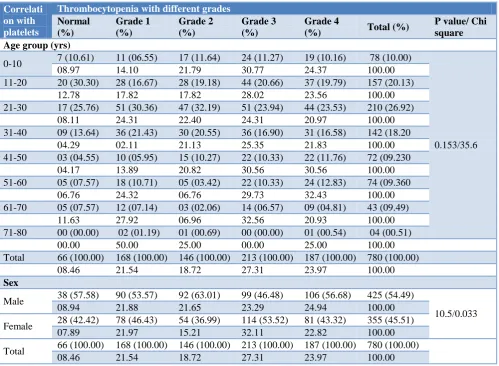
Maximum malarial cases were in 21-30 years age group and minimum cases were in 71-80 years age group. Maximum cases of malaria were found among males.
Maximum thrombocytopenia of all grades was found in 21-30 yrs age group. Maximum thrombocytopenia in males was of grade IV and maximum thrombocytopenia of grade III was found in females.
Maximum P. vivax cases was found in 21-30 yrs age group while maximum cases of P. falciparum was found in 11-20, 21-30 and 41-50 yrs age group and maximum cases of mixed infection in 31-40 yrs age group.
Maximum cases of P. vivax and mixed infection were found among males while P. falciparum was found equal in both sexes.
Table 1: Demographic profile (n=780).
Demographic profile Number %
Age group (yrs)
0-10 78 10.00
11-20 157 20.13
21-30 210 26.92
31-40 142 18.20
41-50 72 09.23
51-60 74 09.49
61-70 43 05.52
71-80 04 00.51
Sex
Male 425 54.50
Female 355 45,50
Table 2: Correlation of demographic profile with platelets.
Correlati on with platelets
Thrombocytopenia with different grades Normal
(%)
Grade 1 (%)
Grade 2 (%)
Grade 3 (%)
Grade 4
(%) Total (%)
P value/ Chi square Age group (yrs)
0-10 7 (10.61) 11 (06.55) 17 (11.64) 24 (11.27) 19 (10.16) 78 (10.00)
0.153/35.6
08.97 14.10 21.79 30.77 24.37 100.00
11-20 20 (30.30) 28 (16.67) 28 (19.18) 44 (20.66) 37 (19.79) 157 (20.13)
12.78 17.82 17.82 28.02 23.56 100.00
21-30 17 (25.76) 51 (30.36) 47 (32.19) 51 (23.94) 44 (23.53) 210 (26.92)
08.11 24.31 22.40 24.31 20.97 100.00
31-40 09 (13.64) 36 (21.43) 30 (20.55) 36 (16.90) 31 (16.58) 142 (18.20
04.29 02.11 21.13 25.35 21.83 100.00
41-50 03 (04.55) 10 (05.95) 15 (10.27) 22 (10.33) 22 (11.76) 72 (09.230
04.17 13.89 20.82 30.56 30.56 100.00
51-60 05 (07.57) 18 (10.71) 05 (03.42) 22 (10.33) 24 (12.83) 74 (09.360
06.76 24.32 06.76 29.73 32.43 100.00
61-70 05 (07.57) 12 (07.14) 03 (02.06) 14 (06.57) 09 (04.81) 43 (09.49)
11.63 27.92 06.96 32.56 20.93 100.00
71-80 00 (00.00) 02 (01.19) 01 (00.69) 00 (00.00) 01 (00.54) 04 (00.51)
00.00 50.00 25.00 00.00 25.00 100.00
Total 66 (100.00) 168 (100.00) 146 (100.00) 213 (100.00) 187 (100.00) 780 (100.00)
08.46 21.54 18.72 27.31 23.97 100.00
Sex
Male 38 (57.58) 90 (53.57) 92 (63.01) 99 (46.48) 106 (56.68) 425 (54.49)
10.5/0.033
08.94 21.88 21.65 23.29 24.94 100.00
Female 28 (42.42) 78 (46.43) 54 (36.99) 114 (53.52) 81 (43.32) 355 (45.51)
07.89 21.97 15.21 32.11 22.82 100.00
Total 66 (100.00) 168 (100.00) 146 (100.00) 213 (100.00) 187 (100.00) 780 (100.00)
08.46 21.54 18.72 27.31 23.97 100.00
DISCUSSION
Age distribution
In our study, out of total 780 study population maximum 210 (26.92%) cases were in 21-30 year age group, followed by 157 (20.13%) in 11-20 year age group and minimum 04 (0.51%) in 71-80 year age group. Similar
Sex distribution
In our study, out of 780 malarial patients 54.50% were males while 45.50% were females. 52% males and 48% females was observed by Ahmad et al, 63.33% males and 36.66% females was found by Gill et al, 65.22% males and 34.78% females by Gupta et al, 67% males and 33% females was found by Aundhakar et al, 69% males and 31% females was reported by Jairajpuri et al, 77.15% males and 22.85% females was reported by Kalavathi et al.29,30,32-35 The males thought to be at a higher risk due to more outdoor activity and less protection from mosquito bites.
Type of malaria
Out of 780 cases of malaria we found 86.28% cases of P.
vivax, 02.31% cases of P. falciparum and 11.41% mixed
infection.
Similar finding P. vivax (87.74%) P. falciparum (03.77%) and mixed infection (08.49%) was found by Jairajpuri et al.30 P. vivax (57.14%, 56.51%, 51.69%, 41.00% and 40.00%), P. falciparum (37.14%, 39.13%, 01.12%, 59.00% and 50.00%) mixed (05.72%, 05.72%, 04.34%, 47.19%, 0.90% and 10.00%), was reported by Kalavathi et al,Gupta et al, Faseela et al,Patel et al,and Kashikunti et al respectively and higher P. falciparum (70.6%) and
P. vivax (28.7%) was found by Agravat et al.29,35-39
Correlation of malaria with platelets
In our study out of 780 malarial cases thrombocytopenia was found in 91.54% cases. Normal platelet was observed in 08.46% out of which 07 .18% were in P. vivax, 0.26%
in P. falciparum and 01.03% in mixed infection.
Maximum 27.31% thrombocytopenia grade III was found in our study followed by 23.97% grade IV than 21.54% in grade I and minimum18.72% in grade II.
Table 3: Correlation of demographic profile with type of malaria parasite.
Correlation with platelets
Malaria parasite with different types
P. vivax P. falciparum Mixed Total P value/ Chi
square Age group (yrs)
0-10 67 (09.96) 01 (00.06) 10 (11.24) 78 (10.00)
0.051/23.625
85.90 1.28 12.82 100.00
11-20 134 (19.91) 04 (22.22) 19 (21.35) 157 (20.13)
85.35 2.55 12.10 100.00
21-30 198 (29.42) 03 (16.67) 09 (10.11) 210 (26.92)
94.29 01.43 04.29 100.00
31-40 117 (17.3) 04 (22.22) 21(23.60) 142 (18.21)
82.39 2.82 14.79 100.00
41-50 58 (08.62) 04 (22.22) 10 (11.24) 72 (09.23)
80.56 5.56 13.89 100.00
51-60 62 (09.210 01 (05.56) 11 (12.36) 74 (09.49)
83.78 01.35 14.86 100.00
61-70 33 (04.90) 01 (05.56) 09 (10.11) 43 (05.51)
76.74 02.33 20.93 100.00
71-80 04 (00.06) 00 (00.00) 00 (00.00) 04 (00.51)
100.00 00.00 00.00 100.00
Total 673 (100.00) 18 (100.00) 89 (100.00) 780 (100.00)
86.28 2.31 11.41 100.00
Sex
Male 363 (53.94) 09 (50.00) 53 (59.55) 425 (54.49)
0.563/1.148
85.41 02.12 12.47 100.00
Female 310 (46.06) 09 (50.00) 36 (40.45) 355 (45.51)
87.32 02.54 10.14 100.00
Total 673 (100.00) 18 (100.00) 89 (100.00) 780 (100.00)
86.28 02.31 11.41 100.00
In our study we found thrombocytopenia (91.54%). Similar finding 92% thrombocytopenia was observed by Jairajpuri et al, 81.9% thrombocytopenia was found by Agravat et al, 77.83% by Gupta et al, 71.61% was found by Akthar et al.29,30,39,40 In our study we found normal
(05.23%, 13.04%, and 10.81%),in P. falciparum normal platelets (13.24%, 08.69%, 13.51%) and in mixed infection 00.00%, 00.43% and 2.70%) by Agravat et al, Gupta et al and Akthar et al.29,39,40 In our study we found grade I thrombocytopenia (21.54%) while 16.51% grade I thrombocytopenia was observed by Jairajpuri et al and 26.13% by Agravat et al.30,39 In our study grade II
thrombocytopenia was found (18.72%) while 31.01%
grade II thrombocytopenia was found by Agravat et al and 2.83% by Jairajpuri et al, in our study Grade III thrombocytopenia (27.31%) was found, while 21.31% was observed by Agravat et al and and 41.03% was found by Jairajpuri et al, Grade IV thrombocytopenia was found in our study (23.97%). 21.23% by Jairajpuri et al and in contrast 3.14% was found by Agravat et al.30,39
Table 4: Correlation of type of malaria parasite with platelets.
Malaria parasite
Grade
normal Grade I Grade II Grade III Grade IV Total
P value/ Chi square
P. vivax 56 (84.85) 137 (81.55) 126 (86.30) 180 (84.51) 174 (93.05) 673 (86.28)
0.076/ 14.218
08.32 32.47 18.72 26.75 25.85 100.00
P.
falciparum
02 (03.03) 07 (04.17) 01 (00.69) 06 (02.82) 02 (01.07) 18 (02.31)
11.11 38.89 05.56 33.33 11.11 100.00
Mixed 08 (12.12) 24 (14.28) 19 (13.01) 27 (12.67) 11 (05.88) 89 (11.41)
00.90 26.97 21.35 30.34 12.36 100.00
Total 66 (100.00) 168 (100.00) 146 (100.00) 213 (100.00) 187 (100.00) 780 (100.00)
08.46 21.54 18.72 27.31 23.97 100.00
Table 5: Correlation of malaria with platelets.
Our study (n=780) (%)
Jairajpuri et al30 (n=230) (%)
Agravat et al39 (n=287) (%)
Gupta et al29 (n=230) (%)
Akthar etal40 (n=74) (%)
Thrombocytopenia 91.54 92 81.5 77.83 71.61
Normal platelets
P. vivax 07.18 07.80 05.23 13.04 10.81
P. falciparum 00.26 00.00 13.24 08.69 13.51
Mixed 1.03 00.00 - 00.43 2.70
Total 08.46 7.80 18.47 22.17 27.03
Grade I
P. vivax 17.56 14.62 08.71
P. falciparum 00.90 00.47 17.42
Mixed 03.07 01.42 -
Total 21.54 16.51 26.13
Grade II
P. vivax 16.15 18.40 19.86
P. falciparum 00.13 00.94 11.15
Mixed 02.44 01.89 -
Total 18.72 02.83 31.01
Grade III
P. vivax 23.08 37.26 02.09
P. falciparum 00.77 00.94 19.13
Mixed 03.46 02.83 -
Total 27.31 41.03 21.31
Grade IV P. vivax 22.30 17.45 01.05
P. falciparum 00.26 01.42 02.09
Mixed 01.41 02.36 -
Total 23.97 21.23 3.14
CONCLUSION
Mostly developing countries are with limited resources and trained health personnel. The haematological aspects of malarial infection constitute a very interesting area and may be used in addition to the clinical assessment, to heighten the suspicion of disease. Thrombocytopenia is associated with both P. vivax as well as P. falciparum
infections. So thrombocytopenia with acute febrile illness in the tropics, increases probability of malaria, as in our study we found thrombocytopenia in about 92% in malarial cases.
Funding: No funding sources Conflict of interest: None declared
REFERENCES
1. White NJ, Breman JG. Harrison’s Principles of Internal Medicine. 18th edition. Volume. I US: The McGraw Hill Companies, Inc; 2012: 1688.
2. Sitprija V. Nephropathy in falciparum malaria. Kidney Int. 1988;34(6):867-77.
3. Naqvi R, Ahmad E, Akhtar F, Naqvi A, Rizvi A. Outcome in severe acute renal failure associated
with malaria; Nephrol Dial Transplant.
2003;18(9):1820-3.
4. World Health Organization, World Malaria Report; 2015. Available at: http://www.who.int/malaria/ media/world_ malaria_report_2015. Accessed on 26 August 2019.
5. World Health Organization, World Malaria Report; 2011. Available at: http://www.who.int/malaria/ media/world_ malaria_report_2011. Accessed on 26 August 2019.
6. Yadav D, Chandra J, Dutta AK. Benign tertian malaria; how benign it is today? Indian J Pediatr. 2012;79(4):525-7.
7. Beale PJ, Cormack JD, Oldrey TB.
Thrombocytopenia in malaria with immunoglobulin (IgM) changes. Br Med J. 1972;1(5796):345-9. 8. Agarwal SS, Nath A, Sharma P, Srivastava IK,
Dwivedi SR, Dutta GP. Comparative evaluation of Plasmodium knowlesi and P. cynomology antigens in the indirect fluorescent antibody test for human malaria. Indian J Med Res. 1983;77:616-22.
9. Lacerda MVG, Mourao MPG, Coelho HCC, Santos
JB. Thrombocytopenia in malaria: who cares? Mem Inst Oswaldo Cruz. 2011;106(1):52-63.
10. Angchaisuksiri P Coagulopathy in malaria. Thromb Res. 2014;133:5-9.
11. Erhart LM, Yingyuen K, Chuanak N, Buathong N, Laoboonchai A, Miller RS, et al. Hematologic and clinical indices of malaria in a semi-immune population of Western Thailand. Am J Trop Med Hyg. 2004;70:8-14.
12. Moulin F, Lesage F, Legros AS, Maroga C,
Moussavou A, Guyon P, et al. Thrombocytopenia and Plasmodium falciparum malaria in children with different exposures. Arch Dis Child. 2003;88:540-1. 13. Genton B, D'Acremont V, Rare L, Baea K, Reeder JC, Alpers MP, et al. Plasmodium vivax and mixed infections are associated with severe malaria in children: a prospective cohort study from Papua New Guinea. PLoS Med. 2008;5(6):881-9.
14. Kochar DK, Tanwar GS, Khatri PC, Kochar SK, Sengar GS, Gupta A, et al. Clinical features of children hospitalized with malaria: a study from Bikaner, Northwest India. Am J Trop Med Hyg. 2010;83(5):981-9.
15. Tjitra E, Anstey NM, Sugiarto P, Warikar N, Kenangalem E, Karyana M, et al. Multidrug – resistant Pv malaria associated with high morbidity and mortility. PLoS Med. 2008;5(6):e128.
16. Bakhubaira S. Hematological parameters in severe complicated Plasmodium falciparum malaria among adults in Aden. Turk J Haematol. 2013;30:394-9.
17. Mania RN, Walsh D, Gaddy C, Hongo G, Waitumbi
J, Otineo L, et al. Impact of plasmodium falciparum infection on haematological parameters in children living Western Kenya. Malar J. 2010:9(3):4. 18. Van Wolfswinkel ME, Vliegenthart –Jongbloed K,
De Mendonca Melo M, Wever PC, McCall MB, Koelewijn R, et al. Predictive value of lympho-cytopenia and the neutrophil- lymphocyte count ratio for severe imported malaria. Malar J. 2013;12:101.
19. Luxemburger C, Ricci F, Nosten F, Raimond D, Bathet S, White NJ. The epidemiology of severe malaria in an area of low transmission in Thailand. Trans R Soc Trop Med Hyg. 1997;91:256-262.
20. WHO. World Malaria Report 2010. Geneva: World
Health Organization; 2011.
21. Bisoffi Z, Van den EJ. Costs of treating malaria according to test results. BMJ. 2008;336:168-9. 22. Bisoffi Z, Gobbi F, Angheben A, Van den EJ. The
role of rapid diagnostic test in managing malaria. PLoS Med. 2009;6:e1000063.
23. Acremont VD, Lengeleler C, Mshinda H, Mtasiwa D, Tanner M, Genton B. Time to move from
presumptive malaria treatment to laboratory
confirmed diagnosis and treatment in African children with fever. PLoS Med. 2009;6:252.
24. English M, Reyburn H, Goodman C, Snow RW.
Abandoning presumptive antimalarial treatment for febrile children aged less than five years-a case of running before we can walk? PLos Med. 2009;6:e1000015.
25. Gerstl S, Dunkley S, Mukhtar A, De SM, Baker S, Maikere J. Assessment of two malaria rapid diagnostic test in children under five years of age, with follow up of false positive pLDH test results in
a hyperendemic falciparum malaria area,
SierraLeone. Malar J. 2010;9:28.
26. WHO. Guidelines for the treatment malaria.
Geneva: World Health Organization; 2010.
27. Dacie SJ, Lewis SM. Reference ranges and normal values, Practical haematology. 10th edition, UK, Churchill Livingstone Publication; 2006: 14-17. 28. Bethesda. U.S Department of Health and Human
Services; National Cancer Institute Criteria for Adverse Events. Version 3; 2006: 4
29. Gupta NK, Bansal SB, Jain UC, Sahare K. Study of
Thrombocytopenia in Patients of Malaria. Trop Parasitol. 2013;3(1):58-61.
30. Jairajpuri Z, Rana S, Jaseem S, Jetley S.
Thrombocytopenia and Malaria: A Coincidental Co-Existence or a significant Association? An Analysis. Ann Pathol Lab Med. 2015;2(2):48-53.
32. Gill MK, Makkar M, Bhat S, Kaur T, Jain K, Dhir G. Thrombocytopenia in Malaria and its Correlation with different types of Malaria. Ann Trop Med Public Health. 2013;6(2):197-200.
33. Ahmad S, Adil F, Shahzad T, Yahiya Y. Severe malaria in children: Factors predictive of outcome and response to Quinine. J Pak Med Assoc. 2011;61(1):54-8.
34. Aundhakar S, Prajapati P, Prajapati S, Aundhakar A, Kothia D, John D, et al. Study of Clinical and Hematological Profile of Plasmodium vivax Malaria in a Tertiary Care Hospital in Western Maharashtra. Int J Scienti Study. 2017;5(3):257-60.
35. Kalavathi GP, Kumar SD. Clinical, Haematological and Biochemical Profile of Malaria Cases.: Int J Med Res. 2016;1(4):50-5.
36. Faseela T, Roche S, Anita KB, Malli CS, Rai Y. Diagnostic value of platelet count in malaria. J Clin Diagn Res. 2011;5:464–6.
37. Patel A, Jain S, Patel B, Modi B. Hematological changes in P. falciparum and P. vivax malaria; Nat J Med Res. 2013;3(2):130-3.
38. Kashinkunti M, Alevoor S. Clinical, Haematological and Coagulation Profile in Malaria. Sch J App Med Sci. 2014;2(2):584-8.
39. Agravat AH, Dhruva GA. Haematological Changes in Patients of Malaria. J Cell Tissue Res. 2010;10(3):2325-9.
40. Akhtar S, Gumashta R, Mahore S, Maimoon S. Hematological Changes in Malaria: A Comparative Study. J Pharm Biol Sci. 2012;2(4):15-9.