A Survey on the Amount of Positive Result of DWI in the
Patients Suffering from TIA and the Correlation of this
Patients With ABCD Scoring
NOOSHIN YAMANI and MOHAMMADREZA GHEINI*
Department of Neurology, Tehran University of Medical Sciences, Tehran, Iran *Corresponding author E-mail: gheini@gmail.com
http://dx.doi.org/10.13005/bpj/934
(Received: January 11, 2016; accepted: February 20, 2016) ABSTRACT
Stroke is the third major reason of death in developed countries and the most prevalent disabling neurologic disorder. One of the approaches to recognize the brain damage is MRI. Presence of an acute damage in MRI of brain with DWI (diffusion weighted imaging) in patients suffering from TIA (transient ischemic attack). Therefore, cognition of the risk factors which can clinically interfere the DWI, might be helpful for making the best decision about the patients suffering from TIA. The purpose of this study is evaluation of the amount of positivity of DWI in the patients suffering from TIA and the correlation of these patients with ABCD scoring. Contributors were patients suffering from TIA hospitalized in neurology department of Sina hospital, who have been randomly selected among patients who had no problem with being studied. These patients were surveyed for one week. After the results of DWI were submitted, the study cases were divided into two groups, one the persons with positive DWI and the other one with typical DWI, and ABCD2 scorings and effecting parameters in ABCD2 scores in these two groups were compared. Raw data were entered in the SPSS software ver.17, and then the non-parametric statistic Chi-squared test was used to analysis the data and the meaningful levels were surveyed (P-Value> 0.05).In this study, 240 male took part, 73.8% of these persons had AC-TIA and 26.2% had PC-TIA. Among 177 persons with AC-TIA, 79.7% of them were normal DWI and 20.3% equal to 36 persons were positive DWI. Then continuing the study, the same score of positive DWI person (36 persons) were selected from normal DWI persons randomly. The average of the age in the normal DWI group was 65.5±10.13, and in the positive DWI group was 68.2±8.3 years old. According to the results obtained from variable statistical tests, totally the ABCD2 scores have no meaningful relationship to the results obtained from DWI (p-Value<0.05), whilst the results depict a meaningful statistical correlation between results from DWI and two other parameters of ABCD2 which are Clinical Features and Symptom Duration (p-Value>0.05). However, discoveries of this study demonstrated no meaningful relationship between results of DWI (normal/positive) and other parameters of ABCD2 scoring such as age, blood pressure, and diabetes in the patients suffering from TIA (p-Value <0.05). The results of this study demonstrates that the total score of ABCD2 have no correlation with DWI results, but some of the parts defining the ABCD2 scores, have meaningful correlation with the results obtained from DWI.
Key words: Passive ischemic attack, stroke, TIA, DWI, ABCD2 scoring, MRI.
INTRODUCTION
One of the most prevalent neurologic disabling diseases in adults and the third reason of death, is stroke (Cerebro Vascular Accident)1. 750000 strokes happen yearly and about 150000 persons die because of stroke2. 88% of the strokes are the ischemic type, which 8 to 12 percent of them
blood vessel ( hemorrhagic). Ischemic stroke takes place because of congestion of one of the arteries carrying oxygen and nutrition to the cerebral cells. TIA (Transient Ischemic Attack) is the passive and unstable conditions of the malfunction of the brain or a part of brain5, or TIA is a bad neurologic period of time, caused from suffering a special part of brain , spinal cord, as well as acute retinal infarction5, 6. The main reason of TIA is presence of an embolus (embolus=a bulk segregated and moving in the vein) and congestion of one of the veins with the embolus. Embolus can be created because of a vascular plaque formed from atherosclerosis or a thrombosis remained from a fibrillation in the heart. This congestion happens in a short period of time and the resulted suffer is not permanent7. TIA is similar to a stroke. Some of the reasons of TIA are high blood pressure, smoking, high levels of cholesterol, diabetes and migraine. Symptoms of TIA are blurry vision, double vision, aphasia, problems in talking, one-sided weakness and numbness, and vertigo. These symptoms disappear after one hour8. In the case of observing the symptoms of stroke in the patient, some experiments are necessary to be done to measure the intensity and extension of the stroke. CT Scan is one of the methods to recognize the stroke. This approach demonstrates a 2D imaginary of the brain using X-ray whereby strokes such as ischemic and hemorrhagic strokes can be recognized. CT scan is capable to demonstrate size, the place of the brain suffered from tumor, damaged veins, and blood clots. Injecting some materials to enhance the quality of the image, and using X-ray may cause allergic reactions and are the most major dangers about using CT scan. MRI is a very more precise technique to depict more details comparing to CT scan, whereby very clear and precise images of brain and its veins are being provided employing magnetic fields, without any X-ray or colorful materials9. MRI is a very effective tool to recognition of damages in the brain, especially ischemic strokes. Images obtained from MRI have three axis (coronal, sagittal, axial), and the images can be taken from the inaccessible spots of brain which CT scan cannot take images from them. There are a variety of imaging in MRI, DWI (Diffusion Weighted Imaging) in which the computer orders are being designed how they react to the apparent diffusion coefficient (the sum of movement of molecules inside the cell)10. The most prevalent
with higher ABCD2 score. Also they believe that evaluating the ABCD2 score is very essential for all the people exposed to ischemic stroke15.
METHODS AND MATERIALS
This is a partial study and it uses random sampling. The statistic society of this study are 240 patients suffering from TIA who referred to Sina Hospital. Patients were informed about the study and the patients who had a tendency to take part in the program, started to collaborate. In this study, the patients who did not have the excluding criteria consisting of the former stroke, not to knowing the exact time of starting the symptoms, and reference to the doctor after 24 hours (of starting symptoms), were brought in the study. In this study, after the initial sampling, 26 women with a age ranging from 55-62 years old who were dismissed from the study case after evaluation of conditions and the ratio of DWI, and male patients added instead of them. The reasons of this dismissing was as below: according to former studies, researchers believe that on the one hand being a male, itself is a risk factor to be stricken by TIA16 and on the other hand, the researches show that positive DWI is being observed in males rather than females17 . Also the statistics show that the rate of TIA was observed in the females aging from 65 to 90, which is not compatible with the age range of the female in our study16 (55 to 62). The rate of TIA is higher especially in the males aging from 65 to 75 comparing with females. Therefore, evaluating the symptoms and relations between them in this group is more real. In the studies in which the ABCD scoring is studied, there has been no pointing to the differences in the results of these parameters17, 18. Also in this study, the number of women referred to be brought in the study was by far less than men, and regarding the standards of the study (which one of the most important ones was the age between 65 to 75), dividing the participants into two groups of male and female was impossible. Therefore, the female participants were dismissed. After that, the sampling continued until the number of participants raised from 214 to 240 persons. Then, in this study patients suffering from TIA consisting of AC-TIA (anterior circulation) and PC-TIA (posterior circulation) were studied. The A and P patients were divided into groups according to symptoms written in valid
dysarthria without weakness of the body (1 score), duration of the symptoms like 10 to 60 minutes (1 score), more than 60 minutes (2 score), diabetes (1 score). According to some references (17, 24), the surveyed factors for the ABCD2 scoring such as Symptom Duration and Clinical Features each one have three states (Clinical Features: talking disorder, one-sided weakness, and Motor + Speech. And about Symptom Duration (less than 10 minutes, 10-59 minutes, and more than 60 minutes). In this study, all of these cases were evaluated and analyzed. Whilst other references in order to calculate the ABCD2 scoring have considered only two states for clinical features (talking disorder, one-sided weakness) and two states for Symptom Duration (10-59 minutes, and more than 60 minutes) (25, 26). Therefore, in order to implementation of this study, to calculate the ABCD2 score and achieving the most precise state, the results of all the three standard states which were defined for Clinical Features and Symptom Duration, were evaluated. Patients were exposed to MRI with the 1.5 tesla power within less than 7 days from the start of the symptoms. These images verified and the results reported by two specialists of MRI. Features of the images are consisting of slice, ¡ field¡ size of matrix¡ gap¡ slice thickness¡ eco time¡ repetition time¡ and the B value . After collecting required data from the patients with TIA, the data were input into the SPSS software ver.17. In order to analysis of the data the Chi-squared test and T-test were used and the meaningful surfaces (P-Value<0.05) were evaluated.
RESULTS
First the variables were introduced then the raw data were analyzed by proper statistical programs. This study is performed on 240 patients suffering from TIA who referred to neurological department of Sina hospital. 26 persons of the participants are female and the rest (214 persons) are male. According to the normal and abnormal profusion (positivity) of the DWI imaging, only 3 persons equal to 11.5 percent of whole the population of females had positive DWI. Due to the limited time and standards defined for the participants and insufficient number of females comparing to males, and also the reasons mentioned in the Method section, females were
Variables DWI
Total Samples n=72 Value
Positive
Normal DWI
n=36 n=36
Subnumbers of ABCD2
scores Age
10.13 ± 65.5 68.2 ±8.3
67.1 ±5.2 753
Blood Pressure 20 persons
22 persons 42 persons
(58%) 856
(55%) (61%)
Clinical Features 12 persons
10 persons 32 persons
(44.4%) 0.022
Talking disorder (33.3%)
(27.78 %)
One-sided Weakness 13 persons
12 persons 25 persons
0.011
(36.11%) (33.33%)
(34.72%)
11 persons 14 persons
25 persons (34.72%) 0.075
Motor + Speech (30.56%)
(38.889%)
Diabetes 8 persons
7 persons 15 persons
148
(22%) (19%)
(20%)
Symptoms Duration <10 min
9 persons 6 persons
15 0.074
(25%) (16.7%)
persons(20.83%)
10-59 min 11 persons
12 persons 23 persons
0.314
(30.6%) (33.3%)
(31.94%)
>60 min 16 persons
18 persons 34 persons
0.042
(44.4%) (50%)
(47.22)
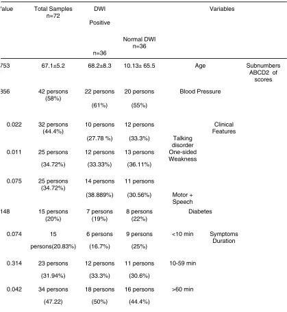
Table 1: The relationship between DWI (normal/positive) of the patients with each parameters of ABCD2 according to the table 1, it can be declared for the
Symptom Duration parameter that the longer duration of symptoms, the more probability of more positive results of DWI. Also, considering meaningful relationship between results of DWI and two indexes of talking disorder and one-sided weakness and considering table 1, this can be included that occurrence and increase of these two clinical parameters (Clinical Features and Symptom
meaningful relationship with results of DWI. While in the previous sections we realized that some sub collections of ABCD2 such as Clinical Features and
Symptom Duration have relations with results of DWI.
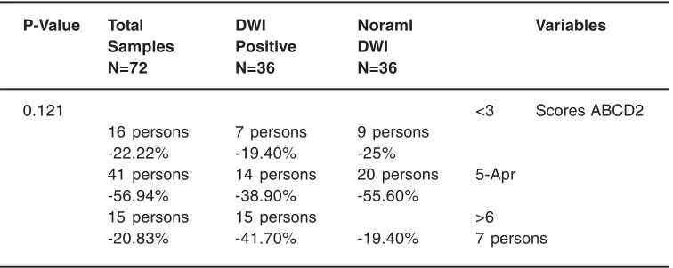
Table 2: The relationship between DWI (normal/positive) with ABCD2 scoring of the patients
P-Value Total DWI Noraml Variables
Samples Positive DWI
N=72 N=36 N=36
0.121 <3 Scores ABCD2
16 persons 7 persons 9 persons
-22.22% -19.40% -25%
41 persons 14 persons 20 persons 5-Apr
-56.94% -38.90% -55.60%
15 persons 15 persons >6
-20.83% -41.70% -19.40% 7 persons
DISCUSSION
Presence of passive neurologic symptoms without acute infarction is called acute ischemic attack (TIA) (27). TIA is a prevalent risk factor for the stroke (28, 29). Some of the prevalent symptoms of TIA are one-sided numbness, talking disorder, and one eye blindness. These symptoms are temporary. Immediate recognition of TIA and determining it from other imitative conditions is very essential, because fast reactions can reduce the risk factor of occurrence of upcoming strokes. TIA is companied by a sudden start, neurologic defection, and talking disorders. Immediate evaluation of TIA in the patients consists these items11, 12: neurologic imaging, vein imaging of head and neck, heart evaluations, verifying blood pressure and other routine clinical tests. The ABCD scoring (age, blood pressure, clinical symptoms, diabetes and duration of symptoms) should be determined during initial examinations, which can be very beneficent for evaluating the risk factor of repetition of ischemic and stroke. The patients with high ABCD scores should be hospitalized, while patients with lower scores are less prone to upcoming strokes and they can be treated as outpatient. The value of ABCD scoring has been investigated in different articles regarding its ability of prediction of disorders in the brain and veins. The rate of occurrence of stroke one week after TIA in different studies shows
stroke in both AC-TIA and PC-TIA patients. In that study, 369 patients suffering from TIA were selected continuously in June of 2009 and December of 2012. Totally 273 patients with AC-TIA and 96 patients with PC-TIA were studied. 21 patients with AC-TIA and 7 patients with PC-TIA faced heart attack in 7 days ( 7.7 versus 7.3 percent, p-Value=0.8). They reported that the ABCD scoring in AC-TIA
group had better predictions comparing to PC-TIA group. Age, one-sided weakness, and blood pressure were indexes in the AC-TIA patients who faced stroke. While in the PC-TIA group, the diabetes was more prominent20. The results of current study showed that the meaningful relationship between some parameters of ABCD2 scoring and DWI results and total score of ABCD2 can not reflect the results of DWI.
REFERENCES
1. Simon R, Greenberg D, Aminoff M. Clinical neurology: McGraw-Hill Prof Med/Tech; 2009. 2. Thom T, Haase N, Rosamond W, Howard VJ, Rumsfeld J, Manolio T, et al. Heart disease and stroke statistics—2006 update a report from the American Heart Association Statistics Committee and Stroke Statistics Subcommittee. Circulation. 2006; 113(6): e85-e151.
3. Bradley WG. Neurology in clinical practice: principles of diagnosis and management: Taylor & Francis; 2004.
4. Amarenco P, Labreuche J, Lavallée P, Touboul P-J. Statins in stroke prevention and carotid atherosclerosis systematic review and up-to-date meta-analysis. Stroke. 2004;35(12):2902-9.
5. Easton JD, Saver JL, Albers GW, Alberts MJ, Chaturvedi S, Feldmann E, et al. Definition and Evaluation of Transient Ischemic Attack A Scientific Statement for Healthcare Professionals From the American Heart Association/American Stroke Association Stroke Council; Council on Cardiovascular Surgery and Anesthesia; Council on Cardiovascular Radiology and Intervention; Council on Cardiovascular Nursing; and the Interdisciplinary Council on Peripheral Vascular Disease: The American Academy of Neurology affirms the value of this statement as an educational tool for neurologists. Stroke. 2009;40(6):2276-93. 6. Albers GW, Caplan LR, Easton JD, Fayad
PB, Mohr J, Saver JL, et al. Transient ischemic attack—proposal for a new definition. The New England journal of medicine. 2002;347(21):1713.
7. h t t p : / / w w w. s t r o k e a s s o c i a t i o n . o r g /
STROKEORG/AboutStroke/TypesofStroke/ T I A / T I A T r a n s i e n t I s c h e m i c -Attack_UCM_310942_Article.jsp#.TrzHVfQkD7Y. 8. http://stroke.emedtv.com/stroke-articles.html. 9. Hollingworth W, Todd CJ, Bell MI, Arafat Q, Girling S, Karia KR, et al. The diagnostic and therapeutic impact of MRI: an observational multi-centre study. Clinical radiology. 2000;55(11):825-31.
10. McRobbie DW, Moore EA, Graves MJ, Prince MR. MRI–From Picture to Proton (2003). Cambridge University Press, Cambridge. 11. Rothwell P, Giles M, Flossmann E, Lovelock
C, Redgrave J, Warlow C, et al. A simple score (ABCD) to identify individuals at high early risk of stroke after transient ischaemic attack. The Lancet. 2005;366(9479):29-36. 12. Rothwell PM, Giles MF, Chandratheva A,
Marquardt L, Geraghty O, Redgrave JN, et al. Effect of urgent treatment of transient ischaemic attack and minor stroke on early recurrent stroke (EXPRESS study): a prospective population-based sequential comparison. The Lancet. 2007;370(9596):1432-42.
13. Crisostomo RA, Garcia MM, Tong DC. Detection of diffusion-weighted MRI abnormalities in patients with transient ischemic attack correlation with clinical characteristics. Stroke. 2003;34(4):932-7. 14. Oppenheim C, Stanescu R, Dormont D,
Crozier S, Marro B, Samson Y, et al. False-negative diffusion-weighted MR findings in acute ischemic stroke. American journal of neuroradiology. 2000;21(8):1434-40. 15. Asimos AW, Johnson AM, Rosamond WD,
accuracy for predicting early ischemic stroke in admitted patients with transient ischemic attack. Annals of emergency medicine. 2010;55(2):201-10. e5.
16. Sacco RL, Benjamin EJ, Broderick JP, Dyken M, Easton JD, Feinberg WM, et al. Risk factors. Stroke. 1997;28(7):1507-17. 17. Anticoli S, Pezzella FR, Pozzessere C,
Gallelli L, Bravi MC, Caso V, et al. Transient Ischemic Attack Fast-track and Long-Term Stroke Risk: Role of Diffusion-Weighted Magnetic Resonance Imaging. Journal of Stroke and Cerebrovascular Diseases. 2015;24(9):2110-6.
18. Rovira A, Rovira-Gols A, Pedraza S, Grivé E, Molina C, Alvarez-Sabín J. Diffusion-weighted MR imaging in the acute phase of transient ischemic attacks. American journal of neuroradiology. 2002;23(1):77-83. 19. De Marchis GM, Kohler A, Renz N, Arnold
M, Mono M-L, Jung S, et al. Posterior versus anterior circulation strokes: comparison of clinical, radiological and outcome character istics. Jour nal of Neurology, Neurosurgery & Psychiatr y. 2010:jnnp. 2010.211151.
20. Wang J, Wu J, Liu R, Gao F, Hu H, Yin X. The ABCD2 score is better for stroke risk prediction after anterior circulation TIA compared to posterior circulation TIA. International Journal of Neuroscience. 2015;125(1):50-5.
21. Engelter ST, Wetzel SG, Bonati LH, Fluri F, Lyrer PA. The clinical significance of diffusion-weighted MR imaging in stroke and TIA patients. Swiss Med Wkly. 2008;138(49-50):729-40.
22. Welch K, Windham J, Knight RA, Nagesh V, Hugg JW, Jacobs M, et al. A model to predict the histopathology of human stroke using diffusion and T2-weighted magnetic resonance imaging. Stroke. 1995;26(11):1983-9.
23. Zuo L, Zhang Y, Xu X, Li Y, Bao H, Hao J, et al. A retrospective analysis of negative diffusion-weighted image results in patients with acute cerebral infarction. Scientific reports. 2015;5.
24. Asimos AW, Rosamond WD, Johnson AM, Price MF, Rose KM, Murphy CV, et al. Early
diffusion weighted MRI as a negative predictor for disabling stroke after ABCD2 score risk categorization in transient ischemic attack patients. Stroke. 2009;40(10):3252-7.
25. Giles MF, Albers GW, Amarenco P, Arsava MM, Asimos A, Ay H, et al. Addition of brain infarction to the ABCD2 score (ABCD2I) a collaborative analysis of unpublished data on 4574 patients. Stroke. 2010;41(9):1907-13.
26. Merwick Á, Albers GW, Amarenco P, Arsava EM, Ay H, Calvet D, et al. Addition of brain and carotid imaging to the ABCD 2 score to identify patients at early risk of stroke after transient ischaemic attack: a multicentre observational study. The Lancet Neurology. 2010;9(11):1060-9.
27. Ay H, Koroshetz WJ, Benner T, Vangel MG, Wu O, Schwamm LH, et al. Transient ischemic attack with infarction: a unique syndrome? Annals of neurology. 2005;57(5):679-86. 28. Hill M, Yiannakoulias N, Jeerakathil T, Tu J,
Svenson L, Schopflocher D. The high risk of stroke immediately after transient ischemic attack A population-based study. Neurology. 2004;62(11):2015-20.
29. Sacco RL, Adams R, Albers G, Alberts MJ, Benavente O, Furie K, et al. Guidelines for Prevention of Stroke in Patients With Ischemic Stroke or Transient Ischemic Attack A Statement for Healthcare Professionals From the American Hear t Association/ American Stroke Association Council on Stroke: Co-Sponsored by the Council on Cardiovascular Radiology and Intervention: The American Academy of Neurology affirms the value of this guideline. Circulation. 2006;113(10):e409-e49.
30. Ay H, Arsava EM, Johnston SC, Vangel M, Schwamm LH, Furie KL, et al. Clinical-and imaging-based prediction of stroke risk after transient ischemic attack the CIP model. Stroke. 2009;40(1):181-6.
31. Fothergill A, Christianson TJ, Brown RD, Rabinstein AA. Validation and refinement of the ABCD2 score a population-based analysis. Stroke. 2009;40(8):2669-73. 32. Brazzelli M, Chappell FM, Miranda H, Shuler
weighted imaging and diagnosis of transient ischemic attack. Annals of neurology. 2014;75(1):67-76.
33. Carpenter CR, Keim SM, Crossley J, Perry JJ, Group BEiEMI. Post-Transient Ischemic Attack Early Stroke Stratification: The ABCD 2 Prognostic Aid. The Journal of emergency medicine. 2009;36(2):194-200.