R E S E A R C H
Open Access
Serum arterial lactate concentration predicts
mortality and organ dysfunction following liver
resection
Matthew G Wiggans
1,2†, Tim Starkie
3†, Golnaz Shahtahmassebi
4, Tom Woolley
3, David Birt
3, Paul Erasmus
3,
Ian Anderson
3, Matthew J Bowles
1, Somaiah Aroori
1and David A Stell
1,2*Abstract
Background:The aim of this study was to determine if the post-operative serum arterial lactate concentration is associated with mortality, length of hospital stay or complications following hepatic resection.
Methods:Serum lactate concentration was recorded at the end of liver resection in a consecutive series of 488 patients over a seven-year period. Liver function, coagulation and electrolyte tests were performed post-operatively. Renal dysfunction was defined as a creatinine rise of >1.5x the pre-operative value.
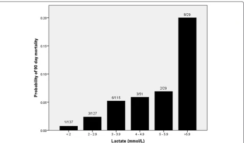
Results:The median lactate was 2.8 mmol/L (0.6 to 16 mmol/L) and was elevated (≥2 mmol/L) in 72% of patients. The lactate concentration was associated with peak post-operative bilirubin, prothrombin time, renal dysfunction, length of hospital stay and 90-day mortality (P< 0.001). The 90-day mortality in patients with a post-operative lactate≥6 mmol/L was 28% compared to 0.7% in those with lactate≤2 mmol/L. Pre-operative diabetes, number of segments resected, the surgeon’s assessment of liver parenchyma, blood loss and transfusion were independently associated with lactate concentration.
Conclusions:Initial post-operative lactate concentration is a useful predictor of outcome following hepatic resection. Patients with normal post-operative lactate are unlikely to suffer significant hepatic or renal dysfunction and may not require intensive monitoring or critical care.
Keywords:Liver, Hepatectomy, Post-operative care
Background
Despite advances in both operative technique and peri-operative care, liver resection is associated with post-operative mortality rates of 0% to 22% (median 3.7%) [1] and morbidity rates of 12.5% to 66% including liver dysfunction [2,3], renal dysfunction [4] and bile leak [5,6]. Factors associated with peri-operative complications and death include patient age [7,8] and gender [9,10], hospital annual number of liver resections undertaken [9,11], pathologic origin of liver tumour [9,11], pre-operative liver and renal dysfunction [8,10], diabetes [12,13], chronic liver
disease [7,9], and the peripheral neutrophil to lymphocyte ratio (NLR) [14]. Operative factors associated with outcome include blood loss [8,10] and transfusion [15,16], extent of liver resection [15,17], duration of surgery [18], simultaneous extrahepatic procedures [15,19], and the use of the Pringle manoeuvre [16,20].
Therefore, many factors affect outcome after liver surgery which have not been incorporated into a single scoring system. The American Society of Anesthesiologists (ASA) grade and Portsmouth Physiologic and Operative Severity Score for the enUmeration of Mortality and morbidity (P-POSSUM) scores are used in the risk prediction of many types of surgery [21,22] including liver surgery [23]. However, these scores may not be applicable to the unique stresses of liver resection. One of the main reported causes of mortality following liver resection is post-hepatectomy liver failure (PHLF) [24]. Although * Correspondence:[email protected]
†Equal contributors
1
Hepatobiliary Surgery, Plymouth Hospitals NHS Trust, Derriford Hospital, Derriford Road, Plymouth, Devon PL6 8DH, UK
2
Peninsula College of Medicine and Dentistry, University of Exeter and Plymouth University, Research Way, Plymouth, Devon PL6 8BU, UK Full list of author information is available at the end of the article
the ‘50-50 criteria’ of serum bilirubin of >50 μmol/L and prothrombin index (laboratory’s calculated mean normal prothrombin time (PT) divided by the patient’s observed PT) of <50% measured on the fifth post-operative day have been shown to be associated with death due to PHLF [2], an earlier prediction system may be clinically more useful in guiding therapy. Furthermore, failure of multiple organ systems may contribute to death following liver resection and there is a need for a global peri-operative measure to predict the risk of developing significant post-operative morbidity and death.
Lactic acid is a by-product of anaerobic metabolism that is subsequently metabolised in the liver during gluconeogenesis [25]. Hyperlactataemia has been shown to be associated with increased mortality and morbidity in a critical care setting [26,27], in patients with liver failure [28], sepsis [29] and following trauma [30]. Similar re-lationships have been shown in the post-operative set-ting following pancreatic resection [31] and other major abdominal surgery [32], cardiac surgery [33] and after hepatic transplantation [34].
The primary aim of this study was to determine if the first post-operative arterial lactate concentration (‘initial lactate’) is associated with adverse outcomes following liver resection including 90-day mortality, length of hospital stay (LOS), and renal and hepatic dysfunction. The secondary aim was to determine which pre- and intra-operative risk factors are associated with initial lactate concentration following liver resection.
Methods
This study was a retrospective analysis of a prospectively maintained database of all patients undergoing liver resection since July 2005. Routine patient characteristics, laboratory data and intra-operative details were retrieved. Pre-operative liver-directed chemotherapy was adminis-tered to selected patients following discussion at a regional multidisciplinary team meeting. A period of recovery of at least six weeks was allowed following cessation of chemotherapy before undertaking surgery. The P-POSSUM scoring system was used to calculate the physiological score [21]. Prior to resection, the operating surgeon makes a vis-ual assessment of the condition of the liver parenchyma and records this as normal or abnormal. Liver resections were performed using standard techniques with a Cavitron Ultrasonic Surgical Aspirator™ (CUSA; Tyco Healthcare, Mansfield, MA, USA) dissector. Hepatic inflow occlusion was used in a minority of cases where there was excessive blood loss. Anaesthetic techniques include the routine use of invasive arterial blood pressure monitoring, central venous pressure monitoring (CVP) (using a target CVP of <5 cm H20) and epidural anaesthesia. Liver resections were defined according to the Brisbane classification [35] and the number of removed segments recorded.
Intravenous fluid replacement was minimised during the resection phase to decrease venous pressure. After removal of the surgical specimen, a pause in surgical activity is routinely planned to allow haemostasis and intravenous volume replacement with 0.9% saline or Hartmann’s solution at the anaesthetist’s discretion. Patients are usually returned to the High Dependency Unit (HDU) after surgery with full invasive monitoring, except for minor resections in fit patients who are returned to the general ward.
The serum lactate was recorded from an arterial blood sample taken immediately prior to abdominal closure or immediately on arrival in the HDU. The arterial lactate in the normal population is below 1.6 mmol/L whereas in a critical care setting <2 mmol/L is more commonly accepted in acutely stressed patients [36].
Serum biochemistry tests and coagulation assays were performed on all patients in the first 24 hours post-operatively and the tests repeated according to clinical course. The peak measurement of bilirubin and PT were recorded and used for analysis. A PT index of <50% corre-sponds to a PT >24 s. Similarly peak post-operative cre-atinine levels were obtained and renal dysfunction was defined according to the Risk, Injury, Failure, Loss, and End-stage kidney disease (RIFLE) criteria [37]. Renal dys-function in categorical analyses was defined as any increase in serum creatinine of ≥1.5-fold from the pre-operative baseline. The length of hospital stay was measured from day of surgery to day of discharge and was expressed as a natural logarithm. Ninety-day mortality was recorded.
The association between initial serum lactate concentra-tion and continuous outcomes was investigated using a multiple linear regression model as well as Spearman’s rank correlation. To overcome increasing variance with the mean a natural log transformation was used. Binary vari-ables were investigated using univariate regression. Poten-tial associations between iniPoten-tial lactate concentration and pre- and intra-operative factors were tested using univariate regression or chi-square test at the level ofP< 0.25 [38], as appropriate. Significant variables in the univariate analysis were included in the multivariate regression model and were considered to be significant if P< 0.05. All analyses were carried out using the statistical package R 2.1.14 [39].
Confirmation was obtained from the South West Health Research Authority that under the harmonised Guidance Approval for Research Ethics Committees (REC), REC re-view was not required because patient data was collected in the course of their normal hospital care and was anonymised for research purposes. No patient consent was required for this study.
Results
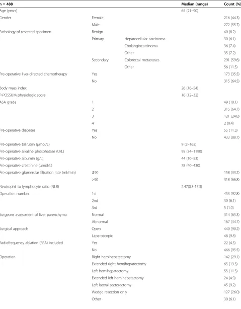
Table 1 Pre-operative and intra-operative characteristics of 488 patients undergoing liver resection
n = 488 Median (range) Count (%)
Age (years) 65 (21–90)
Gender Female 216 (44.3)
Male 272 (55.7)
Pathology of resected specimen Benign 40 (8.2)
Primary Hepatocellular carcinoma 30 (6.1)
Cholangiocarcinoma 36 (7.4)
Other 35 (7.2)
Secondary Colorectal metastases 291 (59.6)
Other 56 (11.5)
Pre-operative liver-directed chemotherapy Yes 173 (35.5)
No 315 (64.5)
Body mass index 26 (16–54)
P-POSSUM physiologic score 16 (12–32)
ASA grade 1 49 (10.1)
2 315 (64.7)
3 121 (24.8)
4 2 (0.4)
Pre-operative diabetes Yes 55 (11.3)
No 433 (88.7)
Pre-operative bilirubin (μmol/L) 9 (2–162)
Pre-operative alkaline phosphatase (U/L) 95 (34–1190)
Pre-operative albumin (g/L) 44 (10–53)
Pre-operative creatinine (μmol/L) 78 (40–430)
Pre-operative glomerular filtration rate (ml/min) ≤90 158 (33.2)
>90 318 (66.8)
Neutrophil to lymphocyte ratio (NLR) 2.47(0.3-17.3)
Operation number 1st 453 (92.8)
2nd 30 (6.1)
3rd 5 (1.0)
Surgeons assessment of liver parenchyma Normal 314 (65.3)
Abnormal 167 (34.7)
Surgical approach Open 440 (90.2)
Laparoscopic 48 (9.8)
Radiofrequency ablation (RFA) included Yes 22 (4.5)
No 466 (95.5)
Operation Right hemihepatectomy 142 (29.1)
Extended right hemihepatectomy 65 (13.3)
Left hemihepatectomy 55 (11.3)
Extended left hemihepatectomy 24 (4.9)
Left lateral sectorectomy 45 (9.2)
Wedge resection only 127 (26.0)
488. The indications for surgery, pre-operative and opera-tive details are shown in Table 1. Results of blood tests are shown in Table 2 and the main post-operative outcome measures are summarised in Table 3. The median number of biochemistry tests performed per patient in the first five post-operative days was 4 (0 to 6) and coagulation assays was 3 (0 to 6). It was not necessary to administer clotting factors to any surviving patients between postoperative days 1 to 5. Peak abnormalities in PT and bilirubin usually occurred early in the post-operative course and tended to improve over five days (Table 2). Post-operatively, 118 patients (24.1%) had a serum bilirubin≥50μmol/L. Minor abnormalities in PT were commonly noted, though only 15 patients (3.1%) developed a PT >24 s. Although a small number of patients remained jaundiced at the time of discharge, only one patient fulfilled the ‘50-50 criteria’ at day five. The median length of hospital stay was seven days (range 2 to 78) with 90% of patients having a LOS be-tween two and 15 days. Twelve patients (2.5%) died within 30 days of surgery and 23 died within 90 days of surgery (4.7%). The most common cause of death was liver failure, which occurred in 11 of 23 patients. Four patients died from ongoing malignancy (of whom three had undergone non-curative resections) and two patients died from sepsis without evidence of liver failure. The remaining deaths
were attributed to pulmonary embolus, heart failure, anas-tomotic leak following colonic resection, bleeding peptic ulcer, strangulated hernia and peritonitis.
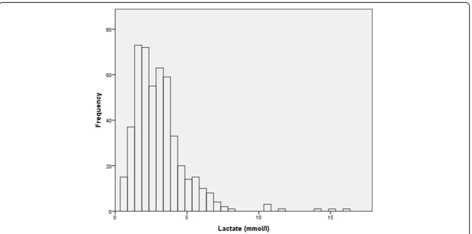
The median initial lactate concentration was 2.8 mmol/ L (inter-quartile range = 1.9 to 3.9) and 350 patients (72%) had an elevated serum lactate concentration (≥2 mmol/L) (Figure 1). There was no difference in the lactate concen-tration taken prior to abdominal closure (n = 380, median 2.8 mmol/L, range 0.6 to 16.0) or immediately on arrival in the HDU (n = 108, median 2.8 mmol/L, range 0.6 to 14.0). The initial lactate concentration was noted to be associated with all recorded outcome measures (Table 4). Although major abnormalities of serum bilirubin and PT were rare in our series there was a weak correlation with initial lactate for both bilirubin (coefficient 0.41,P< 0.001) and PT (coefficient 0.37,P< 0.001), which was stronger for bilirubin. Similarly, there was a weak correlation with length of hospital stay (coefficient 0.28,P< 0.001). Of note the values for length of hospital stay include only survi-vors, and therefore exclude some patients who are likely to have high post-operative lactate levels. Renal dys-function after liver resection was rare in this series (7.0%) but there was a correlation with lactate concen-tration (Table 4). Three of 137 patients (2.2%) with an initial lactate concentration less than 2 mmol/L who
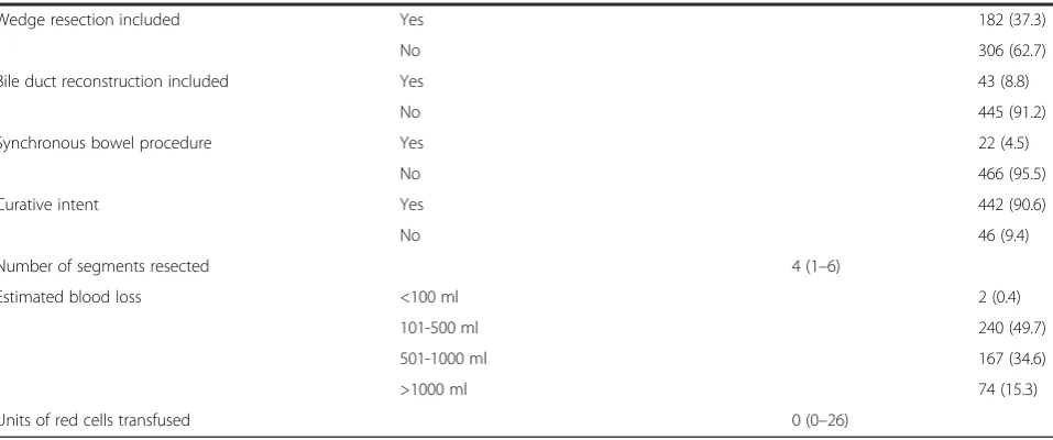
Table 1 Pre-operative and intra-operative characteristics of 488 patients undergoing liver resection(Continued)
Wedge resection included Yes 182 (37.3)
No 306 (62.7)
Bile duct reconstruction included Yes 43 (8.8)
No 445 (91.2)
Synchronous bowel procedure Yes 22 (4.5)
No 466 (95.5)
Curative intent Yes 442 (90.6)
No 46 (9.4)
Number of segments resected 4 (1–6)
Estimated blood loss <100 ml 2 (0.4)
101-500 ml 240 (49.7)
501-1000 ml 167 (34.6)
>1000 ml 74 (15.3)
Units of red cells transfused 0 (0–26)
Table 2 Post-operative blood tests for 488 patients undergoing liver resection
n = 488 POD 0 POD 1 POD 2 POD 3 POD 4 POD 5
Bilirubin Tested (%) 393 (81) 385 (79) 324 (66) 255 (52) 213 (44) 200 (41)
Median (range) 21 (5–170) 27 (6–211) 21 (4–195) 19 (3–167) 18 (4–179) 19 (1–186)
Prothrombin time Tested (%) 387 (79) 317 (65) 233 (48) 170 (35) 135 (28) 107 (22)
Median (range) 16.3 (12.2-32.4) 18.0 (12–200) 18.0 (12.6-39.4) 16.1 (11.2-37.2) 15.3 (11.6-30.6) 15.4 (12.0-26.4)
Creatinine Tested (%) 425 (87) 458 (94) 374 (77) 288 (59) 241 (49) 226 (46)
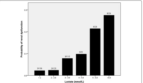
had creatinine measured developed renal dysfunction (negative predictive value (NPV) = 0.98) compared to 8 of 29 (27.5%) patients with an initial lactate greater than 6 mmol/L (positive predictive value (PPV) = 0.28) (P = < 0.001) (Figure 2). In 322 patients with a lactate concentration ≥2 and <6 mmol/L 23 developed renal dysfunction (7.1%).
Similarly, there was a correlation between mortality in the 90-day period following liver resection and initial lactate concentration (Table 4). One of 138 patients (0.7%) with an initial lactate concentration <2 mmol/L died within this period, due to an anastomotic leak following colonic resection (NPV = 0.99), compared to eight of 29 patients with initial lactate≥6 mmol/L (PPV = 0.28) (P= < 0.001) (Figure 3). The deaths in patients with lactate≥6 mmol/L
were due to liver failure in four patients, sepsis without liver failure in two patients, cardiac failure in one patient and ongoing malignancy in the other. Of the remaining 322 patients with lactate concentration≥2 and <6 mmol/L there were 14 deaths within 90 days of surgery (4.3%).
Comparison of patients with initial lactate concentra-tions <2 mmol/L and≥6 mmol/L revealed there were sig-nificantly more major resections performed (P < 0.001) and more patients with pre-operative diabetes (P< 0.001) in patients with a lactate concentration ≥6 mmol/L (Table 5). There was no significant difference in the use of pre-operative chemotherapy between these two groups (P= 0.351). The proportion of patients with both renal dysfunction and who died within 90 days was significantly higher in those with lactate concentrations ≥6 mmol/L (P< 0.001).
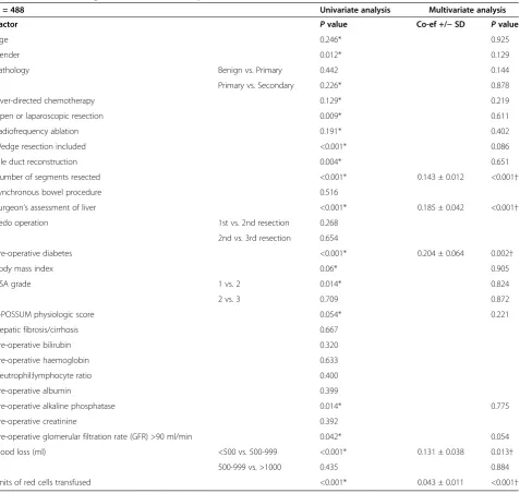
Regression analysis revealed that a pre-operative diag-nosis of diabetes mellitus, the number of liver segments resected, the operating surgeon’s assessment of the health of the liver parenchyma, the operative blood loss and num-ber of units of red cells transfused were all independently associated with initial lactate concentration at closure (Table 6). The only pre-operative factor associated with the post-operative lactate concentration was the presence of diabetes. On average, this increased the post-operative lactate concentration at any level by 20% compared to non-diabetics.
Discussion
The principal findings of this study are that higher ini-tial serum lactate concentration after liver resection is
Table 3 Post-operative outcomes for 488 patients undergoing liver resection
n = 488 Median (range) Count (%)
Peak bilirubin (μmol/L) 29 (4–445)
Peak prothrombin time (s) 17.6 (12.4-200)
Length of stay (days) 7 (2–78)
Renal dysfunction None 450 (92.2)
Risk
(>1.5x pre-operative creatinine) 17 (3.5)
Injury
(>2x pre-operative creatinine)
12 (2.5)
Failure
(>3x pre-operative creatinine) 5 (1.0)
90-day mortality 23 (4.7)
associated with an increased risk of mortality and renal and liver dysfunction. Both the 90-day mortality rate and the rate of renal dysfunction in patients with initial lactate concentrations greater than 6 mmol/L were 28% compared to those patients with initial lactate concentra-tions less than 2 mmol/L where they were 0.7% and 2.2% respectively. Similarly, higher lactate concentration was associated with higher post-operative peaks in serum bilirubin concentration and PT, as well longer lengths of hospital stay.
These findings support and extend those of an earlier study [40] by demonstrating the association of post-operative lactate with renal and hepatic dysfunction and length of hospital stay in addition to mortality. Pre-operative diabetes mellitus, the surgeon’s assessment of
the liver at laparotomy, the extent of liver resection, blood loss and the number of units of blood transfused are also shown to be associated with post-operative serum lactate concentration.
During cellular hypoxia pyruvate is diverted from the citric acid cycle and converted to lactate, reducing the amount of adenosine triphosphate (ATP) generated. This occurs in all metabolically active tissues including muscle, gut, liver, brain, erythrocytes and skin [41-43] and is exacer-bated by intra-operative stresses including blood loss [42], endogenous release of stress hormones [44] and adminis-tration of pressor agents [45]. Liver ischaemia induced by handling of the liver during surgery and temporary inflow occlusion has been shown to lead to a rise in lactate [46]. Serum lactate can also be increased by transfusion of stored blood, which contains a higher concentration of lactate than fresh blood depending on length of storage [47]. Administration of Hartmann’s solution has been shown to have a small effect on serum lactate concentration [48]. A potential weakness of this study is that details of pressor agents were not recorded, which could affect the lactate concentration. Similarly precise details regarding intravenous fluid type and volume of fluid (colloid and crystalloid) were not recorded.
In addition to being a potential source of lactate the liver is the principle location of lactate metabolism, where it is converted back to glycogen, accounting for 70% of
Table 4 Univariate analysis of the association between lactate and postoperative outcomes for 488 patients undergoing liver resection
n = 488 Co-efficient ± SD Pvalue
Peak bilirubin 0.146 ± 0.017 <0.001*
Peak prothrombin time 0.055 ± 0.002 <0.001*
Length of stay 0.046 ± 0.006 <0.001*
Renal dysfunction 0.324 ± 0.072 <0.001*
90-day mortality 0.373 ± 0.079 <0.001*
*Significant at level of P <0.05.
whole body lactate clearance [42]. No change in lactate metabolism has been demonstrated following recovery from partial hepatectomy in either rats [49] or humans [25], implying that the liver has a large functional reserve under physiological conditions of lactate production. However, the effects of intra-operative stress on hepatic glucose homeostasis have not been assessed, particularly when in combination with an extended hepatectomy. It is possible that inflow occlusion during resection and intra-operative handling of the liver lead to a temporary impairment of the ability of the liver to metabolise lactate. The finding of an association between the number of liver segments resected and the initial post-operative lactate supports this hypothesis. Diabetes is also known to be asso-ciated with impaired lactate metabolism via gluconeogenesis
[42] and may account for the strong association with post-operative lactate in this series. Furthermore, the use of metformin in non-insulin-dependent diabetes has also been shown to increase lactate concentration [50]. The rise in serum lactate at the end of liver resection therefore may be due to a failure of lactate metabolism in addition to increased production during surgery.
Significantly, the use of pre-operative chemotherapy was not shown to be associated with elevation of post-operative lactate. This may be due to a policy of allowing a period of recovery after completion of pre-operative chemotherapy before undertaking surgery. Interestingly, the operating surgeon’s assessment of the liver parenchyma was associ-ated with the post-operative lactate concentration. This finding suggests that patient co-morbidity was a more common cause of abnormal liver parenchyma than the use of liver-directed chemotherapy.
An important observation of this study is the relative rarity of major hepatic dysfunction following liver resection in this series with only one patient fulfilling the‘50-50’ criteria [2], who subsequently recovered. Despite the infre-quency of major disturbances of post-operative bilirubin and PT, there was an independent association with increas-ing concentration of post-operative lactate, demonstratincreas-ing that even a minor degree of liver injury can lead to im-paired lactate clearance or increase its production.
Renal dysfunction was also rare in this series, affecting 34 patients (7%) compared to 15% in a similar series
Figure 3Probability of 90-day mortality after liver resection according to lactate concentration in 488 patients.
Table 5 Distribution of risk factors and outcomes in 138 patients with lactate <2 mmol/L and 29 patients with lactate≥6 mmol/L undergoing liver resection
Lactate <2 mmol/L (n = 138) ≥
6 mmol/L (n = 29) P
value
Major resection (%) 26 (18.8) 26 (89.7) <0.001*
Pre-operative chemotherapy (%) 38 (27.5) 5 (17.2) 0.351
Pre-operative diabetes (%) 6 (4.3) 8 (27.6) <0.001*
Post-operative renal dysfunction (%) 3 (2.2) 8 (27.6) <0.001*
90-day mortality (%) 1 (0.7) 8 (27.6) <0.001*
[51]. The risk factors for post-operative renal dysfunction are likely to be similar to those in other forms of abdom-inal surgery, including blood loss and sepsis, which are also initiating factors for anaerobic metabolism and lactate production. This supports the value of initial lactate as an early predictor of renal dysfunction. Of note, the risk of renal dysfunction appeared to rise more rapidly when the post-operative lactate rose above 5 mmol/L (Figure 2). This suggests that the kidneys are able to tolerate a degree of oxidative stress to a threshold level beyond which the risk of damage rises rapidly.
There was a weak association between initial lactate concentration and length of hospital stay in the study
(Table 4). However, this may also be affected by other factors such as post-operative complications, particularly bile leaks, and degree of social support.
The strongest association demonstrated was between lactate concentration and the risk of mortality. In a similar manner to renal dysfunction, there seems to be a threshold level of post-operative lactate of approximately 6 mmol/l above which the risk of 90-day mortality rises rapidly (Figure 3). Organ dysfunction was a major contributor to mortality in the series and initial lactate concentration is a valuable global marker of poor organ function in the early post-operative period, including cardiovascular, renal and hepatic dysfunction.
Table 6 Univariate and multivariate analysis of pre- and intra-operative factors associated with serum lactate concentration following liver resection in 488 patients
N = 488 Univariate analysis Multivariate analysis
Factor Pvalue Co-ef +/−SD Pvalue
Age 0.246* 0.925
Gender 0.012* 0.129
Pathology Benign vs. Primary 0.442 0.144
Primary vs. Secondary 0.226* 0.878
Liver-directed chemotherapy 0.129* 0.219
Open or laparoscopic resection 0.009* 0.611
Radiofrequency ablation 0.191* 0.402
Wedge resection included <0.001* 0.086
Bile duct reconstruction 0.004* 0.651
Number of segments resected <0.001* 0.143 ± 0.012 <0.001†
Synchronous bowel procedure 0.516
Surgeon’s assessment of liver <0.001* 0.185 ± 0.042 <0.001†
Redo operation 1st vs. 2nd resection 0.268
2nd vs. 3rd resection 0.654
Pre-operative diabetes <0.001* 0.204 ± 0.064 0.002†
Body mass index 0.06* 0.905
ASA grade 1 vs. 2 0.014* 0.824
2 vs. 3 0.709 0.872
P-POSSUM physiologic score 0.054* 0.221
Hepatic fibrosis/cirrhosis 0.667
Pre-operative bilirubin 0.320
Pre-operative haemoglobin 0.633
Neutrophil:lymphocyte ratio 0.400
Pre-operative albumin 0.399
Pre-operative alkaline phosphatase 0.014* 0.775
Pre-operative creatinine 0.392
Pre-operative glomerular filtration rate (GFR) >90 ml/min 0.042* 0.054
Blood loss (ml) <500 vs. 500-999 <0.001* 0.131 ± 0.038 0.013†
500-999 vs. >1000 0.435 0.884
Units of red cells transfused <0.001* 0.043 ± 0.011 <0.001†
Conclusions
These findings are of value in clinical practice as it may be possible to use the initial post-operative lactate concentration to determine the patient pathway in the early operative period. Patients with an initial post-operative lactate of less than 2 mmol/L have low rates of mortality and organ dysfunction and we are currently evaluating this criterion as a determinant of the need for post-operative critical care. In addition the correlation of post-operative lactate with subsequent organ dysfunction and mortality may allow its use as a single measure of the impact of innovations in operative technique or peri-operative care.
Abbreviations
ASA:American society of anesthesiologists; ATP: Adenosine triphosphate; CUSA: Cavitron ultrasonic surgical aspirator; CVP: Central venous pressure; HDU: High dependency unit; LOS: Length of stay; NLR: Neutrophil to lymphocyte ratio; NPV: Negative predictive value; PHLF: Post-hepatectomy liver failure; P-POSSUM: Portsmouth physiologic and operative severity score for the enUmeration of mortality and morbidity; PPV: Predictive value; PT: Prothrombin time; REC: Research ethics committee; RIFLE: Risk, injury, failure, loss, and end-stage kidney disease.
Competing interests
The authors declare that they have no competing interests.
Authors’contributions
MW and TS designed the study, collected data, analysed the data and drafted the manuscript. GS participated in the design of the study and performed the statistical analysis. TW, DB, PE, IA, SA and MB participated in the design of the study, collected data and drafted the manuscript. DS conceived the study, supervised its design and coordination and helped to draft the manuscript. All authors read and approved the final manuscript.
Authors’information
M.G. Wiggans and T. Starkie: joint first authors.
Acknowledgements
Thanks to Dr. C. Seavell, Dr. P. Davis and Dr. D. Lunn (Consultant Anaesthetists) for their work in study design and collecting lactate data.
Author details
1
Hepatobiliary Surgery, Plymouth Hospitals NHS Trust, Derriford Hospital, Derriford Road, Plymouth, Devon PL6 8DH, UK.2Peninsula College of
Medicine and Dentistry, University of Exeter and Plymouth University, Research Way, Plymouth, Devon PL6 8BU, UK.3Department of Anaesthetics,
Plymouth Hospitals NHS Trust, Derriford Hospital, Derriford Road, Plymouth, Devon PL6 8DH, UK.4Centre for Health Statistics, Tamar Science Park, Davy
Road, Plymouth, Devon PL6 8BX, UK.
Received: 5 March 2013 Accepted: 19 September 2013 Published: 7 October 2013
References
1. Mann CD, Palser T, Briggs CD, Cameron I, Rees M, Buckles J, Berry DP:A review of factors predicting perioperative death and early outcome in hepatopancreaticobiliary cancer surgery.HPB2010,12:380–388. 2. Balzan S, Belghiti J, Farges O, Ogata S, Sauvanet A, Delefosse D, Durand F:
The“50-50 criteria”on postoperative day 5: an accurate predictor of liver failure and death after hepatectomy.Ann Surg2005,242:824–829. 3. Schreckenbach T, Liese J, Bechstein WO, Moench C:Posthepatectomy liver
failure.Dig Surg2012,29:79–85.
4. Saner F:Kidney failure following liver resection.Transplant Proc2008,
40:1221–1224.
5. Savage AP, Malt MD:Elective and emergency hepatic resection determinants of operative mortality and morbidity.Ann Surg1991,
214:689–695.
6. Koch M, Garden OJ, Padbury R, Rahbari NN, Adam R, Capussotti L, Fan ST, Yokoyama Y, Crawford M, Makuuchi M, Christophi C, Banting S, Brooke-Smith M, Usatoff V, Nagino M, Maddern G, Hugh TJ, Vauthey J-N, Greig P, Rees M, Nimura Y, Figueras J, DeMatteo RP, Büchler MW, Weitz J:Bile leakage after hepatobiliary and pancreatic surgery: a definition and grading of severity by the International Study Group of Liver Surgery.
Surgery2011,149:680–688.
7. Alfieri S, Carrierio C, Caprino P, Di Giorgio A, Sgadari A, Crucitti F, Doglietto G:Avoiding early postoperative complications in liver surgery. A multivariate analysis of 254 patients consecutively observed.Dig Liv Dis
2001,33:341–346.
8. Jarnagin WR, Gonen M, Fong Y, DeMatteo RP, Ben-Porat L, Little S, Corvera C, Weber S, Blumgart LH:Improvement in perioperative outcome after hepatic resection: analysis of 1,803 consecutive cases over the past decade.Ann Surg2002,236:397–406.
9. Dixon E, Schneeweiss S, Pasieka JL:Mortality following liver resection in US medicare patients: does the presence of a liver transplant program affect outcome?J Surg Oncol2007,95:194–200.
10. Melendez J, Ferri E, Zwillman M, Fischer M, DeMatteo R, Leung D, Jarnagin W, Fong Y, Blumgart LH:Extended hepatic resection: a 6-year retrospective study of risk factors for perioperative mortality.J Am Coll Surg2001,192:47–53.
11. Dimick J, Cowan J, Knol J, Upchurch G:Hepatic resection in the United States: indications, outcomes, and hospital procedural volumes From a nationally representative database.Arch Surg2003,138:185–191. 12. Shimada M, Matsumata T, Akazawa K, Kamakura T, Itasaka H, Sugimachi K,
Nose Y:Estimation of risk of major complications after hepatic resection.
Am J Surg1994,167:399–403.
13. Shimada M, Takenaka K, Fujiwara Y, Gion T, Shirabe K, Yanaga K, Sugimachi K:Risk factors linked to postoperative morbidity in patients with hepatocellular carcinoma.Br J Surg1998,85:195–198.
14. Halazun KJ, Aldoori A, Malik HZ, Al-Mukhtar A, Prasad KR, Toogood GJ, Lodge JPA:Elevated preoperative neutrophil to lymphocyte ratio predicts survival following hepatic resection for colorectal liver metastases.Eur J Surg Oncol2008,34:55–60.
15. Poon RT, Fan ST, Lo CM, Liu CL, Lam CM, Yuen WK, Yeung C, Wong J:
Improving perioperative outcome expands the role of hepatectomy in management of benign and malignant hepatobiliary diseases: analysis of 1222 consecutive patients from a prospective database.Ann Surg
2004,240:698–708. discussion 708–710.
16. Benzoni E, Cojutti A, Lorenzin D, Adani GL, Baccarani U, Favero A, Zompicchiati A, Bresadola F, Uzzau A:Liver resective surgery: a multivariate analysis of postoperative outcome and complication.
Langenbecks Arch Surg2007,392:45–54.
17. Bolder U, Brune A, Schmidt S, Tacke J, Jauch KW, Löhlein D:Preoperative assessment of mortality risk in hepatic resection by clinical variables: a multivariate analysis.Liver Transpl Surg1999,5:227–237.
18. Redaelli CA, Dufour J-F, Wagner M, Schilling M, Hüsler J, Krähenbühl L, Büchler MW, Reichen J:Preoperative galactose elimination capacity predicts complications and survival after hepatic resection.Ann Surg
2002,235:77–85.
19. Karoui M, Penna C, Amin-Hashem M, Mitry E, Benoist S, Franc B, Rougier P, Nordlinger B:Influence of preoperative chemotherapy on the risk of major hepatectomy for colorectal liver metastases.Ann Surg2006,
243:1–7.
20. Benzoni E, Lorenzin D, Baccarani U, Adani GL, Favero A, Cojutti A, Bresadola F, Uzzau A:Resective surgery for liver tumor: a multivariate analysis of causes and risk factors linked to postoperative complications.
Hepatobiliary Pancreat Dis Int2006,5:526–533.
21. Copeland GP, Jones D, Walters M:POSSUM: a scoring system for surgical audit.Br J Surg1991,78:355–360.
22. Whiteley M, Prytherch D, Higgins B, Weaver P, Prout W:An evaluation of the POSSUM surgical scoring system.Br J Surg1996,83:812–815. 23. Lam CM, Fan ST, Yuan AW, Law WL, Poon K:Validation of POSSUM scoring
systems for audit of major hepatectomy.Br J Surg2004,91:450–454. 24. Van den Broek MAJ, OldeDamink SWM, Dejong CHC, Lang H, Malagó M,
25. Chioléro R, Tappy L, Gillet M, Revelly JP, Roth H, Cayeux C, Schneiter P, Leverve X:Effect of major hepatectomy on glucose and lactate metabolism.Ann Surg1999,229:505–513.
26. Husain FA, Martin MJ, Mullenix PS, Steele SR, Elliott DC:Serum lactate and base deficit as predictors of mortality and morbidity.Am J Surg2003,
185:485–491.
27. Khosravani H, Shahpori R, Stelfox HT, Kirkpatrick AW, Laupland KB:
Occurrence and adverse effect on outcome of hyperlactatemia in the critically ill.Crit Care2009,13:R90.
28. Macquillan GC, Seyam MS, Nightingale P, Neuberger JM, Murphy N:Blood lactate but not serum phosphate levels can predict patient outcome in fulminant hepatic failure.Liver Transpl2005,11:1073–1079.
29. Bernardin G, Pradier C, Tiger F, Deloffre P, Mattei M:Blood pressure and arterial lactate level are early indicators of short-term survival in human septic shock.Intensive Care Med1996,22:17–25.
30. Manikis P, Jankowski S, Zhang H, Kahn RJ, Vincent JL:Correlation of serial blood lactate levels to organ failure and mortality after trauma.
Am J Emerg Med1995,13:619–622.
31. Gruttadauria S, Marino IR, Vitale CH, Mandala L, Scott VL, Doria C:
Correlation between peri-operative serum lactate levels and outcome in pancreatic resection for pancreatic cancer, preliminary report.J Exp Clin Cancer Res2002,21:539–545.
32. Li SH, Liu F, Zhang YT:[Initial serum lactate level as predictor of morbidity after major abdominal surgery].Zhonghua Yi Xue Za Zhi2008,
88:2470–2473.
33. Murtuza B, Wall D, Reinhardt Z, Stickley J, Stumper O, Jones TJ, Barron DJ, Brawn WJ:The importance of blood lactate clearance as a predictor of early mortality following the modified Norwood procedure.Eur J Cardiothorac Surg2011,40:1207–1214.
34. Nishimura A, Hakamada K, Narumi S, Totsuka E, Toyoki Y, Ishizawa Y, Umehara M, Yoshida A, Umehara Y, Sasaki M:Intraoperative blood lactate level as an early predictor of initial graft function in human living donor liver transplantation.Transplant Proc2004,36:2246–2248.
35. IHPBA:The Brisbane 2000 terminology of hepatic anatomy and resections.HPB2000,2:333–339.
36. Mizock BA:Lactic acidosis.Dis Mon1989,35:233–300. 37. Bellomo R, Ronco C, Kellum JA, Mehta RL, Palevsky P:Acute renal
failure - definition, outcome measures, animal models, fluid therapy and information technology needs: the second international consensus conference of the Acute Dialysis Quality Initiative (ADQI) group.Crit Care
2004,8:R204–R212.
38. Agresti A:An Introduction to Categorical Data Analysis Second Edition.
Hoboken, New Jersey: John Wiley & Sons; 2002.
39. R Foundation for Statistical Computing.http://www.r-project.org/. 40. Watanabe I, Mayumi T, Arishima T, Takahashi H, Shikano T, Nakao A, Nagino
M, Nimura Y, Takezawa J:Hyperlactemia can predict the prognosis of liver resection.Shock2007,28:35–38.
41. Handy J:Lactate - the bad boy of metabolism, or simply misunderstood?
Curr Anaesth Crit Care2006,17:71–76.
42. Phypers B, Pierce JM:Lactate physiology in health and disease.Contin Educ Anaesth Crit Care Pain2006,6:128–132.
43. Theodoraki K, Arkadopoulos N, Fragulidis G, Voros D, Karapanos K, Markatou M, Kostopanagiotou G, Smyrniotis V:Transhepatic lactate gradient in relation to liver ischemia/reperfusion injury during major hepatectomies.
Liver Transpl2006,12:1825–1831.
44. James JH, Luchette FA, McCarter FD, Fischer JE:Lactate is an unreliable indicator of tissue hypoxia in injury or sepsis.Lancet1999,354:505–508. 45. Luchette FA, Jenkins WA, Friend LA, Su C, Fischer JE, James JH:Hypoxia is not the sole cause of lactate production during shock.J Trauma2002,
52:415–419.
46. Pietsch UC, Herrmann ML, Uhlmann D, Busch T, Hokema F, Kaisers UX, Schaffranietz L:Blood lactate and pyruvate levels in the perioperative period of liver resection with Pringle manoeuver.Clin Hemorheol Microcirc
2010,44:269–281.
47. Uvizl R, Klementa B, Adamus M, Neiser J:Biochemical changes in the patient’s plasma after red blood cell transfusion.Signa Vitae2011,
6:64–71.
48. Shin WJ, Kim YK, Bang JY, Cho SK, Han SM, Hwang GS:Lactate and liver function tests after living donor right hepatectomy: a comparison of solutions with and without lactate.Acta Anaesthesiol Scand2011,
55:558–564.
49. Petenusci SO, Freitas TC, Roselino ES, Migliorini RH:Glucose homeostasis during the early stages of liver regeneration in fasted rats.Can J Physiol Pharmacol1983,61:222–228.
50. Davis TM, Jackson D, Davis WA, Bruce DG, Chubb P:The relationship between metformin therapy and the fasting plasma lactate in type 2 diabetes: The Fremantle Diabetes Study.Br J Clin Pharmacol2001,
52:137–144.
51. Slankamenac K, Breitenstein S, Held U, Beck-Schimmer B, Puhan MA, Clavien P-A:Development and validation of a prediction score for postoperative acute renal failure following liver resection.Ann Surg2009,250:720–728.
doi:10.1186/2047-0525-2-21
Cite this article as:Wigganset al.:Serum arterial lactate concentration predicts mortality and organ dysfunction following liver resection.
Perioperative Medicine20132:21.
Submit your next manuscript to BioMed Central and take full advantage of:
• Convenient online submission
• Thorough peer review
• No space constraints or color figure charges
• Immediate publication on acceptance
• Inclusion in PubMed, CAS, Scopus and Google Scholar
• Research which is freely available for redistribution