R E S E A R C H A R T I C L E
Open Access
Investigating the contribution of IL-17A and
IL-17F to the host response during
Escherichia
coli
mastitis
Perrine Roussel
1,2, Patricia Cunha
1,2, Adeline Porcherie
1,2, Wolfram Petzl
3, Florence B Gilbert
1,2, Céline Riollet
1,2,
Holm Zerbe
3, Pascal Rainard
1,2and Pierre Germon
1,2*Abstract
Mastitis remains a major disease of cattle with a strong impact on the dairy industry. There is a growing interest in understanding how cell mediated immunity contributes to the defence of the mammary gland against invading mastitis causing bacteria. Cytokines belonging to the IL-17 family, and the cells that produce them, have been described as important modulators of the innate immunity, in particular that of epithelial cells. We report here that expression of IL-17A and IL-17F genes, encoding two members of the IL-17 family, are induced in udder tissues of cows experimentally infected withEscherichia coli. The impact of IL-17A on the innate response of bovine mammary epithelial cells was investigated using a newly isolated cell line, the PS cell line. We first showed that PS cells, similar to primary bovine mammary epithelial cells, were able to respond to agonists of TLR2 and to LPS, provided CD14 was added to the culture medium. We then showed that secretion of CXCL8 and transcription of innate immunity related-genes by PS cells were increased by IL-17A, in particular when these cells were stimulated with liveE. coli bacteria. Together with data from the literature, these results support the hypothesis that IL-17A and IL-17 F could play an important role in mediating of host-pathogen interactions during mastitis.
Introduction
Despite decades of research, mastitis remains a major concern in dairy farming. Mastitis are mainly due to bacterial infections (Gram-positive pathogens such as
Staphylococcus aureus and Streptococcus uberis, or Gram-negative pathogens such asEscherichia coli) [1].
Previous studies have allowed the identification of key molecular events that ultimately lead to the re-cruitment of neutrophils in the mammary gland upon bacterial colonization. When bacteria enter the udder, the host responds by secreting chemokines (such as CXCL8, CCL20) and other immune components such as antimicrobial peptides (LAP—lingual antimicrobial peptide, TAP—tracheal antimicrobial peptide) and pro-inflammatory cytokines (TNF-α, IL-1β) [2-4]. Production of pro-inflammatory cytokines such as IL-1β, IL-6 and TNF-αand the chemokine CXCL8 have been detected in milk from clinically affected animals and are supposed to contribute to the inflammation observed in mastitis [2,4].
In vivo experiments indicate that the severity ofE. coli
mastitis mainly depends on host factors and that a quick and efficient response is important for an efficient clear-ance of the bacteria [5]. This process relies heavily on the recruitment of neutrophils during infection: a delay in the recruitment of neutrophils aggravates the infec-tion [6,7]. It is therefore expected that any mechanism that modulates the immune response of the host could participate in the defence againstE. colimastitis.
IL-17A and IL-17F are two cytokines that have been described as playing a significant role in the recuitment of neutrophils in other inflammatory diseases. Along with four other structurally related cytokines, IL-17B, IL-17C, IL-17D and IL-17E, they form the IL-17 family [8].
Although expression of IL-17A and IL-17F may be detrimental to the host, in particular in the case of autoimmune diseases, they have been shown to be beneficial to the host to fight against bacterial pathogens such asCitrobacter rodentium,Klebsiella pneumoniae or
Escherichia coli[8,9].
Production of IL-17A duringS. uberismastitis was re-cently demonstrated [10]. Tao and Mallard also reported that IL-17A gene expression was slightly increased
* Correspondence:pierre.germon@tours.inra.fr
1INRA, UMR1282, Infectiologie et Santé Publique, F-37380 Nouzilly, France
2
Université François Rabelais de Tours, UMR1282, Infectiologie et Santé Publique, F-37000 Tours, France
Full list of author information is available at the end of the article
(approx. 1.5-fold in milk) in somatic cells fromS. aureus
infected cows [11]. Microarray analyses of MEC stimu-lated with S. aureus culture supernatant also showed induction of the IL-17A pro-inflammatory pathway [12].
In addition, we recently demonstrated that, in vitro, IL-17A increases the ability of mammary epithelial cells (MEC) to respond to agonists similar to that produced by S. aureus [13]. These cells are thought to play a sig-nificant role in the defence against invading pathogens by producing antimicrobial peptides as well as cytokines and chemokines such as CXCL8 and IL-6. Indeed, in vitro grown primary bovine MEC (pbMEC) have been shown to respond to the presence of bacteria, such asE. coliorS. aureus, or to different purified bacterial agonists by pro-ducing different cytokines, chemokines or antimicrobial peptides [12,14-18].
Based on these data, one could hypothesize that IL-17 cytokines contribute to the innate response of the host duringE. colimastitis; but this remains to be studied.
In the present report, we thus decided to investigate, under controlled conditions, whether expression of genes encoding cytokines of the IL-17 family was induced upon intra-mammary infection of cows by E. coli. We then explored how this over-expression of IL-17A gene could translate in terms of defence against the invading patho-gen. Using a new cell line, called the PS cell line, which we characterized in details in terms of phenotypic markers and response to bacterial agonists, we analysed the influ-ence of IL-17A on the innate immune response of MEC to infection byE. coli.
This report brings a new perspective on the cellular features and molecular parameters involved in the ability of MEC to take part in the udder defence against mastitis.
Materials and methods Ethics statement
Experimental infection of animals were conducted at the Clinic for Ruminants (Munich, Germany) with the ap-proval of the ethics committee of the regional government of upper Bavaria, Germany (No. 55.2-1-54-2531-108-05).
Experimental infection of cows
Five healthy German Holstein Frisian first lactation cows in mid lactation (3-6 months after parturition) fulfilling criteria as previously described [19] were inoculated in one quarter with 500 cfuE. colistrain 1303. Five heifers that received no treatment served as untreated controls. Only animals without previous diagnosis of clinical or subclinical mastitis and a reported somatic cell count <50 000/mL were included in the study. Quarter milk samples were collected and tested weekly before the trial to ensure that they contained <50 000 somatic cells/mL and were free of mastitis pathogens. Animals were randomly assigned to both groups and the experiments
were carried out between March and December. All E. coliinoculated animals developed clinical mastitis in the affected quarter 12 h after inoculation as previously de-scribed [19]. Animals were slaughtered 24 hours post-inoculation (hpi). Liquid nitrogen snap frozen udder samples of lobulo-alveolar tissue 7 cm dorsal of the milk cistern were obtained immediately after slaughtering. RNA was isolated from approx. 100 mg of frozen udder tissue using Trizol (Invitrogen). The sample was placed in a 2 mL tube containing 1.4 mm beads (MP Biomedi-cals) and one mL of Trizol was added. Tissue lysis was obtained by shaking the tubes twice in a FastPrep apparatus (MP Biomedicals) for 45 s at speed 6. The homogenate was further processed as recommended by the manufacturer. RNA quality was checked using an Agilent Bioanalyzer and only samples with a RNA Integrity number above 7 were used. Controls included RNA samples from the uninoculated quarters from inoculated cows as well as samples from quarters of non-inoculated cows.
Isolation and culture of PS cells
3 days of culture, epithelial cells were enriched by selective trypsinization (0.25% trysin-EDTA). Detached cells after 5 min of trypsinization were discarded and fresh medium was added on the cells that remained attached to the plastic of the culture flasks. This selective trypsinization was re-peated on day 6 and a first passage was performed two days later. Cells were then frozen under nitrogen after 2 pas-sages. The PS cell line was obtained by selective trypsiniza-tion at passage 4. The cells were subjected to 2 additrypsiniza-tional passages and frozen.
Cells were thereafter maintained in MEC growth medium (GM): Advanced-DMEM/F12 (Gibco) contain-ing 20 mM HEPES, 2 mM L-glutamine (Gibco), 1 μg/ mL hydrocortisone (Sigma-Aldrich), 10 ng/mL Insulin-like growth factor (IGF)-1, 5 ng/mL Fibroblast growth factor (FGF), 5 ng/mL Epidermal growth factor (EGF).
Cytology of the PS cell line
In order to visualize the morphological features of the PS cell line in culture, a May-Grünwald Giemsa staining was performed on cells grown on 6-well plate. Cells were covered with May-Grünwald dye for 3 min. The same volume of distilled water was added for 1 min. After draining, Giemsa dye (8% in distilled water) was incubated for 20 min and the cells were washed with tap water.
Flow cytometry analysis of PS cells
Antibodies used in the present study are described in Additional file 1. Labellings were done on PS cells at passages 9, 14 and 18. Intracellular labelling for cytoker-atins expression study was performed when cells formed a confluent monolayer. Cells were harvested after trypsin treatment (0.05% trysin-EDTA -Gibco), and the cell pel-let was resuspended in 500μL of permeabilization buffer Cytofix Cytoperm (BD Biosciences), and incubated in the dark at 4 °C. After 20 min, the cell suspension was washed with 1 mL of Perm Wash 1X buffer (BD Biosci-ences). The cells were collected, resuspended in Perm Wash 1X buffer, and were split into each well (3 × 10⁵/ well in 20 μL) of a 96-well U-bottom well plate. The cells were then incubated with 20 μL of primary anti-body for 15 min at 4 °C. The plate was washed with 100μL of Perm Wash buffer, and the cell pellet was re-suspended in 50 μL of the secondary antibody for 15 min at 4 °C when required.
Extracellular labelling was performed when MEC formed a confluent monolayer. Cells were harvested and distributed (20 μL, 3 × 10⁵ cells/well) into 96-well U-bottom well plates. The cell suspension was resuspended in 20 μL of antibodies against CD45, CD14 and CD282 (TLR2), and incubated for 15 min at 4 °C. The cells were then washed with 100 μL of buffer (DPBS without Ca and Mg, Lonza; 2% goat serum, Gibco; 2 mM EDTA). The plate was centrifuged for 10 min at 200g, and, if
necessary, the cell pellet was resuspended in 50 μL of secondary antibodies, and incubated for 15 min at 4 °C. Then the cells were washed and fixed with 150 μL of FACS lysing solution (BD Biosciences). Peripheral blood mononuclear cells (PBMC) isolated from bovine blood were used as positive control for CD45 labelling. Fluorescence-activated cell sorting (FACS) analysis was performed on a FACSCalibur cytometer using CellQuest software (BD Biosciences) and data were analyzed with FlowJo software (Tree Star, Ashland, OR, USA).
Stimulation of cells with live bacteria or purified bacterial agonists
l’Orfrasière (INRA Tours). IL-17A was used as described previously at a concentration of 100 ng/mL [13]. Primary cells were isolated and grown as previously described and used at their third passage [16,21,22].
ELISA assays
CXCL8, IL-17A and TNFα production by PS cells was quantified by sandwich ELISA, with reference to stand-ard as described previously [23,24]. IL-1βwas quantified by ELISA using the Bovine IL-1 beta ELISA Reagent Kit (Thermo Scientific).
Transcript analysis by RT-PCR and RT-qPCR
Total RNA (1 μg) was reverse transcribed to cDNA using random hexamers and SuperScript RT III (Invitro-gen) according to manufacturer’s instructions. Primers used in this study are listed in Additional file 2 and [13]. Expression of PRRs was analyzed by RT-PCR using 40 amplification cycles. Relative quantities of transcripts of interest were analyzed by RT-qPCR as described previously [13]. Briefly, after normalization using the expression of 3 reference genes (Actb, PPIA, 18S RNA), expression of each gene was calculated relative to the values obtained from unstimulated cells arbitrarily set to 1.
Statistical analysis
In vivo gene expression results were analysed by a per-mutation test using the StaXact software (v5.0—Cytel software) to obtain exactp-values. For the analysis of re-sponses of MEC cells to the different bacterial agonists or to live bacteria, data were first compared using the Kruskal and Wallis test followed by exact permutation tests using StatXact. A p-value < 0.05 was considered significant.
Results
IL-17A and IL-17 F gene expression is induced upon infection of the udder byE. coli
Previous reports showed that experimental intra-mammary inoculation ofEscherichia coli1303 induced a significant host response [25]. Based on microarray ana-lyses, expression of cytokines and chemokines such as CCL20 and CXCL8 was shown to be increased uponE. coli 1303 infection 514 and 58-fold, respectively [15]. Our RT-qPCR data confirmed that, upon infection with
E. coli 1303, expressions of CCL20 and CXCL8 genes were induced, approx. 16 000-fold and 108-fold, respect-ively (Figures 1A and B). A higher expression, compared to uninfected cows, was also detected, particularly with CCL20, in uninfected quarters of three out of fiveE. coli
infected cows which is consistent with a systemic effect as previously reported [19,26].
Microarray experiments had failed to detect a signifi-cant expression of IL-17 family genes in udder tissue
after E. coli infection, presumably because of a low ex-pression of these genes [15]. In the present study, we thus used RT-qPCR to evaluate the expression of IL-17 family genes. No expression was detected for IL-17C and IL-17E (data not shown). On the contrary, expres-sion of IL-17A and IL-17F was significantly increased in
E. coliinfected quarters, compared to non-infected quar-ters (Figures 1C and D): the mean fold-change for IL-17A was 72 in samples from cows infected by E. coli. Mean induction of IL-17F was 109-fold in the same samples (Figures 1C and D). Although induction of 17A gene was clearly observed in our tissue samples, IL-17A levels in milk samples from E. coli infected cows were below the detection limit of the ELISA assay used, that is below 200 pg/mL in milk.
Despite this lack of detection of IL-17A in milk from infected quarters, gene expression results suggest that IL-17A and IL-17F may play a role in the immune re-sponse of cows to intra-mammary infection, either at the onset of or during the inflammatory response.
Considering that IL-17A has generally more potent effects than IL-17F on epithelial cells [13], we then ex-plored if IL-17A in particular could contribute to the de-fence of the host against E. coliinfection by modulating the innate immune response of MEC. Indeed, these cells are key players in mounting an efficient early innate im-mune response and IL17-A has been shown to target a variety of cell types, in particular epithelial cells [27-29].
PS cells as a useful tool to study the innate immune response of mammary epithelial cells
Although pbMEC are often used to study the response of MEC to infection, the use of primary epithelial cell cultures has a number of drawbacks. Most importantly, variability of response of pbMEC from different animals on the one hand and lack of reproducibility of experi-ments on the other hand, can be associated with con-tamination of primary cell cultures with other cell types, and the amount of these contaminating cells can vary from one primary culture to another. Considering that these contaminating cells could exacerbate or lower the innate immune response of MEC through cooperation, we decided to use a new mammary epithelial cell line showing innate immune response capabilities to perform these assays.
This cell line, which we called the PS cell line, stems from spontaneously immortalized cells that emerged from a culture of primary bovine MEC isolated from the
surface markers associated with mammary epithelial cells. Results indicated a percentage of CD45-positive cells similar to background level (0.6%) and expression of cytokeratin 18 by more than 99.5% of cells. Cytokeratin 14 was only weakly expressed by PS cells (Figures 2A-F).
RT-PCR data indicated that these cells expressed several genes encoding Pattern Recognition Receptors (PRR) such as TLR1, TLR2, TLR4, TLR6, NOD1 and
In order to use these cells to study the innate immune response, we analysed their capacity to respond to stim-uli such as lipopolysaccharide (LPS), lipoteichoic acid (LTA), lipoproteins or peptidoglycan degradation products. The synthetic molecules Pam2-CSK4 and Pam3-CSK4 are analogs of di-acylated and tri-acylated lipoproteins that signal through the TLR2/TLR6 or TLR1/TLR2
expression is induced upon stimulation of mammary epi-thelial cells [15], and induced also the secretion of CXCL8 (Figure 3E). PS cells also responded to Pam2-CSK4 when used at 10 ng/mL (data not shown). A significant but lower response was observed with Pam3-CSK4, both in terms of increased expression of CCL20, CXCL8, TAP and LAP and increased secretion of CXCL8 (Figure 3). No re-sponse was observed with both MDP and C12-iE-DAP (Figure 3). In a separate experiment, a concentration of flagellin of 100 ng/mL only induced a weak increase in TAP expression (3-fold) but failed to induce significant se-cretion of CXCL8 and increased expression of CCL20
production also occurred when the LPS stimulation was performed in fresh milk (Figure 3). This is in keeping with the occurrence of sCD14 in serum and bovine milk [30].
Compared to the response of primary cells to these same agonists, PS cells responded to a similar array of bacterial compounds, although their response was some-what lower than that of pbMEC (Additional file 4). Fur-thermore, stimulation experiments performed at different passages indicated that the PS cell line is stable at least up to passage 24 (Additional file 4).
PS cells could thus be used as a surrogate to pbMEC to monitor the innate response of mammary epithelial cells and, in the present report, how this response can be modulated by IL-17A.
IL-17A increases the response of PS cells to Pam3-CSK4
In a first attempt to investigate how IL-17A could modu-late the response of MEC, we analysed the response of PS cells to purified bacterial agonists in the presence or not of IL-17A. We selected LPS and Pam3-CSK4, activating TLR4 and TLR1/2, respectively, as they are likely to repre-sent the most relevant agonists in vivo through whichE. colistimulates MEC and they induce a strong innate re-sponse in pbMEC [16] and PS cells (see Figure 3).
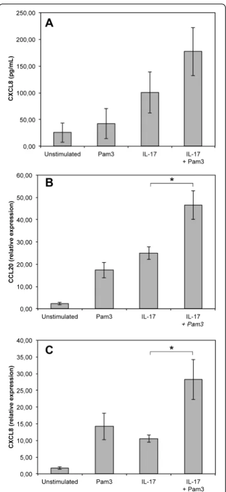
When PS cells were stimulated with both Pam3-CSK4 and IL-17 for 5 hours, the response in terms of CCL20 and CXCL8 gene expression was significantly increased compared to the response observed only in the presence of IL-17A or Pam3-CSK4 (Figure 4). The increase in terms of CXCL8 secretion, although significant statisti-cally, was less important. The impact of IL-17A on the response of PS cells to LPS was also statistically signifi-cant but of lower magnitude. A clear response of PS cells to LPS was observed in the presence of CD14 with high level of secretion of CXCL8 and strong induction of CCL20 and CXCL8 gene expression (Figure 5). Secretion of CXCL8 was increased 1.5 fold when IL-17 was present along with LPS and CD14. In the same conditions, IL-17A induced only a small increase of CCL20 gene expres-sion while no significant impact on CXCL8 gene expression was observed. Interestingly, although PS cells responded very weakly to LPS in the absence of CD14, we could observe that IL-17 increased slightly the re-sponse of PS cells when they were stimulated with LPS alone (Figure 5).
IL-17A increases the response of MEC to liveE. coli
We next analysed if IL-17A could also increase the re-sponse of MEC to live bacteria. We used a protocol pre-viously described which allowed stimulation of cells with live bacteria while avoiding a change in pH of the cul-ture medium due to bacterial outgrowth [21]: briefly, cells were incubated in the presence of bacteria for three hours. Bacteria were then washed away, medium containing Figure 4IL-17A effect on the response of PS cells to Pam3-CSK4.
gentamycin was added with or without IL-17A and incuba-tion was prolonged for 5 h (for RT-qPCR analyses) or 21 h (for ELISA assays of CXCL8 secretion).
Results indicated that IL-17A increased the response of PS cells when they were stimulated with eitherE. coli
strain 1303 or E. coli P4, a prototypical mastitis isolate. Expression of CCL20, CXCL8 and TAP were signifi-cantly increased upon addition of IL-17A in the culture medium (Figure 6). Similarly, secretion of CXCL8 was increased 1.5 fold and 3.1 -fold in PS cells stimulated with strains 1303 and P4, respectively (Figure 6D).
We observed a similar increase in innate response me-diated by IL-17A using primary cells (Figures 6E-F). An increase in CXCL8 secretion was observed with primary cells from two different cows using E. coli P4 and, to a somewhat lower extent, withE. coli 1303. These results indicate that the increased pro-inflammatory response triggered by IL-17A is not restricted to PS cells. In addition, the fact that these primary cells do not require addition of CD14 in the medium to respond to LPS [16] suggests that the effect of IL-17A does not depend on supplementation of the medium with CD14.
Because synergies involving IL-17A have been de-scribed with IL-1β and TNFα we quantified IL-1β and TNFαin the supernatant of stimulated PS cells: amounts of IL-1βand TNFαwere below the detection limit of the ELISA assays used (50 pg/mL for TNFα and 30 pg/mL for IL-1β).
Discussion
The main objective of this report was to investigate if cytokines of the IL-17 family could play a role in the defence of cows against intramammary infections. This question is of particular relevance given the important role of IL-17 proteins in the defence of different hosts against a range of bacterial infections [28,31]. In addition, several reports indicate that IL-17 can be detected in bo-vine milk in different experimental settings. Production of IL-17A was recently demonstrated in the course of S. uberis infection [10]. Furthermore, vaccination of cows with a model antigen could induce a Th17 cell-mediated immune response characterized by the production, in par-ticular, of IL-17A in the mammary gland [24]. The udder has therefore the ability to secrete IL-17A but its contribu-tion to the defence against E. coli mastitis has not been investigated.
From the data presented in this report, we can clearly conclude that expression of IL-17A and IL-17F genes is induced approx. 100-fold upon infection by E. coli. We were however unable to detect any secretion of IL-17A in milk fromE. coliinfected quarters. So far, production of IL-17A in an infected udder was only reported in clin-ical mastitis after experimental Streptococcus uberis in-fection. Yet, IL-17A only reached significant levels after 56 h (peak level at 81 h), long after other cytokines had reached the peak level [10]. It is therefore possible that IL-17A levels could be detected in milk at later time points in the case of E. coli infections, explaining our failure to measure IL-17A in milk from E. coli infected cows 24 hours post-infection. Preliminary results indi-cates that expression of IL-17A and IL-17F genes is also induced in samples from quarters infected by theS. aur-eus strain 1027 (data not shown). Overall, expression of IL-17A and IL-17F genes during infection raise the point of the contribution of these cytokines to the defence of the udder against bacterial infection.
These cytokines are generally considered to act on epithelial cells as well as on endothelial and stromal cells by inducing, for example, expression of anti-microbial peptides and neutrophil-activating molecules [31]. We recently showed that, in vitro, 17A and IL-17F were likely to improve the innate defences of the host through modulation of gene expression patterns of MEC [13]. More precisely, primary MEC express IL-17A and IL-17F receptor genes and respond to the presence of IL-17A by increased expression of different
pro-inflammatory mediators such as CXCL8, CCL20, IL-6 or IL-1β[13].
We therefore chose to investigate in more details if IL-17A could increase the response of the host toE. coli in-fection by modulating the pro-inflammatory response of MEC. To achieve this goal, we used a new cell line that we recently isolated. Prior to performing experiments with IL-17A, we characterized these cells in terms of expression of markers typical for MEC and in terms of response to purified compounds, as we had already done with primary cells [16].
The PS cell line was found to be stable, in terms of cell morphology (data not shown) and response to MAMPs, and displays a cobblestone aspect which is consistent with epithelia characteristics. The strong surface expres-sion of CK18 and the low expresexpres-sion of CK14 allowed us to establish that PS cells are effectively from epithelial origin and of luminal nature [32-35]. The luminal char-acter of PS cells is also indicated by the fact that PS cells formed unorganized clones and express PRRs [17,36]. The high percentage of CK18-positive cells, along with the lack of CD45 labelling, attested to the purity of this cell line and the absence of leukocytes. Additionally, these cells were shown to expressα–casein gene CSN1S1, a proof of their secretory capacity, in accordance with their origin (i.e. secretory parenchyma) (E. Devinoy, M. Nguyen, personal communication).
Similar to pbMEC, PS cells express several PRRs, in-cluding TLR1, TLR2, TLR4, TLR6, NOD1 and NOD2 [16,25,36,37]. Consistent with the expression of TLR1, TLR2, TLR4 and TLR6, PS cells were able to respond to the presence of bacterial agonists that stimulates through these receptors such as the analogs of lipoproteins Pam 2-CSK4 and Pam 3-CSK4, and LPS provided CD14 is added in the culture medium.
Actually, the response of PS cells to LPS in the ab-sence of CD14 was rather weak compared to what had already been described [16,37-39] and given the strong inflammation triggered by LPS infusion in the udder [40-42]. Supplementing our stimulation medium with purified CD14, or raw milk, resulted in a substantial in-crease of the immune response of PS cells to LPS. Others have reported similar findings with pbMEC [30]. These data suggest that the presence of CD14 in milk contributes to the response of MEC to LPS and concur with the observations that 1) concentrations of CD14 in milk are less than optimal and 2) that response of the host can be improved when CD14 is infused into the udder along with theE. coliinoculum [43].
the inflammatory response of the udder toE. coli infec-tion [16,44].
The possibility that PS cells would de-differentiate after several passages, as was observed by Gunther et al. [15], was ruled out by showing that PS cells were still capable of inducing LAP expression upon stimula-tion with MAMPs.
Altogether, these results indicate that PS cells are use-ful surrogates to pbMEC in the understanding of innate immune response of MEC to MAMPs.
After having verified that PS cells expressed the genes encoding for the two sub-units of the IL-17A re-ceptor, we used PS cells to investigate whether IL-17A could contribute to an increased innate immune response of MEC upon stimulation with purified bacterial com-pounds. Our results demonstrate that this could be the case as secretion of CXCL8, as well as expression of CCL20 and CXCL8 genes, are significantly increased when cells are incubated for 5 hours with IL-17A and purified Pam3-CSK4 or, to a lower extent, LPS. We were only able to detect an additive effect with no real synergy.
More interestingly, a synergy between IL-17A and in-nate response of MEC was observed when stimulation was performed with live bacteria. These results, initially obtained with PS cells, were confirmed with primary cells and extend the ones we obtained previously with purified bacterial compounds related to Gram-positive bacteria, i.e. MDP and LTA [13].
Our results are reminiscent to synergy observed with other mediators such as TNFα or IL-1β. [45-47]. Yet, we did not detect any IL-1βnor TNFα after stimulation of PS cells with live E. coli. It is therefore unlikely that production of these pro-inflammatory mediators could explain the synergies we observed. To explain such re-sults, one should take into account the multiple activities of IL-17A both on induction of expression and on stabilization of mRNA transcripts.
The increased response of PS cells is most likely due to the stabilization of mRNA transcripts following IL-17A recognition as was observed for CXCL1 [48-50]. It could also be due to an increased expression of PRR genes such as TLR2 or TLR4. A third alternative is that expression of the IL-17A receptor at the surface of PS cells is increased after infection byE. coli. Yet, in the ab-sence of suitable reagent, such a possibility cannot be tested. The origin of the synergy observed will have to be further investigated.
Another major issue concerns the characterization of the cells that overexpress the genes for IL-17A and IL-17F upon E. coli infection. Immunocytochemistry analyses suggested that MEC or interstitial cells, pre-sumably lymphocytes, could produce IL-17A upon inflammatory challenge [24]. Interestingly, one of the
genes assayed in our work, CCL20, encodes a ligand for the CCR6 receptor which is expressed by effector memory T cells, including Th17 cells [51]. As a conse-quence, the increased expression of CCL20 that we detected in our assays could also participate to the amplification of the host response to infection by attracting more IL-17-producing T cells to the site of infection.
In conclusion, our results show a marked overexpres-sion of the genes encoding IL-17A and IL-17F in the mammary gland during E. coli infections which, by ex-tending previous observation of overexpression during S. uberis udder infections, strongly suggest that these cyto-kines and the cells that produce them are important players of host-pathogen interactions during mastitis. Most interestingly, we were able to demonstrate that a po-tential role for IL-17A is to increase the pro-inflammatory and antimicrobial responses of MEC afterE. coliinfection. These results are also interesting in the scope of vaccine development. As a matter of fact, the protocol used in the present manuscript using live bacteria mimics the in vivo situation encountered by the bacteria entering the udder of vaccinated animals. In such cases, it is ex-pected that the first response of MEC occurs in the ab-sence of IL-17A, IL-17A coming into play only in a second step when memory Th17 cells have been acti-vated. Our results are thus a strong incentive to further investigate the contribution of IL-17A to the defence of the mammary gland.
Additional files
Additional file 1:Antibodies list.List of antibodies used in this study. Additional file 2:Primer table.List of primers used in this study. Additional file 3:Microscopic examination of PS cells.A, PS cells were photographed under phase contrast (100x). B, PS cells after May Grünwald-Giemsa straining (200x).
Additional file 4:Innate immune response of PS cells and pbMEC to different purified bacterial agonists.pbMEC from two cows (white bars) and PS cells at passages 10 and 24 (grey bars) were incubated for 5 h with the indicated concentrations of different purified agonists. Response was analyzed in terms of expression of CCL20, TAP, LAP and CXCL8 genes by RT-qPCR. Data are mean values of stimulations performed in duplicates.
Competing interests
The authors declare that they have no competing interests.
Authors’contributions
Acknowledgments
Dr P. Roussel was recipient of a grant from the“Microbiology and Food Chain”and“Animal Health”divisions of INRA. We wish to thank Dr Eve Devinoy and Dr Minh Nguyen for sharing preliminary data.
Author details
1INRA, UMR1282, Infectiologie et Santé Publique, F-37380 Nouzilly, France.
2
Université François Rabelais de Tours, UMR1282, Infectiologie et Santé
Publique, F-37000 Tours, France.3Clinic for Ruminants with Ambulatory and
Herd Health Services at the Centre for Clinical Veterinary Medicine, Ludwig-Maximilians University Munich, Oberschleissheim, Germany.
Received: 23 October 2014 Accepted: 5 May 2015
References
1. Schukken YH, Gunther J, Fitzpatrick J, Fontaine MC, Goetze L, Holst O, Leigh J, Petzl W, Schuberth HJ, Sipka A, Smith DG, Quesnell R, Watts J, Yancey R, Zerbe H, Gurjar A, Zadoks RN, Seyfert HM (2011) Host-response patterns of intramammary infections in dairy cows. Vet Immunol Immunopathol 144:270–289
2. Riollet C, Rainard P, Poutrel B (2000) Differential induction of complement fragment C5a and inflammatory cytokines during intramammary infections withEscherichia coliandStaphylococcus aureus. Clin Diagn Lab Immunol 7:161–167
3. Shuster DE, Kehrli ME Jr, Rainard P, Paape M (1997) Complement fragment C5a and inflammatory cytokines in neutrophil recruitment during intramammary infection withEscherichia coli. Infect Immun 65:3286–3292 4. Bannerman DD, Paape MJ, Lee JW, Zhao X, Hope JC, Rainard P (2004)
Escherichia coliandStaphylococcus aureuselicit differential innate immune responses following intramammary infection. Clin Diagn Lab Immunol 11:463–472
5. Burvenich C, Van Merris V, Mehrzad J, Diez-Fraile A, Duchateau L (2003) Severity ofE. colimastitis is mainly determined by cow factors. Vet Res 34:521–564
6. Jain NC, Schalm OW, Lasmanis J (1971) Experimentally induced coliform (Aerobacter aerogenes) mastitis in normal cows and in cows made neutropenic by an equine anti-bovine leukocyte serum. Am J Vet Res 32:1929–1935
7. Vangroenweghe F, Rainard P, Paape M, Duchateau L, Burvenich C (2004) Increase ofEscherichia coliinoculum doses induces faster innate immune response in primiparous cows. J Dairy Sci 87:4132–4144
8. Iwakura Y, Ishigame H, Saijo S, Nakae S (2011) Functional specialization of interleukin-17 family members. Immunity 34:149–162
9. Sivick KE, Schaller MA, Smith SN, Mobley HL (2010) The innate immune response to uropathogenicEscherichia coliinvolves IL-17A in a murine model of urinary tract infection. J Immunol 184:2065–2075
10. Tassi R, McNeilly TN, Fitzpatrick JL, Fontaine MC, Reddick D, Ramage C, Lutton M, Schukken YH, Zadoks RN (2013) Strain-specific pathogenicity of putative host-adapted and nonadapted strains ofStreptococcus uberisin dairy cattle. J Dairy Sci 96:5129–5145
11. Tao W, Mallard B (2007) Differentially expressed genes associated with
Staphylococcus aureusmastitis of Canadian Holstein cows. Vet Immunol Immunopathol 120:201–211
12. Gilbert FB, Cunha P, Jensen K, Glass EJ, Foucras G, Rupp R, Rainard P (2013) Differential response of bovine mammary epithelial cells toStaphylococcus
aureusorEscherichia coliagonists of the innate immune system. Vet Res 44:40 13. Bougarn S, Cunha P, Gilbert FB, Harmache A, Foucras G, Rainard P (2011)
Staphylococcal-associated molecular patterns enhance expression of immune defense genes induced by IL-17 in mammary epithelial cells. Cytokine 56:749–759
14. Gunther J, Esch K, Poschadel N, Petzl W, Zerbe H, Mitterhuemer S, Blum H, Seyfert HM (2011) Comparative kinetics ofEscherichia coli- and
Staphylococcus aureus-specific activation of key immune pathways in mammary epithelial cells demonstrates thatS. aureuselicits a delayed response dominated by interleukin-6 (IL-6) but not by IL-1A or tumor necrosis factor alpha. Infect Immun 79:695–707
15. Gunther J, Koczan D, Yang W, Nurnberg G, Repsilber D, Schuberth HJ, Park Z, Maqbool N, Molenaar A, Seyfert HM (2009) Assessment of the immune capacity of mammary epithelial cells: comparison with mammary tissue after challenge withEscherichia coli. Vet Res 40:31
16. Porcherie A, Cunha P, Trotereau A, Roussel P, Gilbert FB, Rainard P, Germon P (2012) Repertoire ofEscherichia coliagonists sensed by innate immunity receptors of the bovine udder and mammary epithelial cells. Vet Res 43:14 17. Strandberg Y, Gray C, Vuocolo T, Donaldson L, Broadway M, Tellam R (2005)
Lipopolysaccharide and lipoteichoic acid induce different innate immune responses in bovine mammary epithelial cells. Cytokine 31:72–86 18. Wellnitz O, Kerr DE (2004) Cryopreserved bovine mammary cells to model
epithelial response to infection. Vet Immunol Immunopathol 101:191–202 19. Mitterhuemer S, Petzl W, Krebs S, Mehne D, Klanner A, Wolf E, Zerbe H,
Blum H (2010)Escherichia coliinfection induces distinct local and systemic transcriptome responses in the mammary gland. BMC Genomics 11:138 20. Bramley AJ (1976) Variations in the susceptibility of lactating and non-lactating
bovine udders to infection when infused withEscherichia coli. J Dairy Res 43:205–211
21. Lahouassa H, Moussay E, Rainard P, Riollet C (2007) Differential cytokine and chemokine responses of bovine mammary epithelial cells toStaphylococcus
aureusandEscherichia coli. Cytokine 38:12–21
22. Bougarn S, Cunha P, Gilbert FB, Meurens F, Rainard P (2011) Technical note: Validation of candidate reference genes for normalization of quantitative PCR in bovine mammary epithelial cells responding to inflammatory stimuli. J Dairy Sci 94:2425–2430
23. Rainard P, Riollet C, Berthon P, Cunha P, Fromageau A, Rossignol C, Gilbert FB (2008) The chemokine CXCL3 is responsible for the constitutive chemotactic activity of bovine milk for neutrophils. Mol Immunol 45:4020–4027 24. Rainard P, Cunha P, Bougarn S, Fromageau A, Rossignol C, Gilbert FB,
Berthon P (2013) T helper 17-associated cytokines are produced during antigen-specific inflammation in the mammary gland. PLoS One 8:e63471 25. Petzl W, Zerbe H, Gunther J, Yang W, Seyfert HM, Nurnberg G, Schuberth HJ
(2008)Escherichia coli, but notStaphylococcus aureustriggers an early increased expression of factors contributing to the innate immune defense in the udder of the cow. Vet Res 39:18
26. Jensen K, Gunther J, Talbot R, Petzl W, Zerbe H, Schuberth HJ, Seyfert HM, Glass EJ (2013)Escherichia coli- andStaphylococcus aureus-induced mastitis differentially modulate transcriptional responses in neighbouring uninfected bovine mammary gland quarters. BMC Genomics 14:36
27. Rainard P, Riollet C (2006) Innate immunity of the bovine mammary gland. Vet Res 37:369–400
28. Gu C, Wu L, Li X (2013) IL-17 family: cytokines, receptors and signaling. Cytokine 64:477–485
29. Song X, Qian Y (2013) The activation and regulation of IL-17 receptor mediated signaling. Cytokine 62:175–182
30. Wang Y, Zarlenga DS, Paape MJ, Dahl GE (2002) Recombinant bovine soluble CD14 sensitizes the mammary gland to lipopolysaccharide. Vet Immunol Immunopathol 86:115–124
31. Gaffen SL (2011) Recent advances in the IL-17 cytokine family. Curr Opin Immunol 23:613–619
32. Pantschenko AG, Woodcock-Mitchell J, Bushmich SL, Yang TJ (2000) Establishment and characterization of a caprine mammary epithelial cell line (CMEC). In Vitro Cell Dev Biol Anim 36:26–37
33. Li P, Wilde CJ, Finch LM, Fernig DG, Rudland PS (1999) Identification of cell types in the developing goat mammary gland. Histochem J 31:379–393 34. Schmid E, Schiller DL, Grund C, Stadler J, Franke WW (1983) Tissue type-specific
expression of intermediate filament proteins in a cultured epithelial cell line from bovine mammary gland. J Cell Biol 96:37–50
35. Bartek J, Durban EM, Hallowes RC, Taylor-Papadimitriou J (1985) A subclass of luminal epithelial cells in the human mammary gland, defined by antibodies to cytokeratins. J Cell Sci 75:17–33
36. Goldammer T, Zerbe H, Molenaar A, Schuberth HJ, Brunner RM, Kata SR, Seyfert HM (2004) Mastitis increases mammary mRNA abundance of beta-defensin 5, toll-like-receptor 2 (TLR2), and TLR4 but not TLR9 in cattle. Clin Diagn Lab Immunol 11:174–185
37. Yang W, Zerbe H, Petzl W, Brunner RM, Gunther J, Draing C, von Aulock S, Schuberth HJ, Seyfert HM (2008) Bovine TLR2 and TLR4 properly transduce signals fromStaphylococcus aureusandE. coli, butS. aureusfails to both activate NF-kappaB in mammary epithelial cells and to quickly induce TNFalpha and interleukin-8 (CXCL8) expression in the udder. Mol Immunol 45:1385–1397
39. Gunther J, Petzl W, Zerbe H, Schuberth HJ, Koczan D, Goetze L, Seyfert HM (2012) Lipopolysaccharide priming enhances expression of effectors of immune defence while decreasing expression of pro-inflammatory cytokines in mammary epithelia cells from cows. BMC Genomics 13:17
40. Mattila T, Frost AJ (1989) Induction by endotoxin of the inflammatory response in the lactating and dry bovine mammary gland. Res Vet Sci 46:238–240
41. Paape MJ, Schultze WD, Desjardins C, Miller RH (1974) Plasma corticosteroid, circulating leukocyte and milk somatic cell responses toEscherichia coli endotoxin-induced mastitis. Proc Soc Exp Biol Med 145:553–559 42. Jain NC (1979) Common mammary pathogens and factors in infection and
mastitis. J Dairy Sci 62:128–134
43. Lee JW, Paape MJ, Elsasser TH, Zhao X (2003) Recombinant soluble CD14 reduces severity of intramammary infection byEscherichia coli. Infect Immun 71:4034–4039
44. Metcalfe HJ, La Ragione RM, Smith DG, Werling D (2014) Functional characterisation of bovine TLR5 indicates species-specific recognition of flagellin. Vet Immunol Immunopathol 157:197–205
45. Andoh A, Takaya H, Makino J, Sato H, Bamba S, Araki Y, Hata K, Shimada M, Okuno T, Fujiyama Y, Bamba T (2001) Cooperation of interleukin-17 and interferon-γon chemokine secretion in human fetal intestinal epithelial cells. Clin Exp Immunol 125:56–63
46. LeGrand A, Fermor B, Fink C, Pisetsky DS, Weinberg JB, Vail TP, Guilak F (2001) Interleukin-1, tumor necrosis factorα, and interleukin-17 synergistically up-regulate nitric oxide and prostaglandin E2 production in explants of human osteoarthritic knee menisci. Arthritis Rheum 44:2078–2083
47. Granet C, Miossec P (2004) Combination of the pro-inflammatory cytokines IL-1, TNF-αand IL-17 leads to enhanced expression and additional recruitment of AP-1 family members, Egr-1 and NF-κB in osteoblast-like cells. Cytokine 26:169–177
48. Datta S, Novotny M, Pavicic PG Jr, Zhao C, Herjan T, Hartupee J, Hamilton T (2010) IL-17 regulates CXCL1 mRNA stability via an AUUUA/tristetraprolin-independent sequence. J Immunol 184:1484–1491
49. Hartupee J, Liu C, Novotny M, Sun D, Li X, Hamilton TA (2009) IL-17 signaling for mRNA stabilization does not require TNF receptor-associated factor 6. J Immunol 182:1660–1666
50. Hartupee J, Liu C, Novotny M, Li X, Hamilton T (2007) IL-17 enhances chemokine gene expression through mRNA stabilization. J Immunol 179:4135–4141 51. Sallusto F, Zielinski CE, Lanzavecchia A (2012) Human Th17 subsets. Eur J
Immunol 42:2215–2220
Submit your next manuscript to BioMed Central and take full advantage of:
• Convenient online submission
• Thorough peer review
• No space constraints or color figure charges
• Immediate publication on acceptance
• Inclusion in PubMed, CAS, Scopus and Google Scholar
• Research which is freely available for redistribution