Open Access
Research article
Studying bacteria in respiratory specimens by using conventional
and molecular microbiological approaches
Geraint B Rogers
1, Thomas WV Daniels
2, Andrew Tuck
3, Mary P Carroll
4,
Gary J Connett
5, Gondi JP David
1and Kenneth D Bruce*
1Address: 1King's College London, Molecular Microbiology Research Laboratory, Pharmaceutical Science Division, 150 Stamford Street,
Franklin-Wilkins Building, King's College London, London, SE1 9NH, UK, 2Lung Transplant Unit, Dept of Thoracic Medicine, Prince Charles Hospital, Rode
Road, Chermside, Brisbane, Queensland 4032, Australia, 3Health Protection Agency South East, Southampton Laboratory, Level B, South
Laboratory Block, Southampton General Hospital, Southampton, S016 6YD, UK, 4Cystic Fibrosis Unit, Southampton University Hospitals NHS
Trust, Tremona Road, Southampton, SO16 6YD, UK and 5Department of Paediatrics, Southampton University Hospitals NHS Trust, Southampton
S016 6YD, UK
Email: Geraint B Rogers - geraint.b.rogers@gmail.com; Thomas WV Daniels - thomaswvd@hotmail.com; Andrew Tuck - Andrew.Tuck@suht.swest.nhs.uk; Mary P Carroll - mary@cjwing1.demon.co.uk;
Gary J Connett - Gary.Connett@suht.swest.nhs.uk; Gondi JP David - jai.david@kcl.ac.uk; Kenneth D Bruce* - kenneth.bruce@kcl.ac.uk * Corresponding author
Abstract
Background: Drawing from previous studies, the traditional routine diagnostic microbiology evaluation of samples from chronic respiratory conditions may provide an incomplete picture of the bacteria present in airways disease. Here, the aim was to determine the extent to which routine diagnostic microbiology gave a different assessment of the species present in sputa when analysed by using culture-independent assessment.
Methods: Six different media used in routine diagnostic microbiology were inoculated with sputum from twelve patients. Bacterial growth on these plates was harvested and both RNA and DNA extracted. DNA and RNA were also extracted directly from the same sample of sputum. All nucleic acids served as templates for PCR and reverse transcriptase-PCR amplification of "broad range" bacterial 16S rRNA gene regions. The regions amplified were separated by Terminal Restriction Fragment Length Polymorphism (T-RFLP) profiling and compared to assess the degree of overlap between approaches.
Results: A mean of 16.3 (SD 10.0) separate T-RF band lengths in the profiles from each sputum sample by Direct Molecular Analysis, with a mean of 8.8 (SD 5.8) resolved by DNA profiling and 13.3 (SD 8.0) resolved by RNA profiling. In comparison, 8.8 (SD 4.4) T-RF bands were resolved in profiles generated by Culture-derived Molecular Analysis. There were a total of 184 instances of T-RF bands detected in the direct sputum profiles but not in the corresponding culture-derived profiles, representing 83 different T-RF band lengths. Amongst these were fifteen instances where the T-RF band represented more than 10% of the total band volume (with a mean value of 23.6%). Eight different T-RF band lengths were resolved as the dominant band in profiles generated directly from sputum. Of these, only three were detected in profiles generated from the corresponding set of cultures.
Conclusion: Due to their focus on isolation of a small group of recognised pathogens, the use of culture-dependent methods to analyse samples from chronic respiratory infections can provide a restricted understanding of the bacterial species present. The use of a culture-independent molecular approach here identifies that there are many bacterial species in samples from CF and COPD patients that may be clinically relevant.
Published: 15 April 2009
BMC Pulmonary Medicine 2009, 9:14 doi:10.1186/1471-2466-9-14
Received: 17 August 2008 Accepted: 15 April 2009
This article is available from: http://www.biomedcentral.com/1471-2466/9/14
© 2009 Rogers et al; licensee BioMed Central Ltd.
BMC Pulmonary Medicine 2009, 9:14 http://www.biomedcentral.com/1471-2466/9/14
Page 2 of 11 (page number not for citation purposes)
Background
Sputum culture has been used by the respiratory physician to provide insight into the bacteria present in many air-way diseases such as pneumonia, Cystic Fibrosis (CF) and Chronic Obstructive Pulmonary Disease (COPD) [1,2]. In COPD for example, the presence of bacteria in the lower airways has been correlated with exacerbation frequency [3], airways inflammation [4], and indirectly with decline in lung function [5], Moreover in CF, the first identifica-tion of Pseudomonas aeruginosa from the lower airways has been negatively correlated with decline in lung function and survival [6,7]. Despite this, the results of conventional sputum culture and sensitivity tests are often not used to alter management in the chronic phase of these condi-tions [8]. To exemplify this, in a review of outcomes fol-lowing pulmonary exacerbations in the placebo control arm of a large inhaled tobramycin trial, Smith et al [9] found CF patients with resistant P. aeruginosa fared no worse than those with sensitive strains (both groups were treated with standardised intravenous antibiotics). Fur-thermore, for such a key diagnostic tool, it would be hoped that the conventional sputum culture and sensitiv-ity tests as performed by routine Diagnostic Microbiology Laboratories would have good measures of inter- and intra-operator reproducibility. For CF sputum analysis at least, Foweraker et al have demonstrated this not to be the case [10].
Thus, despite the central importance of bacteria to pulmo-nary medicine, this standard tool for bacterial identifica-tion does not appear to be useful or to perform as well as would be desired. To environmental microbiologists, this may not come as a surprise. In environments such as soil and sea water, most of the bacteria present cannot be cul-tured [11]. The process of the derivation of pure cultures
in vitro on solidified medium prior to identification, as
first developed by Robert Koch in the late nineteenth cen-tury [12], is still however the means by which routine Diagnostic Microbiology analyses clinical samples. Con-ceptually, there appears to be no reason why bacteria that inhabit the environment of the human lung should be necessarily different from this culture bias. Routine diag-nostic microbiology uses specific growth protocols to iso-late species considered to be significant in disease. Whilst this process can provide efficient assays for known aetio-logical agents, when applied uncharacterised, mixed infections, it can preclude the identification of novel path-ogens and species that would not typically be expected in airway samples. As such, it is important to develop at very least parallel systems of analysis.
One such approach has used nucleic acids extracted directly from clinical samples to detect and identify bacte-rial species. In this culture-independent approach, nucleic acid extracts serve as templates for the PCR amplification
of 16S ribosomal RNA genes spanning all Bacteria ("broad range") [13]. This PCR uses conserved regions of the gene to serve as primers, with the variable sequence between these primer sites serving to identify the bacterial species. One such method, Terminal Restriction Fragment Length Polymorphism (T-RFLP) analysis, resolves multi-ple bacterial species in a single sammulti-ple as a discrete set of bands formed by the species-characteristic lengths of the first cut position of a single restriction endonuclease in ribosomal gene PCR products [14]. Through comparison to predicted cut lengths, the bacterial species in a sample can be assigned tentatively as a series of species identities. We have previously used this approach to study the bacte-rial communities present in CF sputum [15,16], with the most abundant (or dominant) species present identified through the analysis of the intensity and width of each band formed. Also, the presence of metabolically active bacteria can be detected through the reverse transcription of 16S rRNA extracted and analysed also by T-RFLP (RT-T-RFLP). Again, previously we have used this approach to show that CF sputum samples contain metabolically active bacteria [17].
In this study, we extend this work to compare the bacterial species detected in twelve CF and COPD sputum samples by culture-dependent and culture independent analysis. Each sample was divided in two. One portion was ana-lysed directly by T-RFLP and RT-T-RFLP (Direct Molecular Analysis). The other portion was cultured in vitro on a selection of media that would form the typical range used for respiratory samples by a diagnostic microbiology lab-oratory. To allow comparison of species recovered, all cul-ture plate growth was also analysed by both T-RFLP and RT-T-RFLP (Culture-derived Molecular Analysis). The sub-stantial differences found in terms of species detected by culture-dependent and independent strategies are dis-cussed subsequently.
Methods
Sample collection and processing
medium for B. cepacia complex (CEP). All media were supplied by E & O Laboratories Limited, Burnhouse, Bon-nybridge, Scotland.
All cultures were handled and incubated in accordance with routine microbial surveillance practices. Following incubation, all growth present on the culture media was scraped off and placed in sterile tubes with 2.5 ml 0.9% saline (a separate tube being used for each culture plate). These tubes were stored at -80°C prior to nucleic acid extraction.
Nucleic acid extraction
Prior to DNA extraction, sputum samples were washed in sodium phosphate buffer to remove adherent saliva. DNA and RNA extraction from sputum samples and the cul-tured organisms was then carried out as previously described [16].
All reagents, glassware and plastics used in RNA work were DEPC-treated prior to use. RNA was extracted as fol-lows: 0.75 ml of Tri Reagent (Sigma-Aldrich, Dorset, UK) were added to approximately 0.2 ml of each sample and vortexed for 1 min. Samples were incubated at room tem-perature for 5 min prior to the addition of 0.2 ml chloro-form. Samples were vortexed for 15 sec. and incubated at room temperature for 5 min. Phases were separated by centrifugation at 12,000 × g for 15 min at 4°C.
Isolation of DNA
0.3 ml of 100% ethanol was added to precipitate the DNA from the lower phase. The sample was mixed by inver-sion, incubated at room temperature for 3 min and centri-fuged at 12,000 × g for 5 min at 4°C. The pellet was washed in 0.1 M sodium citrate, 10% ethanol solution
(during each wash the pellet was allowed to stand for at least 30 min). Pellets were centrifuged at 12,000 × g for 5 min at 4°C and washed twice in 75% ethanol. The DNA was vacuum dried, with the pellet resuspended in 50 μl H2O and stored at -20°C.
Isolation of RNA
The upper phase was transferred to a fresh microfuge tube and 0.5 ml of propan-2-ol was added. Samples were incu-bated for 10 min at room temperature and RNA was pel-leted by centrifugation at 12,000 × g for 10 min at 4°C. The supernatant was removed and the RNA pellet washed once in 75% ethanol and re-pelleted by centrifugation at 7,500 × g for 5 min at 4°C. Pellets were air-dried for 10 min, resuspended in 30 μl distilled water and incubated for 10 mins at 55°C. Purified RNA samples were stored as aliquots at -70°C. Prior to reverse transcription, any resid-ual DNA was removed using DNAseI (Epicentre, Madi-son, USA) in accordance with the manufacturer's instructions, with PCR amplification controls performed as appropriate.
Reverse transcription
cDNA was generated from the isolated RNA using the reverse primer 926r (see below) and AMV reverse tran-scriptase (Promega, Southampton, UK) in accordance with the manufacturer's instructions. Double stranded DNA was generated using 1 μl of this cDNA as template in a 50 μl PCR reaction containing both primers (8f700 and 926r). PCR products amplified were verified by Tris-Ace-tate- EDTA (TAE)-agarose gel electrophoresis on 0.8% (wt/vol) TAE-agarose gels stained in ethidium bromide (0.5 mg/L) with images, viewed on a UV transilluminator (Herolab, Wiesloch, Germany), captured by using a Hero-lab image analyser with E.A.S.Y STOP win 32 software (Herolab).
PCR amplification and restriction endonuclease digestion
The oligonucleotide primers used to amplify a region of the 16S rRNA gene for members of the Domain Bacteria, 8f700 (5'-AGA GTT TGA TCC TGG CTC AG-3') and 926r (5'-CCG TCA ATT CCT TTR AGT TT-3') were as described previously [14]. Primer 8f700 was labelled at the 5' end with IRD700 (TAGN, Gateshead, UK); primer 926r was unlabeled. PCR mixtures comprised 1× PCR buffer, 1.5 mM MgCl2, each deoxynucleoside triphosphate at a con-centration of 0.2 mM, each primer at a concon-centration of 0.2 mM, and 1 U of REDTaq DNA polymerase (Sigma-Aldrich, Gillingham, UK), in a final volume of 50 μl. The final concentration of the template DNA used was approx-imately 50 ng. An initial denaturation step of 94°C for 2 min was followed by 32 cycles of denaturation at 94°C for 1 min, annealing at 56°C for 1 min, and extension at 72°C for 2 min, with a final extension step at 72°C for 10
Table 1: Background clinical in formation regarding the patients involved in the study.
patient condition FEV1 (% predicted) age
1 CF 60% 30
2 CF 57% 45
3 CF 37% 47
4 CF 35% 22
5 CF 49% 55
6 CF 45% 21
7 CF 48% 40
8 CF 20% 22
9 COPD 60% 77
10 COPD 14% 69
11 COPD 16% 74
12 COPD 37% 70
BMC Pulmonary Medicine 2009, 9:14 http://www.biomedcentral.com/1471-2466/9/14
Page 4 of 11 (page number not for citation purposes) min. Amplification was carried out by using a GeneAmp
PCR System 2400 (Perkin-Elmer, Beaconsfield, UK). PCR products amplified were verified as described above.
PCR products (ca. 200 ng) were digested by using the restriction endonuclease CfoI (Roche, Lewes, United King-dom) for 3 h at 37°C with the reaction buffer supplied by the manufacturer. All restriction endonuclease digestions were carried out to complete digestion as shown by com-paring PCR products after various digestion incubation times (data not shown). The restriction endonuclease was inactivated by heating at 90°C for 20 min. An approxi-mately 100 ng portion of digested PCR products for T-RFLP analysis was separated by length by using a 25 cm SequagelXR denaturing polyacrylamide gel (National Diagnostics) prepared in accordance with the manufac-turer's instructions, with the addition of 8.3 M urea and 10% (final concentration v/v) formamide, using a LI-COR IR2 automated DNA sequencer (LI-COR Biosciences) at 55°C and 1,200 V.
T-RFLP profile analysis
T-RFLP profiles were analyzed using Phoretix 1D Advanced software v.5.10 (Nonlinear Dynamics, Newcas-tle upon Tyne, UK). The sizes of the bands resolved by T-RFLP were determined by comparing their relative posi-tion with known size markers, comprised of bands equiv-alent to 75, 100, 150, 200, 250, 300, 350, 400, 450, 500, 600, 700, 800, 900 and 1000 bases of single-stranded DNA (microSTEP 15a [700 nm], Microzone, Lewes, UK). Phoretix 1D Advanced software was also used to deter-mine the volume of each band (with band volume the product of the area over which a band was detected and the intensity of signal recorded over that area). Band vol-ume was expressed as a percentage of the total volvol-ume of bands detected in a given electrophoretic profile. T-RFLP bands were resolved over the region between 50 and 958 bases. No bands shorter than 50 bases in length were recorded as they were in the region susceptible to high lev-els of signal stemming from the IR tag on unattached 8f700IR primer. In this study, the threshold used to detect bands was 0.01% of the total signal between the 50 and 958 base region.
Results
Direct Molecular Analysis – overall assessment
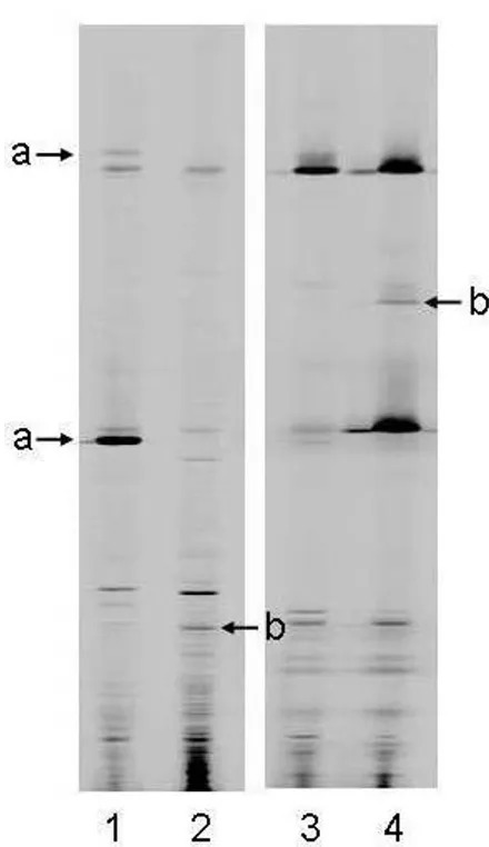
Examples of the ways in which T-RFLP profiles, generated either directly from sputum samples (Direct Molecular Analysis), or from cultures derived from those samples (Culture-derived Molecular Analysis), differ are shown in Figure 1. Here, each T-RFLP profile contains a varying number of T-RF bands of different lengths. T-RF bands also allow an assessment of the relative abundance of the species present. Examples of the ways in which profile pat-terns differ depending on whether they are generated from
Two examples of pairs of DNA profiles generated from two sputum samples (A and B), one from Direct Molecular Analy-sis and one from the total pool of colonies isolated from that sputum using routine surveillance media
Figure 1
the DNA or RNA component of a sample are shown in Figure 2. These differences in T-RF band intensities can be used to obtain a relative measure of metabolic activity.
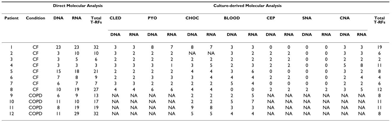
A breakdown of numbers of T-RF bands resolved in the DNA-derived and RNA-derived profiles generated from the sputum samples directly and following cultivation on different media are shown in Table 2. Overall, a total of 266 T-RF bands were resolved in the DNA and RNA-derived T-RFLP Direct Molecular Analysis profiles
gener-ated directly from these sputum samples, representing 104 different T-RF band lengths. The most frequent band length was identified 19 times, with 66 band lengths found only once. Of these 266 T-RF bands, a total of 106 were resolved in the DNA-based T-RFLP profiles alone, representing 65 different T-RF band lengths. 196 T-RF bands were detected in both the DNA and RNA-derived profiles, representing 96 different band lengths, and a total of 160 bands were detected in the RNA-based T-RFLP profiles alone representing 79 band lengths as shown schematically in Figure 3.
Direct Molecular Analysis – assessment per sample
On average, 8.8 (Standard Deviation 5.8) and 13.3 (SD 8.0) T-RF bands were resolved from the DNA and RNA isolated directly from sputum respectively, representing a mean of 16.3 (SD 10.0) separate T-RF band lengths in the profiles from each sputum sample. On average, 5.7 (SD 4.1) T-RF bands were detected in both the DNA and the RNA profile from the sample set as a whole, 3.2 (SD 3.4) were detected in the DNA profile alone, and 7.6 (SD 6.3) were detected in the RNA profile alone.
Culture-derived Molecular Analysis
A composite of "all" Culture-derived Molecular Analysis T-RF bands was formed representing the species detected on all types of media. Within this, a total of 40 different T-RF band lengths were resolved. In the sample set, an aver-age of 2.9 separate T-RF lengths were detected in the pro-files generated from the CLED agar, 3.5 from PYO agar, 4.3 from chocolate agar, 4.7 from BLOOD agar, 1.3 from CEP agar, 0.4 from SNA agar, 3.8 from CNA agar. The highest number of separate band lengths was resolved from the bacteria cultured on chocolate agar (21), fol-lowed by BLOOD agar (18), CNA agar (15), PYO agar (13), CLED agar (10), CEP agar (4) and SNA agar (1). On average, 8.8 (SD 4.4) separate T-RF bands were resolved by Culture-derived Molecular Analysis per patient.
Routine diagnostic microbiology
Analysis of "historical" culture-based routine diagnostic microbiology surveillance data revealed that, for these 12 patients, eight were reported as being infected with
Pseu-domonas spp., with six reported as having (normal) oral
flora. Escherichia coli and Staphylococcus aureus were each reported in a single case.
Comparing the approaches
The difference in the average number of T-RF bands detected in the samples between the Direct Molecular Analysis and Culture-derived Molecular Analysis groups was determined to be highly statistically significant (P = 0.008, two-tailed paired T-test). For 77.9% (SD 20.9%) of the T-RF band lengths resolved from the Culture-derived Molecular Analysis, a band of the same length was Two examples of pairs of DNA and RNA profiles generated
from two sputum samples (C and D), by Direct Molecular Analysis
Figure 2
BMC Pulmonary Medicine
200
9,
9
:14
http
://www.bio
m
e
dcent
ral.com/147
1-246
6/9/1
4
Pa
ge 6 of
1
1
(page number not for citation purposes)
Table 2: Number of T-RF bands resolved in T-RFLP profiles generated from the culture-independent and culture-based approaches.
Direct Molecular Analysis Culture-derived Molecular Analysis
Patient Condition DNA RNA Total
T-RFs CLED PYO CHOC BLOOD CEP SNA CNA T-RFsTotal
DNA RNA DNA RNA DNA RNA DNA RNA DNA RNA DNA RNA DNA RNA
1 CF 23 23 32 3 3 8 7 8 7 3 3 0 0 0 0 3 3 19
2 CF 3 10 10 3 2 2 2 NA NA 3 2 2 2 0 0 3 3 6
3 CF 3 5 6 2 2 2 2 2 2 2 2 2 2 0 0 2 2 2
4 CF 3 3 3 3 3 3 1 3 5 2 3 2 2 0 0 5 8 11
5 CF 15 18 21 2 2 2 2 4 4 3 6 0 0 0 0 3 2 8
6 CF 7 8 9 2 2 3 3 3 4 4 4 2 2 0 0 2 4 4
7 CF 6 7 7 3 3 2 2 2 2 5 4 0 0 0 0 2 2 6
8 CF 10 19 27 4 4 6 6 4 4 0 0 2 2 2 2 3 5 12
9 COPD 6 9 13 NA NA NA NA 2 2 2 5 NA NA NA NA NA NA 8
10 COPD 11 10 17 NA NA NA NA 2 2 5 7 NA NA NA NA NA NA 11
11 COPD 8 19 19 NA NA NA NA 9 8 3 3 NA NA NA NA NA NA 11
12 COPD 11 29 32 NA NA NA NA 5 5 4 4 NA NA NA NA NA NA 8
detected at the same position in both the DNA-derived and RNA-derived T-RFLP profiles from the same sample. In 15.4% (SD 16.8%) of instances, a band was detected in the RNA profile alone, and in 6.7% (SD 7.2%) of instances, a band was detected in the DNA profile alone. Overall, of all the T-RF band lengths resolved from the Culture-derived Molecular Analysis, a match to at least one T-RF from DNA-derived or RNA-derived Direct Molecular Analysis was 92.5%. For comparison of the results obtained using the different strategies, see Table 3.
There were a total of 184 instances of T-RF bands detected in the direct sputum profiles alone, representing 83 differ-ent T-RF band lengths. Amongst these were fifteen instances where the T-RF band represented more than 10% of the total band volume for the profile (with a mean value of 23.6%). These instances were spread between 12 patients and represented 12 different T-RF band lengths. There were 46 instances of T-RF bands being resolved in the RNA profiles generated from cultures but having no corresponding band in the RNA-derived profile generated directly from the sputum sample. These T-RF bands repre-sented 34 different band lengths. The degree to which the different T-RF band lengths were detected in the Direct Molecular Analysis and Culture-derived Molecular Analy-sis is illustrated in Figure 4.
The number of T-RF bands resolved in T-RFLP profiles generated from the independent and culture-based approaches are shown in Table 2. In four instances, material cultured from inoculated bacterial media plates
yielded no T-RF bands in either the DNA or the RNA pro-files (Patient 8 – BLOOD agar, Patient 1 – CEP agar, Patient 5 – CEP agar, Patient 7 – CEP agar,). No T-RF bands were detected in either the DNA-derived or RNA-derived profiles from any of the SNA (yeast) agar, except in the case of Patient 8. In both the DNA-derived and RNA-derived profiles generated from Patient 8, two T-RF bands, both consistent with Pseudomonas species were detected. In one instance, a chocolate medium culture was not available for analysis (Patient 2).
Five different T-RF band lengths (155, 564, 582, 598, and 373 bases) were resolved as the most abundant ("domi-nant") band in DNA-derived profiles generated directly from sputum. The first four of these bands are consistent with those generated from P. aeruginosa, Pseudomonas sp.,
Streptococcus constellatus and Lactobacillus sp., respectively,
as determined by analysis of published sequence data. It was not possible to assign an identity to the 373 base T-RF band due to multiple species being predicted to generate a band of this length. Strategies for resolving this problem are discussed below. Five different T-RF bands (155, 376, 564, 583, and 592 bases) were resolved in the equivalent RNA-derived profiles. These T-RF bands are consistent with those generated from P. aeruginosa, Actinomyces sp.,
Pseudomonas sp., Streptococcus constellatus and
Carnobacte-rium sp., respectively. Overall, eight different T-RF band
lengths were resolved as the most abundant ("dominant") band in profiles generated directly from sputum. Of these, only three were detected in profiles generated from the corresponding set of cultures (155, 373 and 376 bases).
On average, the most abundant T-RF band length detected in the DNA-derived profiles not resolved in any of the cor-responding Culture-derived Molecular Analysis repre-sented 34.0% (SD 29.3%) of the total signal volume. On average, the most abundant T-RF band length detected in the RNA-derived profiles not resolved in any of the corre-sponding Culture-derived Molecular Analysis represented 21.0% (SD 15.5%) of the total signal volume. The average volumes of bands detected in the DNA-derived and RNA-derived profiles, but not in the corresponding Culture-derived Molecular Analysis profiles were 9.0% (SD 17.0%) and 4.9% (SD 7.7%) respectively.
Discussion
The process of bacterial culture in vitro has been shown to be selective and provides a distorted representation of the bacteria present in a clinical sample. Diagnostic microbi-ology has in fact for many years exploited this through the use of media for the selective growth of particular species or groups of species whilst excluding others leading to their isolation from complex contexts. This ability to exclude species makes diagnostic microbiology a useful tool when assaying for particular aetiological agents, such Venn diagram showing the overlap between the T-RF bands
detected in the DNA and RNA profiles generated by Direct Molecular Analysis from sputum samples
Figure 3
BMC Pulmonary Medicine 2009, 9:14 http://www.biomedcentral.com/1471-2466/9/14
Page 8 of 11 (page number not for citation purposes) as those that are known to be responsible for some acute
airway infections. However, when applied to the analysis of chronic airway infections, such as those associated with CF and COPD, such approaches may fail to identify the many opportunistic pathogens that could potentially col-onise the airways. In an effort however to avoid biases associated with culture-based analysis, culture-independ-ent techniques are being increasingly used to characterise bacteria found in chronic airways diseases. We set out to determine the extent to which routine diagnostic micro-biological culture was masking the bacterial species present in a set of respiratory samples. Here, we report sig-nificant differences in the composition of bacterial com-munities in sputa as characterised by Direct Molecular Analysis and Culture-derived Molecular Analysis per-formed on the same samples. Differences both in the number and identity of the organisms resolved, their rela-tive prevalence and their relarela-tive levels of metabolic activ-ity were identified. Overall, these findings were considered as a series of questions that a respiratory phy-sician would ask (Table 3) and are discussed in that fash-ion as below.
In terms of the bacteria "missed" by culture, the findings were marked. Overall, there were a total of 184 instances of bands detected in the Direct Molecular Analysis profiles but not in Culture-derived Molecular Analysis profiles, representing 83 different T-RF band lengths. Of course, this in itself does indicate whether these organisms were clinically significant, however, two factors suggest that
they might be. Firstly, two of the five T-RF band lengths identified as dominant in T-RFLP profiles generated from sputum were not resolved in the profiles generated from the corresponding cultures. This suggests that in some cases, species that represent a significant proportion of the bacteria in the sample might be missed. Secondly, meta-bolically active bacteria, as detected by RT-T-RFLP profil-ing, were amongst those responsible for these dominant T-RF bands. Assuming that these were not acquired in transit through the upper airway [19], this means that metabolically active bacteria were present in the lower air-ways in significant numbers. As such, they are likely to have elicited an immune response of some form and may have potential roles as lung pathogens. Furthermore, such species are likely to be involved in complex inter-species communication that impacts on the bacterial community [20,21].
In relation to these marked differences, it is likely that one group that may be highly represented are those bacteria that require either anaerobic, microaerophilic or similar conditions for growth. No diagnostic microbiology serv-ice that we are aware of would routinely test for the pres-ence of bacteria requiring anaerobic conditions for growth in respiratory samples of this kind. Despite this, the importance of micro-anaerobic environments within the lower airways of patients suffering from CF is being increasingly recognised [22,23]. This is all the more important given the wide range of clinically important anaerobic species, for example within genera such as
Table 3: Summary of a comparison of the results of Direct Molecular Analysis (DMA), Culture-derived Molecular Analysis (CMA), and Routine Diagnostic Microbiology (RDM)
Patient Condition Were all species reported in RDM represented in DMA?
Was the dominant band detected by DMA also detected by
RDM?
Were there bands detected by DMA that were not detected by CMA?
Was the dominant band detected by DMA also detected
CMA?
Was the dominant band detected by RT DMA also detected by
CMA?
DNA RNA DNA RNA DNA RNA DNA RNA DNA RNA
1 CF N* N* N N Y Y Y Y Y Y
2 CF Y Y Y Y Y Y Y Y Y Y
3 CF Y Y Y Y Y Y Y Y Y Y
4 CF Y Y Y Y N N Y Y Y Y
5 CF Y Y N Y Y Y Y Y Y Y
6 CF Y Y Y Y Y Y Y Y Y Y
7 CF Y Y Y Y Y Y Y Y Y Y
8 CF Y Y Y N Y Y Y Y Y Y
9 COPD Y Y N N Y Y Y Y Y Y
10 COPD Y Y N/A N/A Y Y Y Y Y Y
11 COPD Y Y N/A N/A Y Y N N Y Y
12 COPD Y Y N/A N/A Y Y Y Y Y Y
Totals (%) 11/12
(91.7)
11/12 (91.7)
6/9 (66.7) 6/9 (66.7) 11/12 (91.7)
11/12 (91.7)
11/12 (91.7)
11/12 (91.7)
12/12 (100)
12/12 (100)
Bacteroides, Fusobacterium, Porphyromonas, Prevotella, and
Peptostreptococcus, that have previously been identified by
16S rRNA clone sequence analysis in respiratory samples from CF patients [16,24].
Whilst the assignment of species identities to T-RF band lengths was not the focus of this study, comparison of the T-RF band lengths generated from the different sample types allowed assessment of the degree to which data from culture-dependent and culture-independent methodolo-gies overlapped. In only three cases, culture-based diag-nostics generated a T-RF length that was not detected by molecular means. This is analogous to the situation regarding the results of the routine diagnostic microbiol-ogy where there was only one instance of a species being reported by conventional diagnostics that was not resolved by direct T-RFLP profiling. On this one occasion, T-RFLP profiling of Culture-derived Molecular Analysis resulted in the detection of a band tentatively identified as being S. aureus. There are many explanations of this including whether cultivation over-represents this species, the impact of detection thresholds in T-RFLP profiling, and possible contamination of growth media. Incorporat-ing specific PCR based assays e.g. Alarcón et al [25] into the next phase of work will be valuable in determining the likely origin of such discrepancies. Equally however, to form such a small part of all the species present, this again raises important questions over the clinical significance of the other species that were much more common in the sample. Therefore, it must be questioned whether or not species that are "missed" by culture are really present at
levels that make them of clinical significance. In relation to this, of the band lengths detected as the dominant band in either the DNA or RNA profiles generated directly from the sputum, only three of the 8 were detected in any of the corresponding Culture-derived Molecular Analysis pro-files. The concept of over representation was taken further. It was found that the typical band volume of bands detected in the direct DNA profile, but not in culture, was approximately 10% of the total lane volume. This means that some of the most numerically significant species present in the sample are, in many cases, going undetected by culture.
The restriction enzyme, CfoI, was selected because it is able to differentiate between the recognised key species associated with CF and COPD respiratory infections (P. aeruginosa, S. aureus, B. cepacia, H. influenzae, S.
mal-tophilia, S. pneumoniae, M. catarrhalis). Whilst other
restric-tion enzymes have been shown to provide greater levels of resolution [26,27], no single restriction enzyme is able to resolve all bacterial species. Therefore, it must be recog-nised that in many instances, T-RF bands of the same length will be generated from different bacterial species. This may result in an underestimation of species richness, lower confidence in ascribing species to T-RF bands on the basis of T-RF band length alone, and an overestimation of the proportion of the total bacterial community repre-sented by these bacterial species. Steps can be taken to off-set the failure of a particular restriction enzymes to resolve all species present, including generating multiple profiles with different restriction enzymes, and such approaches may need to be applied were the techniques described here to be applied to a wider study of chronic respiratory infections.
SNA agar is routinely used to isolate yeasts from respira-tory samples. Although the T-RFLP profiling used here was designed to resolve bacterial species alone, SNA cul-tures were included. The fact that they provided no signal in all but one instance indicated that this medium was highly selective for fungal species. Although no attempt was made to do so in this study, other studies have shown that fungal communities can be studied by Direct Molec-ular Analysis [28]. This would clearly be important to assess more generally in airway specimens.
This study also considered whether the bacteria detected were metabolically active. Unlike culture-based methods, the detection of bacteria in clinical samples by DNA-based methodologies does not indicate whether the bacteria in question are viable. Although activity does not necessarily imply pathogenicity, the presence of actively metabolising bacteria in sputum samples does suggest further investiga-tion is warranted. The use of rRNA-based analyses to char-acterise active microbial communities is based on the Venn diagram showing the overlap between the T-RF bands
detected in the profiles generated by Direct Molecular Anal-ysis and those detected using Culture-derived Molecular Analysis
Figure 4
BMC Pulmonary Medicine 2009, 9:14 http://www.biomedcentral.com/1471-2466/9/14
Page 10 of 11 (page number not for citation purposes) assumption that active or growing cells have increased
lev-els of rRNA relative to dormant, or intact dead cells. Sim-ilar comparisons have been made of bacteria in environments as diverse as soil [29], dairy fermentations [30] and CF sputum [18]. For these respiratory samples, approximately a quarter of the T-RF bands were detected in the direct analysis of sputum were found only in lanes generated from DNA, with 42% were found in both DNA and RNA and the remainder present in RNA alone. It is quite possible that those found only as a DNA signal were from inactive or dead cells. It is also possible that, given the variation in ribosomal operon numbers between bac-terial species (from one to 15) [31-33], that this influ-enced the relative amounts of signal generated for any given band position. It should be noted that because rRNA transcripts are many times more common in bacte-rial cells than are rRNA genes, RNA-based analysis may provide a greater sensitivity in the detection of uncom-mon species within samples. This may well explain the significantly greater number of T-RF bands in RNA-derived profiles. Molecular based methods are not them-selves without bias, with such bias known in the amplifi-cation process which will impact on both PCR and RT-PCR steps [34,35]. Despite this, whilst these data may be influenced by these technical issues, the findings suggest that three quarters of the bacteria present in these respira-tory samples were metabolically active.
Conclusion
There are still limitations in terms of molecular microbi-ology. For example, this study did not focus on species identification, rather on the degree to which culture-inde-pendent methodologies may preclude the identification of organisms that could have a causative association with a particular pathology or disease when applied to chronic respiratory conditions such as COPD or CF. Whilst some of the bacteria present in sputum samples may result from contamination during expectoration, it is also possible that they represent populations colonising the lower res-piratory tract. Inclusive, culture-independent approaches, such those described here, provide a means by which fur-ther study could determine the degree to which these sit-uations is the case.
Whilst culture-dependent diagnostics will continue to play an important role in the detection of known respira-tory pathogens, the deployment of culture-independent profiling techniques will help to identify respiratory path-ogens whose clinical significance is not yet recognised in these conditions. Clearly however, much more is needed to improve methodologies and more fundamentally understand the potential significance of the species detected in terms of airways disease. In particular, studies to determine whether the bacterial community profiles
are stable over time, and how they change and respond to antimicrobial interventions, are now needed.
Abbreviations used
(CF): Cystic Fibrosis; (COPD): Chronic Obstructive Pul-monary Disease; (T-RFLP): Terminal Restriction Fragment Length Polymorphism; (RT-T-RFLP): Reverse Tran-scriptase Terminal Restriction Fragment Length Polymor-phism; (DMA): Direct Molecular Analysis; (CMA): Culture-derived Molecular Analysis; (RDM): Routine Diagnostic Microbiology.
Competing interests
The authors declare that they have no competing interests.
Authors' contributions
GBR performed all molecular microbiological analyses and wrote the initial draft of the paper. AT directed all conventional microbiology. TTD directed patient liason, sample collection and participated in study design. GBR, KDB and MPC conceived the question. KDB directed writ-ing and analysis. KDB and MPC obtained fundwrit-ing for the project. GJC and GJPD participated in funding, data col-lection, data analysis and interpretation, and editing. All authors have read and approved the final manuscript.
Acknowledgements
This research was funded by the Anna Trust.
References
1. Wilson R: Bacteria, antibiotics and COPD. Eur Respir J 2001,
17:995-1007.
2. Miravitlles M: Exacerbations of chronic obstructive pulmonary disease: when are bacteria important? Eur Respir J 2002,
20:9S-19S.
3. Patel IS, Seemungal TA, Wilks M, Lloyd-Owen SJ, Donaldson GC, Wedzicha JA: Relationship between bacterial colonisation and the frequency, character, and severity of COPD exacerba-tions. Thorax 2002, 57:759-64.
4. Hurst JR, Wilkinson TM, Perera WR, Donaldson GC, Wedzicha JA:
Relationships among bacteria, upper airway, lower airway, and systemic inflammation in COPD. Chest 2005, 127:1219-26. 5. Donaldson GC, Seemungal TA, Patel IS, Bhowmik A, Wilkinson TM, Hurst JR, MacCallum PK, Wedzicha JA: Airway and systemic inflammation and decline in lung function in patients with COPD. Chest 2005, 128(4):1995-2004.
6. Frederiksen B, Hoiby N, Koch C: Age at onset of chronic pulmo-nary infection with P. aeruginosa infection is a predictor for survival in CF. Pediatric Pulmonology 1998, 26:325.
7. Frederiksen B, Koch C, Hoiby N: Antibiotic treatment of initial colonization with Pseudomonas aeruginosa postpones chronic infection and prevents deterioration of pulmonary function in cystic fibrosis. Pediatric Pulmonology 1997, 23:330-5. 8. National Collaborating Centre for Chronic Conditions. Chronic
obstructive pulmonary disease: National clinical guideline on management of chronic obstructive pulmonary disease in adults in primary and secondary care. Thorax 2004, 59(Suppl 1):1-232.
9. Smith AL, Fiel SB, Mayer-Hamblett N, Ramsey B, Burns JL: Suscepti-bility testing of Pseudomonas aeruginosa isolates and clinical response to parenteral antibiotic administration – Lack of association in cystic fibrosis. Chest 2003, 123(5):1495-1502. 10. Foweraker JE, Laughton CR, Brown DF, Bilton D: Phenotypic
Publish with BioMed Central and every scientist can read your work free of charge "BioMed Central will be the most significant development for disseminating the results of biomedical researc h in our lifetime."
Sir Paul Nurse, Cancer Research UK
Your research papers will be:
available free of charge to the entire biomedical community
peer reviewed and published immediately upon acceptance
cited in PubMed and archived on PubMed Central
yours — you keep the copyright
Submit your manuscript here:
http://www.biomedcentral.com/info/publishing_adv.asp
BioMedcentral
impact on the validity of antimicrobial susceptibility testing. J Antimicrob Chemother 2005, 55(6):921-927.
11. Amann RI, Ludwig W, Schleifer KH: Phylogenetic identification and in situ detection of individual microbial cells without cul-tivation. Microbiol Rev 1995, 59:143-169.
12. Atlas RM, Bartha R: Microbial ecology, fundamentals and applications 3rd edition. San Fransisco: The Benjamin/Cummings Publishing Company; 1992.
13. Weng L, Rubin EM, Bristow J: Application of sequence-based methods in human microbial ecology. Genome Res 2006,
16:316-22.
14. Liu WT, Marsh TL, Cheng H, Forney LJ: Characterization of microbial diversity by determining terminal restriction frag-ment length polymorphisms of genes encoding 16S rRNA. Appl Environ Microbiol 1997, 63(11):4516-4522.
15. Rogers GB, Hart CA, Mason JR, Hughes M, Walshaw MJ, Bruce KD:
Bacterial diversity in cases of lung infection in cystic fibrosis patients: 16S ribosomal DNA (rDNA) length heterogeneity PCR and 16S rDNA terminal restriction fragment length polymorphism profiling. J Clin Microbiol 2003, 41(8):3548-3558. 16. Rogers GB, Carroll MP, Serisier DJ, Hockey PM, Jones G, Bruce KD:
Characterization of bacterial community diversity in cystic fibrosis lung infections by use of 16S ribosomal DNA termi-nal restriction fragment length polymorphism profiling. J Clin Microbiol 2004, 42(11):5176-5183.
17. Rogers GB, Carroll MP, Serisier DJ, Hockey PM, Jones G, Bruce KD:
Bacterial activity in cystic fibrosis lung infections. Respir Res
2005, 6:49.
18. Health Protection Agency: Investigation of bronchoalveolar lav-age, sputum and associated specimens, BSOP 57. Issue 2.1 Issued by Standards Unit, Evaluations and Standards Laboratory, Cen-tre for Infections; 2008.
19. Rogers GB, Carroll MP, Serisier DJ, Hockey PM, Jones G, Kehagia V, Connett GJ, Bruce KD: Use of 16S rRNA gene profiling by ter-minal restriction fragment length polymorphism analysis to compare bacterial communities in sputum and mouthwash samples from patients with cystic fibrosis. J Clin Microbiol 2006,
44(7):2601-2604.
20. Duan K, Dammel C, Stein J, Rabin H, Surette MG: Modulation of Pseudomonas aeruginosa gene expression by host micro-flora through interspecies communication. Mol Microbiol 2003,
50(5):1477-1491.
21. Keller L, Surette MG: Communication in bacteria: an ecological and evolutionary perspective. Nat Rev Microbiol 2006,
4(4):249-58.
22. Worlitzsch D, Tarran R, Ulrich M, Schwab U, Cekici A, Meyer KC, Birrer P, Bellon G, Berger J, Weiss T, Botzenhart K, Yankaskas JR, Randell S, Döring G: Effects of reduced mucus oxygen concen-tration in airway Pseudomonas infections of cystic fibrosis patients. J Clin Invest 2002, 109(3):317-325.
23. Yoon SS, Hennigan RF, Hilliard GM, Ochsner UA, Parvatiyar K, Kam-ani MC, Allen HL, DeKievit TR, Gardner PR, Schwab U, Rowe JJ, Iglewski BH, McDermott TR, Mason RP, Wozniak DJ, Hancock RE, Parsek MR, Noah TL, Boucher RC, Hassett DJ: Pseudomonas aer-uginosa anaerobic respiration in biofilms: Relationships to cystic fibrosis pathogenesis. Dev Cell 2002, 3(4):593-603. 24. Kolak M, Karpati F, Monstein HJ, Jonasson J: Molecular typing of
the bacterial flora in sputum of cystic fibrosis patients. Int J Med Microbiol 2003, 293(4):309-317.
25. Alarcon B, Vicedo B, Aznar R: PCR-based procedures for detec-tion and quantificadetec-tion of Staphylococcus aureus and their application in food. J Appl Microbiol 2006, 100:352-64.
26. Moyer CL, Tiedje JM, Dobbs FC, Karl DM: A computer-simulated restriction fragment length polymorphism analysis of bacte-rial small-subunit rRNA genes: efficacy of selected tetra-meric restriction enzymes for studies of microbial diversity in nature. Appl Environ Microbiol 1996, 62:2501-7.
27. Engebretson JJ, Moyer CL: Fidelity of select restriction endonu-cleases in determining microbial diversity by terminal-restriction fragment length polymorphism. Appl Environ Micro-biol 2003, 69:4823-9.
28. Singh BK, Nazaries L, Munro S, Anderson IC, Campbell CD: Use of multiplex terminal restriction fragment length polymor-phism for rapid and simultaneous analysis of different com-ponents of the soil microbial community. Appl Environ Microbiol
2006, 72:7278-85.
29. Pesaro M, Nicollier G, Zeyer J, Widmer F: Impact of soil drying-rewetting stress microbial communities and activities and on degradation of two crop protection products. Appl Environ Microbiol 2004, 70:2577-87.
30. Sanchez JI, Rossetti L, Martinez B, Rodríguez A, Giraffa G: Applica-tion of reverse transcriptase PCR-based T-RFLP to perform semi-quantitative analysis of metabolically active bacteria in dairy fermentations. J Microbiol Methods 2006, 65:268-77. 31. Bercovier H, Kafri O, Sela S: Mycobacteria possess a surprisingly
small number of ribosomal RNA genes in relation to the size of their genome. Biochem Biophys Res Commun 1986, 136:1136-41. 32. Andersson SG, Zomorodipour A, Winkler HH, Kurland CG: Unu-sual organization of the rRNA genes in Rickettsia prow-azekii. J Bacteriol 1995, 177:4171-5.
33. Rainey FA, WardRainey NL, Janssen PH, Hippe H, Stackebrandt E:
Clostridium paradoxum DSM 7308(T) contains multiple 16S rRNA genes with heterogeneous intervening sequences. Microbiology-Uk 1996, 142:2087-95.
34. Suzuki MT, Giovannoni SJ: Bias caused by template annealing in the amplification of mixtures of 16S rRNA genes by PCR. Appl Environ Microbiol 1996, 62:625-30.
35. Polz MF, Cavanaugh CM: Bias in template-to-product ratios in multitemplate PCR. Appl Environ Microbiol 1998,
64(10):3724-3730.
Pre-publication history
The pre-publication history for this paper can be accessed here: