S Y S T E M A T I C R E V I E W
Open Access
The efficacy of muscle energy techniques
in symptomatic and asymptomatic
subjects: a systematic review
Ewan Thomas
1,2*, Antonio Rosario Cavallaro
2, Diba Mani
3,4, Antonino Bianco
1and Antonio Palma
1Abstract
Background:Muscle energy techniques are applied to reduce pain and increase range of motion. These are applied to a variety of pathological conditions and on asymptomatic subjects. There is however limited knowledge on their effectiveness and which protocol may be the most beneficial.
Objective:The aim of this review is to determine the efficacy of muscle energy techniques (MET) in symptomatic and asymptomatic subjects.
Design:Systematic Review.
Methods:A literature search was performed using the following database: Cochrane Library, MEDLINE, NLM Pubmed and ScienceDirect. Studies regarding MET in asymptomatic and symptomatic patients were considered for investigation. The main outcomes took into account range of motion, chronic and acute pain and trigger points. Two trained investigators independently screened eligible studies according to the eligibility criteria, extracted data and assessed risk of bias. Randomized control trials (RCT’s) were analyzed for quality using the PEDro scale.
Results:A total of 26 studies were considered eligible and included in the quantitative synthesis: 14 regarding symptomatic patients and 12 regarding asymptomatic subjects. Quality assessment of the studies through the PEDro scale observed a“moderate to high”quality of the included records.
Conclusions:MET are an effective treatment for reducing chronic and acute pain of the lower back. MET are also effective in treating chronic neck pain and chronic lateral epicondylitis. MET can be applied to increase range of motion of a joint when a functional limitation is present. Other techniques seem to be more appropriate compared to MET for trigger points.
Keywords:Manipulative therapies, Pain, Range of motion
Introduction
Muscle energy techniques (MET) were originally devel-oped by two osteopathic physicians, Fred Mitchell, Sr. and Fred Mitchell, Jr., to treat soft tissue, mobilize joints, stretch tight muscles and fascia, reduce pain and to
im-prove circulation and lymphatic drainage [1,2]. MET are
defined as a manual treatment in which a patient pro-duces a contraction in a precisely controlled position and direction against a counterforce applied by a manual
therapist [3]. It could be advocated that MET are similar to proprioceptive neuromuscular facilitation stretching (PNF) [4]; however, the execution of MET is usually per-formed with lower forces compared to those of PNF in order to recruit tonic muscle fibers that are associated with tonic motor units which require lower action potentials in order to be recruited than phasic muscle fibers. These latter are activated during PNF and
typi-cally occur at forces greater than 25% of the person’s
maximal force [5]. Another difference between MET and PNF is that the contraction during MET is performed at the initial barrier of tissue resistance, rather than at the end of the range of motion (ROM) of a joint [6].
© The Author(s). 2019Open AccessThis article is distributed under the terms of the Creative Commons Attribution 4.0
International License (http://creativecommons.org/licenses/by/4.0/), which permits unrestricted use, distribution, and
reproduction in any medium, provided you give appropriate credit to the original author(s) and the source, provide a link to the Creative Commons license, and indicate if changes were made. The Creative Commons Public Domain Dedication waiver (http://creativecommons.org/publicdomain/zero/1.0/) applies to the data made available in this article, unless otherwise stated. * Correspondence:ewan.thomas@unipa.it
1Sport and Exercise Sciences Research Unit, University of Palermo, Via
Giovanni Pascoli 6, 90144 Palermo, Italy
2International Academy of Osteopathic Medicine, AISeRCO, Palermo, Italy
Many studies have applied MET in patients with acute
and chronic low back pain (LBP) [7–10], latent trigger
points [11,12], cervical pain [13,14] and other
musculo-skeletal dysfunctions [15–17]. MET have been also used
in asymptomatic subjects in order to increase mobility [18–20]. There is varying evidence that when a joint has a functional limitation, the application of a MET can increase its ROM [15]. In particular, Lenehan et al. [21] applied MET on the thoracic spine in asymptomatic subjects presenting with a restricted and a non-restricted side. MET were able to increase mobility only on the re-stricted side.
MET protocols developed differ in paradigms, such as in the number of repetitions, strength of contraction, duration of stretch phase and duration of relaxation
phase [11,15,17,19]. Two of the most prominent MET
typologies of application are those advocated by Green-man [3] and Chaitow [5]. The first involves the applica-tion of three to five repetiapplica-tions with a relaxaapplica-tion phase long enough to reduce the tension of the targeted tissue (usually 5 to 7 s), whereas the second involves the appli-cation of four repetitions with a relaxation/stretch phase of 30 to 60 s after each contraction [19]. Other authors
have also used similar MET applications [21–23],
al-though a consensus of which protocol could be more effective still needs to be investigated [1]. Aside from the form of application, the main physiological mechanism pro-posed for MET [5] involves two general principles: (1) post-isometric relaxation [24], which causes a reduction in the tone of a muscle following an isometric contraction and (2) reciprocal inhibition [25], which involves the reduction in tone of the antagonist muscle following the isometric contraction of the agonist muscle through inhibition of the alpha motor neuron. Post-isometric relaxation is the most frequently applied approach, while reciprocal inhibition is used when a tissue has severe limitations or has become fibrotic, as a treatment modality associated to post-isometric relaxation [5]. Although many texts advocate these as the principal mechanisms responsible for muscle relaxation, it has been seen in studies analyzing joint extensibility that including a preisometric contraction, does not alter resting EMG activity, notwithstanding increased ROM [26]. In addition, another study evaluating motor neuron activity, has shown increases in EMG activity also following concen-tric and eccenconcen-tric contractions. A greater increase was seen in the first case while such increase was less pronounced in the latter [27]. The exact mechanism for MET-induced pain relief is still unknown, although it has been proposed that MET act on joint proprioceptors and mechanoreceptors that will result in an effect on descending pathways, changing the motor programming of the target joint [1,22,28,29]. It has also been advocated that the reduction of pain and increased mobility are due to changes in the viscoelastic properties of the soft tissue followed by the application of
the technique; the mechanism for increased flexibility has been attributed to an increase in stretch tolerance [4, 22]. Very few studies investigating the effects of the different typologies and efficacies of MET have been performed; therefore, the aim of this review is to understand the efficacy of MET specifically on pain and joint range of motion and to understand the differences between the different MET protocols in symptomatic and asymptomatic subjects.
Materials and methods
This systematic review aims to determine if muscle en-ergy technique may be effective on pain or may increase range of motion of a joint and if such techniques are ap-plied using different protocols. The PRISMA (Preferred Reporting Items for Systematic Reviews and Meta-Ana-lyses) statement was adopted [30]. Studies that include randomized control trials (RCT) analyzing symptomatic patients with various conditions and studies applied on asymptomatic subjects with range of motion limitations
were reviewed. All the manuscripts that were RCT’s
were assessed for methodological quality using the Physiotherapy Evidence Database (PEDro) scale.
Inclusion and exclusion criteria
Studies that met the following criteria were included or excluded in this systematic review.
Study designs
This review includes studies that have applied MET in both asymptomatic subjects and symptomatic patients.
In particular, RCT’s analyzing patients who have been
treated for their condition using MET and which include a control group, were selected for the symptomatic population. Studies that do not have a control group or
a comparator were not considered for inclusion. RCT’s,
pretest-posttest and quasi-experimental studies analy-zing the effect of MET were also included for the asymptomatic subjects. Reviews, systematic reviews and meta-analysis were not considered.
Participants
All the analyzed participants were adults to whom a MET had been applied. Children were not considered for analysis.
Interventions
to the nature of the variables of each study, pain was measured through different means.
Comparators
Comparators were control groups for the symptomatic population and control groups or other interventions for the asymptomatic participants.
Outcomes
The primary outcomes were changes in range of motion (ROM) and pressure pain thresholds (PPT) after the MET application in the asymptomatic participants and change in the pain and disability indexes after the MET application in the symptomatic population. PPT, the pain indexes and disability indexes, for this review varied across studies. Another outcome that was analyzed across the review for both asymptomatic and sympto-matic participants was the MET protocol used. Each protocol will be considered in terms of number of con-tractions, seconds which the contraction is applied to the targeted joint, the contraction force applied, the re-laxation phase and the stretch, if applicable.
Search strategy
The literature search was conducted on the following databases: Cochrane Library, MEDLINE, NLM Pubmed, and ScienceDirect. Due to the extensive nature and variety of topics covered by the search, there was no limit to the search period, however the search ended on June 2018. The search strategy was conducted using the following keywords: muscle, energy, technique and MET. These keywords were used as follows: muscle AND energy AND technique OR MET.
After the initial title screening, inclusion and exclusion criteria above described were applied for abstract selec-tion. The inclusion criteria were: (1) studies had to be peer-reviewed, (2) studies had to be performed on adult subjects, (3) studies had to clearly specify the MET pro-cedure used, (4) studies had to report an objective out-come and (5) studies concerning symptomatic patients
had to be RCT’s. The exclusion criteria were: (1)
Non-English manuscripts, (2) studies performed on
chil-dren, (3) non-RCT’s studies, (4) studies that did not
specify a MET procedure. Following the inclusion of the selected manuscripts, these were divided into two groups: those applying MET on asymptomatic pants and those applying MET on symptomatic partici-pants. Duplicate records, abstracts and unpublished materials were removed. Full-text copies of the retrieved records were screened for the same criteria. If the full text copy was not retrievable through database or elec-tronic search, the corresponding authors of the studies were contacted. If no response was received or the authors were not able to provide a copy of the selected
manuscript, the article was excluded from the inves-tigation. Reference lists of relevant publications were also screened.
Selection of studies
Selection was conducted independently by two re-viewers. Any disagreement was resolved through negoti-ation. All of the identified records from each database were combined into a single End Note file (End Note Version X7.5; Thompson Reuters, New York, USA) and subsequently screened for relevance using title and abstract. The full text of relevant studies was retrieved and assessed for eligibility against the inclusion criteria
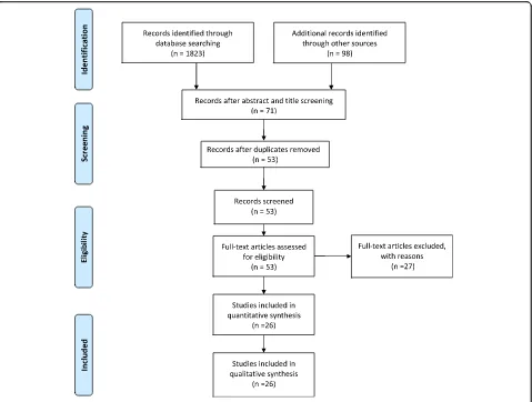
set above. The PRISMA flow diagram (Fig.1) illustrates
the process by which the manuscripts were selected and included in the final analysis.
Quality assessment
The methodological quality of the randomized con-trolled trials was assessed using the PEDro scale. The PEDro scale is a 10-item tool designed to reliably assess
the quality of physical therapy-based RCT’s, based on a
10-item checklist [31], where trials scoring at least 6/10 are deemed to be of ‘moderate to high quality’, although this cut-off point has yet to be validated [32]. Two authors independently assessed the PEDro score. Both authors successfully completed the PEDro consistency training. Data were entered and reviewed in Microsoft Excel spreadsheets (2016 Microsoft Corporation, Red-mond, WA) and any disparity in scores resolved by discussion and thorough reevaluation.
Statistical analysis
The studies included in the qualitative synthesis were classified according to the screened population (asymp-tomatic vs RCT’s). For each group, descriptive statistics were performed for the main variables and other pre-sented indexes using STATISTICA for Windows (Stat-soft, Inc., Ver. 10.0, Tulsa, OK, USA).
Results
A total number of 1921 manuscripts were initially iden-tified after the first search strategy in the four databases. After the application of the inclusion and exclusion cri-teria on each article’s title and abstract, 71 records were considered eligible, 18 records were removed as dupli-cates and 53 records were screened as full-text. Of the 53 records screened, 27 were removed due to non-MET studies, non-English manuscripts, missing data, not meeting the inclusion criteria or not being RCT’s. A final number of 26 original studies were included in the in the qualitative synthesis (Fig.1and Table1).
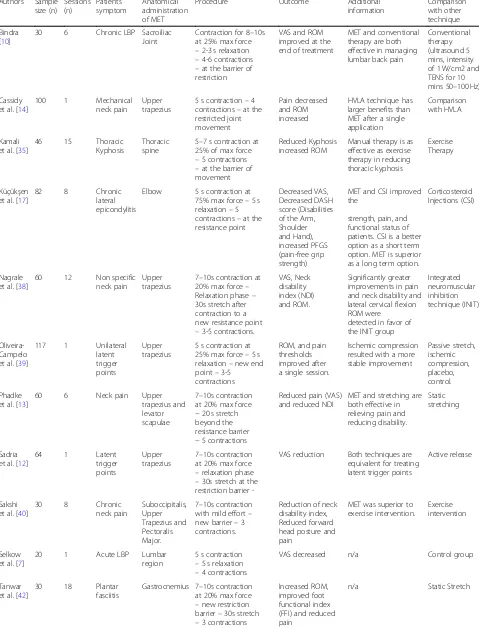
subjects and symptomatic patients. There were 12 stud-ies included in the asymptomatic population group [15, 16, 18–22, 33, 34, 36, 37, 41] (Table2) comprising 485 participants and 14 studies included in the symptomatic
population group [7–14, 17, 35, 38–40, 42] (Table 3)
comprising a total of 954 patients, leading to a total of 1439 subjects analyzed.
All reported data to follow are reported as means for each study. The 14 studies included in the symptomatic
population were all RCT’s and these were subjected to
PEDro quality assessment evaluation. Not all studies
from the asymptomatic population group were RCT’s.
Quality assessment of included RCT’s
The PEDro scores ranged from 2/10 to 9/10, with 10/14 RCT’s achieving ‘moderate to high quality’scores (6/10). The overall PEDro risk of bias score of all the included studies was 6.4/10. The lowest scores for the included studies were achieved in item 6 (blinding of therapist)
and item 7 (blinding of assessors) for scores of 1.4/10 and 2.9/10, respectively. A breakdown of PEDro scores for each trial is shown in Table4.
Asymptomatic population
Of the twelve included studies, ten investigated the
ef-fects of MET on joint ROM [15, 18–22, 33,36, 37, 41],
one the effect of MET on PPT [16] and one the effects of MET on corticospinal and spinal reflex excitability [34].
Three studies targeted the hamstring muscles [19,20,22],
two studies the lumbar region [18,34], one study the thor-acic spine [21], one study the pectoralis minor [36], one study the glenohumeral joint [37] and four studies the
cer-vical spine [15,16,33,41]. The number of treatment
ses-sions retrieved from the studies and included in this review ranged between one and twelve. Seven studies
ap-plied MET within a single session [15, 16,21,22,33,34,
Table 1 Summary of retrieved records included in the qualitative synthesis (Continued) Autho r Sampl e size (n) Popu lation Purpose Protoc ol Duration Anat omi cal adm inistra tion of ME T Outcome Pre- VAS Post- VAS Pre-ROM Post-RO M O ther Saks hi et al. [ 40 ] 30 Patie nts with chron ic nec k pain Effect of techniqu e 8 session s Suboc cipital is, upp er trapezius, and pe ctoralis maj or Reduced Pain 6.4 1.6 n/a n/a D isability index pre 34.9 po st 11 .9 Schenk et al. [ 41 ] 18 Asym pto matic part icipants Effect of techniqu e 7 session s Cer vical region Increased ROM n/a n/a Flexion : 51.8° Extenti on:
69.1° L Rotation
:
35.6° R Rotation
: 35.2° Flexion : 54.4° Extenti on:
71.9° L Rotation
:
40.8° Rrotation: 43.4°
Table 2Summary of evidence of studies with asymptomatic participants
Authors Sample size (n)
Sessions (n)
Anatomical administration of MET
Procedure Outcome Additional
information
Comparison with other technique
Ballantyne et al. [22]
40 1 Hamstring 5 s contraction at 75% of max–3 s relaxation -at a point of discomfort–4 contractions
Increased Rom n/a n/a
Burns et al. [33]
18 1 Cervical spine 3 to 5 s contraction at 0.5 kg of pressure - 3 to 5 s relaxation–to a new barrier of motion–2 to 4 contractions
Increased Rom with MET–Decreased with Sham
n/a With Sham
treatment (stretching for 3 to 5 s –return to neutral position - for 3 stretches)
Fryer et al. [15]
52 1 Atlanto-axial
joint
5 s: 5 s contraction at the first resistance point –5 s relaxation–3 times 20s: 20 s contraction at the first resistance point–5 s relaxation–3 contractions
Increased Rom in the MET groups. With the 5 s group showing the gretest increase.
The increase was greater in the direction of restriction compared to the direction of no restriction
Comparison of a 5 duration contraction with a 20 duration contraction + Sham therapy and control group.
Fryer et al. [34]
12 1 L5/S1
segment bilaterally
5 s contraction at a first tissue tension point - relaxation–new barrier contractions
–3 contractions
Decreased H-reflex and silent period.
MET produces decreased motor excitability in the motor cortex and spinal cord.
Control Group
Hamilton. et al. [16]
90 1 Subocciptal
region
3 to 5 s contraction –5 s relaxation–3 contractions
Pressure pain thresholds increased in the MET compared to the sham but not HVLA procedure
n/a Comparison with
HVLA and Sham treatment
Laudner et at. [36]
39 12 Pectoralis
Minor
3 s stretch–5 s contraction at 25% of max force–4 contractions–no rest
Increased pectoralis length and decreased forward scapular position.
No increase of scapular upward rotation.
n/a
Lenehan et al. [21]
59 1 Thoracic
spine
5 s contraction at the first rotational barrier–no rest –new rotational barrier - four repetitions
Increased Trunk ROM
Restricted direction of treatment increased rotation more than non-restricted rotation
Comparison with control group.
Moore et al. [37]
61 1 Glenohumeral
Joint
5 s contraction at 25% max force - the participant then internally rotated the arm for 30-s and an active assisted stretch was applied - 3 contractions
Increased horizontal adduction and internal rotation ROM
n/a Control group
Schenk et al. [41]
18 7 Cervical
region
5 s contraction –3 s relaxation –increase of direction of limitation–4 contractions
Increased Rom in all six ranges of motion of the cervical region.
provided eight MET sessions [18], only one study provided ten MET sessions [20] and only one study provided twelve MET sessions [36].
All of the above protocols were applied on the restricted side of a targeted joint in the asymptomatic subjects. Dif-ferent procedures for applying MET were presented by different authors. Ballantyne et al. [22] applied four con-tractions of 5 s each at 75% of the participant’s maximal force with a three-second relaxation phase between each contraction on the hamstring muscle. Similarly, Schenk et al. [18] implemented a non-specified force of contraction with a MET treatment applied for 4 weeks on the lumbar region (at the L5 and S1 intersegment junction). The re-sults provided by Ballantyne et al. demonstrate an increase in hamstring passive extensibility, although such an in-crease was also exhibited in the control group. Schenk et al. [18] showed that MET were able to increase lumbar ac-tive extension compared to a control group. Burns et al. [33] applied two to four contractions of 5 s each with 0.5 kg of pressure and a three to 5 s relaxation phase between each contraction applied to the cervical spine. These re-sults showed a significant difference between pre- and post-treatment and between MET and the control group for side-bending and rotation of the cervical spine. Fryer et al. [15] compared two different MET protocols for ROM increases: the first protocol applied three contrac-tions of 5 s each, with a 5 s rest between each contraction
and the second applied three contractions of 20 s each, with a 5 s rest between each contraction applied on the atlanto-axial joint. The results reveal that the 5 s contrac-tion protocol increased active ROM on the restricted side of the atlanto-axial joint to a greater extent than the 20 s contraction protocol and the sham therapy used for com-parisons (stretching). Similar protocols to the 5 s protocol of Fryer and colleagues [15] are those of Hamilton et al. [16] and Fryer et al. [34]. Hamilton and colleagues com-pared the MET technique to a high velocity low amplitude (HVLA) technique, a short, quick thrust over the re-stricted joints with the goal of restoring normal range of motion and a sham treatment to decrease sub-occipital PPT. All the recruited participants were free of: (1) neck
pathologies, (2) long-term cortico-steroid use, (3)
vertebro-basilar insufficiency (4) chronic pain or (5) head-aches. A hand-held electronic algometer consisting of a pressure transducer applied a pressure to the suboccipital area (between C0 and C2) of 30 kPa/s. When the pressure changed into a sensation of pain, the participants pushed a button and stopped the algometer. Such experiment pro-vided evidence that MET is more effective than the sham treatment but equal to the HVLA in reducing PPT.
Fryer et al. [34] investigated the effects of MET on corticospinal and spinal reflex excitability and a single application of MET to the lumbosacral joint produced a significant decrease in corticospinal and spinal reflex
Table 2Summary of evidence of studies with asymptomatic participants(Continued)
Authors Sample size (n)
Sessions (n)
Anatomical administration of MET
Procedure Outcome Additional
information
Comparison with other technique
Schenk et al. [18]
26 8 Lumbar
region
Greenman Protocol. Increased extension of the
Lumbar Spine
No increase of ROM in control group
Control group
Shadmehr et al. [20]
30 10 Knee Rom Hamstrings 10s contraction
at 50% max force –10s relaxation –greater resistance point–3 contractions
Improvement of knee extension.
MET had an early effect on improving muscle’s flexibility compared with the passive stretch.
Passive stretch
Smith et al. [19]
40 2 Hamstrings Chaitow MET: 7–10s contraction at 40% max force–2-3 s relaxation –30 s stretch to the palpated and/or tolerance to stretch –3 contractions. Greenman MET: 7–10s contraction at 40% max force–2-3 s relaxation–leg placed at a new barrier–4 contractions.
Both Greenman and Chaitow approaches produced increases of active knee extension immediately after intervention.
No statistical differences between the two techniques.
Chaitow vs. Greenman protocol
Total
Table 3Summary of evidence of studies with Symptomatic patients Authors Sample size (n) Sessions (n) Patients symptom Anatomical administration of MET
Procedure Outcome Additional
information
Comparison with other technique
Bindra [10]
30 6 Chronic LBP Sacroiliac Joint
Contraction for 8–10s at 25% max force –2-3 s relaxation –4-6 contractions –at the barrier of restriction
VAS and ROM improved at the end of treatment
MET and conventional therapy are both effective in managing lumbar back pain
Conventional therapy (ultrasound 5 mins, intensity of 1 W/cm2 and TENS for 10 mins 50–100 Hz)
Cassidy et al. [14]
100 1 Mechanical
neck pain
Upper trapezius
5 s contraction–4 contractions–at the restricted joint movement
Pain decreased and ROM increased
HVLA technique has larger benefits than MET after a single application
Comparison with HVLA
Kamali et al. [35]
46 15 Thoracic
Kyphosis
Thoracic spine
5–7 s contraction at 25% of max force –5 contractions –at the barrier of movement
Reduced Kyphosis increased ROM
Manual therapy is as effective as exercise therapy in reducing thoracic kyphosis
Exercise Therapy
Küçükşen et al. [17]
82 8 Chronic
lateral epicondylitis
Elbow 5 s contraction at 75% max force–5 s relaxation–5 contractions–at the resistance point
Decreased VAS, Decreased DASH score (Disabilities of the Arm, Shoulder and Hand), increased PFGS (pain-free grip strength)
MET and CSI improved the
strength, pain, and functional status of patients. CSI is a better option as a short term option. MET is superior as a long term option.
Corticosteroid Injections (CSI)
Nagrale et al. [38]
60 12 Non specific
neck pain
Upper trapezius
7–10s contraction at 20% max force– Relaxation phase– 30s stretch after contraction to a new resistance point –3-5 contractions.
VAS, Neck disability index (NDI) and ROM.
Significantly greater improvements in pain and neck disability and lateral cervical flexion ROM were
detected in favor of the INIT group
Integrated neuromuscular inhibition technique (INIT) Oliveira-Campelo et al. [39]
117 1 Unilateral
latent trigger points
Upper trapezius
5 s contraction at 25% max force–5 s relaxation–new end point–3-5 contractions
ROM, and pain thresholds improved after a single session.
Ischemic compression resulted with a more stable improvement Passive stretch, ischemic compression, placebo, control. Phadke et al. [13]
60 6 Neck pain Upper
trapezius and levator scapulae
7–10s contraction at 20% max force
−20 s stretch beyond the resistance barrier
−5 contractions
Reduced pain (VAS) and reduced NDI
MET and stretching are both effective in relieving pain and reducing disability.
Static stretching
Sadria et al. [12]
64 1 Latent
trigger points
Upper trapezius
7–10s contraction at 20% max force –relaxation phase –30s stretch at the restriction barrier
-VAS reduction Both techniques are equivalent for treating latent trigger points
Active release
Sakshi et al. [40]
30 8 Chronic
neck pain Suboccipitalis, Upper Trapezius and Pectoralis Major.
7–10s contraction with mild effort– new barrier–3 contractions.
Reduction of neck disability index, Reduced forward head posture and pain
MET was superior to exercise intervention.
Exercise intervention
Selkow et al. [7]
20 1 Acute LBP Lumbar
region
5 s contraction –5 s relaxation –4 contractions
VAS decreased n/a Control group
Tanwar et al. [42]
30 18 Plantar
fasciitis
Gastrocnemius 7–10s contraction at 20% max force –new restriction barrier–30s stretch –3 contractions
Increased ROM, improved foot functional index (FFI) and reduced pain
excitability, suggesting a decrease in motor excitability. The physiological mechanism was shown to act through an increase of the silent period of motor evoked poten-tial and a reduction of the H-reflex amplitude. Both effects are associated with inhibition of the motor excit-ability of the motor cortex and the spinal cord.
Laudner et al. [36] applied MET using four contrac-tions of 5 s’ duration, each at 25% of the patient’s max-imal force, with a 3 s stretch directed to the pectoralis
minor muscle. There was no relaxation phase between
contractions. The results of Laudner et al.’s study
showed an increase of the pectoralis minor length (length in cm/participant height in cm × 100: pre 8.0 ± 6 0.5 vs. post 8.8 ± 0.5,p< 0.001) after a six-week interven-tion and decreased forward scapular posiinterven-tion when com-pared to a control group which did not receive any intervention (length in cm/participant height in cm × 100: pre 7.9 ± 0.5 vs. post 7.7 ± 0.5,p= .67). The scapular
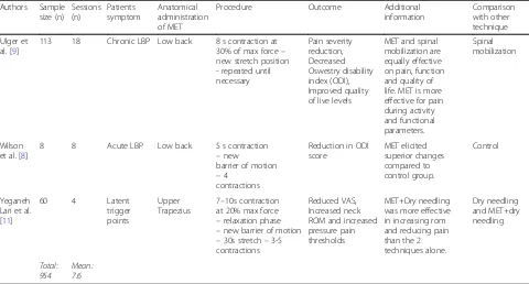
Table 3Summary of evidence of studies with Symptomatic patients(Continued)
Authors Sample size (n)
Sessions (n)
Patients symptom
Anatomical administration of MET
Procedure Outcome Additional
information
Comparison with other technique
Ulger et al. [9]
113 18 Chronic LBP Low back 8 s contraction at 30% of max force– new stretch position - repeated until necessary
Pain severity reduction, Decreased Oswestry disability index (ODI), Improved quality of live levels
MET and spinal mobilization are equally effective on pain, function and quality of life. MET is more effective for pain during activity and functional parameters.
Spinal mobilization
Wilson et al. [8]
8 8 Acute LBP Low back 5 s contraction
–new
barrier of motion –4
contractions
Reduction in ODI score
MET elicited superior changes compared to control group.
Control
Yeganeh Lari et al. [11]
60 4 Latent
trigger points
Upper Trapezius
7–10s contraction at 20% max force –relaxation phase –new barrier of motion –30s stretch–3-5 contractions
Reduced VAS, Increased neck ROM and increased pressure pain thresholds
MET+Dry needling was more effective in increasing rom and reducing pain than the 2 techniques alone.
Dry needling and MET+dry needling
Total:
954 Mean:7.6
Table 4Quality assessment for included studies using the PEDro Score
Authors 1 2 3 4 5 6 7 8 9 10 11 Tot.
Bindra [10] ✓ ✓ ✓ ✓ ✓ ✓ 5
Cassidy et al. [14] ✓ ✓ ✓ ✓ ✓ ✓ ✓ 6
Kamali et al. [35] ✓ ✓ ✓ ✓ ✓ ✓ ✓ ✓ 7
Küçükşen et al. [17] ✓ ✓ ✓ ✓ ✓ ✓ ✓ ✓ ✓ 8
Nagrale et al. [38] ✓ ✓ ✓ ✓ ✓ ✓ ✓ ✓ ✓ 8
Oliveira-Campelo et al. [39] ✓ ✓ ✓ ✓ ✓ ✓ ✓ ✓ 7
Phadke et al. [13] ✓ ✓ ✓ ✓ ✓ ✓ ✓ ✓ 7
Sadria et al. [12] ✓ ✓ ✓ ✓ ✓ ✓ ✓ ✓ ✓ 8
Sakshi et al. [40] ✓ ✓ ✓ ✓ ✓ 4
Selkow et al. [7] ✓ ✓ ✓ ✓ ✓ ✓ ✓ ✓ ✓ 8
Tanwar et al. [42] ✓ ✓ ✓ 2
Ulger et al. [9] ✓ ✓ ✓ ✓ ✓ ✓ ✓ ✓ ✓ ✓ 9
Wilson et al. [8] ✓ ✓ ✓ ✓ ✓ 4
Yeganeh Lari et al. [11] ✓ ✓ ✓ ✓ ✓ ✓ ✓ 6
position was measured with the participant with the shoulders touching a wall. The perpendicular distance from the wall to the anterior portion of the acromion was the calculated forward scapular position. A similar protocol of Laudner et al. [36] was used by Lenehan et al. [21] and was able to achieve an increase in trunk ro-tation on the restricted side of roro-tation in the analyzed population.
Moore et al. [37] applied MET to the glenohumeral joint, for one group to the horizontal abductors and the second group to external rotators for three contractions of 5 s each at 25% of the maximal force of each patient. The horizontal abduction group was then asked to adduce the arm for 30 s after each contraction whereas the external rotator group was asked to actively rotate the arm internally for 30 s after each contraction. The results were then compared to those of a control group which did not receive any intervention. Dominant arm glenohumeral internal and external rotation ROM and glenohumeral horizontal adduction ware passively mea-sured before the MET intervention. The results of Moore et al. [37] showed that MET applied to the hori-zontal abductors increased glenohumeral ROM internal rotation and adduction to a greater extent than MET ap-plied to the external rotators. Both groups had signifi-cant increases of internal rotation and adduction when compared to the control group.
The study performed by Shadmehr et al. [20] applied a MET protocol in women on their hamstring muscles. Each participant prior to the application of the MET protocol was assessed for passive stretch of their ham-string muscles, in order to evaluate hamham-string flexibility. The subsequently applied MET protocol consisted of
three contractions of 10 s each at 50% of the patient’s
maximal force with a ten-second relaxation phase be-tween each contraction. The authors then compared the MET protocol to a static stretch performed for three sets. Each set was composed of three contractions of 10 s each. During each contraction the leg was held for each of the 10 s at the first resistance point of the knee joint perceived by the therapist. The authors reported no significant difference between MET and the static stretching in improving hamstring flexibility.
Two primary protocol types are implemented in the use of MET: the Greenman and Chaitow. The Greenman protocol consists of four contractions of seven to 10 s
each performed at 40% of the patient’s maximal force
with a three-second relaxation phase between tions. The Chaitow Protocol consists of three contrac-tions of seven to 10 s each performed at 40% of the
patient’s maximal force with a three-second relaxation
phase followed by a 30 s stretch applied at the palpated barrier of restriction. Schenk and colleagues [41] applied the Greenman protocol on the cervical region of the
spine. The restriction point of the cervical region was found by the practitioner and if the subject had a limita-tion in extension, left rotalimita-tion and left side bending the practitioner would passively introduce extension, left ro-tation and left side bending to the point of the restric-tion barrier. Each subject was then asked to produce a small isometric force away from the direction of
restric-tion against the practirestric-tioner’s hand. The authors report
that the application of the Greenman protocol for 4 weeks increased ROM of the cervical region in all six ranges of motion. Smith et al. [19] compared the Green-man and the Chaitow protocols described above, applied
to subjects’hamstring muscles to improve their
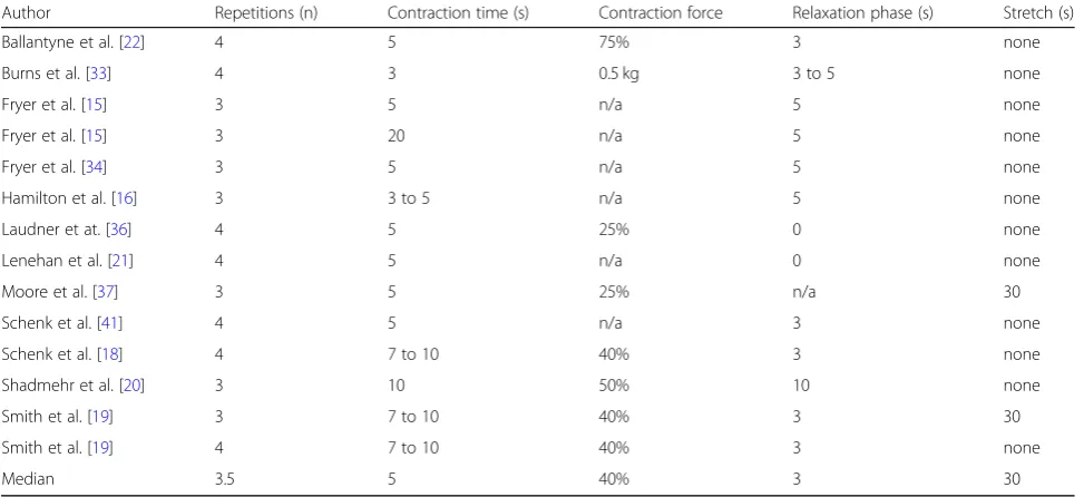
extensi-bility. Each participant was measured for passive ROM of the hamstring muscles. The results obtained from the two protocols highlight that both are effective in increas-ing hamstrincreas-ing extensibility with no significant difference between the two groups. A summary of the MET
proce-dures is shown in Table5.
Symptomatic population
The goal of the prescriptions for MET in the studies evaluated in this review were variable. Of the fourteen included studies, seven investigated the effects of MET
on chronic pain [9, 10, 13, 14, 17, 40, 42], with two of
these intended to treat chronic LBP [9, 10], three
intended to treat chronic neck pain (CNP) [13, 14, 40],
one plantar fasciitis [42] and one chronic lateral epicon-dylitis [17]. Two studies investigated the effects of MET on acute pain [7,8]. Four studies investigated the effects
of MET on trigger points [11, 12, 38, 39], all in the
upper trapezius and one study examined the effects of MET on thoracic kyphosis [35].
The number of sessions for a typical MET prescription varied considerably. The range of those evaluated in this review is one to eighteen. Of the included studies, four
applied MET within a single session [7, 12, 14,39], one
study provided four MET sessions [11], two studies
pro-vided six MET sessions [10, 13], three studies provided
eight MET sessions [8, 17, 40], one study provided
twelve MET sessions [38], one study provided fifteen MET sessions [35] and two studies provided eighteen
MET sessions [9,42].
MET and chronic pain
Two of the retrieved records regarding chronic pain investigated the effects of MET on LBP. The study by Bindra et al. [10] applied four to six contractions of eight
to 10 s each at a force of 25% of the patient’s maximal
of the sacroiliac joint. The joint was treated for the ap-propriate dysfunction identified and for each patient, the restriction barrier was found in order to correctly apply the MET technique. The authors compared MET with conventional therapy (ultrasound for 5 min, intensity of 1
W/cm2and TENS for 10 min 50–100 Hz), revealing that
MET was the most beneficial treatment to manage pain and increase ROM. The Visual Analog Scale (VAS) used to assess pain decreased five points compared to baseline in the MET group and three points for the conventional therapy. The Oswestry Disability Index (ODI) also de-creased 18% after six treatments in the MET group and 13% in the conventional therapy group. Ulger et al. [9] aimed to treat patients complaining of LBP. The authors applied an eight-second contraction at 30% of the patient’s maximal force until a relaxation of the targeted muscles was achieved. The MET procedure was applied on the quadratus lumborum and piriformis muscles. The treat-ment was applied for 18 sessions and compared to spinal mobilization. At the end of the treatment, MET was found more effective in reducing pain (VAS: pre 7 compared to post 2,p< 0.001) and the ODI (pre 46.4 compared to post 18.9, p < 0.001) compared to spinal mobilization (VAS: pre
5 compared to post 2, p= 0.979 and ODI pre 43.5
compared to post 23.5,p= 0.083).
Three of the retrieved records regarding chronic pain investigated the effects of MET on CNP. Sakshi and col-leagues [40] investigated the effects of MET applied with three contractions of 7 to 10 s each at a mild effort in order to reduce pain, disability and forward head pos-ition in patients with CNP. The MET procedure was applied on the suboccipitalis, the upper trapezius and the pectoralis major muscles. After eight treatments,
there was a reduction of the Neck Disability Index (NDI) (MET: pre 34.95 ± 9.74 compared to post 11.99 ± 4.42, p< 0.0001, exercise intervention: pre 34.50 ± 5.92 compared post 22.80 ± 6.79,p< 0.01), reported VAS pain (MET: pre 6 ± 1 compared post 2 ± 1, p < 0.0001; exercise intervention: pre 7 ± 1 compared to post 4 ± 1, p < 0.01), and forward head posture (MET: pre 51.70 ± 1.70% com-pared post 47.18 ± 1.63%, p < 0.01, exercise intervention: pre 52.96 ± 3.70% compared post 50.74 ± 3.91%, p < 0.01). The sample had been compared to an exercise intervention, which revealed a significant difference be-tween the two groups in all measures (p < 0.01 for NDI
and reported pain andp= 0.04 for forward head posture,
which was measured sitting on a chair, calculating the distance from the chair backrest to the tip of the chin).
Phadke et al. [13] compared the effects of MET and static stretching on pain and functional disability. The protocol used by Phadke and colleagues involved three contractions of seven to 10 s each using 20% of maximal isometric contraction with a 20-s stretch between each contraction. The stretching protocol involved five repeti-tions of 20 s holds. The MET and stretching protocols ware applied to the upper trapezius and levator scapulae muscles. After six treatments, the VAS score and the NDI
decreased significantly (VAS: pre 6 compared to post 2,p
< 0.001 NDI: pre 17.25 compared to post 8.03, p < 0.001) compared to the stretching group (VAS: pre 5 compared to post 2, p < 0.001 NDI: pre 17.21 compared to post 9.6,
p < 0.001) (Intragroup difference VAS p= 0.020 and NDI
p= 0.024). Cassidy et al. [14] compared the effects of
HVLA manipulation and MET manipulation in a sample of persons with CNP. CNP was assessed through the 101-point numerical rating scale. The results after Table 5Summary of the MET protocols applied in asymptomatic subjects
Author Repetitions (n) Contraction time (s) Contraction force Relaxation phase (s) Stretch (s)
Ballantyne et al. [22] 4 5 75% 3 none
Burns et al. [33] 4 3 0.5 kg 3 to 5 none
Fryer et al. [15] 3 5 n/a 5 none
Fryer et al. [15] 3 20 n/a 5 none
Fryer et al. [34] 3 5 n/a 5 none
Hamilton et al. [16] 3 3 to 5 n/a 5 none
Laudner et at. [36] 4 5 25% 0 none
Lenehan et al. [21] 4 5 n/a 0 none
Moore et al. [37] 3 5 25% n/a 30
Schenk et al. [41] 4 5 n/a 3 none
Schenk et al. [18] 4 7 to 10 40% 3 none
Shadmehr et al. [20] 3 10 50% 10 none
Smith et al. [19] 3 7 to 10 40% 3 30
Smith et al. [19] 4 7 to 10 40% 3 none
treatment were in favor of the MET, in which a reduction of 17 points was achieved, while that the other treatment resulted in a reduction of 10.5 points in the 101-point nu-merical rating scale score. The protocol applied by Cassidy et al. [14] comprised four contractions of 5 s each. The au-thors, however, specify neither the duration of the relax-ation phase nor the force applied to each contraction. The MET was applied on the muscle responsible for restricting joint movement.
One of the studies included in this review investigated the effects of MET in patients with a history of at least a month of plantar fasciitis and compared the effects with those from static stretching. The MET protocol used by Tanwar et al. [42] comprised three contractions of seven to 10 s each at 20% of the patient’s maximal force with a 3 s relaxation phase and a 30 s stretch after the rela-xation phase between each contraction. The contractions were applied to the soleus and gastrocnemius muscles. The authors analyzed the ROM of the ankle, the foot functional index and pain through the numerical pain rating scale. ROM, the foot functional index and the numerical pain rating scale all improved to a
signifi-cantly greater extent (p< 0.05) after the MET protocol
(mean ROM: pre 6.7° compared to post 14.5° foot func-tional index: pre 43.9 compared to post 24.5 numerical pain rating scale: pre 6.5 compared to post 2.2) com-pared to the static stretching protocol (mean ROM: pre 6.4° compared to post 10.5° foot functional index: pre 43.6 compared to post 29.8 numerical pain rating scale: pre 6.3 compared to post 3.3). The protocol was applied for eighteen sessions over a period of 4 weeks.
Küçükşen et al. [17] analyzed the effects of eight MET
treatments compared to a corticosteroid injection for chronic lateral epicondylitis. The MET protocol used by
Küçükşen et al. comprised five contractions of 5 s each
at 75% of the patient’s maximal force with a five-second
relaxation phase between each contraction. Each con-traction targeted the hand pronator muscles.
The study assessed pain-free grip strength, reported
VAS values and the “Disabilities of the Arm, Shoulder
and Hand questionnaire (DASH)”, respectively. The grip
strength of the affected side was presented as a ratio of the maximum grip strength of the unaffected side. The measurements were performed at baseline, 6, 26, and 52 weeks after the treatments (baseline measurements of pain-free grip strength MET: 40.46 ± 17.26% compared
to corticosteroid injection 44.00 ± 18.64%, p= .495, VAS
MET: 7.39 ± 1.07 compared to corticosteroid injection
7.17 ± 1.07, p= .330, DASH MET: 46.73 ± 11.88
com-pared to corticosteroid injection 45.63 ± 10.40,p= .666). At the six-week evaluation, all measurements improved in both the corticosteroid injection group and the MET group, however the values for the corticosteroid group were significantly greater than those of the MET group
(pain-free grip strength MET: 60.95 ± 19.07% compared
to corticosteroid injection 72.4 ± 19.54%, p< 005, VAS
MET: 4.38 ± 2.08 compared to corticosteroid injection
2.98 ± 2.49, p= .004, DASH MET: 26.25 ± 15.40
com-pared to corticosteroid injection 21.10 ± 14.02, p= .113). At 26 and 52 weeks, the MET group scored significantly better in all measurements. The corticosteroid group at 26 and 52 weeks had the tendency to relapse (26 weeks pain-free grip strength MET, 68.90 ± 19.15% compared
to corticosteroid injection 61.45 ± 19.03%, p= .034, VAS
MET, 4.00 ± 2.59 compared to corticosteroid injection
5.29 ± 2.04, p= .016, DASH MET: 23.78 ± 17.50
com-pared to corticosteroid injection 27.84 ± 14.91, p= .079
and 52 weeks pain-free grip strength MET: 75.08 ± 26.19% compared to corticosteroid injection 62.24 ±
21.83%,p= .007, VAS MET: 3.28 ± 2.86 compared to
cor-ticosteroid injection 4.95 ± 2.36, p= .001, DASH MET =
22.56 ± 20.29 compared to corticosteroid injection 27.03
± 15.45, p= .061). No patients in the MET group
reported side effects from the treatment, whereas three participants out of forty-one experienced side effects in the corticosteroid injection group.
MET and acute pain
Only two studies analyzed the effects of MET on acute pain. Wilson et al. [8] evaluated twelve patients with acute LBP. The authors applied a MET protocol consist-ing four contractions for 5 s each. Neither relaxation phase nor force applied were specified. The procedure was applied on each patients restricted side, directly tar-geting L3. The authors assessed the ODI before and after the application of eight treatments and compared the outcomes with a control group that underwent a ma-nipulative sham treatment. The ODI measures were
significantly improved (p< 0.05) in the MET group (pre
45 vs post 7 with a mean decrease of 83%) compared to the control group (pre 44 vs post 15, with a mean de-crease of 65%). Another study by Selkow and colleagues [7] applied four contractions of 5 s each with a 5 s rela-xation phase on the hamstrings and the iliopsoas muscle to treat non-specific LBP. VAS significantly decreased
after a single application from 2.9 to 2.5 (p= .04)
whereas it increased in the control group treated with a sham therapy from 1.4 to 3.5.
MET and myofascial trigger points
Nagrale et al. [38] compared traditional MET to an in-tegrated neuromuscular inhibition technique, a specific type of treatment for trigger points. Both treatments were applied for twelve sessions. The outcome measures were the VAS, the NDI and ROM of the neck. The MET involved three to five contractions for 7 to 10 s each at
20% of the patient’s maximal force with a 2 to 3 s
rela-xation phase and a 30 s stretch between each contrac-tion. The stretch was performed taking the head and neck into increasing degrees of side bending, flexion and rotation to advance the stretch placed on the muscle. The integrated neuromuscular inhibition technique was a sequence of ischemic compressions over the trigger point followed by strain-counter-strain techniques until a position of ease was found. This procedure was re-peated three to five times. Both groups revealed signi-ficant improvements in all the outcome measurements, although the improvements presented by the integrated neuromuscular inhibition technique group were signifi-cantly greater than those of the MET group.
A similar study design implemented by Oliveira-Cam-pelo et al. [39] comprised three to five contractions of 5
s each at 25% of the patient’s maximal force with a
five-second relaxation phase. The MET protocol was compared to an ischemic compression group, a passive stretching group, a placebo and a no-treatment group. ROM, VAS and pain pressure sensitivity were assessed at baseline, after 10 min, 24 h after the treatment and a week later. After a single treatment, pain thresholds and ROM of contralateral lateral flexion and ipsilateral rota-tion improved in both the manipulative treatment groups (MET: pain thresholds pre 1.8 ± 0.4 kg/cm2
com-pared to post 2.3 ± 0.4 2 kg/cm2, p< 0.01; mean ROM
contralateral flexion: pre 39.8 ± 4.6° compared to post 45.2 ± 4.7°, p < 0.01; mean ROM ipsilateral rotation: pre 70.4 ± 5.7° compared to post 73.4 ± 5.1°, p< 0.01. Ische-mic compression: pain thresholds pre 1.7 ± 0.3 kg/cm2 compared to post 2.9 ± 0.4 kg/cm2, p < 0.01; mean ROM contralateral flexion: pre 39.8 ± 5.1° compared to post 46.8 ± 5.4°, p < 0.01, mean ROM ipsilateral rotation: pre 71.2 ± 5.7° compared to post 76.5 ± 6.7°, p < 0.01). How-ever, the improvements measured a week after the treat-ment seem to have been better maintained in the ischemic compression group compared to the MET group.
Sadria et al. [12] compared the effects of MET to a form of ischemic compression (active release). The au-thors measured VAS before and after a single application of the techniques. Active release consisted of a compres-sion of the trigger point followed by an active motion of
the patient’s neck from a shortened position to an
elon-gated position involving a contralateral neck side flexion and ipsilateral neck rotation. The MET protocol con-sisted of four contractions ranging 7 to 10 s in duration
at 20% of the patient’s maximal force with a
three-second relaxation phase. During the relaxation phase, the head and the neck were eased into increasing de-grees of side bending and rotation, this position being held for 30 s. The outcome of the study reports a reduc-tion in the VAS in both groups with no significant dif-ference between the two treatments.
The last included study in this review analyzed the ef-fects of MET on latent trigger points [11] comparing manual treatment to dry needling. The outcome mea-sures reported by the study were reported score on VAS, PPT and range of active contralateral flexion. The pa-tients were divided into three groups: dry needling alone, MET alone and dry needling plus MET. MET was applied with three to five contractions of 7 to 10 s each
at 20% of the patient’s maximal force with a two to 3 s
relaxation phase and a 30-s stretch between each con-traction. All groups significantly improved in ROM, VAS, and pressure pain threshold. However, the combi-nation of MET and dry needling was more effective than either treatment alone.
MET and other dysfunctions
Of the retrieved records, only one study analyzed other types of dysfunctions than those described above. The study of Kamali et al. [35] applied a MET protocol consisting five repetitions of five to 7 s at 25% of the patient’s maximal force in order to treat postural hyperkyphosis. For each pa-tient, the authors analyzed the dorsal tract of the vertebral column and identified which vertebrae presented the great-est movement rgreat-estriction in extension. The therapist then placed his/her hand on the spinous process of the vertebra in order to move it to the end of the extension barrier and applied the MET protocol above described. In addition to applying MET, the therapist applied a massage to the back extensor muscles for 10 min, a mobilization of the thoracic spine and a myofascial release technique. The exercise ther-apy comprised a combination of strengthening and stretch-ing exercises. Both treatments were carried out for a five-week period. The outcome measures were thoracic ky-phosis angle measured by a six-camera motion analysis sys-tem and muscle strength of the back extensor muscles measured through a dynamometer. All measures improved post-treatment in both groups with no significant differ-ences between groups (Kyphosis angle in upright sitting: MET increase from baseline 2.51 ± 1.92°; Exercise interven-tion increase from baseline 3.17 ± 2.35°,p= 0.855. Kyphosis angle in relaxed sitting: MET increase from baseline 5.16 ± 3.90°; Exercise intervention increase from baseline 5.18 ±
4.25°,p= 0.935. Muscle strength: MET increase from
base-line 26.76 ± 22.65 N; Exercise intervention increase from baseline 27.28 ± 16.50 N,p= 0.175).
Discussion
The aim of this review was to understand the efficacy of MET on pain and joint range of motion, and to under-stand the differences between the different MET proto-cols in symptomatic and asymptomatic subjects. The
quality assessment showed a “moderate to high” quality
level of the included RCT’s.
The analyzed protocols for the asymptomatic subjects, comprised three or four contractions (mode: 4 contrac-tions; median: 3.5 contractions) ranging from three to 10 s in duration (mode and median: 5 s) with contraction
forces ranging from 25 to 75% of the patient’s maximal
force (mode and median: 40%), a relaxation phase ranged between 0 and 5 seconds (mode and median: 3 s) and the stretch phase, which was not applied in 10 of the 14 protocols.
The only protocol directly comparing MET with and without a stretch was that of Smith et al. [19], in which the authors concluded that altering the duration of the stretch does not increase the effects of the technique on muscle extensibility. Fryer et al. [15] also compared the effect of a five- and twenty-second contraction time protocol and concluded that the five-second protocol was more effective in reducing the rotational asymmetry of the atlanto-axial joint. Therefore a shorter protocol can be suggested in an asymptomatic population if the aim of the MET is to increase joint ROM.
In symptomatic patients, the protocols comprised three to six contractions (mode and median: 4 contrac-tions) extending in the range of 5 to 10 s, (mode and median: 8 s) with contraction forces ranging from 20 to
75% of the patient’s maximal force (mode and median:
20%), a relaxation phase between 0 and 10 s (mode and
median: 3 s) and a stretch phase that was not present in 9 of 14 protocols. When reported, the stretch phase ranged between 20 to 30 s (mode and median: 30 s).
The range of contraction forces for MET protocols sug-gested by Chaitow [5] ranges between 15 and 40% of a person’s maximal contraction. The first case is usually ap-plied in acute dysfunctions, whereas the second for chronic dysfunctions. In this systematic review the upper range of contraction forces used is 75% of a person’s ma-ximal contraction, which is far from the suggested range of Chaitow. Only two studies however apply such high contraction intensity, Ballantyne et al. [22] in an
asymp-tomatic population and Küçükşen et al. [17] for chronic
lateral epicondylitis. The aim of Ballantyne was to acutely increase hamstring extensibility and such result was achieved by the authors, who attribute the immediate in-crease in ROM to an inin-creased stretch tolerance. This could mean that a high intensity contraction could pro-duce postsynaptic inhibitory mechanisms, resulting in
lower excitation of the cortical and α-motor neurons,
thereby modulating stretch perception [4]. It is unclear
why Küçükşen et al. [17] performed a 75% contraction
on a symptomatic population, however the results pro-vided by the authors highlight that also with a higher contraction intensity it is possible to achieve positive clinical outcomes.
The results of the studies assessing the effects of MET on chronic LBP all showed decreases of the pain and dis-ability indexes (VAS and ODI). In particular, Bindra et al. [10] compared the effects of MET to conventional treat-ment and both treattreat-ments were similarly effective in redu-cing LBP. Other reviews analyzing the effects of MET on
LBP [43,44] concluded that MET are moderately effective
Table 6Summary of the MET protocols applied in symptomatic patients
Author Repetitions (n) Contraction time (s) Contraction force Relaxation phase (s) Stretch (s)
Bindra [10] 4 to 6 8 to 10 25% 2–3 none
Cassidy et al. [14] 4 5 n/a n/a n/a
Kamali et al. [35] 5 5 to 7 25% 0 none
Küçükşen et al. [17] 5 5 75% 5 none
Nagrale et al. [38] 3 to 5 7 to 10 20% 2–3 30
Oliveira-Campelo et al. [39] 3 to 5 5 25% 5 none
Phadke et al. [13] 5 7 to 10 20% n/a 20
Sadria et al. [12] 4 7 to 10 20% 3 30
Sakshi et al. [40] 3 7 to 10 Mild effort n/a none
Selkow et al. [7] 4 5 n/a 5 none
Tanwar et al. [42] 3 7 to 10 20% 3 30
Ulger et al. [9] Until necessary 8 30% n/a none
Wilson et al. [8] 4 5 n/a n/a none
Yeganeh Lari et al. [11] 3 to 5 7 to 10 20% 2–3 30
for chronic and non-specific LBP for managing pain and disability. There is no evidence that MET are ineffective for patients presenting with LBP. However, both reviews [43,44] posit the necessity of producing higher methodo-logical quality studies in the field.
Only two studies analyzed acute LBP [7, 8] having as
total sample a number of 28 patients. Notwithstanding the limited retrieved records, both studies showed that MET was able to decrease pain and disability indexes after the treatment procedure. The targeted anatomical regions were the hamstrings, the iliopsoas and L3.
In regards to CNP, MET were compared to an exercise intervention, a stretching intervention and a mobilization intervention. In all three studies pertaining to CNP, pain and disability indexes were analyzed and showed that MET were the superior treatment compared to the other inter-ventions for reduction in pain and disability. The study intervention periods range between 2 and 8 treatments.
The only study evaluating plantar fasciitis advocated an increase in ROM in parallel with decreased pain scores after the manipulative treatment [42]. Unfortu-nately, the study of Tanwar et al. [42] is of low methodo-logical quality as reported in the PEDro scale, having no form of blinding, inadequate follow-up, no comparisons between groups and no measures of variability. There-fore, future research on the topic of plantar fasciitis is encouraged in order to evaluate the effects of MET.
Küçükşen and colleagues analyzed the effects of MET
and corticosteroid injection after six, 26 and 52 weeks. Interestingly, the early phase of the treatment following the injection of corticosteroids was more beneficial in reducing pain and increasing pain free grip strength. However, the authors demonstrated a relapse after 26 and 52 weeks in the injection group, whereas a continu-ous reduction in elbow pain was shown in the MET group from 6 to 52 weeks.
Four studies evaluated the effects of MET on
myofas-cial trigger points [11, 12, 38, 39] and successfully
pro-vided evidence that pain and disability indexes are reduced after the application of MET. However, other treatments such as ischemic compressions, integrated neuromuscular inhibition technique, Active Release technique and dry needling are equally (active releases and dry needling) or even more effective (ischemic com-pression and integrated neuromuscular inhibition tech-nique) in reducing the negative symptoms of myofascial trigger points. Thus, in comparison to MET, more spe-cific techniques are more appropriate in the treatment of myofascial trigger points.
Limitations
There was a large heterogeneity in the MET protocols uti-lized. Of the 26 included studies, only 15 provided a full
description of the treatment protocol (number of contrac-tions, contraction time and force, relaxation phase if used and stretch duration, magnitude and hold time utilized between the contractions). Therefore, it is difficult to ge-nerally state which protocol is the most beneficial.
Future studies evaluating MET effectiveness are en-couraged in order to identify which procedure may be more beneficial when treating different musculo-skeletal disorders.
Conclusions
MET are effective in improving reported pain, disability and joint range of motion in both asymptomatic subjects and symptomatic patients. The studies evaluated in this review have provided evidence that MET are specifically effective for alleviating chronic pain of the lower back and neck and chronic lateral epicondylitis. There is also evidence supporting MET as a beneficial therapy for re-ducing acute lower back pain and improving the related disability indexes. However, further evidence is needed to confirm MET as an effective treatment for plantar fasciitis and other musculoskeletal disorders. A defini-tive protocol for MET application, due to the hetero-geneity of the results, could not be identified, and a future evaluation of the parameters of MET prescrip-tion is suggested.
Abbreviations
CNP:Chronic Neck Pain; DASH: Disabilities of the Arm, Shoulder and Hand questionnaire; HVLA: High Velocity Low Amplitude; LBP: Low Back Pain; MET: Muscle Energy Techniques; NDI: Neck Disability Index ODI - Oswestry Disability Index; PNF: Proprioceptive Neuromuscular Facilitation; PPT: Pressure Pain Thresholds; RCT’s: Randomized Control Trials; ROM: Range Of Motion; VAS: Visual Analog Scale
Acknowledgements Not applicable.
Funding None.
Availability of data and materials
The datasets used and/or analysed during the current study are available from the corresponding author on reasonable request.
Authors’contributions
ET and ARC Conceptualization; ET and AB Data collection; ET and DM Drafting the article; AB, DM, AP Critical revision of the article; AP Final approval of the version to be published. All authors read and approved the final manuscript.
Ethics approval and consent to participate Not Applicable.
Consent for publication Not Applicable.
Competing interests
Publisher’s Note
Springer Nature remains neutral with regard to jurisdictional claims in published maps and institutional affiliations.
Author details
1Sport and Exercise Sciences Research Unit, University of Palermo, Via
Giovanni Pascoli 6, 90144 Palermo, Italy.2International Academy of Osteopathic Medicine, AISeRCO, Palermo, Italy.3Department of Integrative Physiology, University of Colorado, Boulder, CO, USA.4Department of Applied Physiology and Kinesiology, University of Florida, Gainesville, FL, USA.
Received: 7 January 2019 Accepted: 16 May 2019
References
1. Fryer G. Muscle energy technique: an evidence-informed approach. Int J Osteopath Med. 2011;14(1):3–9.https://doi.org/10.1016/j.ijosm.2010.04.004. 2. Goodridge JP. Muscle energy technique: definition, explanation, methods of
procedure. J Am Osteopath Assoc. 1981;81(4):249–54.
3. DeStefano LA. Greenman’s principles of manual medicine: LWW -Philadelphia; 2011.
4. Thomas E, Bianco A, Paoli A, Palma A. The relation between stretching typology and stretching duration: the effects on range of motion. Int J Sports Med. 2018;39(4):243–54.https://doi.org/10.1055/s-0044-101146. 5. Chaitow L, Liebenson C. Muscle Energy Techniques: Harcourt
publisher - Boston; 2001.
6. Ptaszkowski K, Slupska L, Paprocka-Borowicz M, Kolcz-Trzesicka A, Zwierzchowski K, Halska U, et al. Comparison of the short-term outcomes after Postisometric muscle relaxation or Kinesio taping application for normalization of the upper trapezius muscle tone and the pain relief: a preliminary study. Evid Based Complement Alternat Med. 2015;2015:721938. https://doi.org/10.1155/2015/721938.
7. Selkow NM, Grindstaff TL, Cross KM, Pugh K, Hertel J, Saliba S. Short-term effect of muscle energy technique on pain in individuals with non-specific lumbopelvic pain: a pilot study. J Man Manip Ther. 2009;17(1):E14–8 10. 1179/jmt.2009.17.1.14E.
8. Wilson E, Payton O, Donegan-Shoaf L, Dec K. Muscle energy technique in patients with acute low back pain: a pilot clinical trial. J Orthop Sports Phys Ther. 2003;33(9):502–12.https://doi.org/10.2519/jospt.2003.33.9.502. 9. Ulger O, Demirel A, Oz M, Tamer S. The effect of manual therapy and
exercise in patients with chronic low back pain: double blind randomized controlled trial. J Back Musculoskelet Rehabil. 2017;30(6):1303–9.https://doi. org/10.3233/BMR-169673.
10. Bindra S. A study on the efficacy of muscle energy technique as compared to conventional therapy on lumbar spine range of motion in chronic low Back pain of sacroiliac origin. Human Biol Rev. 2013;2(4):13.
11. Yeganeh Lari A, Okhovatian F, Naimi S, Baghban AA. The effect of the combination of dry needling and MET on latent trigger point upper trapezius in females. Man Ther. 2016;21:204–9.https://doi.org/10.1016/j. math.2015.08.004.
12. Sadria G, Hosseini M, Rezasoltani A, Akbarzadeh Bagheban A, Davari A, Seifolahi A. A comparison of the effect of the active release and muscle energy techniques on the latent trigger points of the upper trapezius. J Bodyw Mov Ther. 2017;21(4):920–5.https://doi.org/10.1016/j.jbmt.2016.10.005. 13. Phadke A, Bedekar N, Shyam A, Sancheti P. Effect of muscle energy
technique and static stretching on pain and functional disability in patients with mechanical neck pain: a randomized controlled trial. Hong Kong Physiother J. 2016;35:5–11.https://doi.org/10.1016/j.hkpj.2015.12.002. 14. Cassidy JD, Lopes AA, Yong-Hing K. The immediate effect of manipulation
versus mobilization on pain and range of motion in the cervical spine: a randomized controlled trial. J Manip Physiol Ther. 1992;15(9):570–5. 15. Fryer G, Ruszkowski W. The influence of contraction duration in muscle
energy technique applied to the atlanto-axial joint. J Osteopath Med. 2004; 7(2):79–84.https://doi.org/10.1016/S1443-8461(04)80016-9.
16. Hamilton L, Boswell C, Fryer G. The effects of high-velocity, low-amplitude manipulation and muscle energy technique on suboccipital tenderness. Int J Osteopath Med. 2007;10(2):42–9.https://doi.org/10. 1016/j.ijosm.2007.08.002.
17. Kucuksen S, Yilmaz H, Salli A, Ugurlu H. Muscle energy technique versus corticosteroid injection for management of chronic lateral epicondylitis:
randomized controlled trial with 1-year follow-up. Arch Phys Med Rehabil. 2013;94(11):2068–74.https://doi.org/10.1016/j.apmr.2013.05.022. 18. Schenk RJ, MacDiarmid A, Rousselle J. The effects of muscle energy
technique on lumbar range of motion. J Man Manip Ther. 1997;5(4):179–83. https://doi.org/10.1179/jmt.1997.5.4.179.
19. Smith M, Fryer G. A comparison of two muscle energy techniques for increasing flexibility of the hamstring muscle group. J Bodyw Mov Ther. 2008;12(4):312–7.https://doi.org/10.1016/j.jbmt.2008.06.011.
20. Shadmehr A, Hadian MR, Naiemi SS, Jalaie S. Hamstring flexibility in young women following passive stretch and muscle energy technique. J Back Musculoskelet Rehabil. 2009;22(3):143–8.https://doi.org/10.3233/ BMR-2009-0227.
21. Lenehan KL, Fryer G, McLaughlin P. The effect of muscle energy technique on gross trunk range of motion. J Osteopath Med. 2003;6(1):13–8.https:// doi.org/10.1016/S1443-8461(03)80004-7.
22. Ballantyne F, Fryer G, McLaughlin P. The effect of muscle energy technique on hamstring extensibility: the mechanism of altered flexibility. J Osteopath Med. 2003;6(2):59–63.https://doi.org/10.1016/S1443-8461(03)80015-1. 23. Feland JB, Myrer JW, Schulthies SS, Fellingham GW, Measom GW. The
effect of duration of stretching of the hamstring muscle group for increasing range of motion in people aged 65 years or older. Phys Ther. 2001;81(5):1110–7.
24. Lewit K. Manipulative therapy in rehabilitation of the locomotor system: Butterworths - Oxford; 1999.
25. Crone C. Reciprocal inhibition in man. Dan Med Bull. 1993;40(5):571–81. 26. Magnusson SP, Simonsen EB, Aagaard P, Dyhre-Poulsen P, McHugh MP,
Kjaer M. Mechanical and physical responses to stretching with and without preisometric contraction in human skeletal muscle. Arch Phys Med Rehabil. 1996;77(4):373–8.
27. Moritani T, Muramatsu S, Muro M. Activity of motor units during concentric and eccentric contractions. Am J Phys Med. 1987;66(6):338–50.
28. Skyba DA, Radhakrishnan R, Rohlwing JJ, Wright A, Sluka KA. Joint manipulation reduces hyperalgesia by activation of monoamine receptors but not opioid or GABA receptors in the spinal cord. Pain. 2003;106(1–2):159–68.
29. Gill KP, Callaghan MJ. The measurement of lumbar proprioception in individuals with and without low back pain. Spine (Phila Pa 1976). 1998; 23(3):371–7.
30. Moher D, Liberati A, Tetzlaff J, Altman DG, Group P. Preferred reporting items for systematic reviews and meta-analyses: the PRISMA statement. PLoS Med. 2009;6(7):e1000097.https://doi.org/10.1371/journal.pmed.1000097. 31. Olivo SA, Macedo LG, Gadotti IC, Fuentes J, Stanton T, Magee DJ. Scales to
assess the quality of randomized controlled trials: a systematic review. Phys Ther. 2008;88(2):156–75.https://doi.org/10.2522/ptj.20070147.
32. Moseley AM, Herbert RD, Sherrington C, Maher CG. Evidence for physiotherapy practice: a survey of the physiotherapy evidence database (PEDro). Aust J Physiother. 2002;48(1):43–9. https://doi.org/10.1016/S0004-9514(14)60281-6.
33. Burns DK, Wells MR. Gross range of motion in the cervical spine: the effects of osteopathic muscle energy technique in asymptomatic subjects. J Am Osteopath Assoc. 2006;106(3):137–42.
34. Fryer G, Pearce AJ. The effect of muscle energy technique on corticospinal and spinal reflex excitability in asymptomatic participants. J Bodyw Mov Ther. 2013;17(4):440–7.https://doi.org/10.1016/j.jbmt.2013.05.006. 35. Kamali F, Shirazi SA, Ebrahimi S, Mirshamsi M, Ghanbari A. Comparison of
manual therapy and exercise therapy for postural hyperkyphosis: a randomized clinical trial. Physiother Theory Pract. 2016;32(2):92–7.https:// doi.org/10.3109/09593985.2015.1110739.
36. Laudner KG, Wenig M, Selkow NM, Williams J, Post E. Forward shoulder posture in collegiate swimmers: a comparative analysis of muscle-energy techniques. J Athl Train. 2015;50(11):1133–9. https://doi.org/10.4085/1062-6050-50.11.07.
37. Moore SD, Laudner KG, McLoda TA, Shaffer MA. The immediate effects of muscle energy technique on posterior shoulder tightness: a randomized controlled trial. J Orthop Sports Phys Ther. 2011;41(6):400–7.https://doi.org/ 10.2519/jospt.2011.3292.
38. Nagrale AV, Glynn P, Joshi A, Ramteke G. The efficacy of an integrated neuromuscular inhibition technique on upper trapezius trigger points in subjects with non-specific neck pain: a randomized controlled trial. J Man Manip Ther. 2010;18(1):37–43.https://doi.org/10.1179/
39. Oliveira-Campelo NM, de Melo CA, Alburquerque-Sendin F, Machado JP. Short- and medium-term effects of manual therapy on cervical active range of motion and pressure pain sensitivity in latent myofascial pain of the upper trapezius muscle: a randomized controlled trial. J Manip Physiol Ther. 2013;36(5):300–9.https://doi.org/10.1016/j.jmpt.2013.04.008.
40. Sakshi N, Suman M, Geetanjali S. Effect of muscle energy technique and deep neck flexors exercise on pain, disability and forward head posture in patients with chronic neck pain. Indian J Physiother Occup Ther. 2014;8(4):6. 41. Schenk R, Adelman K, Rousselle J. The effects of muscle energy technique
on cervical range of motion. J Man Manip Ther. 1994;2(4):6.
42. Tanwar A, Moitra M, Goyal M. Effect of muscle energy technique to improve flexibility of gastro-soleus complex in plantar fasciitis: a randomised clinical, prospective study design. Indian J Physiother Occup Ther. 2014;8(4):5. 43. Franke H, Fryer G, Ostelo RW, Kamper SJ. Muscle energy technique for
non-specific low-back pain. Cochrane Database Syst Rev. 2015;(2):CD009852. https://doi.org/10.1002/14651858.CD009852.pub2.