Pharmacophore
ISSN-2229-5402
Journal home page: http://www.pharmacophorejournal.com
Corresponding Author: Mohammad Motamedifar. Shiraz HIV/AIDS Research Center, Institute of Health & Department of Bacteriology and Virology, Shiraz University of Medical Science, Shiraz, Iran. Email: motamedm @ yahoo.com
ANTIBIOTIC SENSITIVITY PROFILE OF THE BACTERIAL
ISOLATES FROM THE BLOOD SAMPLES OF THE PATIENTS IN
DIFFERENT WARDS OF A MAJOR REFERRAL HOSPITAL, SHIRAZ,
IRAN 2015-2016
Aida Salehi Nobandegani
1, Mohammad Motamedifar
2, 3*
1.
Student research committee, Shiraz University of Medical Sciences, Shiraz, Iran
.
2.
Shiraz HIV/AIDS Research Center, Institute of Health & Department of Bacteriology and
Virology, Shiraz University of Medical Science, Shiraz, Iran
.
3.
Ph.D. in Microbiology, Professor of Microbiology, Shiraz University of Medical Sciences,
Fars, Iran.
To Cite This Article: Aida Salehi Nobandegani, Mohammad Motamedifar (2019), “Antibiotic Sensitivity Profile of the Bacterial Isolates from the Blood Samples of the Patients in Different Wards of A Major Referral Hospital, Shiraz, Iran 2015-2016”, Pharmacophore, 10(2), 30-36.
Introduction
Infectious diseases have always constituted the most serious health issue in the world, at least until the beginning of the 20th century when chronic degenerative diseases started to develop in developed countries. In his classic Plagues and Peoples, 3
McNeill analyzed the importance of infectious diseases in the history of humanity. He concluded that the role of infectious diseases in the course of the historical evolution of human civilization has been underestimated. The explosive characteristics and unpredictability of epidemics are a source of fear, insecurity, and panic even today, as could be clearly seen during the recent SARS epidemic [1].
However, as mentioned before, what is not to be ignored is antimicrobial resistance in bacterial pathogenesis which has become a worldwide challenge associated with high morbidity and mortality. Multidrug-resistant patterns in gram-positive and gram-negative bacteria have caused difficult to treat or even untreatable diseases with conventional antimicrobials. Since the early diagnosis of causative microorganisms and their antimicrobial susceptibility patterns in subjects with bacteremia and other serious infections is lacking in many health care settings, broad-spectrum antibiotics are mostly needlessly utilized.
A R T I C L E I N F O A B S T R A C T
Received:
29th Oct 2018
Received in revised form: 15th Mar 2019
Accepted:
20th Mar 2019 Available online: 25th Apr 2019
Keywords: Antibiotic resistance, Bacteremia, Shiraz, Iran
Introduction: The choice of antimicrobial treatment for septicemia is usually empirical and based on health knowledge of local antimicrobial activity patterns of the most prevalent bacteria causing such bloodstream infections. The present study aimed to investigate the prevalence of bacterial pathogens causing bacteremia and their antimicrobial resistance profiles in hospitalized subjects. Materials and Methods: This cross sectional study was done at a major referral hospital, Faghihi Hospital, Shiraz, Iran. We examined 1262 positive blood cultures from 6300 specimens over a period of twelve months from September 2015 to September 2016. Results: Bacterial strains were isolated from 20% of blood cultures. The identified gram-negative bacteria were Alcaligenes (29.6%), Escherichia coli (7%), Klebsiella (3.7%), Acintobacter baumannii (3.6%), Pseudomonas (2%), Enterobacter (1.2%) and Brucella (1%). Of gram positive strains Staphylococcus aureus (27.2%) Staphylococcus epidermidis (11.4%) Non-hemolytic streptococci (4.6%), Diphtroids (4%), Enterococci (3.6%), and Micrococci (0.6%), were the most frequent isolates. Imipenem, Piperacilin/ tazobactam, Gentamicine and Amikacin were the most effective antibiotics against gram negative agents. Vancomycin, Rifampin, Cephalotin and Cefazolin were the most active antibiotics against gram negative bacterial agents. Conclusion: Resistance to majority of the antimicrobial agents for several pathogens implicated in bloodstream infections, particularly in gram-negative bacteria which cause complications in treatment of septicemia. Since S. epidermidis and Alcaligenes are normal flora of human’s skin and body, contamination of needle during blood sampling must be considered in such positive results. So we recommend considering better infection control precautions in blood sampling in the hospital.
Dramatic increases in emerging resistance happen and, when linked with poor infection control methods, resistant bacteria can easily be distributed to the other patients and environments 2. Availability of renewed epidemiological data on
antimicrobial resistance in frequently encountered bacterial pathogens will be helpful not only for determining treatment strategies but also for devising an efficient antimicrobial stewardship program in hospitals [1, 2].
Resistant bacteria, especially Staphylococci, Enterococci, Klebsiella pneumoniae, and Pseudomonas spp., are becoming common in healthcare institutions. Bacterial resistance often ends in treatment failure, which can have severe outcomes, especially in critically ill patients. Inadequate empiric antibacterial therapy, described as the primary use of an antibacterial agent to which the causative pathogen was not sensitive. It has been associated with increased mortality rates in patients with bloodstream infections due to resistant P. aeruginosa, S. aureus, K. pneumoniae, Escherichia coli, Enterobacter spp., coagulase-negative Staphylococci, and Enterococci [3]. Prolonged therapy with antimicrobial agents, such as vancomycin or linezolid, may also result in the development of low level resistance that compromises therapy, but that may not be detected by routine susceptibility testing methods used in hospital laboratories [4].
Therefore, usually susceptible populations of bacteria may become resistant to antimicrobial agents by mutation and selection, or by acquiring from other bacteria the genetic information that encodes resistance. The last event may happen through one of several genetic mechanisms, including transformation, conjugation, or transduction. By genetic exchange mechanisms, many bacteria have become resistant to various types of antibacterial agents. These bacteria with multidrug
resistance (defined as resistance to ≥ 3 antibacterial drug classes) have become a cause for serious concerns, particularly in hospitals and other healthcare institutions where they tend to occur most regularly [5].
As noted above, susceptible bacteria can gain resistance to an antimicrobial agent via new mutations. Such spontaneous mutations may induce resistance by changing the target protein to which the antibacterial agent binds by modifying or eliminating the binding site (e.g., change in penicillin-binding protein 2b in Pneumococci, which leads to penicillin resistance), upregulating the production of enzymes that inactivate the antimicrobial agent (e.g., erythromycin ribosomal methylase in Staphylococci), down-regulating or changing an outer membrane protein channel that the drug needs for cell entry (e.g., OmpF in E. coli), or up-regulating pumps that discharge the drug from the cell (efflux of fluoroquinolones in S. aureus). In all of these examples, strains of bacteria carrying resistance-conferring mutations are selected by antimicrobial application, which kills the susceptible strains but allows the newly resistant strains to persist and grow. Acquired resistance that develops because of chromosomal mutation and selection is termed vertical evolution [6].
Finding approaches against the development of antibiotic resistance is an important global challenge for the life sciences community and for public health. The past decades have seen a dramatic global rise in human-pathogenic bacteria that are resistant to one or various antibiotics. More infections caused by resistant microorganisms fail to respond to conventional treatment, and even last-resort antibiotics have lost their powers. The appearance of resistant infections caused by these bacteria has led to mortality and morbidity and there is an instant need to discover solutions to resist bacterial resistance [7]. In this study, we aimed to investigate the prevalence of bacterial pathogens causing bacteremia and their antimicrobial resistance profiles in hospitalized patients in Faghihi hospital as a major referral hospital in Shiraz, southwest of Iran.
Materials and Methods
This retrospective study was done on laboratory records of the blood cultures of patients with nosocomial or community acquired infections who were admitted at the different wards of Faghihi Hospital, a tertiary care hospital affiliated to Shiraz University of Medical Sciences, the major referral center of infectious disease in southwest of Iran during a 12-month period (September 2015 to September 2016). This study was approved in the ethic committee of the university. For doing the study a questionnaire form was designed in order to record patients' data including age, sex, culture and antibiotic sensitivity test results. All blood samples were taken in aseptic conditions. Blood samples were gathered after careful cleaning of the puncture site with 70% alcohol and consequently followed by povidone iodine. Under the aseptic conditions 5 mL of blood was drawn by venipuncture and transmitted into culture bottles. The bottles were incubated at 37°C for 10 days aerobically. Routine subculturing was done on MacConkey agar and 5% sheep blood agar after 24 hours, 48 hours, 5th day and 10th day. Microorganisms that recovered from blood culture were identified by cultural features, morphology and variety of standard biochemical tests such as oxidase, TSI, SIM, citrate, OF, Lysine decarboxylase, gelatinase, MR/VP, etc.
The Kirby-Bauer disk diffusion approach was utilized to test antimicrobial susceptibility (in Mueller-Hinton agar medium, Merck, Germany) based on the guidelines of the Clinical Laboratory Standards Institute (CLSI, 1999) [8]. The performed antibiogram disks included vancomycin (V, 30 mcg), ciprofloxacin (CIP, 5 mcg), clindamycin (CD, 2 mcg), trimethoprim-sulfamethoxazole (SXT, 30 mcg), gentamicin (GM, 10 mcg), ofloxacin (OFX, 5 mcg), penicillin G (P, 10 mcg), oxacillin (OXA, 5 mcg), amikacin (AMK, 30 mcg), cefotaxime (CTX, 30 mcg), ceftazidime (CAZ, 30 mcg), ceftriaxone (CTR, 30 mcg), cefazolin (CZ, 30 mcg), chloramphenicol (CHL, 30 mcg), piperacillin (PIP, 100 mcg), imipenem (IPM, 10mcg), nitrofurantoin (FM, 50 mcg), ampicillin (AM, 10 mcg) and rifampin (RIF, 5 mcg).
Results
A total number of 6300 blood samples were requested for culture in our 12-month study time periods. From this numbers just 20% (1262 samples) were positive. 1262 blood culture samples were from patients with mean age of 55.2 years old hospitalized in different wards. They included 686 males (54.4%) and 576 females (45.6%).
The most frequent isolated microorganisms were as follow (Tables 1, 2).
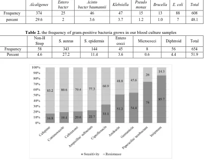
Table 1. the frequency of gram-negative bacteria grows in our blood culture samples
Alcaligenes Entero bacter
Acinto
bacter baumannii Klebsiella
Pseudo
monas Brucella E. coli Total
Frequency 374 25 46 47 15 13 88 608
percent 29.6 2 3.6 3.7 1.2 1.0 7 48.1
Table 2. the frequency of gram-positive bacteria grows in our blood culture samples
Non-H
Strep S. aureus S. epidermis
Entero
cocci Micrococci Diphtroid Total
Frequency 58 343 144 45 8 56 654
Percent 4.6 27.2 11.4 3.6 0.6 4.4 51.9
Figure 1: The sensitivity and resistance of gram-negative microorganisms to different antibiotics
Figure 1 shows in different antibiotics that covered the gram-negative microorganisms, Imipenem followed by Piperacilin/ tazobactam, Gentamicin and Amikacin had the best coverage on gram-negative microorganisms respectively. Sensitivity of microorganism to Imipenem was 5.7%, to Piperacilin/ tazobactam was 74%, to Gentamicin was 54.4%, and to Amikacin was 51.2%. Our results showed that just 16.8% of microorganisms were sensitive to Cefepime.
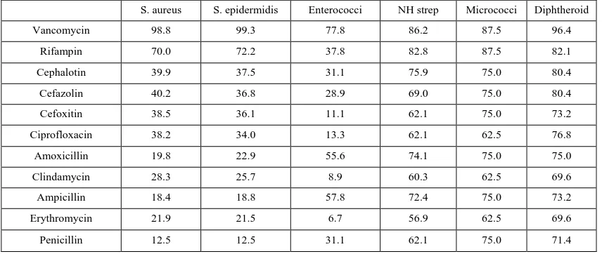
Figure 2 shows in different antibiotics that covered the gram-positive microorganisms, Vancomycin followed by Rifampin, Cephalotin and cefazolin have the best coverage on gram-positive microorganisms, respectively. Sensitivity of microorganism to Vancomycin was 96%, to Rifampin was 70.6%, to Cephalotin was 45.9%, and to Cefazolin was 45.1%. Our results showed that Penicillin has the least coverage on our isolated microorganisms.
Table 3: Sensitivity and resistance profile of gram-positive and -negative microorganisms to Ciprofloxacin.
Sensitivity Resistance
Frequency Percent Frequency Percent
Gram-positives 270 57.3 384 48.5
Gram-negatives 201 42.7 407 51.5
As Table 3 shows, gram-positive microorganisms were more sensitive to Ciprofloxacin than gram-negative ones. We used Chi-Square test in our analysis to investigate the relation between type of microorganisms and their sensitivity to ciprofloxacin, which was statically significant (P-value=0.002).
We investigated the percentages of sensitivity of each microorganism to different antibiotics as follow (Tables 4, 5).
Table 4: Sensitivity profile of gram-negative bacteria to different tested antibiotics.
Entrobacter
(n=25)
Acintobacter baumannii
(n=46)
E. coli
(n=86)
Alcaligenes
(374)
Klebsiella
(n=47)
Pseudomonas
(n=15)
Brucella
(n=13)
Imipenem 84.0 6.5 98.9 93.6 78.7 66.7 100
Ceftriaxone 36.0 0.0 31.8 15.5 34.0 6.7 100
Ampicillin/ sulbactam 16.0 0.0 36.4 19.8 31.9 0.0 100
Piperacilin/ tazobactam 68.0 2.2 78.4 86.9 36.2 53.3 100
Amikacin 68.0 0.0 95.5 40.6 74.5 66.7 100
Gentamicin 80.0 0.0 80.7 50.3 63.8 60.0 100
Cotrimoxazole 52.0 2.2 22.7 14.2 36.2 6.7 100
Cefepime 32.0 0.0 45.5 6.4 25.5 33.3 100
Ciprofloxacin 72.0 2.2 33.0 29.4 42.6 66.7 100
Among the gram-negatives, Alkaligenes was predominant, as shown in Table 4. Alkaligenes exhibited the most sensitivity to imipenem (98.9%) and Piperacilin/ tazobactam (86.9%), and the least sensitivity to cefepime. The second predominant isolated bacterium was E. coli which was mostly sensitive to imipenem (98.9%), Amikacin (95.5%) and Gentamicin (80.7%).
Table 5: Sensitivity pattern of gram-positive bacteria to different tested antibiotics
S. aureus S. epidermidis Enterococci NH strep Micrococci Diphtheroid
Vancomycin 98.8 99.3 77.8 86.2 87.5 96.4
Rifampin 70.0 72.2 37.8 82.8 87.5 82.1
Cephalotin 39.9 37.5 31.1 75.9 75.0 80.4
Cefazolin 40.2 36.8 28.9 69.0 75.0 80.4
Cefoxitin 38.5 36.1 11.1 62.1 75.0 73.2
Ciprofloxacin 38.2 34.0 13.3 62.1 62.5 76.8
Amoxicillin 19.8 22.9 55.6 74.1 75.0 75.0
Clindamycin 28.3 25.7 8.9 60.3 62.5 69.6
Ampicillin 18.4 18.8 57.8 72.4 75.0 73.2
Erythromycin 21.9 21.5 6.7 56.9 62.5 69.6
Penicillin 12.5 12.5 31.1 62.1 75.0 71.4
Discussion and Conclusion
Bacterial antibiotics resistance is remaining as an alarming problem in the therapy of bloodstream infections. Bacterial bloodstream infections are mostly caused by strains that are resistant to a wide range of antimicrobial agents [9]. The current study investigated the antimicrobial resistance profiles of 1262 bacteria isolated from bloodstream infections. The data demonstrated the frequency of antimicrobial resistance among bacterial pathogens isolated from bloodstream infections. In the present analysis, among a total of 6300 specimens that were sent for culture and sensitivity between September 2015 and September 2016, only 1262 cases were positive. This number does not seem to be considerable during 1 year, considering the national and local treatment guidelines which emphasize that empirical treatment might have contributed to high number of bacterial resistance to antibiotics. Deployment and adherence to antimicrobial therapy guidelines and policies by evidence-based generated data might improve the contemporary situation of high prevalence of antibiotic resistance. Blood specimens contributed to more than half of specimens processed were from emergency wards. This could be explained by the local practice in these wards that emphasizes on the need of blood culture to patients presenting with fever.
The result of our study highlighted the key role of rational prescription of antibiotics. This circumstance becomes more important when it involves the appearance of antimicrobial resistance produced by gram-negative bacilli and MRSA strains. So, the discovery of antimicrobial resistance patterns in every hospital plays an essential role in the management of infections.Resistant bacteria may also spread and make broader infection-control problems, not only within healthcare institutions, but in communities as well [10]. Clinically important bacteria, such as methicillin-resistant S. aureus (MRSA) and extended-spectrum-lactamase (ESBL) producing E. coli, are frequently observed in the community. More infections caused by resistant microorganisms fail to respond to standard treatments, and even last-resort antibiotics have lost their power. The emergence of resistant infections caused by these bacteria has led to mortality and morbidity and there is an urgent need to find solutions to combat bacterial resistance [2].
Gram-negative bacteria are the main causes of bloodstream infections in many countries. Also, this type of bacteria has been the most common contributing pathogens of bloodstream infections in the present study. This is important that, different etiological agents of bloodstream infections can be associated with the varying demography of bloodstream infections in developing countries due to different geographical area [11].
Some studies have demonstrated that Acinetobacter species, S. typhi and E. coli were the most common gram-negative bacteria that cause bloodstream infections. In a study conducted in Brazil hospitals, from 3807 samples, E. coli followed by Klebsiella were the most common gram-negative isolated bacteria [12]. In a study conducted in India, the most frequently isolated gram-negative bacteria included P. aeruginosa, E. coli, K. pneumoniae, and S. typhi other than Citrobacter, Acinetobacter, Proteus, and Enterobacter spp. [7].
In our study, Alcaligenes followed by E. coli were the most common gram-negative pathogens. In the mentioned study in Brazil hospitals S. aureus followed by Coagulase-negative Strep were the most common gram-positive bacteria. In a study performed in India, the most frequently isolated gram-positive bacteria was S. aureus, followed by E. feacalis and the other remaining Streptococcus and Staphylococcus spp. [13]. In our study, S. aureus followed by S. epidermidis were the most common causes of gram-positive infections.
Our result is in agreement with the findings of a study in the ICUs in Tabriz hospitals that most common gram-positive organisms recovered were S. aureus followed by S. epidermidis. This result is also in agreement with the findings of Mohammad Taheri et al. (2010) and Khalili et al. (2012) conducted in ICU and infectious ward settings in Iran.
Gram-negative bacteria are more resistant to antibiotics than positive; in our study, the total resistance of gram-negative bacteria to antibiotics were 51.5%, comparing to the 48.5% in gram-positive strains [14]. In our study, among gram-negative strains, Acintobacter baumannii was the most resistant bacterium against antibiotics. 93.5 percent of Acintobacter baumanniis were resistant to Imipenem as the best antibiotic choice against gram-negatives in our study. In Hamishekar et al.’s study, Acintobacter spp. was documented as one of gram-negative multi-drug resistant strains (MDR in 25.8% of Acineobacter baumannii). In our study 87.3% of Acintobacter baumanniis were MDR. Pseudomonas was found in 1.3% of our culture samples. 33.3% of Pseudomonas was resistant to Imipenem. In Hamishekar et al.’s study, Pseudomonas was the third most common strain isolated from samples with MDR in 16.6% of cases. More than 50% gram negatives were resistant to Ceftriaxone, Ampicillin/ sulbactam, Cotrimoxazole, Cefepime. Klebsiella was found in 3.7% of the cases in our study. In Moremi’s study, klebsiella was the second most prevalent strain (14.8%), and 38.5% of them were resistant to third generation Cephalosporins. In our study, 23.3% of these strains were resistant to Imipenem. More than 50% of gram-negatives were resistant to Ceftriaxone, Ampicillin/ sulbactam, Piperacilin/ tazobactam, Cotrimoxazole, Cefepime, and Ciprofloxacin. Klebsiella showed least resistance to carbapenems and moderate resistance to aminoglycosides, tigecycline, and beta-lactam beta-lactamase inhibitor combination.
Entrobacter was found in 2% of the cases in our study, 16.0% was resistant to Imipenem. In Hamishekar’s study, the most frequently isolated microorganisms were Enterobacter spp. with 84% resistance to Imipenem. In Anvarinejhad’s study in Namazi hospital in 2015, the Enterobacteriaceae family was resistant to the majority of antibiotics tested, except colistin, imipenem, amikacin, and meropenem.
blood cultures. A percentage of positive results may be biased in these kind of studies [15]. E. coli was the second most common negative strain isolated from our cultures, just 1.1% of this strain was resistant to Imipenem. Amikacin and Gentamicin were the next most effective antibiotics against these bacteria. In Manjula’s study, Ceftriaxone and Cefotaxim were the most effective in vitro against E. coli. In Anvarinejhad’s study, E. coli was the most frequent organism isolated from cultures in the Namazee hospital. After Imipenem, Piperacilin/ tazobactam, Gentamicin and Amikacin were the antibiotics with most efficacy against gram-negative agents. Among gram-positive strains, Enterococci were the most resistant bacteria against antibiotics. It was found in 3.6% of cultures, 22.2 percent of Enterococci was resistant to Vancomycin as the best antibiotic choice against gram-positives in our study. In Biswas’ study, 12.0% isolates were either vancomycin resistant or vancomycin intermediate. In Anvarinejad’s study, the increased prevalence of Enterococci has emerged as a public health concern. Cristich mentioned that the widespread resistance of Enterococci has had a substantial impact on our use of both empirical and definitive antibiotics for the treatment of Enterococcal infections, a situation that is likely to persist for the foreseeable future.
Non-hemolytic streptococci were the third most common gram-positive strains isolated in our study (4.6%). In Manjula’s study, conducted in a hospital in north India, after Staphylococci and Enterococci it was the third strain isolated from blood cultures. 13.8% of Non-hemolytic streptococci was resistant to vancomycin; in Manjula’s study this strain was resistant in 25.4% of cultures to vancomycin. Rifampin, Cephalotin, and amoxicillin were the next most effective antibiotics against this strain. S. aureus (27.2%) followed by S. epidermdis (11.4%) were the most gram-positive strains isolated from our samples; likewise, in Manjula’s study, S. aureus was the most prevalent gram-positive isolates. In Tung’s study, S. aureus was the most common bacterium among both gram-negative and gram-positive strains. Also, in Hamishekar’s study, S. aureus was the most frequent pathogen among gram-positives (39.7%). Because S. epidermidis is a normal flora of the skin, so contamination of needle should be considered in positive results and probably a percent of positive results may be biased in this study [16]. The rate of methicillin-resistant S. aureus (MRSA) was 87.5%. However, in our study 98.0% and 99.3% of S. aureus and S. epidermidis were sensitive to vancomycin, respectively. It should be noted that disk diffusion method is not a reliable method for the detection of vancomycin susceptibility testing according to CLSI standards [17].
After Vancomycin, Rifampin, Cephalotin and Cefoxitin were the antibiotics with most efficacy against gram-negative agents. In Hamishekar’s study the most active antimicrobials were vancomycin (93.5%) followed by amikacin (71.5%) and gentamicin (46%). In a study conducted in India in 2013, S. aureus showed maximum resistance to amoxicillin (100%), and ampicillin (91.7%). In our study, S. aureus showed maximum resistance to Penicillin (87.5%), and ampicillin (81.6%). In a study conducted by Jopani in Shiraz (2008), Vancomycin and imipenem were the most active antibiotics against gram-positive and gram-negative bacteria, similar to the results of our study [18].
Similar to other studies, extensive frequency of antimicrobial resistance levels was distinguished in our study. The high frequency of antibiotic resistance rates in our medical center might be because of undifferentiated and excess use of treatment in our country due to their easy availability. Another cause could be the altering patterns of antibiotic utilization that varies in lifestyle. In light of our findings, there is an increasing requirement for new agents. Appropriate antimicrobial treatment for bloodstream infections is essential in declining morbidity and mortality among patients with bloodstream infections caused by bacteria. Therefore, accurate microbiological diagnosis and their antimicrobial resistance profile can be very important for rapid initiations of sufficient treatment for bloodstream infections.
Acknowledgements:
This study was related to thesis of Dr. Aida Salehi Nobandegani for receiving MD degree and was financially supported by Shiraz University of Medical Sciences Grant Number11743.
Conflicts of Interest: None.
References
1. Barreto ML, Teixeira MG, Carmo EH. Infectious diseases epidemiology. J Epidemiol Community Health. 2006 Mar;60(3):192-5.
2. Moremi N, Claus H, Mshana SE. Antimicrobial resistance pattern: a report of microbiological cultures at a tertiary hospital in Tanzania. BMC Infect Dis. 2016 Dec 13;16(1):756-56.
3. Rabirad N, Mohammadpoor M, Lari AR, Shojaie A, Bayat R, Alebouyeh M, et al. Antimicrobial susceptibility patterns of the gram-negative bacteria isolated from septicemia in Children's Medical Center, Tehran, Iran. J Prev Med Hyg. 2014 Mar;55(1):23-6.
5. Hsueh PR, Chen WH, Luh KT. Relationships between antimicrobial use and antimicrobial resistance in Gram-negative bacteria causing nosocomial infections from 1991-2003 at a university hospital in Taiwan. Int J Antimicrob Agents. 2005 Dec;26(6):463-72.
6. Gohel K, Jojera A, Soni S, Gang S, Sabnis R, Desai M, et al. Bacteriological profile and drug resistance patterns of blood culture isolates in a tertiary care nephrourology teaching institute. Biomed Res Int. 2014;20(14):153-47. 7. Blair JM, Webber MA, Baylay AJ, Ogbolu DO, Piddock LJ. Molecular mechanisms of antibiotic resistance. Nat Rev
Microbiol. 2015 Jan;13(1):42-51.
8. NCCLS. Laboratory automation: specimen container/specimen carrier; proposed standard. NCCLS Document AUTO1-P [ISBN 1-56238-378-7]. Wayne, PA: NCCLS, 1999:1–20
9. Roach S, Wallinga D. Commentary on genetic mechanisms of antimicrobial resistance in bacteria from U.S. food animals: ESBLs are here. Front Microbiol. 2013 Jul 30;4(2):214-96.
10. Japoni A, Farshad S, Alborzi A, Kalani M, Rafaatpour N, Oboodi B, et al. Epidemiology and antibacterial susceptibility patterns of bloodstream infections, 2001-2004: an experience with BACTEC 9240 in Southern Iran. Pak J Biol Sci. 2008 Feb 1;11(3):422-7.
11. Karlowsky JA, Jones ME, Draghi DC, Thornsberry C, Sahm DF, Volturo GA, et al. Prevalence and antimicrobial susceptibilities of bacteria isolated from blood cultures of hospitalized patients in the United States in 2002. Ann Clin Microbiol Antimicrob. 2004 May 10;3:7.
12. Bhullar K, Waglechner N, Pawlowski A, Koteva K, Banks ED, Johnston MD, et al. Antibiotic resistance is prevalent in an isolated cave microbiome. PLoS One. 2012;7(4): 349-53.
13. Gales AC, Sader HS, Ribeiro J, Zoccoli C, Barth A, Pignatari AC, et al. Antimicrobial susceptibility of gram-positive bacteria isolated in Brazilian hospitals participating in the SENTRY Program (2005-2008). Braz J Infect Dis. 2009 Apr;13(2):90-8.
14. Cloutier M, Mantovani D, Rosei F. Antibacterial Coatings: Challenges, Perspectives, and Opportunities. Trends Biotechnol. 2015 Nov;33(11):637-52.
15. Aisenberg, G, K.V. Rolston and A. Safdar, 2004. Bacteremia caused by Achromobacter and Alcaligenes species in 46 patients with cancer. (1989-2003) Cancer, 101: (9)2134-2140.
16. M. Mohaghegh, K. Ghazvini, R. Jafari, M. Alikhani, G.A. Garamjan, J. Falahi , D. Bordbar .Retrospective Study on the Prevalence and Antibiotic Resistance Pattern of Staphylococcus Aureus and Staphylococcous Epidermidis Among Pattients Suspicious of Bacteremia During 2006-2011. 2015 May;3(2):e22930
17. Jorgensen J.H, Turnidege J . Suseptiblility Test Methods: Dilution and Disk Diffusion Methods. 2015 Manual of Clinical Microbiology, Eleventh Edition. January 2015; 4(3): 1253-1273