Grand Rounds Vol 13 pages 4–11 Specialities: ENT; Head and neck surgery Article Type: Case Report
DOI: 10.1102/1470-5206.2013.0002
ß2013 e-MED Ltd
Leiomyosarcoma arising in the nasal cavity
Mutsuo Kudo and Harumi Suzaki
Department of Otorhinolaryngology, School of Medicine, Showa University, Shinagawa-ku, Tokyo, Japan
Corresponding address: Mutsuo Kudo, PhD, Department of Otorhinolaryngology, School of Medicine, Showa University, 1-5-8 Hatanodai, Shinagawa-ku, Tokyo, 142-8666, Japan.
Email: mkudo@med.showa-u.ac.jp
Date accepted for publication 13 December 2012
Abstract
We report a rare case of leiomyosarcoma arising in the nasal cavity. A 45-year-old woman visited our hospital because of recurrent epistaxis in 1998. A biopsy was performed on a mass in the posterior area of the left nasal cavity, and a histological diagnosis of leiomyosarcoma was made. Although the nasal tumor was resected via left lateral rhinotomy, tumor recurrence was detected. In addition, multiple metastatic tumors were detected in both lungs. Therefore, CyberKnife therapy was performed for the recurrent tumor and radiofrequency ablation for a large tumor among the metastatic lung tumors. Although the patient died of multiple metastasis of the whole body in 2007, tumors in the lungs and the primary site, which were treated with radiofrequency ablation and CyberKnife therapy, did not increase. Leiomyosarcoma has a poor prognosis, and when recurrence or distant metastasis occurs, radical cure is even more difficult. In this patient, CyberKnife therapy and radiofrequency ablation were effective for treating local recurrent and metastatic lesions. Even in patients in whom a radical cure is difficult due to recurrence or distant metastasis, CyberKnife therapy and radiofrequency ablation can relieve symptoms and prolong survival.
Keywords
Leiomyosarcoma; nasal cavity; radiofrequency ablation; CyberKnife therapy.
Introduction
Malignant tumors in the nasal and paranasal sinus areas are mostly epithelial tumors, and the incidence of sarcoma is low. The diagnosis of sarcoma is often difficult clinically and histologically. However, due to advances in immunohistological diagnostic methods, case reports of sarcoma have been increasing. Reports on nasal and paranasal sinus sarcomas, rhabdomyo-sarcoma, fibrorhabdomyo-sarcoma, and malignant fibrous histiocytoma, have been relatively frequent. However, case reports of leiomyosarcoma have been extremely rare since the report by Dobben in 1958[1]. We treated a patient with leiomyosarcoma arising in the nasal cavity, and report the case and review the literature.
Case report
The patient was a 45-year-old woman. Her chief complaint was epistaxis. There was nothing of note in her family history.
Present illness
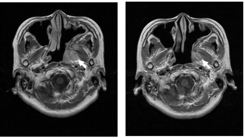
On December 1998, the patient developed epistaxis during sleep and visited the emergency outpatient clinic of our hospital because epistaxis did not stop. Epistaxis had also occurred 1 month earlier but stopped spontaneously, and since then, left nasal obstruction had continued. A magnetic resonance imaging (MRI) examination revealed a tumor in the posterior part of the left nasal cavity (Fig. 1). An endoscopic examination revealed a submucosal mass lesion in the posterior area of the left nasal cavity, from which a biopsy specimen was obtained.
Histopathological findings
Sections of the biopsy specimen stained with hematoxylin and eosin (H&E) showed extensive proliferation by atypical spindle-shaped cells with nuclear pleomorphism (Fig. 2). Mitotic figures were also frequently observed (10/10 high power fields), and multinucleated giant cells were often present. Immunohistochemical staining revealed that the tumor was positive for desmin, vimentin, and a-smooth muscle actin (a-SMA), but was negative for epithelial membrane antigen, leukocyte common antigen, and S-100 protein (Fig. 3). Based on these findings, a diagnosis of leiomyosarcoma was made.
Course
The nasal tumor was resected by left lateral rhinotomy in January 1999. However, tumor recurrence was detected in the left pterygopalatine fossa in August 2001 (Fig. 4). Because the tumor size remained unchanged after completion of radiotherapy at 36 Gy, total resection of the recurrent tumor was performed in December 2001.
Subsequently, the patient was followed up periodically. In November 2002, masses were detected in the lungs (Fig. 5). As a result of a computed tomography (CT)-guided lung biopsy, a diagnosis of lung metastasis of leiomyosarcoma was made. Multiple metastatic tumors were present in the lungs bilaterally. Because surgical resection was impossible, chemotherapy was given repeatedly. The tumor size decreased after the initial course of chemotherapy (CYVADIC; cyclophosphamide 700 mg/m2, vincristine 2 mg/m2, adriamycin 70 mg/m2, dacarbazine (DTIC) 350 mg4 days/m2); however, the tumor gradually increased in size over 6 courses of
chemotherapy (Fig. 6). The patient underwent radiofrequency ablation of a large (2 cm in size) metastatic tumor in the lungs in July 2004.
The recurrent tumor tended to increase more markedly than the metastatic tumor in the lung, and was expected to lead to eating/swallowing disorders in the future. Therefore, in November 2005, CyberKnife therapy was performed on the recurrent tumor (Fig. 7). After CyberKnife therapy, the recurrent tumor decreased in size; local pain was transiently aggravated immediately after this therapy, but gradually improved as the tumor decreased in size.
After these treatment methods, metastasis developed in subcutaneous areas, muscles, the liver, and bones throughout the body and treatment was given to mainly alleviate symptoms. However, her general condition gradually deteriorated and she died in July 2007. Tumors of the lung and the primary site, for which radiofrequency ablation and CyberKnife therapy were performed, respectively, did not increase in size up to her death (Figs. 7 and 8). Without the aggravation of local pain, the patient was able to eat until immediately before death.
Fig. 2. Histopathological findings. H&E stained sections showed extensive proliferation by atypical spindle-shaped cells with nuclear pleomorphism (arrows).
Fig. 3.Immunohistochemical findings. Immunohistochemical staining revealed that the tumor was positive for desmin, vimentin, anda-SMA, but was negative for s-100 protein.
Discussion
Leiomyosarcoma is a malignant tumor that develops in soft tissue, and accounts for approximately 7% of all soft tissue sarcomas[2]. Leiomyosarcomas usually occur in the uterus,
gastrointestinal tract, and retroperitoneal area; occurrence in the head and neck area is rare[3]. Within the head and neck area, leiomyosarcomas frequently develop in the nasal and paranasal sinus areas, although this accounts for only about 3% of all leiomyosarcomas.
The mean age at the onset of leiomyosarcoma in the nasal and paranasal sinus areas was shown to be 53.218.6 years, but this tumor has been observed over a wide age range from 18 to 87 years[3–5]. The male/female ratio is almost 1:1, showing no sex-related differences. Initial symptoms include nasal obstruction, epistaxis, local swelling, and pain. There are few findings characteristic of this disease.
Because leiomyosarcomas are considered to be derived from aberrant undifferentiated mesenchymal tissue, smooth muscles on the vascular wall, and myoepithelial cells in the submucosal glands[6], they can develop anywhere in the nasal and paranasal sinus areas. However, Fig. 4. Axial MRI with T2-weighted images. Tumor recurrence (arrow) was detected in the left pterygopalatine fossa (left). After the completion of radiotherapy at 36 Gy, the size of the tumor remained unchanged (right).
their development in the posterior, rather than anterior area of the nasal cavity has been reported frequently[7,8]; the primary site in our patient was the posterior area of the nasal cavity.
A pathological examination is indispensable for a definitive diagnosis of leiomyosarcoma, but its differentiation from rhabdomyosarcoma, fibrosarcoma, malignant schwannoma, or spindle-cell tumors is difficult using conventional H&E staining alone[9,10]. Therefore, an evaluation using
immunostaining or electron microscopy is necessary. For immunohistochemical diagnosis,a-SMA and muscle-specific actin as smooth muscle markers and vimentin as a mesenchymal marker are useful[11,12].
Leiomyosarcoma still has a poor prognosis. The 5-year survival rate in patients with leiomyosarcoma in the head and neck area is 35–50%. The primary choice of treatment is commonly radical resection, and extensive resection is desirable. However, resection with an adequate safety zone is often difficult in nasal and paranasal sinus areas. Recurrence occurs in 55% of patients, mostly within 1 year[3]. Miyajima et al.[13]analyzed leiomyosarcomas in all areas,
Fig. 6. Chest CT scan. Lung metastatic tumor size decreased after the initial course of chemotherapy (left); however, it gradually increased during 6 courses of chemotherapy (right).
Fig. 7. Axial MRI with T1-weighted image. In April 2005, tumor recurrence (arrow) was detected in the primary site (left). Tumor size in the primary site decreased after CyberKnife therapy. The tumor remained reduced in size until the patient died (right).
and reported the following poor prognostic factors: (1) deeply situated tumor, (2) tumor diameter 5 cm, (3) high mitotic rate, (4) extensive necrotic area of the tumor, and (5) a high American Joint Committee on Cancer (AJCC) stage. Many studies have suggested that tumor size and the degree of local extension are associated with the prognosis in the nasal and paranasal sinus areas. Kurubilla et al.[7] reported that the prognosis is poor for cases extending to the ethmoid sinus, rather than those localized in the nasal cavity. In addition, leiomyosarcomas in the head and neck area rarely metastasize to cervical lymph nodes, and prophylactic cervical dissection may not be necessary in patients in whom preoperative diagnostic imaging shows no clear cervical lymph node metastasis[14]. In this patient, no cervical lymph node metastasis was observed during the course of the disease.
Leiomyosarcoma of the head and neck area is not radiosensitive[3]. In this patient, radiotherapy
(36 Gy) performed at the time of recurrence in 2001 did not reduce the size of the tumor, and secondary surgery was performed. Combination chemotherapy with various drugs such as CYVADIC therapy[15]was also performed, but effective chemotherapy for leiomyosarcoma has not
been standardized. It is difficult to achieve a radical cure for leiomyosarcoma in nasal and paranasal sinus areas using chemotherapy alone. Chemotherapy should be regarded an auxiliary therapy in a multidisciplinary treatment. However, leiomyosarcoma tends to metastasize hematogenously, mainly to the lungs (84%), liver, and skeleton[16]. In our patient, systemic multiple metastases were observed, which suggests the importance of not only local treatment but also chemotherapy. Postoperative chemotherapy may be important in patients who undergo radical resection, and effective chemotherapy in multidisciplinary treatment needs to be established in the future.
CyberKnife therapy may be an effective option for medically inoperable or unresectable patients with leiomyosarcoma.
Metastatic spread is almost exclusively hematogenic. In our patient, lung metastasis was seen 1 year after surgery, and radiofrequency ablation was performed on one of the metastatic lung tumors. Radiofrequency ablation was first used clinically for the treatment of malignant hepatic tumors and subsequently for bone and soft tissue tumors, renal malignant tumors, and mammary gland tumors. In recent years, this method has been performed for solid lung malignancies, and is available for medically inoperable or unresectable cases[17]. Although indications of local therapy, such as surgical resection of lung metastasis, remain very controversial, some studies have reported that intensive local treatment may play some role in prolonging the survival of patients with colorectal cancer, renal cell cancer, and soft tissue sarcoma[18,19]. Radiofrequency ablation induces coagulation necrosis in tumors by a percutaneous approach. High treatment success rates of complete coagulation (90%) were observed when the size of the tumor was smaller than 2 cm. Radiofrequency ablation does not require thoracotomy, is minimally invasive, and has effects similar to those of surgical resection on metastatic lung tumors 2 cm. Bilateral multiple lung metastatic tumors were observed in our patient. Although a radical cure was not expected, due to the patient’s strong wishes, radiofrequency ablation was performed on 1 (2 cm in size) of the lung metastatic lesions. An electrode was inserted into the lung metastatic lesion, and the electrode was heated to 63–798C. This was repeated 4 times, and was effective in halting the growth of the tumor. Favorable clinical effects were also observed compared with other lung metastatic lesions not treated using radiofrequency ablation.
Conclusion
The prognosis for leiomyosarcoma of the nose or paranasal sinuses is poor. The 5-year survival rate is 20%, and approximately 50% of patients do not survive the first year[3]. It is controversial whether local treatment should be performed in patients with distant metastasis or incurable disease. However, we consider that its implementation could extend survival and improve the patient’s quality of life. Radiofrequency ablation and CyberKnife therapy can be useful choices in multidisciplinary treatment for leiomyosarcoma in nasal and paranasal sinus areas.
References
1. Dobben GD. Leiomyosarcoma of the nasopharynx. Arch Otolaryngol 1958; 68: 211–213. 2. Russell WO, Cohen J, Enzinger F, et al. A clinical and pathological staging system for soft
tissue sarcomas. Cancer 1977; 40: 1562–1567. doi:10.1002/1097-0142(197710)40:451562:: AID-CNCR282040042843.0.CO;2-6. PMid:907970.
3. Lippert BM, Godbersen GS, Lu¨ttges J, Werner JA. Leiomyosarcoma of the nasal cavity. ORL J Otorhinolaryngol Relat Spec 1996; 58: 115–120.doi:10.1159/000276810. PMid:8736058. 4. Fu YS, Perzin KH. Nonepithelial tumors of the nasal cavity, paranasal sinuses and
nasopharynx; a clinicopathological study. IV. Smooth muscle tumors (leiomyoma, leiomyo-sarcoma). Cancer 1975; 35: 1300–1308. doi:10.1002/1097-0142(197505)35:55 1300::AID-CNCR282035050843.0.CO;2-Z. PMid:1122481.
5. Josephson RL, Blair RL, Bedard YC. Leiomyosarcoma of the nose and paranasal sinuses. Arch Otolaryngol Head Neck Surg 1985; 93: 270–274.
6. Batsakis JG. Neoplasms of smooth muscle. In: Batsakis JG, editor. Tumors of the head and neck: clinical and pathological considerations, 2nd ed. London: Williams & Wilkins; 1979; p. 354–356.
7. Kuruvilla A, Wenig BM, Humphrey DM, Heffner DK. Leiomyosarcoma of the sinonasal tract: a clinicopathologic study of nine cases. Arch Otolaryngol Head Neck Surg 1990; 116: 1278–1286. doi:10.1001/archotol.1990.01870110050005. PMid:2242259.
8. Richter HJ, Steinert W, Mahn B, Niemczyk HM. [Leiomyosarcoma of the nasal cavity and paranasal sinuses]. HNO 1981; 29: 17–21 (in German). PMid:7204108.
9. Bundtzen JL, Norback DH. The ultrastructure of poorly differentiated rhabdomyosarcomas: a case report and literature review. Hum Pathol 1982; 13: 301–313. doi:10.1016/ S0046-8177(82)80220-7. PMid:7076215.
10. Ji X. Significance of electron microscopy in the diagnosis of undifferentiated neoplasms of the nose and paranasal sinuses. Chung Hua Erh Pi Yen Hou Tsa Chih 1991; 26: 29–31, 62–63.
Res 1980; 64: 93–98.
16. Tokiya R, Imajo Y, Yoden E, et al. A long-term survivor of leiomyosarcoma around the right side of the base of the skull: effective radiotherapy combined with intra-arterial chemother-apy. Int J Clin Oncol 2002; 7: 57–61. PMid:11942051.
17. Dupuy DE, Zagoria RJ, Akerley W, Mayo-Smith WW, Kavanagh PV, Safran H. Percutaneous radiofrequency ablation of malignancies in the lung. Am J Roentgenol 2000; 174: 57–59. 18. Rusch VW. Pulmonary metastasectomy. Current indications. Chest 1995; 107: 322–333.
doi:10.1378/chest.107.6_Supplement.322S.