R E S E A R C H A R T I C L E
Open Access
Dynamics of tear fluid desiccation on a glass
surface: a contribution to tear quality assessment
Leonidas Traipe-Castro
1, Daniela Salinas-Toro
1,2, Daniela López
1, Mario Zanolli
1, Miguel Srur
1, Felipe Valenzuela
1,
Aníbal Cáceres
3, Héctor Toledo-Araya
3and Remigio López-Solís
3*Abstract
Background:Fern-like crystalloids form when a microvolume of tear is allowed to dry out at ambient conditions on a glass surface. Presence of crystalloids in tear“microdesiccates”is used to evaluate patients with Dry-Eye disease. This study aims to examine morphologically the desiccation process of normal tear fluid and to identify changes associated with accelerated tear evaporation. Tear microdesiccates from healthy (Non-Dry Eye) and Dry Eye subjects were produced at ambient conditions. Microdesiccate formation was monitored continuously by dark-field video microscopy. Additionally, accelerated desiccation of tear samples from healthy subjects was conducted under controlled experimental conditions. Particular morphological domains of tear microdesiccates and their progressive appearance during desiccation were compared.
Results:In normal tear microdesiccates, four distinctive morphological domains (zones I, II, III and transition band) were recognized. Stepwise formation of those domains is now described. Experimentally accelerated desiccation resulted in marked changes in some of those zones, particularly involving either disappearance or size reduction of fern-like crystalloids of zones II and III. Tear microdesiccates from Dry Eye subjects may also display those differences and be the expression of a more synchronous formation of microdesiccate domains.
Conclusion:Morphological characteristics of tear microdesiccates can provide insights into the relative rate of tear evaporation.
Keywords:Tear, Tear ferning test, Dry eye, Dark-field microscopy
Background
Fern-like microcrystalloids are formed when some bio-logical fluids are placed as sessile microdrops on glass surfaces and allowed to dry out at ambient conditions [1-3]. In the case of tear fluid, failure in the formation of fern-like structures has been closely associated with Dry Eye disease [4-6]. Up-to date, the Tear Ferning test using the Rolando’s scoring system has proven useful as a la-boratory aid in Dry Eye assessment [4,7-9]. Accordingly, abundance of fern-like tear crystalloids is suggestive of normality (scores I and II) whereas reduced tear ferning (scores III and IV) is usually observed among Dry Eye patients [5,6,10]. Based on our experience with over six hundreds tear ferning assays, desiccation of a tear sessile drop collected from a healthy subject on a glass surface
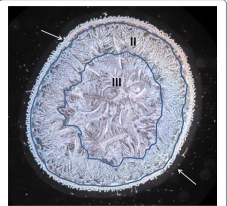
results in circular tear“microdesiccates”comprising four discrete morphological domains or zones, namely, an outer structured hyaline zone I, a band of regularly and centripetally oriented crystalloids that are distinctive of zone II, a central zone III comprising typical randomly distributed fern-like structures differing to each other in robustness, length and branching, and, finally, a transition band, which is a noticeable structure located between zones I and II that seems to serve to anchor the zone II ferns [11]. All these structural or morphological compo-nents of a tear“microdesiccate”, besides the fern-like crys-talloid structures, may also be the expression of a normal tear composition and, probably, a normal tear film.
A number of reports have shown that tear protein profiles may exhibit major alterations when the tear fluid subjected to desiccation has been sampled from Dry Eye patients [12,13]. On the other hand, changes in lipid composition or mucin composition of tear fluid resulting from dysfunctional Meibomium glands or from goblet cell
* Correspondence:rlopez@med.uchile.cl
3
Cellular and Molecular Biology Program, Faculty of Medicine-ICBM, University of Chile, Independencia 1027, Postcode 8380453 Santiago, Chile Full list of author information is available at the end of the article
influenced by or be an expression of the rate of tear evap-oration. The present study was aimed at characterizing the progression of desiccation of tear fluid from healthy sub-jects (absence of Dry Eye) on a glass surface using a mor-phological approach. The study also aimed to evaluate the effect of various experimental conditions that enhance evaporation of tear samples collected from single healthy subjects both on the dynamics of desiccation and the mor-phological features of the resulting tear microdesiccates. Finally, the study characterized morphologically a differing progression of desiccation of tear fluid collected from both eyes of a mild/moderate Dry Eye case.
Results
Rate of tear desiccation
Under the current experimental conditions in this study, full desiccation of 1 μL aliquots of tear fluid collected from healthy (non Dry Eye) donors usually took around 9–10 minutes. A linear relationship between volume of tear sample in the range 0.25-2μL and the time necessary for complete desiccation was observed. In this volume range, desiccation of tear fluid took consistently 15% longer than desiccation of equivalent aliquots of water. Such difference, however, was not statistically significant (p = 0.2738, Mann–Whitney) (Figure 1).
Tear microdesiccates from healthy subjects display a similar four-domain design
Multiple microdesiccates produced from identical ali-quots taken from a single sample of tear fluid were strik-ingly reproducible in terms of size, design and shape of structural elements. By contrast, tear microdesiccates produced simultaneously from identical aliquots of tear fluid sampled from different healthy subjects usually ex-hibited some marked differences in discrete structural components (Figure 2). Those differences were sex- and age-independent. However, all the tear microdesiccates shared a common general design comprising a four-domain organization. For convenience those four-domains have been called zones I, II and III and transition band. Zone I is the outermost band of amorphous material that surrounds the whole circular area of the desiccate. Zone II is a dense band of multiple and identical fern-shaped and leaf-fern-shaped crystalloid structures, which is adjacent to zone I. Zone III, the central zone of the
desiccated tear sample, presents typical fern-like crystal-loid structures displaying a variety of forms differing to each other in robustness, length and branching. Finally, the transition band is a structurally discrete and usually continuous domain located along the whole interphase between zone I and zone II (Figure 3).
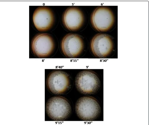
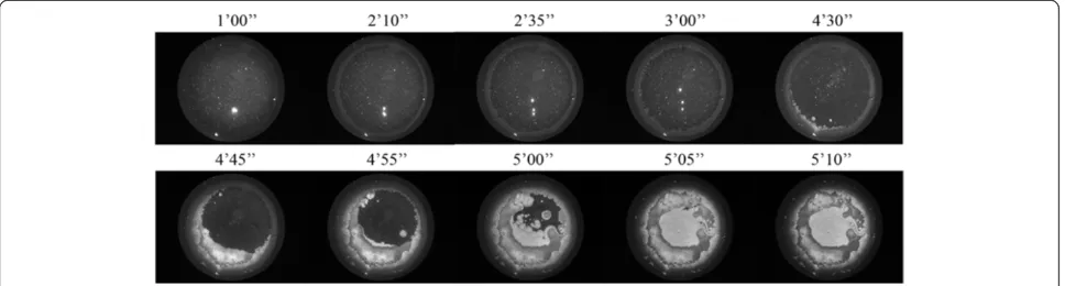
Temporal appearance of the morphological zones occurring in a normal tear microdesiccate
Under the ambient conditions in this study, 1μL of tear was fully desiccated in about 9–10 minutes (desiccation time). Under the dark-field microscope, zone I was the first domain to become unequivocally identifiable. After appearing simultaneously as a thin strip along the whole perimeter of a roughly circular area of the sessile drop of tear, zone I became progressively thicker and reached its maximum width during the first half of the time needed for full desiccation of the tear aliquot, that is, around 4–5 minutes. Then, following a stage of apparent rest spanning most of the second third of the whole time of desiccation, a series of major and rapid crystallization events started to take place. Formation of the zone II crystalloids was clearly the first major noticeable change at this third stage of tear desiccation. These crystalloids started to form by growing centripetally from regularly spaced sites located on a unstructured but distinct narrow strip in proximity to zone I. While zone II crystalloids were still growing,
two events took place at a fourth stage. Firstly, the un-structured narrow strip at the interface between zone I and zone II became more structured and, secondly, a few irregularly disperse sites located close to the center of the tear fluid under desiccation seemed to serve as nucleation points on which morphologically distinct fern-like crystal-loid structures began to grow multidirectionally. Each of the branches of these zone III crystalloids grew continu-ously until they made contact with other growing crystal-loid structures from either zone II or zone III. Complete
convergence of all the growing crystalloids from zones II and III marked the endpoint of tear desiccation. An usu-ally clear-cut boundary between zones II and III became the most visible morphological evidence of this contact inhibition phenomenon (Figure 4).
Effect of experimentally accelerated tear desiccation on the four-domain morphology of tear microdesiccates
Rate of water evaporation depends on a number of en-vironmental factors, such as atmospheric pressure, water vapor pressure and temperature. Thus, it would be ex-pected that the rate of water evaporation from a sample of tear placed on a glass surface would increase either by lowering the air pressure surrounding the sample, by re-moving water vapor from the environment of the sample or by increasing the air temperature. In order to correl-ate the drying rcorrel-ate of a tear sample with the morphology of the corresponding tear microdesiccate, in a first ex-periment two 1-uL aliquots drawn from a single tear sample collected from a healthy subject were placed on two independent glass surfaces (microscope slides). One of the aliquots was allowed to dry at ambient temperature (24°C), air pressure (759 Torr) and relative humidity (41%) while the second aliquot was allowed to dry under re-duced air pressure (360 mm Hg/Torr) inside of a 45 mL Falcon disposable tube. Both samples were placed at a distance of 10 cm. Under these conditions, the time of desiccation of the tear aliquot placed in the Falcon tube was reduced by half. Morphological analysis of both tear microdesiccates showed marked differences. In contrast with the usual four zones occurring in normal tear microdesiccates produced at ambient conditions, the tear microdesiccate obtained at reduced air pressure showed a significantly thinner zone I, a roughly normal transition band, a paucity of disperse small-sized crys-talloids and a central zone strikingly devoid of major Figure 2Intraindividual similarities and interindividual differences in the morphological appearances of tear microdesiccates from single healthy subjects.Microdesiccates from 1-uL aliquots of two successive samples of tear fluid taken from single healthy subjects (same eye) displayed marked morphological similarities (neighbour images). Microdesiccates corresponding to four subjects(A, B, C and D)are shown. Note also some clear differences among microdesiccates from different subjects, although a general design is maintained. Microdesiccates in the figure were rated I-II (normal) in the conventional Rolando’s classification system.
crystalloids (Figure 5). In an analog experiment, two 1-uL aliquots drawn from a single tear sample taken from a healthy subject were placed on two independent glass sur-faces. One of the aliquots was allowed to dry at the same ambient conditions indicated above (24°C, 759 torr) while the second aliquot was allowed to dry in an oven at 30°C. In this experiment, time needed for full desiccation of the tear sessile drop in the oven was also reduced by half. Again, both tear desiccates showed marked mor-phological differences. The abundant fern-like crystalloids occurring in zone II and zone III of the tear desiccate
produced at ambient temperature, at 30°C were re-placed by abundant groups of small crystalloids making contact with each other. In addition, at this higher temperature, differentiation of zones II and zone III dis-appeared. By contrast, the translucid zone I maintained its main characteristics at both temperatures. Thus, substitution of the typical zone II and zone III loids by roughly uniform small but abundant crystal-loids was a common observation in microdesiccates produced under various conditions of accelerated desic-cation (Figure 5).
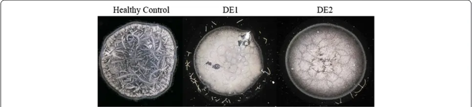
Temporal progression of the formation of tear microdesiccates from cases of patients with Dry Eye
Dry Eye has been frequently associated with tear fluid displaying a lower tendency to form ferns upon desicca-tion on glass surfaces [5,6,10]. Figure 6 shows desiccates of microvolumes of tear samples taken from two random patients with moderate or severe Dry Eye. Times needed for full desiccation of those tear samples were in average 40% shorter than those observed in desiccation of a tear sample from a healthy subject (p < 0.01). Samples from both patients showed several marked changes in the four-domain structure, the most remarkable and common of which was the disappearance of the distinctive zones II and III together with the formation of abundant groups of small crystalloids making contact with each other. Such remarkable differences evoked the morphological features of tear microdesiccates from healthy subjects produced under experimental conditions facilitating desiccation.
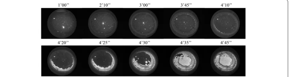
Thus, tear samples collected separately from both eyes of a mild/moderate Dry Eye patient were subjected to video-imaging during tear desiccation. As in the produc-tion of microdesiccates from normal tear fluid, zone I was the first visible structure to appear during desiccation of both tear samples of the Dry Eye patient. At variance with progression of microdesiccation of tear from healthy sub-jects, however, in both tear samples from the Dry Eye pa-tient a variety of crystallization events started to take place simultaneously all over the area of desiccation soon after zone I was apparently complete. Layers of tiny and small centripetally oriented crystalloids started to form and pro-gress rapidly supported on the zone I-transition band complex. Within a few seconds, in the most central area under desiccation many groups of small or tiny fern-like or leaf-like crystalloids started to grow multidirectionally. These crystalloids grew until they entered in contact with neighbor groups of crystalloids of the same morphological Figure 5Changes in the morphological features of tear microdesiccates produced under conditions of accelerated evaporation.
One-microliter aliquots from a single sample of tear collected from a healthy subject were allowed to dry simultaneously either at ambient conditions (left), at reduced pressure (center) or at a higher temperature (right). Under both non-ambient conditions, the time of desiccation was reduced by half and the resulting microdesiccates lacked distinctive zones II and III, preserved the surrounding zone I and exhibited much smaller and more homogeneous groups of crystalloids than those observed in the distinct zones II and III of the control microdesiccate. Figures are representative of five independent experiments.
Discussion
In this study we have described the dynamics of formation of a domain-based structure produced by desiccation of a sessile tear drop on a glass flat surface. Overall, our obser-vations indicate that each fern-like or leaf-like crystalloid in the tear microdesiccate is formed from an origin site and that its orderly lineal growth and branching end when the growth frontlines of the crystalloid get in contact with other forming or already formed crystalloid.
Spontaneous desiccation of a microvolume of tear fluid collected from healthy subjects gives rise to tear microde-siccates consisting of four domains named zones I, II, III and transition band [11]. We have now shown that those four domains of a normal tear desiccate are formed asynchronously. The outermost hyaline zone I of the tear microdesiccate is the first domain to form at an early stage of desiccation. This structure represents a“pinning”stage of the desiccation process by which the sessile drop of the tear fluid becomes firmly bound to the glass surface thus preventing a decrease in the contact surface between the tear and the glass. Later, during a second stage of the des-iccation process, water loss occurs at the water/air inter-face while the tear drop would be undergoing successive changes of shape [25,26]. During this period, only some degree of disordered movement can be hardly seen by
(zone III). Each of these nascent zone III crystalloids emerges from a single site, is usually multibranched and becomes larger, less regular in structure and more robust than zone II crystalloids. The highly diverse extents of these zone III crystalloids, and the ones of their branches, seem to depend stochastically on the contact inhibition phenomenon. Formation of zone III crystalloids would represent a fourth and final stage of the tear desiccation process. Contact between zone II and zone III crystalloids inhibits their growth and demarcates the end of the organization of a tear microdesiccate. Overall, formation of zones II and III are late, separate and partly overlapped events during desiccation of normal tear. Their formation in the final stages of desiccation occurs much faster than the visible events of the previous stages of tear desiccation. Anyhow, formation of a tear microdesiccate from any normal tear fluid sample seems to occur in an orderly sequence of events.
Formation of tear crystalloids can reflect the presence of definite components in the tear fluid, mostly proteins, mucins and salts [27,28]. In support of that view and regardless of significance for desiccation that those tear components may have, in this study we have given some evidence showing that tear microdesiccates from any single subject are essentially identical whereas tear
desiccates produced from tear samples provided by dif-ferent subjects can vary markedly. However, the mor-phological characteristics of tear desiccates produced from a single tear sample are dependent not only upon the composition of the tear fluid but also on the condi-tions under which desiccation takes place [29]. In this study we showed that a single sample of normal tear may produce morphologically different microdesic-cates under different drying conditions. Thus, under drying conditions that enhance tear water evaporation, such as reduced air pressure, reduced partial water pressure or higher ambient temperature, crystalloids would be originated from a higher number of origins. Because the growth of single crystalloids seems to occur continuously until they make contact with other forming or already formed crystalloids, then a faster desiccation will result in smaller crystalloids. Altogether, these experimental observations suggesting a link between morphological features of tear microdesiccates and relative rate of tear evaporation may provide an additional source of valuable information on tear evaporation from single eyes that may help to assess and diagnose ocular surface disorders [4,7-9,12,18,23,30-36]. In our view, the abun-dance of small crystalloids instead of the vigorous zone III crystalloids in a tear microdesiccate would represent the morphologic expression of a rapid evaporation of the tear water. In this regard, images of honeycomb structures occurring in tear ferning tests of Dry Eye patients, as re-ported by other laboratories [29], would be the expres-sion of abundant groups of small crystalloids whose growth from a high number of origin sites was rapidly halted by their contact with similar growing neighbor groups of crystalloids.
Based on the dynamics of the drying process we have suggested that accelerated tear water evaporation would be strongly associated with predominant formation of small tear crystalloids. However, such morphological fea-ture of a tear desiccate produced from a given tear sample can be also described as the lack of formation of large
in normal tear desiccates [11]. Emerging from those sites, zone II fern-like crystalloids grow centripetally until com-ing into contact with other tear crystalloids. All these events represent just a minor fraction of an orderly se-quence of interactions between tear components. Thus, the whole structure of a tear microdesiccate on a glass sur-face would be the final expression of a highly organized supramolecular complex whose morphological characteris-tics would be dependent onin vitrointermolecular interac-tions occurring while tear water is being removed. Some of those characteristics, either normal or abnormal, may be also suggestive of a supramolecular mode of organization of the same tear components structuring the tear film on the wet ocular surface.
Conclusion
Spontaneous desiccation of a microvolume of tear fluid collected from healthy subjects on a flat glass surface gives rise to tear microdesiccates consisting of well-defined morphological domains. Major fern-like crystalloids are a major feature of some of those domains. Formation of the microdesiccate domains occurs in an orderly sequence of asynchronic events. Under experimental conditions of
Methods
Subjects
All nine healthy subjects (age range 20–59 years old) included in this study fulfilled the following criteria: a) Normal visual parameters, b) Schirmer I test over 10 mm at 5 min [21], c) Fluorescein break-up time over 10 seconds [22], d) OSDI questionnaire scoring below 15 [23], e) Tear ferning test score I or II [5,6], f ) No previous eye surgery and g) No medication during the last three months [4]. In addition, three patients (age range 20–55 years old) diag-nosed with Dry Eye according to the DEWS guidelines [8], who had not experienced eye surgery and who were regu-larly attending the Fundación Oftalmológica Los Andes Ophthalmology Clinic in Santiago, Chile, were invited to be volunteer tear donors. Each one of the diagnostic tests was performed by single trained personnel. The study was conducted between March 2011 and June 2013 in ac-cordance with the tenets of the Helsinki Declaration of 1975 and the guidelines of both the Ethics Committee of the Faculty of Medicine, University of Chile and the Ethics Committee of Fondecyt-Chile (Fondo de Desarrollo Científico y Tecnológico-Chile).
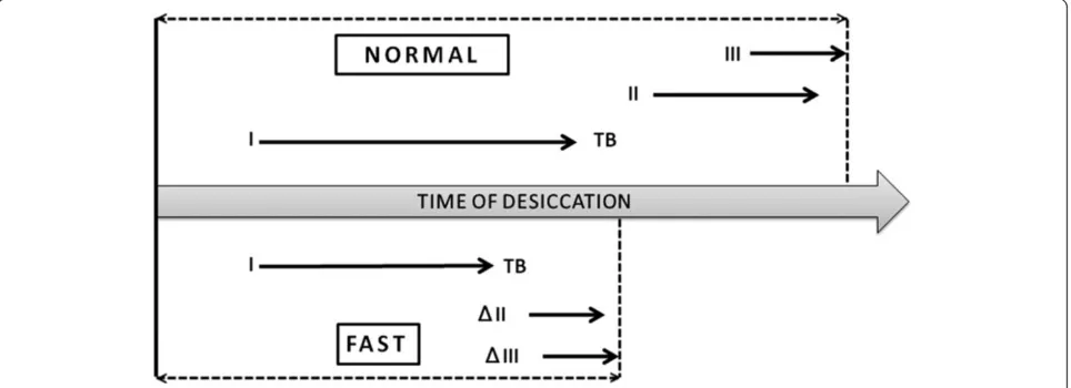
Figure 9Diagram representing the stepwise appearance of the morphological domains of a normal tear microdesiccate and its probable modification in patients with evaporative Dry Eye.Formation of zones I, II, III and transition band during normal tear desiccation
Tear collection
Tear fluid was collected using polyurethane minisponges, as reported elsewhere [24]. Samples were taken always around 9–11 AM to control for eventual circadian varia-tions. For each eye a single 3-minute tear sample was taken. The amount of sample was determined by gravim-etry. Desiccation assays were conducted immediately after tear collection.
Tear desiccation
Excepting specific experiments (see Results section), one-microliter aliquots of fresh samples of tear fluid were taken with a P2-Gilson micropipette fitted with an ultra-fine tip and placed sharply on a point of a microscope slide that was positioned horizontally. Tear aliquots were allowed to dry spontaneously at ambient conditions of temperature (range 15-25°C), relative humidity (range 40-45%) and altitude (520 m above sea level). Micro-graphs of the dry samples were taken under a dark-field microscope (Zeiss Axiostar Plus, objective lens = 2.5X, ocular lens = 10X) fitted with a Canon Powershot G10 14.7 megapixel digital camera. Fern images were classified as types I through IV according to Rolando’s criteria [5,6].
Progression of tear desiccation
Desiccation of one-microliter aliquots of tear fluid was performed under the microscope as described above, ex-cept that to this purpose the digital camera was adjusted for capturing high resolution videos. Video-images (30 frames per second) were taken during 1 second at time-intervals of 5 seconds. The endpoint of desiccation was defined as the one in which growth of tear crystalloids was fully halted. Time for full desiccation of each tear sample was scored. In order to prevent any artifactual effect on tear desiccation produced by heat transfer, the lamp of the microscope was turned on only when video-images were being recorded.
Materials
Absorbing polyurethane mini-sponges (PeleTim®) and microcentrifuge tubes for tear collection were obtained from VOCO, Cuxhaven, Germany and from Axygen Sci-entific, California, USA, respectively. Sterile Schirmer tear test strips and fluorescein strips were acquired from Alcon Laboratories, Santiago, Chile. Microscope slides were purchased from Isolab Laborgeräte, Wertheim am Main, Germany.
Consent
Written informed consent was obtained from the pa-tient for the publication of this report and any accom-panying images. Confidentiality of patient information was protected.
Competing interests
All authors declare that they have no competing interests with any organization.
Authors’contribution
All authors contributed extensively to the work presented in this paper. LT-C, conducted clinical assessments to screen for Dry Eye and for normal ocular surface, and performed analyses of tear microdesiccates; MS and FV conducted clinical assessment of subjects to screen for Dry Eye and for normal ocular surface; DS-T designed and performed video-imaging experiments of tear desiccation and measured tear evaporation rate; DL carried out dark-field microscopy and morphological analysis of tear microdesiccates and produced graphic representation of experimental data; MZ, conducted Dry Eye assessment, performed tear collection from Dry Eye patients and performed video-imaging experiments of tear desiccation; AC, conducted the experiments on accelerated tear evaporation; HT-A designed experiments on controlled desiccation and carried out a critical review of the manuscript and RL-S conducted experiments on normal tear fluid, identification of morphological domains in tear desiccates and manuscript writing. All authors read and approved the final manuscript.
Acknowledgements
This study was partially supported by Grant 1110325 from Fondo Nacional de Ciencia y Tecnología (Fondecyt), Chile.
Author details
1Fundación Oftalmológica Los Andes Ophthalmology Clinic (FOLA), Vitacura,
Santiago, Chile.2School of Medical Technology-Ophthalmology, Faculty of Medicine, University of Chile, Santiago, Chile.3Cellular and Molecular Biology
Program, Faculty of Medicine-ICBM, University of Chile, Independencia 1027, Postcode 8380453 Santiago, Chile.
Received: 19 May 2014 Accepted: 20 May 2014 Published: 4 June 2014
References
1. Bennett SL, Cullen JB, Sherer DM, JR W Jr:The ferning and nitrazine tests of amniotic fluid between 12 and 41 weeks gestation.Am J Perinatol
1993,10:101–104.
2. Pattanasuttinont S, Sereepapong W, Suwajanakorn S:The salivary ferning test and ovulation in clomiphene citrate-stimulated cycles.J Med Assoc Thai2007,90:876–883.
3. Maragou M, Vaikousis E, Ntre A, Koronis N, Georgiou P, Hatzidimitriou E, Sotsiou F, Dantis P:Tear and saliva ferning tests in Sjögren’s syndrome (SS).Clin Rheumatol1996,15:125–132.
4. Bron AJ, Abelson MB, Ousler G, Pearce E, Tomlinson A, Yokoi N, Smith JA, Begley C, Caffery B, Nichols K, Schaumberg D, Schein O, Calonge M, Baudouin C, Goto E, Grus F, Paugh J:Methodologies to diagnose and monitor Dry Eye disease: report of the diagnostic methodology subcommittee of the international dry eye workshop.Ocul Surf2007, 5:108–123.
5. Rolando M:Tear mucus ferning test in normal and keratoconjunctivitis sicca eyes.Chibret Int J Ophthalmol1984,2:33–41.
6. Rolando M, Baldi F, Calabria G:Tear mucus crystallization in children with cystic fibrosis.Ophthalmologica1988,197:202–206.
7. Albach KA, Lauer M, Stolze HH:Diagnosis of keratoconjunctivitis sicca in rheumatoid arthritis. The value of various tests.Ophthalmologe1994, 91:229–234.
8. Lemp MA, Baudouin C, Baum J, Dogru M, Foulks GN, Kinoshita S, Laibson P, Mcculley J, Murube J, Pflugfelder SC, Rolando M, Toda I:Definition and classification of Dry Eye disease: report of the diagnosis and
classification subcommittee of the international dry eye workshop.Ocul Surf2007,5:75–92.
9. Julio G, Lluch S, Cardona G, Fornieles A, Merindano D:Item by item analysis strategy of the relationship between symptoms and signs in early dry eye.Curr Eye Res2012,37:357–364.
10. Guillon J:Current clinical techniques to study the tear film and tear secretions.InThe tear film: structure, function and clinical examination.
15. Dartt DA:Tear lipocalin: structure and function. Review.Ocul Surf2011, 9:126–138.
16. Roncone M, Bartlett H, Eperjesi F:Essential fatty acids for dry eye: a review.Cont Lens Ant Eye2010,33:49–54.
17. Gipson IK, Spurr-Michaud SJ, Senchyna M, Ritter R 3rd, Schaumberg D: Comparison of mucin levels at the ocular surface of postmenopausal women with and without a history of Dry Eye.Cornea2011,30:1346–1352. 18. Corrales RM, Narayanan S, Fernández I, Mayo A, Galarreta D, Fuentes-Páez G, Chaves FJ, Herreras JM, Calonge M:Ocular mucin gene expression levels as biomarkers for the diagnosis of dry eye syndrome.Invest Ophthalmol Vis Sci2011,52:8363–8369.
19. Arciniega JC, Wojtowicz JC, Mohamed EM, McCulley JP:Changes in the evaporation rate of tear film after digital expression of Meibomian glands in patients with and without Dry Eye.Cornea2011,30:843–847. 20. Tong L, Chaurasia SS, Mehta JS, Beuerman RW:Screening for meibomian
gland disease: its relation to dry eye subtypes and symptoms in a tertiary referral clinic in Singapore.Invest Ophthalmol Vis Sci2010, 51:3449–3454.
21. van Bijsterveld OP:Diagnostic tests in the Sicca syndrome.Arch Ophthalmol1969,82:10–14.
22. Vitali C, Moutsopoulos HM, Bombardieri S:The European Community Study Group on diagnostic criteria for Sjogren’s syndrome. Sensitivity and specificity of tests for ocular and oral involvement in Sjogren’s syndrome.Ann Rheum Dis1994,53:637–647.
23. Schiffman RM, Christianson MD, Jacobsen G, Hirsch JD, Reis BL:Reliability and validity of the ocular surface disease index.Arch Ophthalmol2000, 118:615–621.
24. López-Cisternas J, Castillo-Díaz J, Traipe-Castro L, López-Solís RO:Use of polyurethane minisponges to collect human tear fluid.Cornea2006, 25:312–318.
25. Duggal R, Hussain F, Pasquali M:Self-assembly of single-walled carbon nanotubes into a sheet by drop drying.Adv Mater2006,18:29–34. 26. Sharfrin E, Zisman WA:Constitutive relations in the wetting of low energy
surfaces and the theory of the retraction method of preparing monolayers.J Phys Chem1960,64:519–524.
27. Del Rosario D:Prueba de cristalización lagrimal. Capítulo 29.InOjo seco.
Edited by del Castillo Murube J. Granada: Sociedad Española de Oftalmología; 1997.
28. Kogbe O, Liotet S, Tiffany JM:Factors responsible for tear ferning.Cornea
1991,10:433–444.
29. Horwath J, Ettinger K, Bachernegg M, Schmut O:Ocular ferning test. Effect of temperature and humidity on tear ferning patterns.Ophthalmologica
2001,215:102–107.
30. Lemp MA:Report of the National Eye Institute/Industry Workshop on clinical trials in dry eyes.CLAO J1995,21:221–232.
31. Behrens A, Doyle JJ, Stern L, Chuck RS, McDonnell PJ, Azar DT, Dua HS, Hom M, Karpecki PM, Laibson PR, Lemp MA, Meisler DM, Murube Del Castillo J, O’brien TP, Pflugfelder SC, Rolando M, Schein OD, Seitz B, Tseng SC, Van Setten G, Wilson SE, Yiu SC:Dysfunctional tear syndrome: a Delphi approach to treatment recommendations.Cornea2006,25:900–907. 32. Dogru M, Stern ME, Smith JA, Foulks GN, Lemp MA, Tsubota K:Changing
trends in the definition and diagnosis of dry eyes.Am J Ophthalmol2005, 140:507–508.
33. Matoba AY, Harris DJ, Meisler DM, Pflugfelder SC, Rapuano CJ, Weiss JS, Musch DC:Diagnostic Tests.InDry Eye Syndrome: Preferred Practice Pattern.
San Francisco, California: American Academy of Ophthalmology; 2003:14–16. 34. Versura P, Nanni P, Bavelloni A, Blalock WL, Piazzi M, Roda A, Campos EC:
Tear proteomics in evaporative dry eye disease.Eye (Lond)2010, 24:1396–1402.
doi:10.1186/0717-6287-47-25
Cite this article as:Traipe-Castroet al.:Dynamics of tear fluid desiccation
on a glass surface: a contribution to tear quality assessment.Biological
Research201447:25.
Submit your next manuscript to BioMed Central and take full advantage of:
• Convenient online submission
• Thorough peer review
• No space constraints or color figure charges
• Immediate publication on acceptance
• Inclusion in PubMed, CAS, Scopus and Google Scholar
• Research which is freely available for redistribution