R E S E A R C H A R T I C L E
Open Access
Diagnostic efficacy of multiple MRI
parameters in differentiating benign
vs. malignant thyroid nodules
Hao Wang
1, Ran Wei
1, Weiyan Liu
2, Yongqi Chen
3and Bin Song
1*Abstract
Background:Diffusion weighted imaging (DWI) has a good diagnostic value for malignant thyroid nodules, but the published protocols suffer from flaws and focus on the apparent diffusion coefficient (ADC). This study investigated the diagnostic performance of multiple MRI parameters in differentiating malignant from benign thyroid nodules.
Methods:This was a retrospective study of 181 consecutive patients (148 benign and 111 malignant nodules, confirmed by pathological results). The patients underwent conventional MRI, DWI, and dynamic contrast-enhanced MRI before surgery. The chi-square test and the Student t test were used to compare the conventional features and ADC value between malignant and benign groups. Multivariate logistic regression was used to identify the independent predictors and to construct a model. Receiver operator characteristic (ROC) curve analysis was used to assess the diagnostic performance of the independent variables and model.
Results:Tumor diameter, ADC value, cystic degeneration, pseudocapsule sign, high signal cystic area on T1-weighted imaging, ring sign in the delayed phase, and irregular shape showed significant differences between two groups (allP< 0.05). The multivariable analysis revealed that ADC value (OR = 694.006,P< 0.001), irregular shape (OR = 32.798,P< 0.001), ring sign in the delayed phase (OR = 20.381,P= 0.004), and cystic degeneration (OR = 8.468,P= 0.016) were independent predictors. Among them, ADC performed the best in discriminating benign from malignant nodules, with an area under the curve (AUC) of 0.95, 0.90 sensitivity, and 0.91 specificity. When the independent factors were combined, the diagnostic performance was improved with an AUC of 0.99, 0. 97 sensitivity, and 0.95 specificity.
Conclusions:ADC value could discriminate between benign and malignant thyroid nodules with a good performance. Subjective features such as the ring sign, irregular shape, and cystic degeneration associated with malignant thyroid nodules could provide complementary information for differentiation.
Keywords:Diffusion weighted imaging, Magnetic resonance imaging, Thyroid carcinoma, Thyroid nodule
Background
About, 19–67% of the healthy, asymptomatic individuals
are diagnosed with nodules of the thyroid [1]. Malignant
thyroid nodules account for 5–15% of all thyroid nodules
[2, 3]. With the rapid economic development of China,
healthcare access and screening are improving in China
and the detection and prevalence of thyroid nodules is reaching epidemics proportions, from 8% in 2002 to
25% in 2013 [4], as has been observed in developed
countries [5].
Although ultrasound (US) is widely used to detect thyroid nodules and determine their malignant potential, the US features and index of each grade of the Thyroid Imaging Reporting and Data System (TI-RADS) remain
controversial [6, 7]. US-guided fine-needle aspiration
biopsy (FNAB) is widely used for the diagnosis of
thy-roid nodules [7]. Nevertheless, up to 7% of the nodules
* Correspondence:[email protected]
1Department of Radiology, Minhang Branch, Zhongshan Hospital, Fudan
University, Shanghai, China
Full list of author information is available at the end of the article
yield non-diagnostic cytology and an additional 15–30% of fine-needle aspiration cytology (FNAC) show an inde-terminate cytology [7,8].
DWI reflects the random Brownian motion of water molecules within a voxel of tissue. The movement of the tissue water molecules between the two opposing gradi-ents will result in dephasing, which is seen as signal loss. ADC can be quantified calculated with repeating the se-quence in different magnetic field strengths [9]. The ADC depends largely on the presence of barriers to diffusion within the water microenvironment, namely, cell
mem-branes and macromolecules [10]. Malignant thyroid
nodules usually have a lower ADC value compared to benign nodules [11–15], but this is controversial [16] and most of these studies used relatively low b values (< 500 × 10−3/m2) [11, 14]. DWI has been shown to be
of diagnostic value for Graves’ Disease [17], but DWI
studies of thyroid nodules are relatively rare because the head and neck region is very heterogeneous and contains a variety of tissues that include fat, muscle, air, soft tissues, glands, and bones. In addition, MRI is more expensive and less available than US.
Neverthe-less, US has 40–80% sensitivity and 40–96% specificity
for malignant thyroid nodules, highlighting the need for complementary examinations. In addition, previous DWI studies of thyroid nodules focused on the ADC [11,12,14,16,18].
Therefore, in the present study, we sought to improve the image quality with a special coil on the neck surface and a breath-hold technique, and to investigate the value of multiple MRI parameters for the diagnosis of thyroid carcinoma. Quantitative and qualitative parameters were examined.
Methods
Patients
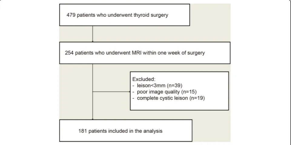
This was a retrospective diagnostic study. Between January 2013 and December 2016, 479 consecutive patients under-went thyroidectomy at our hospital. Among them, 254 pa-tients had undergone MRI examination within one week before surgery at the Department of Radiology. This study was approved by the institutional review board of our hos-pital. The requirement for individual written informed consent was waived.
The inclusion criteria were: 1) thyroid nodule that underwent surgery; and 2) available MRI data obtained within one week of surgery. The exclusion criteria were: 1) lesion size < 3 mm by US (n= 39); 2) poor image quality
deemed to be non-diagnostic after review (n= 15); or 3)
lesions with complete cystic changes (n= 19).
Among the 181 patients included, 140 patients under-went subtotal thyroidectomy, 25 underunder-went lobectomy, and 16 underwent total thyroidectomy.
Multiparametric MRI protocol
MRI was performed with a 1.5 T scanner (EXCITE HD GE Healthcare, Waukesha, WI, USA) equipped with an 8-channel special neck surface coil (Chenguang Medical Technology Ltd., Shanghai, China). All patients were ex-amined using the same machine, coil, and scan series.
The MR imaging protocol included T1-weighted ima-ge(T1WI), T2-weighted image(T2WI), DWI, and contrast enhanced T1WI of thyroid. Coronal fat-suppressed T2WI (repetition time (TR)/ echo time (TE), 1280 ms/85 ms; slice thickness; 4 mm; gap, 1 mm; matrix, 288 × 192; num-ber of excitations (NEX), 4; field of view (FOV), 18 cm); axial T1WI (TR/TE, 460 ms/8 ms; slice thickness, 4 mm; gap, 0.5 mm; NEX, 2; FOV, 14 cm; matrix, 288 × 192); axial fat-suppressed T2WI (TR/TE, 3000 ms/85 ms, slice thick-ness, 4 mm; gap, 0.5 mm; NEX, 4;FOV, 14 cm; matrix, 320 × 224). DWI was performed using a single-shot echo planar imaging sequence with the diffusion gradient b
factor = 800 s/mm2. Imaging parameters for DWI were:
TR/TE, 6550 ms/minimum; FOV, 14 cm; NEX, 4; matrix, 128 × 128; slice thickness, 4 mm; and gap = 0.5 mm. Contrast enhancement studies were implemented using axial T1WI obtained with a fast-spoiled gradient recalled echo (TR/TE, 5.7 ms/1.7 ms; FOV, 14 cm; matrix, 192 × 256; NEX, 1). Gadolinium (Magnevist, Bayer HealthCare Pharmaceuticals, Montville, NJ, USA) was intravenously injected at 0.2 ml/kg body weight and 3 ml/s, followed by a 20 ml saline flush. In each patient, a phase was performed prior to the injection of contrast medium. Six phases were obtained after injection of the contrast agent at 30, 60, 120, 180, 240, and 300 s. In the contrast-enhanced protocol, breath-hold was performed during each phase. Spatial saturation bands were also used to remove signal from overlying fat and adjacent tissues. All patients received training on the breath-hold technique before the MRI examination.
Image analysis
Two radiologists (13 and 18 years of MRI experience) reviewed all images using an AW4.5 workstation (GE Healthcare, Waukesha, WI, USA). Each radiologist was blinded to histopathology results. All images were reviewed independently by the two radiologists. Discrepancies were solved by discussion.
ADC maps. Meanwhile, obvious areas of cystic changes, hemorrhage, calcification, and lesion margins need to avoid based on combined T2WI, contrast enhanced T1WI and DWI. The size of ROIs was determined to correspond with the darkest portion of lesions on ADC maps ranged
10-50 mm2. The ADC values were measured twice and
then the average value was taken.
In this study, blood vessels, thyroid tissue, and muscles were used to assess subjectively the degree of early en-hancement of the nodule, which was categorized as: mild (the enhancement was similar to that of adjacent muscle tissues); moderate (the enhancement was higher than that of adjacent muscle tissues but lower than that of blood vessels); and marked (the enhancement approached that of blood vessels). The pattern of enhancement reflected the dynamic contrast enhancement of thyroid nodules after the injection of the contrast agent in the delayed phase with‘wash in’or ‘wash out’. The ring sign was de-fined as the central area of the nodule showing‘wash out’, whereas the peripheral area showed persistent enhance-ment. The pseudocapsule sign was defined as the tumor area showing a clear capsule after contrast agent adminis-tration in the delayed phase.
Statistical analysis
Categorical data were presented as percentage and ana-lyzed using the chi-square test. Continuous data were tested using the Kolmogorov-Smirnov test. Normally dis-tributed continuous data were presented as means ± standard deviation and analyzed using the Student t test. Non-normally distributed data were presented as medians
(range). All statistical analyses were performed with SPSS
23 (IBM, Armonk, NY, USA). Two-sided P-values < 0.05
were considered statistically significant. Binary logistic regression was used to identify the features that were independently predictive of malignant thyroid tumor. Var-iables demonstrating a significant association with malig-nant thyroid lesions were entered into the model in a forward stepwise method. The final model was selected based on variables withP< 0.05. The odds ratios (OR) and 95% confidence intervals (95%CI) were used as a measure of the relative magnitude of an association between pre-dictor variables and malignant tumor. ROC curves were used to determine the cutoff value to differentiate the pa-rameters of malignant from benign tumors.
Results
Characteristics of the patients
Figure 1 presents the patient flowchart. From 479
pa-tients who underwent thyroid surgery, 254 papa-tients underwent MRI examination within 1 week before sur-gery and 181 patients met the eligibility criteria. There were 38 males and 143 females, with 148 benign thyroid nodules (137 nodular goiters, seven follicular adenomas,
and four Hashimoto’s thyroiditis) and 111 malignant
nod-ules (107 papillary thyroid carcinomas, three follicular car-cinomas, and one medullary carcinoma). The patients were 50.6 ± 14.2 (range, 12–84) years. No significant dif-ferences in patient age (P= 0.700) or gender (P= 0.419) were found between the benign and malignant nodule
groups (Table 1). There were 140 nodules in the right
lobe, 104 in the left lobe and 15 in the isthmus. A single
nodule was found in 126 patients and multiple nodules in 55 patients. Fourteen patients had concurrent benign and malignant nodules.
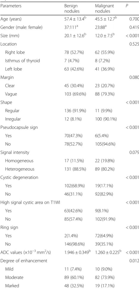
Association between MRI parameters and malignant nodules
Table1 shows the univariable analyses of the association
between MRI features and malignant nodules. The benign
nodules (20.1 ± 12.6 mm, range: 3.8–64.6 mm) were
sig-nificantly larger than the malignant thyroid nodules (12.0 ± 7.5 mm, range: 4.3–47.3 mm) (P< 0.001). The ADC of the benign group (1.95 ± 0.35 × 10⁻³mm2/s, range: 0.99– 3.16 × 10⁻³mm2/s) was significantly higher than that of
the malignant group (1.26 ± 0.23 × 10⁻³mm2/s, range:
0.77–2.22 × 10⁻³mm2/s) (P< 0.001). Cystic degeneration, the pseudocapsule sign, and high signal cystic area on T1WI were more common in benign thyroid nodules (all
P< 0.001). The ring sign in the delayed phase and irregu-lar shape after contrast agent was significantly more com-mon in the malignant group (both P< 0.001) (Figs. 2, 3
and 4). There were no significant differences regarding
margin and signal heterogeneity between the malignant and benign groups (bothP> 0.05).
Multivariable analysis
Table 2 shows the results of the final logistic regression
model. The ADC value (OR = 694.006, P< 0.001),
irregu-lar shape (OR = 32.798,P< 0.001), ring sign in the delayed
phase (OR = 20.381, P= 0.004), and cystic degeneration
(OR = 8.468, P= 0.016) were independently associated
with malignant thyroid nodules, with 0.961 accuracy.
ROC curve analysis
The ROC curve analysis revealed that the best cut-off of ADC values achieved AUC of 0.95, 0.90 sensitivity and 0.91 specificity. Cystic degeneration, ring sign, and ir-regular shape had AUC of 0.76, 0.82, and 0.91, respect-ively. When the independent factors were combined, the diagnostic performance was improved to an AUC of 0.99, 0.97 sensitivity, and 0.95 specificity (Fig.5).
Discussion
DWI has a good diagnostic value for thyroid disease.
Previous studies [17, 19] showed that ADC values of
the thyroid gland can be used to assess the activity of Graves’disease and to differentiate Graves’disease from painless thyroiditis in patients with untreated thyrotoxi-cosis. The ADC value is a noninvasive imaging ap-proach used for differentiating malignant from benign solitary thyroid nodules, but the published protocols suffer from flaws and previous studies focus on the ADC [11, 12, 14–16, 18]. Therefore, the aim of the present study was to investigate the diagnostic performance of multiple MRI parameters in differentiating malignant from benign thyroid nodules. The results showed that ADC, irregular shape, ring sign, and cystic degeneration were independently associated with malignant thyroid nodules. While the irregular shape, ring sign, and cystic degeneration can be subjective and dependent upon the
radiologist’s experience, ADC can provide quantitative
information to differentiate thyroid carcinoma from be-nign thyroid nodules. The present study suggests that Table 1Characteristics of the patients and MRI features of the
thyroid nodules
Parameters Benign
nodules
Malignant nodules
P
Age (years) 57.4 ± 13.4b 45.5 ± 12.7b 0.700 Gender (male: female) 37:111a 23:88a 0.419 Size (mm) 20.1 ± 12.6b 12.0 ± 7.5b < 0.001
Location 0.525
Right lobe 78 (52.7%) 62 (55.9%)
Isthmus of thyroid 7 (4.7%) 8 (7.2%)
Left lobe 63 (42.6%) 41 (36.9%)
Margin 0.080
Clear 45 (30.4%) 23 (20.7%)
Vague 103 (69.6%) 88 (79.3%)
Shape < 0.001
Regular 136 (91.9%) 11 (9.9%)
Irregular 12 (8.1%) 100 (90.1%)
Pseudocapsule sign < 0.001
Yes 70(47.3%) 6(5.4%)
No 78(52.7%) 105(94.6%)
Signal intensity 0.079
Homogeneous 17 (11.5%) 22 (19.8%)
Heterogeneous 131 (88.5%) 89 (80.2%)
Cystic degeneration < 0.001
Yes 102(68.9%) 19(17.1%)
No 46(31.1%) 92(82.9%)
High signal cystic area on T1WI < 0.001
Yes 63(42.6%) 9(8.1%)
No 85(57.4%) 102(91.9%)
Ring sign < 0.001
Yes 2(1.4%) 72(64.9%)
No 146(98.6%) 39(35.1%)
ADC values (×10−3mm2/s) 1.946 ± 0.349b 1.260 ± 0.225b < 0.001
Degree of enhancement 0.012
Mild 11 (7.4%) 10 (9.0%)
Moderate 89 (60.1%) 82 (73.9%)
Marked 48 (32.5%) 19 (17.1%)
Data are numbers of patients, unless indicated otherwise. Numbers in parentheses are percentages
a
Gender ratio
b
combining subjective MRI features to a quantitative meas-urement could improve the diagnostic yield of MRI for malignant thyroid nodules.
The incidence of thyroid cancer is rapidly increasing, with a 3% estimated annual increase in the United States
[20]. Similar patterns have been reported in Canada,
Australia, China, and Western Europe [20,21]. US is the
primary imaging modality to assess thyroid nodules [22].
FNAB is an accurate and cost-effective method for evaluating thyroid nodules, with high diagnostic
sensitiv-ity and specificsensitiv-ity [23]. Nevertheless, US-guided FNAB
is an invasive procedure and cannot distinguish between
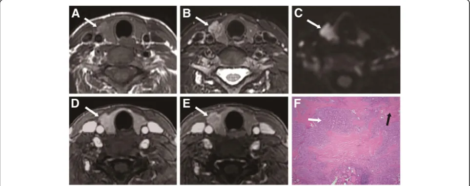
Fig. 2A 47-year-old woman with thyroid nodular goiter in the left thyroid lobe.aAxial T1-weighted image showing a heterogeneous isointense nodule (long arrowhead) with patchy hyperintense signal (white arrow) in the left lobe.bAxial T2-weighted image showing a heterogeneous hyperintense nodule with cystic change (white arrow) in the left lobe.cAxial DWI image showing a hyperintense nodule (white arrow) with ADC value of 1.990 × 10⁻³mm2/s.dAxial contrast-enhanced image showing a heterogeneous hyperintense lesion with regular shape and clear margin in the left thyroid lobe during early phase.eAxial contrast-enhanced image showing the pseudocapsule sign (white arrow) in the left thyroid lobe during delayed phase.fHistopathological hematoxylin and eosin (H&E, × 40) staining showing heterogeneous follicular hyperplasia with colloid and hemorrhage (white arrow)
Fig. 3A 44-year-old woman with thyroid papillary carcinoma in the right thyroid lobe.aAxial T1-weighted image showing an isointense lesion (white arrow) in the right thyroid lobe.bAxial T2-weighted image showing a heterogeneous hyperintense lesion with extrathyroidal extension (white arrow) in the right thyroid lobe.cAxial DWI image showing a hyperintense nodule (white arrow) with ADC value of 1.070 × 10⁻³mm2/s.
benign and malignant non-papillary follicular and oxy-philic cell lesions [6,12]. MRI is an effective noninvasive modality to differentiate malignant from benign tumors
[24]. A number of MRI studies examined the ADC
values of thyroid nodules [8, 22, 25], but image quality was considered to be relatively poor because of suscepti-bility artifacts, motion artifacts, and low signal-to-noise ratio with previous head and neck joint coil. One previ-ous study showed that only 26/40 patients had images
that could be interpreted because of distortion [8].
An-other study showed that patient motion was the major factor of exclusion due to breathing, swallowing, and coughing [2]. In addition, the b value is a critical factor affecting image quality and ADC values. When low b value is used, the ADC value tends to be higher due to the contribution of perfusion. Applying high maximum b values may be preferable when ADC measurements are performed to differentiate malignant from benign tis-sues, exclusively based on their water diffusion character-istics. Nevertheless, the signal-to-noise ratio decreases as the b value increases, thus limiting the maximum b value.
In addition, we used a neck surface coil to increase the signal-to-noise ratio. Indeed, because the coil was close to the surface of neck, it could minimize the air-tissue boundary for reducing susceptibility artifacts. Therefore, a
relatively high b value (800 × 10−3s/mm2) was used,
which could better reflect the actual diffusion characteris-tics in this study. In addition, we used special techniques to improve image quality. A relatively small FOV (14 × 14 cm) was used to reduce susceptibility artifacts. Shim blocks were used to optimize magnetic homogeneity in the thyroid region. All patients received respiratory train-ing to improve movement-related problems. We used a breath-hold technique on dynamic contrast-enhanced MRI phase to reduce breathing motion artifacts and added saturated zone to reduce carotid artery pulsatile artifacts. Therefore, 239 of the 254 patients showed excellent image quality in this study.
Some studies have shown that DWI can differentiate benign from malignant thyroid nodules [7, 12, 14, 26]. Nevertheless, the numbers of cases included in these studies were relatively small and the findings were
some-times inconclusive [16, 26]. The sample size of the
present study was relatively large, with 181 patients and 259 thyroid lesions. Malignant nodules in this study showed lower ADC values compared with benign nod-ules. Logistic regression showed that ADC values had a high prediction value for the malignant status of thyroid lesions. Cytological features of malignant thyroid nod-ules in this study included enlarged and irregular nuclei, increased cell density, and relatively severe desmoplastic
Fig. 4A 53-year-old man with thyroid follicular carcinoma in the right thyroid lobe.aAxial T1-weighted image showing a heterogeneous isointense lesion (white arrow) in the right thyroid lobe.bAxial T2-weighted image showing a heterogeneous hyperintense lesion with regular shape and clear margin (white arrow) in the right thyroid lobe.cAxial DWI image showing a markedly hyperintense nodule (white arrow) with ADC value of 0.998 × 10⁻³mm2/s.dAxial contrast-enhanced image showing a markedly enhanced lesion with regular shape and clear margin (white arrow) in the right thyroid lobe during early phase.eAxial contrast-enhanced image shows a wash-out enhanced lesion with regular shape and clear margin (white arrow) in the right thyroid lobe during delayed phase.fHistopathological hematoxylin and eosin (H&E, × 40) staining showing abundant follicular hyperplasia (white arrow) and tumor cell invasion in the peripheral stroma (black arrow)
Table 2Independent Variables in the regression equation
Parameters OR 95% CI P
Lower Upper
ADC 694.006 49.769 9677.615 < 0.001
Irregular shape 32.798 6.495 165.619 < 0.001
Ring sign 20.381 2.668 155.717 0.004
response, whereas abundant follicles, extracellular fluid and smaller cell density resulted in higher ADC values in adenoma and nodular goiter. These results were con-sistent with previous studies [18,26].
Dynamic contrast enhancement can play a complemen-tary role in the diagnosis of thyroid carcinoma. During the delayed phase, the ring sign (with a central washout enhancement) was seen in a large number of malignant thyroid nodules, which was not reported in previous stud-ies. The central tumor area with washout indicates active growth of tumor cells, whereas the peripheral area is mainly composed of loose connective tissue with abun-dant intercellular matrix. Peripherally enhanced areas in malignant thyroid tumors during the delayed phase may also be related to the fibrous stroma of the tumor and the presence of vascular fibrotic stroma. Malignant thyroid nodules in the present study showed irregular shape after contrast agent. The histopathological characteristics of thyroid carcinoma indicate the invasive and
heteroge-neous growing pattern. One recently published study [27]
also showed that irregular margins on US were strong pre-dictor of malignancy.
In the present study, cystic degeneration, high signal cystic area on T1WI, and the pseudocapsule sign were significantly more frequent in benign thyroid nodules
than in malignant nodules. Nodular goiter was the main pathological type of benign thyroid nodules in this study. Due to the relative abundance of colloid follicles and hemorrhage, nodular goiter showed cystic changes and high signal intensity in cystic areas. Shi et al. [26] showed similar results. Na et al. [28] showed that the risk of malig-nancy of partially cystic nodules was lower than the risk of malignancy of purely solid nodules. Similar to previous studies [28], the present study showed that 68.9% of thy-roid benign nodules showed cystic changes, but only 17.1% of the malignant lesions showed cystic changes. The pseudocapsule sign was not the real capsule of tumor, but showed a clear capsule after contrast agent adminis-tration because the tumor compressed the peripheral thyroid parenchyma and caused fibrosis. Therefore, the pseudocapsule sign indicates a chronic and benign patho-logical process.
In this study, the enhancement degree between the two groups was significantly different, but there was major overlap between the two groups. Nodular goiter and adenoma showed moderate or marked enhance-ment with abundant hyperplasia of thyroid follicles. Follicular thyroid carcinoma showed marked enhance-ment because of abundant hyperplasia of thyroid folli-cles and neovascularity. Papillary thyroid carcinomas
demonstrated moderate or marked enhancement with increased cell density, severe desmoplastic response, and cell proliferation, which were consistent with pre-vious studies [26,29].
Taken together, the present study strongly suggests that multiple MRI parameters should be considered when evaluating thyroid nodules. While irregular shape, ring sign, and cystic degeneration can be subjective and
dependent upon the radiologist’s experience, ADC can
provide quantitative information to differentiate thyroid carcinoma from benign thyroid nodules. On the other hand, whether the parameters observed in the present study are better than other modalities such as US,
com-puted tomography, and scintigraphy [30] require
add-itional studies.
This study has several limitations. Firstly, this study was retrospective in design, leading to selection bias and therefore undermining the validity of the results. Prospective studies with larger sample size would increase the credibility of the results. Secondly, thyroid nodules measuring < 3 mm were not included. Improvements in MRI software and using smaller slice gaps may facilitate the detection of smaller lesions in future studies. Import-antly, it has been shown that small thyroid lesions are at higher risk of malignancy than larger ones [21]. Therefore, the present study probably underreported the number of malignant lesions. Thirdly, in this study, the major malig-nant pathological type was papillary carcinoma, while the major benign type was nodular goiter, similar to previous studies [18, 26]. Nevertheless, the underrepresentation of rarer pathological types could bias the results. We need to enlarge the samples in the following studies. Fourthly, our center only has a 1.5-T MRI scanner and differences in imaging parameters for malignant thyroid nodules could not be compared with a 3-T scanner. In addition, 1.5 T MRI scanners cannot implement multiple b values and we applied a high b value to better reflect the value of diffu-sion. Meanwhile, with technological and software develop-ment, some advanced diffusion imaging like diffusion tensor imaging has been used for the differentiation be-tween malignant and benign tumors of the head and neck
[31]. Our center could not implement those different
ad-vanced diffusion imaging modules. Furthermore, we could not determine the K-trans value because dynamic contrast enhancement at our center is routinely done at 30 s, 60 s, and then every minute, and only the trend of dy-namic enhancement could be extracted. Fifthly, we used a neck surface coil to increase signal noise ratio and many techniques to reduce artifacts, but we did not compare the differences among the coils and tech-niques. In addition, we did not compare the difference among different pathological types. Finally, the irregular shape, ring sign, and cystic degeneration are indeed subjective, but these parameters had nevertheless high
sensitivity and specificity. These parameters have not been reported before, and could have some value for the management of patients with thyroid nodule.
Conclusions
Multiple MRI parameters could be helpful to differentiate malignant thyroid nodules from benign nodules. The lo-gistic regression showed that ADC value could discrimin-ate between benign and malignant thyroid nodules with a good performance. Subjective signs such as the ring sign, irregular shape, and cystic degeneration associated with malignant thyroid nodules could provide complementary information for differentiation. Combining subjective MRI features to a quantitative measurement could improve the diagnostic performance of MRI for malignant thyroid nodules.
Abbreviations
ADC:Apparent diffusion coefficient; AUC: Area under the curve; DWI: Diffusion weighted imaging; FNAB: Fine-needle aspiration biopsy; FNAC: Fine-needle aspiration cytology; FOV: Field of view; NEX: Number of excitations; ROC: Receiver operator characteristic; ROI: Region of interest; T1WI: T1-Weighted imaging; T2WI: T2-weighted images; TE: Echo time; TI-RADS: Thyroid Imaging Reporting and Data System; TR: Repetition time; US: Ultrasound
Acknowledgments
We thank all members of the Department of Radiology, Minhang Branch, Zhongshan Hospital, Fudan University, and all members of the Department of Pathology and General Surgery for helpful discussions and invaluable help in manuscript preparation. We also express our most sincere thanks to Jiliang Ren for revising this manuscript.
Funding
This project was funded by the Shanghai Municipal Commission of Health and Family Planning,China (No. 201740242) and the Science and Technology Commission of Minhang District, Shanghai,China (No. 2015MHZ026).
Availability of data and materials
The data set supporting the results of this article are included within the article. Data and materials are available at request. Please contact the author for data requests.
Authors’contributions
Guarantor of integrity of the entire study: BS. Study concepts and design: BS, HW. Literature research: HW, RW. Clinical studies: HW, WYL, YQC.
Experimental studies / data analysis: HW. Statistical analysis: HW, BS. Manuscript preparation: RW, HW. Manuscript editing: HW, RW. All authors read and approved the final manuscript.
Ethics approval and consent to participate
This study had approval from the Institutional Review Board of Minhang Branch, Zhongshan Hospital, Fudan University, and written informed consent was obtained from patients.
Consent for publication
Not applicable.
Competing interests
All authors declare that they have no competing interests.
Publisher’s Note
Author details
1Department of Radiology, Minhang Branch, Zhongshan Hospital, Fudan
University, Shanghai, China.2Department of General Surgery, Minhang
Branch, Zhongshan Hospital, Fudan University, Shanghai, China.3Department of Pathology, Minhang Branch, Zhongshan Hospital, Fudan University, Shanghai, China.
Received: 30 September 2018 Accepted: 21 November 2018
References
1. Yoon JH, Lee HS, Kim EK, Moon HJ, Kwak JY. Malignancy risk stratification of thyroid nodules: comparison between the thyroid imaging reporting and data system and the 2014 American Thyroid Association management guidelines. Radiology. 2016;278(3):917–24.
2. Shi R, Yao Q, Wu L, Zhou Q, Lu Q, Gao R, Hu J, Kao L, Bains A, Yan Z, et al. T2 * mapping at 3.0T MRI for differentiation of papillary thyroid carcinoma from benign thyroid nodules. J Magn Reson Imaging. 2016;43(4):956–61. 3. Haugen BR. 2015 American Thyroid Association management guidelines for
adult patients with thyroid nodules and differentiated thyroid Cancer: what is new and what has changed? Cancer. 2017;123(3):372–81.
4. Liu Y, Lin Z, Sheng C, Zhu Y, Huang Y, Zhong N, Jia Z, Qu S. The prevalence of thyroid nodules in Northwest China and its correlation with metabolic parameters and uric acid. Oncotarget. 2017;8(25):41555–62.
5. Force USPST, Bibbins-Domingo K, Grossman DC, Curry SJ, Barry MJ, Davidson KW, Doubeni CA, Epling JW Jr, Kemper AR, Krist AH, et al. Screening for thyroid Cancer: US preventive services task force recommendation statement. Jama. 2017;317(18):1882–7.
6. Noda Y, Kanematsu M, Goshima S, Kondo H, Watanabe H, Kawada H, Bae KT. MRI of the thyroid for differential diagnosis of benign thyroid nodules and papillary carcinomas. AJR Am J Roentgenol. 2015;204(3):W332–5. 7. Sasaki M, Sumi M, Kaneko K, Ishimaru K, Takahashi H, Nakamura T.
Multiparametric MR imaging for differentiating between benign and malignant thyroid nodules: initial experience in 23 patients. J Magn Reson Imaging. 2013;38(1):64–71.
8. Brown AM, Nagala S, McLean MA, Lu Y, Scoffings D, Apte A, Gonen M, Stambuk HE, Shaha AR, Tuttle RM, et al. Multi-institutional validation of a novel textural analysis tool for preoperative stratification of suspected thyroid tumors on diffusion-weighted MRI. Magn Reson Med. 2016;75(4): 1708–16.
9. Razek AA. Diffusion-weighted magnetic resonance imaging of head and neck. J Comput Assist Tomogr. 2010;34(6):808–15.
10. Abdel Razek AA, Elkammary S, Elmorsy AS, Elshafey M, Elhadedy T. Characterization of mediastinal lymphadenopathy with diffusion-weighted imaging. Magn Reson Imaging. 2011;29(2):167–72.
11. Dilli A, Ayaz UY, Cakir E, Cakal E, Gultekin SS, Hekimoglu B. The efficacy of apparent diffusion coefficient value calculation in differentiation between malignant and benign thyroid nodules. Clin Imaging. 2012;36(4):316–22. 12. Nakahira M, Saito N, Murata S, Sugasawa M, Shimamura Y, Morita K, Takajyo
F, Omura G, Matsumura S. Quantitative diffusion-weighted magnetic resonance imaging as a powerful adjunct to fine needle aspiration cytology for assessment of thyroid nodules. Am J Otolaryngol. 2012;33(4):408–16. 13. Mutlu H, Sivrioglu AK, Sonmez G, Velioglu M, Sildiroglu HO, Basekim CC, Kizilkaya E. Role of apparent diffusion coefficient values and diffusion-weighted magnetic resonance imaging in differentiation between benign and malignant thyroid nodules. Clin Imaging. 2012;36(1):1–7.
14. Wu Y, Yue X, Shen W, Du Y, Yuan Y, Tao X, Tang CY. Diagnostic value of diffusion-weighted MR imaging in thyroid disease: application in differentiating benign from malignant disease. BMC Med Imaging. 2013; 13:23.
15. Razek AA, Sadek AG, Kombar OR, Elmahdy TE, Nada N. Role of apparent diffusion coefficient values in differentiation between malignant and benign solitary thyroid nodules. AJNR Am J Neuroradiol. 2008;29(3):563–8. 16. Schueller-Weidekamm C, Kaserer K, Schueller G, Scheuba C, Ringl H, Weber
M, Czerny C, Herneth AM. Can quantitative diffusion-weighted MR imaging differentiate benign and malignant cold thyroid nodules? Initial results in 25 patients. AJNR Am J Neuroradiol. 2009;30(2):417–22.
17. Abdel Razek AA, Sadek AG, Gaballa G. Diffusion-weighed MR of the thyroid gland in Graves' disease: assessment of disease activity and prediction of outcome. Acad Radiol. 2010;17(6):779–83.
18. Bozgeyik Z, Coskun S, Dagli AF, Ozkan Y, Sahpaz F, Ogur E. Diffusion-weighted MR imaging of thyroid nodules. Neuroradiology. 2009;51(3):193–8. 19. Abdel Razek AAK, Abd Allah SS, El-Said AAE. Role of diffusion-weighted
magnetic resonance (MR) imaging in differentiation between Graves' disease and painless thyroiditis. Pol J Radiol. 2017;82:536–41.
20. Morris LG, Sikora AG, Tosteson TD, Davies L. The increasing incidence of thyroid cancer: the influence of access to care. Thyroid. 2013;23(7):885–91. 21. Davies L, Ouellette M, Hunter M, Welch HG. The increasing incidence of
small thyroid cancers: where are the cases coming from? Laryngoscope. 2010;120(12):2446–51.
22. Chen L, Xu J, Bao J, Huang X, Hu X, Xia Y, Wang J. Diffusion-weighted MRI in differentiating malignant from benign thyroid nodules: a meta-analysis. BMJ Open. 2016;6(1):e008413.
23. Thoeny HC, De Keyzer F, King AD. Diffusion-weighted MR imaging in the head and neck. Radiology. 2012;263(1):19–32.
24. Luczynska E, Heinze-Paluchowska S, Domalik A, Cwierz A, Kasperkiewicz H, Blecharz P, Jereczek-Fossa B. The utility of diffusion weighted imaging (DWI) using apparent diffusion coefficient (ADC) values in discriminating between prostate Cancer and Normal tissue. Pol J Radiol. 2014;79:450–5.
25. Lu Y, Moreira AL, Hatzoglou V, Stambuk HE, Gonen M, Mazaheri Y, Deasy JO, Shaha AR, Tuttle RM, Shukla-Dave A. Using diffusion-weighted MRI to predict aggressive histological features in papillary thyroid carcinoma: a novel tool for pre-operative risk stratification in thyroid cancer. Thyroid. 2015;25(6):672–80.
26. Shi HF, Feng Q, Qiang JW, Li RK, Wang L, Yu JP. Utility of diffusion-weighted imaging in differentiating malignant from benign thyroid nodules with magnetic resonance imaging and pathologic correlation. J Comput Assist Tomogr. 2013;37(4):505–10.
27. Liu J, Zheng D, Li Q, Tang X, Luo Z, Yuan Z, Gao L, Zhao J. A predictive model of thyroid malignancy using clinical, biochemical and sonographic parameters for patients in a multi-center setting. BMC Endocr Disord. 2018;18(1):17.
28. Na DG, Kim JH, Kim DS, Kim SJ. Thyroid nodules with minimal cystic changes have a low risk of malignancy. Ultrasonography. 2016;35(2):153–8. 29. Gupta N, Norbu C, Goswami B, Chowdhury V, RaviShankar L, Gulati P, Kakar A. Role of dynamic MRI in differentiating benign from malignant follicular thyroid nodule. Auris Nasus Larynx. 2011;38(6):718–23.
30. Zandieh S, Muin D, Bernt R, Hittmair K, Haller J, Hergan K. Characteristics of incidentally found thyroid nodules in computed tomography: comparison with thyroid scintigraphy. BMC Med Imaging. 2017;17(1):8.