Dr. Aamer Ali Khattak (Corresponding Author) Assistant Professor
Department of Medical Lab Technology
University of Haripur, Near Swat Chock Hattar Road, Haripur, KPK, Pakistan
Cell No: +92-333-5140556 Email: amir.khattak@hotmail.com Date Received: January 17, 2017 Date Revised: March 24, 2017 Date Accepted: May 10, 2017
INTRODUCTION
In burns patients, infections make morbidity and mortality higher. Burns are one of the most common and
MICROBIOLOGICAL PROFILE AND COMMONLY
USED ANTIBIOTICS SUSCEPTIBILITY PATTERN OF
ISOLATES AMONG BURN PATIENTS AT A
TERTIARY CARE HOSPITAL
Aamer Ali Khattak1, Usman Ayub Awan1, Mohsina Haq2, Salma Khalid3, Fouzia Ashraf4, Muhammad Faisal Nadeem4 1Department of Medical Lab Technology, University of Haripur - Pakistan
2Department of Pathology, PMC, Riphah Intermational University Islamabad - Pakistan
3Department of Public Health, PMC, Riphah Intermational University Islamabad - Pakistan
4Department of Pathology, Allama Iqbal Medical College, Lahore - Pakistan
ABSTRACT
Objective: To assess frequency of different pathogens in burn patients and susceptibility pattern of commonly used antibiotics.
Material and Methods: In this prospective study, burn patients (admitted or visited OPD) of Ayub Teaching Hospital, Abbottabad, Pakistan from January 2013 to December 2015 were registered. Mainly wound swab and blood specimen (along with all sort of culturing isolates) were collected from burn patients and were cultured by aseptic conventional method with the intention of observing microbial pathogens. Microbiological profile and antibiotic susceptibility pattern was collected using standard collection techniques and analyzed at local private laboratory.
Results: Among total 491 clinical isolates, 241 (49%) were observed positive. Frequency of pathogens isolated; Pseu-domonas aeruginosa 163 (68%), Staphylococcus aureus 43(18%), E.coli 14(6%), Candida albicans 10(4%), Klebsiella pneumoniae 6 (2%), Salmonella species 2(1%), Proteus species 2(1%) and 1(0.4%) Streptococcus pyogenes. Among these isolates Pseudomonas aeruginosa, Escherichia coli and Staphylococcus Aureus revealed 80±10 % resistant to Ampicillin, Co-Amoxiclav, Amoxicillin, Clarithromycin, Methicillin, and Vancomycin. The rate of bacteremia was 49% among burn patients isolates.
Conclusion: Regular antibiotic sensitivity testing should be done for each patient in order to select an appropriate antimicrobial agent.
Key Words: Burns, Antibiotic Resistance, MRSA, Multidrug Resistant, Microbial Infections.
This article may be cited as: Khattak AA, Awan UA, Haq M, Khalid S, Ashraf F, Nadeem MF. Microbiological profile and commonly used antibiotics susceptibility pattern of isolates among burn patients at a tertiary care hospital. J Med Sci 2017; 25: (2) 200-204.
Different patient groups such as burn patients and trauma patients have unique predisposition to different infections. In a Swedish study, the most common infec-tion was burn wound infecinfec-tion (60%) followed by blood stream infection (20%), urinary tract infection (20%), and pneumonia (10%) respectively6.
The organisms that predominate as causative agents of burn wound infection in any burn treatment facility changes over time. Gram-positive organisms are primarily dominant as compared to Gram-negative op-portunistic organisms7. However, the common isolated pathogens are Pseudomonas aeruginosa, Staphylococ-cus aureus, Klebsiella Spp, various Coliform bacilli and Fungi (Candida albicans, Aspergillus fumigatus)8,9. Fur-thermore their frequencies reported were Pseudomonas aeruginosa (38%), Staphylococcus aureus (20%), and Acinetobacter baumanni (10%)10. Multidrug resistant bacteria have frequently been reported as the cause of nosocomial outbreaks of infection in burn units or as colonizers of wounds of burn patients11,12. Pseudomonas aeruginosa develops resistance against antimicrobial drugs more rapidly by producing exopolysaccharide that binds with water and form gels making it resistant to many antibiotics hence making treatment even more complicated13-17.
To evaluate newer therapies for wound infec-tions, it becomes essential to qualify and quantify bacteria’s which are involved in causing different in-fections. Large numbers of clinical studies are carried out to determine the flora of burn wounds in different parts of the world. It showed that the general bacterial flora of burn wounds may be different in different clin-ical settings18.Regarding the significance of infection in burn victims, it is necessary for every burn unit to rule out the pattern and incidence of dominant flora of burn patients. In this perspective the present study was planned to sort out the pattern of burn wounds microbial colonization in our population, in order to determine the most frequent microbial isolates in our burn patients along their antibiotic sensitivity patterns towards commonly used antibiotics.
MATERIAL AND METHODS
This study was conducted in the department of Pathology, Ayub Teaching Hospital, Abbottabad, Pakistan from January 2013 to December 2015. In this 2 year prospective study total 491 isolates were collected from all admitted burns cases and cases with minor burn dealt in emergency at Ayub Teaching Hospital during 2013 and 2015 after taking proper consent. Specimens of their wound swabs and blood
(along with urine, throat swab, CVP tips, nasal swab, dressing gauze, urinary catheter, ETT tips, oral swab) were collected, labeled and transported following stan-dard microbiology sample collection and transportation procedures culture and sensitivity. The collected swabs were inoculated onto Blood agar and MacConkey agar. Selective media like Mannitol Salt Agar was also used in order to differentiation between S. aureus and P. aeruginosa. After the incubation for 24 hours at 37˚C the isolates were identified and separated on basis of their colony characteristics, pigment formation for P. aeruginosa (Pyocyanin and Pyoverdine) along with biochemical tests (Catalase and Coagulase) in addition to Gram staining as described by Parvin et al. 200919. Antibiotic sensitivity of different isolates was done using disc diffusion method on Muller–Hinton agar plate17. For the antibiotic sensitivity discs were placed on the culture inoculated agar plate surface. After overnight incubation at 37˚C, the zone of inhibition exhibited by each antibiotic was measured in millimeter (mm).
RESULTS
This study was conducted on 491 wound swab and blood culture for microbiological profiling and com-monly used drug susceptibility. In 491 burn patients 211 males (43%) and 280 females (57%) with age ranges of burn patients were 1 year to 71 years with a median age of 28.
About 241 isolates were positive for microbial growth with 49% rate of bacteremia. In 241 microbial positive isolates the frequency of pathogens isolated was Pseudomonas aeruginosa 163(68%) followed by Staphylococcus aureus 43(18%), E. coli 14(6%), Candida 10(4%), Klebsiella 6(2%), Salmonella 2(1%), Proteus 2(1%) and Strep pyogenes 1(0.4%). Among these isolates Pseudomonas aeruginosa, E. coli and Staphylococcus aureus revealed 80±10% resistant
to Ampicillin, Augmentin, Amoxicillin, Clarithromycin, Methicillin, and Vancomycin.
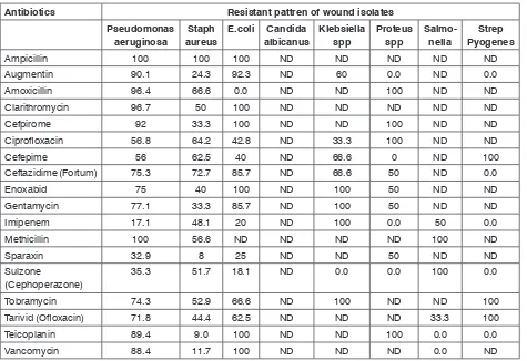
The proportion of these isolate were predominant-ly increased in samples from Pus 254(52%), Blood 79 (16%), Urine 47(10%), Throat Swab 28(6%), CVP Tips 21(4%), Nasal swab 17(3%), Dressing Gauze 14(3%), Catheter 12(2%), ETT Tips 12(2%), Axillary Swab 06(1%), Oral Swab 01 (0.2%). It is worth mentioning that in our samples Pseudomonas aeruginosa and Staph aureus are frequent in pus while E. coli in urine and Salmonella species in blood samples. Antibiotic Sensitivity of wound isolates are shown in Table 1.
DISCUSSION
Burn wound is considered to be one of the major health problem worldwide20. Regardless of significant improvement in the survival of burn patients, infectious complications still continue to be the major cause of morbidity and mortality in burn patients21. In our current study female population are being more effected by burns (57%) as compared to males (43%). Our results are in line with results reported by a study in which they revealed that incidence of burn cases are much higher
in females 53.2% as compared to males 39.2%22. This may due to domestic and household responsibilities of females in our setup which make them more prone to burns.
In our study P. aeruginosa was the most frequent microbe found in our patients. These result are similar to other studies that were conducted on burns patients23-24. In striking contrast to our finding, some published studies have reported S. aureus as their predominant microbe of burn wound infections25-26. Klebsiella spp was reported only (2%) in current study which is too much lower than the study which shown (31%)27.
In our study 57% Staph aureus were resistant to Methicillin this support the study conducted in Iran which revealed Methicillin resistant were 58%14 but opposing to study conducted in Hyderabad revealed 33% respectively27. Pseudomonas aeruginosa revealed 80±10% resistant to Ampicillin, Co-Amoxiclav, Amoxicil-lin, Clarithromycin, MethicilAmoxicil-lin, and Vancomycin which confirmed the previous studies14-28.
The present study should prompt well designed local studies to confirm and improve our findings. P. Table 1: Antibiotic sensitivity of wound isolates
Antibiotics Resistant pattren of wound isolates
Pseudomonas
aeruginosa aureusStaph E.coli Candida albicanus Klebsiella spp Proteus spp Salmo-nella PyogenesStrep
Ampicillin 100 100 100 ND ND ND ND ND
Augmentin 90.1 24.3 92.3 ND 60 0.0 ND 0.0
Amoxicillin 96.4 66.6 0.0 ND ND 100 ND ND
Clarithromycin 96.7 50 100 ND ND ND ND ND
Cefpirome 92 33.3 100 ND ND 100 ND ND
Ciprofloxacin 56.8 64.2 42.8 ND 33.3 100 ND ND
Cefepime 56 62.5 40 ND 66.6 0 ND 100
Ceftazidime (Fortum) 75.3 72.7 85.7 ND 66.6 50 ND 0.0
Enoxabid 75 40 100 ND 100 50 ND ND
Gentamycin 77.1 33.3 85.7 ND 100 50 ND ND
Imipenem 17.1 48.1 20 ND 100 0.0 50 0.0
Methicillin 100 56.6 ND ND ND ND 100 ND
Sparaxin 32.9 8 25 ND ND 50 ND ND
Sulzone
(Cephoperazone) 35.3 51.7 18.1 ND 0.0 0.0 100 0.0
Tobramycin 74.3 52.9 66.6 ND 100 ND ND 100
Tarivid (Ofloxacin) 71.8 44.4 62.5 ND ND ND 33.3 100
Teicoplanin 89.4 9.0 100 ND ND 100 0.0 0.0
aeruginosa, S. aureus constituted the most common bacterial microbes of burn wounds in our patients. A variable antibiotic susceptibility pattern was observed among the grown microbes. Early excision of deep burns and coverage with skin graft can help to effec-tively reduce the burden of these infections. With this evidence base in mind, we can revisit our policy of empiric antibiotic cover for our burn injury patients with sepsis.
CONCLUSION
The two predominant microbes of in burn patients were Pseudomonas aeruginosa and Staphylococcus aureus. Multidrug-resistant microbial infections are becoming gradually common and difficult to treat.
RECOMMENDATIONS
Selection of appropriate antibiotics after sensi-tivity testing is very important in treating not only burn wound infection but any bacterial infection. Secondly hospital should have strict antibiotic policy for burn patients in order to treatment properly and prevent the spread of multi-drug resistant and community acquired wound infections pathogens. It’s the era of molecular analysis so it is recommended that genes associated with different antibiotics should be evaluated mutations conferring resistance.
REFERENCES
1. Church D, Elsayed S, Reid O, Winston B, Lindsay R. Burn wound infections. Clinical microbiology reviews 2006;19(2):403-34.
2. Rastegar LA, Alaghehbandan R. Epidemiological study of self-inflicted burns in Tehran, Iran. The Jour-nal of burn care & rehabilitation 2002;24(1):15-20. 3. Gang RK, Bang RL, Sanyal SC, Mokaddas E, Lari
AR. Pseudomonas aeruginosa septicaemia in burns. Burns 1999;25(7):611-6.
4. Lari AR, Alaghehbandan R, Akhlaghi L. Burn wound infections and antimicrobial resistance in tehran, iran: an increasing problem. Annals of burns and fire disasters 2005;18(2):68-70.
5. Oncul O, Yuksel F, Altunay H, Açikel C, Celikoz B, Cavuslu S.The evaluation of nosocomial infection during 1-year-period in the burn unit of a training hospital in Istanbul, Turkey. Burns 2002;28(8):738-44.
6. Tancheva D, Hadjiiski O. Effect of early nutritional support on clinical course and septic complications in patients with severe burns. Annals of burns and fire disasters 2005;18(2):74-83.
7. Appelgren P, Bjornhagen V, Bragderyd K, Jonsson
CE, Ransjo U. A prospective study of infections in burn patients. Burns 2002;28(1):39-46.
8. Ahmad M, Hussain SS, Khan MI, Malik SA. Pattern of bacterial invasion in burn patients at the Pakistan Institute of Medical Sciences, Islamabad. Annals of burns and fire disasters 2006;19(1):18-21.
9. Revathi G, Puri J, Jain BK. Bacteriology of burns. Burns 1998;24(4):347-49.
10. Skoll PJ, Hudson DA, Simpson JA. Aeromonas hy-drophila in burn patients. Burns 1998;24(4):350-53. 11. Lawrence JC. Burn bacteriology during the last 50
years. Burns 1992;18:23-29.
12. Bagdonas R, Tamelis A, Rimdeika R, Kiudelis M. Analysis of burn patients and the isolated pathogens. Lithuanian Surg 2004;2(3):190-93.
13. Shahid M, Malik A. Resistance due to aminoglyco-side modifying enzymes in Pseudomonas aerugi-nosa isolates from burns patients. Indian Journal of Medical Research 2005;122(4):324-29.
14. Ekrami A, Kalantar E. Bacterial infections in burn patients at a burn hospital in Iran. Indian Journal of Medical Research 2007;126(6):541-43.
15. Karlowsky JA, Jones ME, Draghi DC, Thornsberry C, Sahm DF, Volturo GA. Prevalence and antimicro-bial susceptibilities of bacteria isolated from blood cultures of hospitalized patients in the United States in 2002. Annals of clinical microbiology and antimi-crobials 2004;3:61-65.
16. Agnihotri N, Gupta V, Joshi RM. Aerobic bacterial isolates from burn wound infections and their anti-biograms a five-year study. Burns 2004;30(3):241-43. 17. Irfan M, Ahmed I, Shafee M, Tareen AM, Rehman
MFU, Khan SA. Microbiological Investigation of Burn Patients in Burn Intensive Units, in Quetta, Pakistan. Gas 2014;29:29-31.
18. Shaikh Diln, Di Sah, Shaikh Khur, Shaikh Muni, Naqvi BS, Shaikh MR, et al. Incidence and Resistance Pat-tern of Bacteria Associated With Burn Wound Sepsis. Pakistan Journal of Pharmacology 2004;21(2):39-47. 19. Hassanzadeh P, Motamedifar M, Hadi N. Prevalent
bacterial infections in intensive care units of Shiraz University of medical sciences teaching hospitals, Shiraz, Iran. Jpn J Infect Dis 2009;62(4):249-53. 20. Rajput A, Singh KP, Kumar V, Sexena R, Singh RK.
Antibacterial resistance pattern of aerobic bacteria isolates from burn patients in tertiary care hospital. Biomedical research 2008;19(1):1-4.
21. David W, Albert T, Basil A. Infection of burn wounds. Bennett J, Brachman P, Hospital Infections, 4th ed Philadelphia: Lippincott-Raven 1998;587-601. 22. Alghalibi SMS, Humaid AA, Alshaibani EAS, Alhamzy
AUTHOR’S CONTRIBUTION
Following authors have made substantial contributions to the manuscript as under: Khattak AA: Concept, design acquisition of data ,manuscript Writing
Awan UA: Sample collection and lab Analysis Haq M: literature review and statistical analysis Khalid S: Bibliography Statistical analysis Ashraf F: data analysis and result interpretation Nadeem MF: sample collection and reference collection
Authors agree to be accountable for all aspects of the work in ensuring that questions related to the accuracy or integrity of any part of the work are appropriately investigated and resolved.
CONFLICT OF INTEREST: Authors declare no conflict of interest GRANT SUPPORT AND FINANCIAL DISCLOSURE NIL
infection in Sanaa, Yemen. Egypt. Acad. J biolog Sci 2011;3(1):19-25.
23. Noorbakhsh Sabet N, Japoni A, Mehrabani D, Japoni S. Multi-drug resistance bacteria in Qom hospitals, Central Iran. Iran Red Crescent Med J 2010;12:501-03. 24. Oncul O, Ulkur E, Acar A, Turhan V, Yeniz E, Kara-caer Z, Yildiz F. Prospective analysis of nosocomial infections in a burn care unit, Turkey. 2009.130(6): 758-64.
25. Altoparlak U, Erol S, Akcay MN, Celebi F, Kadanali A. The time-related changes of antimicrobial resistance patterns and predominant bacterial profiles of burn wounds and body flora of burned patients. Burns 2004;30(7):660-4.
26. Erol S, Altoparlak U, Akcay MN, Celebi F, Parlak M. Changes of microbial flora and wound colonization in burned patients. Burns 2004;30(4):357-61. 27. Rao SR, Lakshmi LJ, Pavani S, Kawle V, Prakash
SJ. Bacteriological Profile, Antibiogram of burn wound isolates and detection of MRSA And ESBL production at Tertiary Care Hospital, Hyderabad. 2014; 3(10): 1691-98.
28. Estahbanati HK, Kashani PP, Ghanaatpisheh F. Frequency of Pseudomonas aeruginosa serotypes in burn wound infections and their resistance to antibiotics. Burns 2002;28(4):340-48.