HISTOPATHOLOGICAL EFFECT OF AZATHIOPRINE ON LIVER, INTESTINE AND SPLEEN OF ALBINO MICE
Dina Khudhair Hussein Ali*
Department of Biology, College of Science, Baghdad University, Baghdad, Iraq E. mail: *dinakhudhair4@gmail.com.
Article received 01.12.2017, Revised 01.08.2018, Accepted 12.8.2018 ABSTRACT
This study was carried out to study the effect of azathioprine drug on some histological structure in albino mice. sixty adult female albino mice (20mice/group), were used in this study, the average body weights are 30-32 gm. two doses of azathioprine (50 mg/kg/b wt. and 100 mg/kg/b wt.) were given by the oral route daily for 40 days to the second and third group respectively, while control group was given normal saline. The histopathological examination of the treated animal for liver showed congestion with dilation of portal vein, Dilation of sinusoids and Mono-nuclear cell aggregation for the second group while for the third treated group showed small granulomatous lesion of mononuclear cells, Enlargement and proliferation of kupper cells, Severe necrosis. Sever vacuolar degeneration of hepatocytes. The histopathological section of spleen was showed Infiltration of megakaryocyte for the second group while for the third group showed hypertrophy of central arteriole with dilation and infiltration of mono-nuclear cell mainly macrophage and plasma cell in red pulp. The histological changes in intestine tissue of the treated animals showed vacular degeneration of epithelial cell with hyperplasia of goblet cell which produce allots of mucin for the second group while for the third group showed infiltration of mononuclear cells between glands. From this study we concluded that there was harmful effect of azathioprine on different tissues of the experimental animal dependent on increasing dose of this drug.
Keywords: azathioprine, spleen, mice, Histology, liver, harmful, spleen
INTRODUCTION
Azathioprine (Imuran) belongs to the chemical class of purine homologues, it is an imidazole tra-nsformed to 6- thioionsinic acid and 6-mercaptop- urine in body [Day et al., 2005]. in organ trans- plantation. It is act to prevent rejection of organs as an immunosuppressive drug and auto-immune diseases. It is used also to treat some medical dis-eases like Crohn's disease and rheumatoid arthritis [Patel and McCall, 2006] to treat an array of auto-immune diseases likedermatitisandrestrictivelung disease with prevent rejection of transplanted org-ans thus Azathioprine is used alone or with other immunosuppressive drug. It is also important for inflammatory bowel disease [Evans, 2004]. since it stop the action of amido-phosphoribosyl trans-ferase enzyme, So it block the synthesis of RNA and DNA by cells [Sami, 2016]. In spite of that its mediated the action of Thioguanosine triphosph-ate (TGTP) which is incurportriphosph-ated in to RNA syn-thesis [Maltzman and Koretzky, 2003]. The mode of action to azathioprine is send activated monon-uclear cells and T cells into apoptosis (program-med cell death) by interacts with the GTP-binding protein, stopping the regulation of specific protein Bcl-xL [Steinhilber, et al., 2005] when the pers-ons begins to use this drug, they have common side effects including Nausea, dizziness, vomiting, skin rashes, fatigue and diarrhea. Win transplant patients receiving this drug hair loss is usually
seen. One of side effect of azathioprine is suppres-ses the bone marrow synthesis so patients can develop anemia. Acute pancreatitis can occur in patients with Crohn's disease [Weersma, et al., 2004].
MATERIALS AND METHODS
ANIMALBREEDING:Fortyadults,15-16weeks
old, female albino mice (Mus musculus), weighing 30-32gm were obtained from animal’s house of the College of Science, University of Baghdad, Iraq. The animals were kept in a well-ventilated room, in standard plastic cages at 24-28°C tempe-rature with 12 hrs natural light and 12 hrs dark-ness. The mice had free access to tap water and dry pellets which obtained from local market ad libitum. The mice were allowed to acclimatize for few days.
TREATMENT: Animals where divided into 3
groups, 20 per group as follows:
Group1: animals were treated with normal saline. Group 2 animal were treated with 50mg/ kg body weight of azathioprine.
Group 3: animals were treated with 100 mg/kg/ body weight of azathioprine.
ANIMALS KILLING AND SPECIMENS COLLECTION: By cervical dislocation [Siddi-que, et al., 2015], all mice were killed after 40 days. Liver, spleen and intestine were removed and fixed in Bouinʼs fluid for 12-24 hours then washed several times with ethanol 70% by dehyd-rated process and embedded in paraffin and kept until use. Serial sections were cutted of 5 microns thickness using a rotatory microtome and then stained with hematoxylin and eosin (H&E) then examined using light microscope. Photo-graphs were taken by digital camera [Bancroft and Stevens, 1982].
RESULT AND DISCUSSION
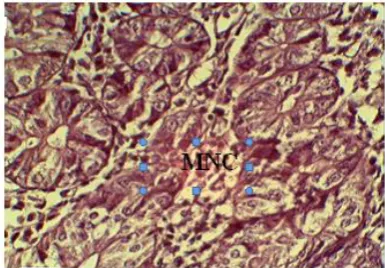
Azathioprine is an immune suppressive drug, it is the most important drug used in the therapy of rheumatoid arthritis; inflammatory bowel disease; acute lymphoblastic and leukemia. The histologi- cal examination of liver, spleen and intestine in control group shows normal histology as show in Fig 1, 4 and 8. The histopathological examination of liver for treated animals with 50 mg/kg body weight (second group) of azathioprine showed dil-ation of portal vein with congestion, mononu-clear cell aggregation (MNC) and dilation of sinu-soids Fig 2 ,while for the third group which trea-ted with100mg/kg body weight showed small gra-nulomatous lesion of mononuclear cells, Enlarge- ment and proliferation of kupper cells, severe necrosis. sever vacuolar degeneration of hepato-cytes Fig 3.
The histopathological section of spleen was sho-wed Infiltration of megakaryocyte for the second group which treated with 50 mg/kg body weight while for the third group which treated with100 mg/kg body weight showed hypertrophy of central arteriole with dilation and infiltration of mononuc-lear cell (MNC) mainly macrophage and plasma cell in red pulp Fig 5, 6 and 7.
Histological changes in of tissue of intestine for the second treated animals with50 mg/kg body weight showed vascular degeneration of epithelial cell with hyperplasia of goblet cell witch produce allotsof mucin while for the third group which tre- ated with100 mg/kg body weight showed infil-tration of mononuclear cells between glands Fig 9 and 10.
The harmful effect of azathioprine on the infe-cted tissues may be due to azathioprine toxicity, Functional polymorphisms of several enzymes involved in the metabolism of thiopurines like azathioprine have been linked with toxicity [Sch-maier, 2008]. Mion et al., 1991] demonstrated that azathioprine induces hepatotoxity, since it causes lesions in the liver, hyperplasia and peri sinusoidal
fibrosis in it and this agree with our study (Fig 2, 3) [César
et al.,
2004]. The production free radi-cal in organs and tissues are the cause of toxic effects of azathioprine since its reduce to stop the synthesis of different specialized cell like hepato-cytes by selective inhibiting the synthesis of aden-ine which belong to puraden-ine nucleotides and this agree with our study Fig 3 [Weersmaet al.,
2004]. Azathioprine cause acute pancreatitis, so it has harmful effect on liver cell which is responsible for detoxification [Machalinska et al.,2002] And kuffer cell in liver play role in detoxification also [Niels et al., 2016, Hascheck and Rausseaux, 1998]. The severity of these negative effects of azathioprine on animal organs may be related to the duration of exposure [Maltzman and Kore-tzky, 2003].No study available reported the effect of aza-thioprine on the tissue of spleen and intestine, but theresults of histological examination in this study may support the negative effect of azathioprine on these tissues. The harmful effect of azathioprine on tissue of spleen and intestine especially vascu-lar degeneration may be due to Azathioprine inhi-bits the regeneration of cells by interrupting with DNA synthesis [Dearden and Nicholson, 1984] as in Fig 7 & 9, Since azathioprine belongs to NSA-IDs, there is direct relation-ship between format-ion of gastric ulcer and anti-inflammatory of NSAIDs [Bjarnason et al., 1987]. The clinical implications in some patient receiving NSAIDs drugs may bleed from the small intestine and lose protein, contributing to iron deficiency and hypo-albuminaemia [Gately and Li, 2004] and this agree with our study. NSAIDs also induce apop-tosis, cell growth inhibition and antiangiogenesis such as azathioprine [Patel, et al.,2006].
After patients take azathioprine, the absorption completely occurred in digestive tract very quic-kly [Jewell and Truelove, 1972]. Jewell and Truelove [1972] demonstrated that azathioprine cause gastrointestinal disturbance, the azathiopr-ine treated patients suffered from relapses of the ulcerativecolitis(reoccurrence of symptoms which include diarrhea and inflammation in this state) [Casey, 1968].
chi-ldren treated with immunosuppressive drugs (aza-thioprine), anemia was diagnosed due to changes in the upper gastrointestinal tract, the location of the ileum, intestinal villous atrophy. And this study was agreed with our result Fig 10.
Fig 1: Section of liver of control group showed nor-mal hepatocytes, nornor-mal central vein, and nornor-mal arrangement of liver plate (40X)
Fig 2: Histopathological section of liver of mice
treated with 50mg/kg body weight showed: Conges-tion with dilaConges-tion of portal vein (CP), DilaConges-tion of sinusoids (DS). Mononuclear cell aggregation (M).
Fig. 3: Histopathological section of liver of mice treated with 100 mg / kg body weight showed: small granulomatous lesion of mononuclear cells (M), Enlaregement and proliveration of kupper cells (EK),
Severe necroseis (N). Sever vacuolor degene-ration of hepatocytes (DH).
Fig 4: Section of spleen of control group showed normal white pulp and normal red pulp (40X).
Fig 5: Histopathological section of spleen of mice treated with 50mg/kg body weight showed: Infiltra- tion of mega karyocyte
Fig 7: Histopathological section of spleen of mice treated with 100 mg / kg body weight showed: red pulp infiltration of mononuclear cell (MNC) mainly macrophage and plasma cell.
Fig 8: Section of intestine for control group showed normal histology (40X).
Fig 9:Histopathological section of intestine of mice
treated with 50 mg / kg body weight showed vacular degeneration of epithelial cell (V) with hyperplasia of goblet cell (H) which produce allots of mucin(M).
Fig 10:Histopathological section of intestine of mice
treated with 100mg/kg body weight showed: infiltr- ation of inflammatory cells between glands (MNC).
REFERENCES
Bancroft, J. and A. Stevens, Theory and practic of histological technique. 2nded. Churchill Living stone, London Pp. 662 (1982).
Bjarnason, I., Zanelli, G. and P. Prouse, Blood and proteinloss via small-intestinal inflamma-tion induced by nonsteroidal anti-inflamma-tory drugs. Lancet Pp. 711–714 (1987). Casey, T.P., Azathioprine (Imuran) administration
and the development of malignant lymphomas in NZB mice. Clinical and Experimental Immunology 3: 305 (1968).
César, M., María, D. and M. Fernández, Azathio-prine Acts upon Rat Hepatocyte Mitochondria and Stress-Activated Protein Kinases Leading to Necrosis: Protective Role of NAcetyl-L-cysteine. Journal of Pharmacology and Expe-rimental Therapeutics 104: 269-86 (2004). Day. R.O., Furst, D., E., Van riel, L. and B.
Bresnihan, Antirheumatic therapy: Actions and outocomes, USA. Springer science + Bus-iness Media Pp. 4-5 (2005).
Dearden, M.J.C. and R.M. Nicholson, Correlation between gastric imtancy and tory activity of non-steroidal anti-inflamma-tory drugs. J. Pharin. Pharinacol. 36: 713-715 (1984).
Evans W.E., Pharmacogenetics of thiopurine S-methyltransferase and thiopurine therapy. Ther. Drug Monit. 26(2): 186–1891 (2004). Gately, S. and W. Li, Multiple roles of COX-2 in
tumor angiogenesis: a target for antiangioge-nic therapy. Semin. Oncol. 31: 2–11 (2004). Gearry, R.B.1. and M.L. Barclay, Azathioprine
and 6-mercaptopurine pharmacogenetics and metabolite monitoring in inflammatory bowel disease. J. Gastroenterol. Hepatol. 20(8): 1149 - 57 (2005).
Hascheck, W.M. and Rausseaux, Fundamentals of Toxicologic Pathology. Academic Press, San Diego (1998).
Jewell, D.P. and S.C. Truelove, Azathioprine in Ulcerative Colitis: An Interim Report on a Controlled Therapeutic Trial. British Medical Journal 1: 709-712 (1972).
Machalinska, A., Nowak, Jarema, A., Wisznie-wska and B. Machalinski, In vivo effects of sodium fluoride on bone marrow transplan-tation in lethally irradiated mice. Floride 35(2): 81 – 89 (2002).
Maltzman, J.S. and G.A. Koretzky, Azathioprine: Old drug, new action. Journal of Clinical Inv-estigation 111(8): 1122–1124 (2003).
hyperplasia of the liver and perivenous fibro-sis in a patient treated for multiple sclerofibro-sis. Gut. 32: 715-717 (1991).
Niels, T., Wolfgang M., Bernd B., Burkhard B., Jürgen B., Stephan M., Dietrich H., Christian M., Tobias K., Wolfgang K., Britta S., Ulf H., Joseph W., Attyla D. and S. Andreas, Aza-thioprine-induced Acute Pancreatitis in Patie-nts with Inflammatory Bowel Diseases—A Prospective Study on Incidence and Severity. J. Crohns. Colitis. 10(1): 61–68 (2016). Patel, A.A., Swerlick, R.A. and C.O. McCall,
Azathioprine in dermatology: The past, the present and the future. Journal of the Ameri-can Academy of Dermatology 55(3): 369–389 (2006).
Pytrus, T.1., Flis A.,IwańczakF. and B. Iwańczak, Thefrequencyofanemiainchildren with newly diagnosed Crohn's disease in children. Pol. Merkur. Lekarski. 34(203): 263-268 (2013) Sami Naveed, Autoimmune Bullous Diseases:
Approach and Management, Springer Pp. 83 (2016).
Schmaier A.H., Laboratory evaluation of hemos-tatic and thrombotic disorders. In: Hoffman R., Benz E.J. Jr, Shattil S.J. Hoffman, Hema-tology: Basic Principles and Practice. 5th Edi., Philadelphia, Elsevier Pp. 122 (2008).
Siddique, T., Rabbi A.F., Najam, S.S., Khan, A., Qureshi, J.A., Khurshid, M., Islam, M. and M. Zain, Amplification, cloning and expression of
the reg3 δ gene from mouse pancreas. Pak. J.
Biotechnol. 12(1): 55-61 (2015).
Steinhilber, D., Schubert-Zsilavecz, M. and H.J. Roth, Medizinische Chemie (in German). Stu-ttgart: Deutscher Apotheker Verlag Pp. 340 (2005).