See related article on page 96.
Abstract
Background: Partial nephrectomy (PN) is now the gold standard
for the surgical treatment of small renal masses. We evaluated the effect of WIT and other factors on RDF assessed by preoperative and postoperative renal scintigraphy.
Methods: Between 2003 and 2008, 182 consecutive laparoscopic
PN (LPN) were performed in an academic centre. Among those, 56 had mercaptoacetyl triglycine (MAG3) lasix renal scintigraphy preoperatively and postoperatively.
Results: Medians for age, preoperative estimated glomerular
filtra-tion rate and computed tomography scan tumour size were 62
years, 82 mL/min/1.73m2 and 26 mm, respectively. Median WIT
and preoperative RDF were 30 minutes and 50%, respectively. Median loss of RDF after surgery was 14%. Linear regression curves showed that loss in RDF rate was 0.2% per minute when WIT was <30 minutes and 0.7% per minute when WIT was ≥30 minutes. In multivariate analysis, length of WIT and endophytic tumour location were associated with a statistically significant loss of RDF (p < 0.05), but only in the group who experienced >30 minutes of WIT.
Interpretation: Our results suggest that the factors associated with
loss of RDF are not the same before and after 30 minutes of WIT and that the rate of loss in RDF increases after 30 minutes. Since, the effect of WIT is small up to 30 minutes, we believe that sur-gery should focus on limiting the resection of normal parenchyma and to ensure negative margins and hemostasis, rather than on premature unclamping.
Cite as: Can Urol Assoc J 2011;5(2):89-95;DOI:10.5489/cuaj.10044
Résumé
Contexte : La néphrectomie partielle (NP) est maintenant la norme
thérapeutique pour le traitement chirurgical des petites masses rénales. Nous avons évalué l’effet du temps d’ischémie chaude et d’autres facteurs sur la fonction rénale différentielle par scintigra-phie rénale préopératoire et postopératoire.
Méthodologie : Entre 2003 et 2008, 182 NP par laparoscopie ont
été effectuées de façon consécutive dans un centre universitaire. De ces cas, 56 ont subi une scintigraphie rénale Lasix au MAG3
(mercaptoacétyltriglycine) avant et après l’opération.
Résultats : Les valeurs médianes pour l’âge, le taux de filtration
glomérulaire préopératoire évalué et la taille de la tumeur mesurée par tomodensitométrie étaient de 62 ans, 82 mL/min/1,73 m2 et 26 mm, respectivement. Le temps médian d’ischémie chaude et la fonction rénale différentielle avant l’opération étaient de 30 min-utes et de 50 %, respectivement. La perte médiane de fonction rénale différentielle après l’intervention était de 14 %. Les courbes de régression linéaire ont révélé que la perte de fonction rénale différentielle était de 0,2 % par minute lorsque le temps d’ischémie chaude était inférieur à 30 minutes et de 0,7 % par minute lorsque le temps d’ischémie chaude était ≥ 30 minutes. Dans l’analyse multivariée, la durée de l’ischémie chaude et une tumeur endophy-tique ont été associées à une perte de fonction rénale différentielle significative sur le plan statistique (p < 0,05), mais seulement dans le groupe ayant subi une ischémie chaude de plus de 30 minutes.
Interprétation : Nos résultats portent à croire que les facteurs
asso-ciés à la perte de fonction rénale différentielle diffèrent avant et après 30 minutes d’ischémie chaude, et que le taux de perte de fonction rénale différentielle augmente après 30 minutes. Comme l’effet de l’ischémie chaude est minimal lorsque celle-ci est de 30 minutes ou moins, nous croyons qu’il faut chercher à limiter la résection du parenchyme normal et assurer des marges néga-tives et l’hémostase plutôt que de viser un déclampage prématuré.
Introduction
The annual number of patients with renal cell carcinoma (RCC) has increased in the last decade secondary in part
due to an increase in incidentally found tumours;1 the
high-est increase has been observed in the incidence of
local-ized tumours.2,3 This trend has led to the more frequent
use of partial nephrectomy (PN), which has now become a standard, if not the preferred treatment, for smaller renal
masses.4 One of the modifiable factors associated with the
loss of renal function (RF) during PN is warm ischemia time (WIT), but the safe duration of clamping time is unkown. Some studies based on creatinine or estimated glomerular filtration rate (eGFR) suggest that WIT should be kept under 20 minutes, while others have suggested 40 minutes as a
cut-off.5-11 This discrepancy between studies underscores the
Multivariate analysis of the factors involved in loss of renal
differential function after laparoscopic partial nephrectomy:
a role for warm ischemia time
Frédéric Pouliot, MD, PhD, FRCSC;
*†Allan Pantuck, MD, MS, FACS;
†Annie Imbeault, MD;
*Brian Shuch, MD;
†need for homogenous data using RF tools that evaluate the damage to the operated kidney.
To evaluate the impact of WIT after laparoscopic PN (LPN), we analyzed its effect on the loss of renal differential function (RDF) on the operated kidney in a series of LPN. In a small cohort of patients, we demonstrate that the factors associated with loss in RDF before and after 30 minutes are different.
Methods
Data collection and patient selection
We included 56 patients who had a MAG3-lasix renal scin-tigraphy performed both preoperatively and postoperative-ly from a cohort of 182 patients who underwent LPN for organ-confined enhancing tumours on preoperative imaging. All procedures were performed by 1 surgeon in a single institution. Tumour size was defined by the longest single dimension of the lesion on the computed tomography scan. The localization of the tumour was defined as endophytic if more than 5 mm of the tumour was located in the renal
parenchyma consistent with the Johntson classification.12
Transperitoneal laparoscopic partial nephrectomy technique
A 4-port approach was used. Initial dissection was directed toward defining the renal hilum. The renal hilum was cross-clamped with a Aesculap (Aesculap, Inc., Center Valley, PA) noncrushing laparoscopic bulldog clamp or with an Aesculap Satinsky clamp. The lesion with a rim of normal parenchyma was sharply resected. Renal repair began with runs of sutures to close the collecting system. Subsequently, 2 or 3 parenchymal buttress sutures were placed. FloSeal (Baxter Healthcare Corporation, Hayward, CA) and polygla-ctin mesh rolls (Surgicel, Ethicon 360, Inc.) were placed to fill the parenchymal void and stabilized by tying down the buttress sutures. A Jackson-Pratt drain was placed.
Follow-up
Serum creatinine (Cr) was measured preoperatively and postoperatively at day 1, at 3 months and at the last date of follow-up. Renal function was evaluated by assessing the split RF using MAG-3-lasix scintigraphy at a median of 10 days.
Statistical analysis
Discrete variables were reported as a percentage and con-tinuous variables as a median and mean ± standard devia-tion. Differences between patient groups among the discrete
variables were tested using the chi-square test, while dif-ferences among continuous variables were tested using the two-sample t-test or the one-way ANOVA. The association of RDF with each characteristic was investigated using a univariate linear regression model. When investigating the relationship of RDF across different WIT intervals, the one-way ANOVA model was used and a linear contrast was used to test for a linear trend. When investigating the rela-tionship between RDF and WIT (measured continuously), a four-degree polynomial curve was fitted. Derivatives of the curve’s equation were calculated to obtain the WIT where the maximal change in the RDF loss rate occurred. To adjust for possible confounders, multivariate linear regression mod-els were also used. All analyses were conducted using SAS version 9.1.3 (SAS Institute Inc., Cary, NC).
Results
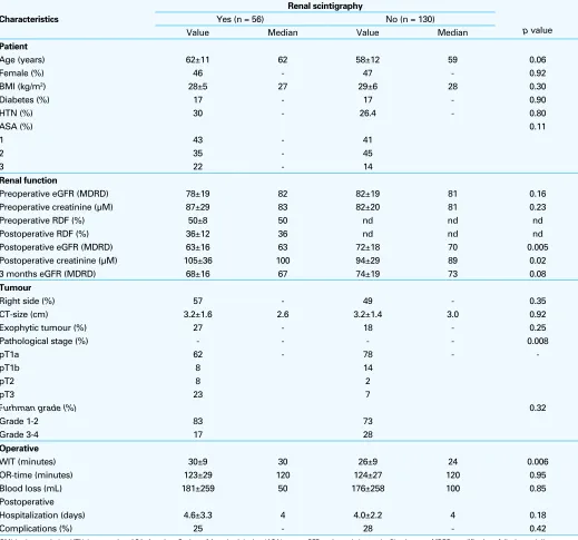
We detailed the characteristics of the patients (Table 1). No statistical difference was observed between the 56 patients selected for the study and the other 130 patients of our series, with the exception of tumour stage, WIT, postopera-tive creatinine and eGFR. Of note, the WIT in our cohort
of 56 patients was on average 29.8 ±8.8 minutes (range
9-60 minutes) and loss of RDF was on average 14.2 ±
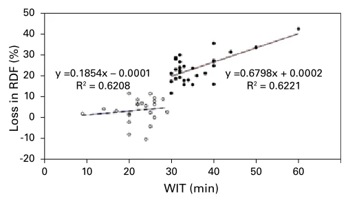
9.7 %. Endophytic location of the tumour, WIT, postopera-tive eGFR, operapostopera-tive time and blood loss were associated with a loss of RDF (Table 2). Since WIT was the modifiable factor with the strongest association, we first analyzed the effect of WIT at different intervals on loss of RDF. As the WIT increases, there is a statistically significant increase in loss of RDF in univariate analysis (Table 3). Next, we evaluated whether the loss of WIT was the same over time by plotting the effect of WIT as a continuous variable on the RDF of the operated kidney (Fig. 1). A polynomial curve drawn from the relation between WIT and loss in RDF illustrates that the rate of loss of RDF (% per minute) before and after 30 minutes is not the same. Almost two separate linear curves are observed before and after 30 minutes. The calculated point of inflection (point of maximal change in the rate of loss in RDF) of this polynomial curve is 32 minutes. We next analyzed the relation between WIT and loss in RDF using linear regression analysis before and after 30 minutes (data
not shown). The coefficient of determination (r2) of the two
linear regression curves was not the same (r2 = 0.06 before
vs. r2 = 0.39 after 30 minutes). Therefore, the discrepancy
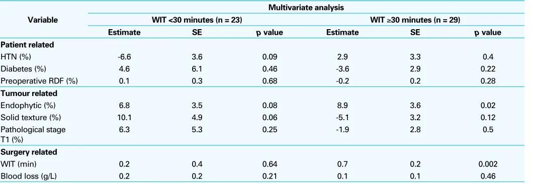
When the data were analyzed using the multivariate regression models, the factors with the strongest association with a loss in RDF (but not statistically significant) for the
group WIT<30 were HTN (p= 0.09), solid texture (p= 0.06)
and endophytic location of the tumour (p= 0.08) (Table
5). The WIT was not associated with loss in RDF. In the
group WIT≥30, WIT was the main factor associated with
a loss in RDF (p= 0.002), while the endophytic location
of the tumour was the other statistically significant factor
(p= 0.02). We next determined the rate of loss of RDF when
only considering WIT as a factor. We used our models to correct the loss in RDF for every factor other than WIT. By comparing the linear relation between WIT and loss of RDF between the two groups, we observed that the adjusted rate
of RDF-loss was 3.5-fold higher in the group with WIT≥30
minutes than the group <30 minutes. The calculated rate of loss in RDF was 0.2% per minute in the group WIT <30 as
compared to 0.7% per minute in the WIT≥30 group (Fig. 2).
Table 1. Characteristics of patients in our series of laparoscopic partial nephrectomy with or without renal scintigraphy
Characteristics
Renal scintigraphy
p value Yes (n = 56) No (n = 130)
Value Median Value Median
Patient
Age (years) 62±11 62 58±12 59 0.06
Female (%) 46 - 47 - 0.92
BMI (kg/m2) 28±5 27 29±6 28 0.30
Diabetes (%) 17 - 17 - 0.90
HTN (%) 30 - 26.4 - 0.80
ASA (%) 0.11
1 43 - 41
2 35 - 45
3 22 - 14
Renal function
Preoperative eGFR (MDRD) 78±19 82 82±19 81 0.16
Preoperative creatinine (µM) 87±29 83 82±20 81 0.23
Preoperative RDF (%) 50±8 50 nd nd nd
Postoperative RDF (%) 36±12 36 nd nd nd
Postoperative eGFR (MDRD) 63±16 63 72±18 70 0.005
Postoperative creatinine (µM) 105±36 100 94±29 89 0.02
3 months eGFR (MDRD) 68±16 67 74±19 73 0.08
Tumour
Right side (%) 57 - 49 - 0.35
CT-size (cm) 3.2±1.6 2.6 3.2±1.4 3.0 0.92
Exophytic tumour (%) 27 - 18 - 0.25
Pathological stage (%) - - - - 0.008
pT1a 62 - 78 -
-pT1b 8 14
pT2 8 2
pT3 23 7
Furhman grade (%) 0.32
Grade 1-2 83 73
Grade 3-4 17 28
Operative
WIT (minutes) 30±9 30 26±9 24 0.006
OR-time (minutes) 123±29 120 124±27 120 0.95
Blood loss (mL) 181±259 50 176±258 100 0.85
Postoperative
Hospitalization (days) 4.6±3.3 4 4.0±2.2 4 0.18
Complications (%) 25 - 28 - 0.42
Discussion
In this study, we have used preoperative and postopera-tive MAG-3-lasix renal scintigraphy to evaluate the factors most responsible for a loss of RDF after laparoscopic partial nephrectomy. We have identified WIT as the factor with the strongest association with a loss of RDF in multivariate analysis. We also demonstrated that the effect of WIT varies over time, being 3.5-fold higher after 30 minutes than before. Accumulating evidence from population-based analyses demonstrates that impaired renal function has adverse effects
on several health outcomes.13 Moreover, it has been shown
that 24% of patients with cT1a tumours have an eGFR of
less than 60 mL/min/1.73m2.14 The evidence heightens the
interest in techniques that have the potential to preserve RF. Indeed, the identification of modifiable factors that could alter renal function during or after nephron-sparing
nephrec-tomy is of the utmost importance to minimize the effect of the intervention on the operating kidney. Most studies have based their conclusions on serum creatinine and eGFR, two parameters that do not accurately reflect the damage to the
operating kidney.15 For instance, Porpiglia and colleagues
have reported that WIT was associated with loss in RDF, while it did not affect the eGFR, showing the higher sensi-tivity of RDF to detect renal damage. Since the goal of PN is to preserve RF on the operating kidney, we believe that looking at loss of RDF or eGFR by renal scintigraphy is the best method to identify damage to the operating kidney. On the other hand, RF assessment using eGFR would be better to delineate the patients that are at risk for global RF deterioration and subsequent renal insufficiency. In multi-variate analysis, only the endophytic location of the tumour and WIT were statistically associated with loss of RDF. We believe that the association of tumour location and loss of RDF occurs due to extirpation of normal parenchyma when there is more than more than 5 mm of parenchymal incursion (our definition of endophytic). Interestingly, the association between loss of RDF and the size of normal parenchymal margins resected almost achieved statistical
significance (p= 0.07). A study with more power would
likely achieve significance. Three other studies have used renal scintigraphy to evaluate factors that can predict loss of RF on the operating kidney (Appendix 1). The removal of normal parenchyma seems to be a common factor
associ-ated with loss in RF.15,16 We also showed that WIT is strongly
associated with loss in RDF. On average, the absolute loss in RDF was 14% and relative loss in RDF was 29% (relative loss = absolute loss RDF/preoperative RDF × 100). This find-ing concurs with three recent studies that showed that loss in
RF by renal scintigraphy was of 29%,16 29 %17 and 24%15 in
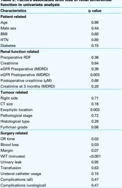
the early postoperative period when using renal scintigraphy Table 2. Factors associated with loss in renal differential
function in univariate analysis
Characteristics p value
Patient related Creatinine at 3 months (MDRD) 0.28
Tumour related
BMI: body mass index; HTN: hypertension; eGFR: estimated glomerular filtration rate; MDRD: modification of diet in renal disease; RDF: renal differential function; CT: computed tomography; WIT: warm ischemia time; OR: operating room.
0
(Appendix 1). Based on these series and the current study, the estimated loss of RDF on the operating kidney after a LPN with a WIT of 30 minutes for a tumour of about 3 cm should be expected to be between 25% and 30%.
The difference in the factors responsible for a loss of RDF before and after 30 minutes is interesting from a clinical perspective. We have shown in a multivariate regression model that the factors associated with loss of RDF before 30 minutes are patient- or tumour -related, while those associated loss of RDF after 30 minutes are tumour- and surgery-related. An important finding of this study is that the impact of WIT before 30 minutes is minimal, whereas after 30 minutes, it is the main factor associated with a loss of RDF. Since our series is not sufficiently powered to conclude that WIT has no impact on RDF before 30 minutes, it does show that its effect is minimal when loss of RDF is corrected for other patient- and tumour-related factors. Therefore, we believe that surgeons should focus on minimizing the removal of normal parenchyma,
ensur-ing negative margins and careful hemostasis as opposed to an unnecessary premature unclampling of the renal artery before 30 minutes. By analyzing loss in RDF as a continu-ous variable, we also demonstrated that there is no specific time at which the renal artery clamp should be removed, but instead that the rate of loss of RDF changes around 30 minutes. We have estimated that the rate of loss in RDF due to WIT increases to 0.7% per minute after 30 minutes. For a kidney with a 50% RDF, this translates into a loss of 1.4 % per minute of functional parenchyma. This estimated rate may help surgeons plan their surgeries based on the pre-operative RF of the patient, the characteristics of the tumour and the estimated clamping time. Many techniques have been described to limit ischemic damages during LPN. such as renal cooling, early unclamping, on-demand clamping and parenchymal compression with a Kaufman clamp, all of which should be considered if the WIT is anticipated to be
longer than 30 minutes.18 Open PN should also be strongly
considered with or without ice cooling in cases where LPN is anticipated to be difficult.
Our study has the limitation of using an early postopera-tive renal scintigraphy and therefore, the ischemic damage observed may be more reflective of acute renal ischemic
0
Fig. 2. Effect of WIT on the loss of RDF after adjusting for confounding factors using multivariate analysis regression models.
Table 3. Loss in renal differential function according to warm ischemia time intervals
WIT (min) n RDF loss*± WIT: warm ischemia time; RDF: renal differential function; SD: standard deviation.
Table 4. Comparative analysis of characteristics of the patients with warm ischemia times < or ≥ than 30 minutes that were used for the multivariate analysis
Variable
Descriptive statistics
p value
WIT <30 minutes (n = 23) WIT ≥30 minutes (n = 29)
Mean SD Mean SD
Change in RDF 10.6 9.4 17.6 8.9 0.01
Patient related
HTN (%) 46 - 17 - 0.04
Diabetes (%) 13 - 21 - 0.49
Preoperative RDF (%) 49 - 50.5 9.0 0.42
Tumour related
Endophytic (%) 64 - 80 - 0.23
Solid texture (%) 80 - 73 - 0.75
Pathological stage T1 (%) 80 - 73 - 0.75
Surgery related
WIT (min) 22.5 4.5 35.8 6.6 <0.001
Blood loss (g/L) 20.6 11.0 24.6 15.8 0.30
injuries than the chronic damage responsible for late renal insufficiency. However, since loss of RDF is greater imme-diately after surgery, this period probably allows for a more sensitive assessment of factors involved. Hence, we have shown that WIT has little impact on renal function before 30 minutes in the early postoperative period (the period of maximal acute injury), which might reinforce our conlusion that surgery should focus on surgical precision rather than WIT before 30 minutes.
Another limitation of our study is the use of RDF as the measure of renal function. Because it is relative to the func-tion of the other kidney, this measure cannot be extended to every patient, especially those with an asymmetric renal function, solitary kidney or comorbidities which affect renal function. Finally, the small number of patients included in the series might have limited our ability to detect significant factors involved in loss of RDF despite our efforts to perform a multivariate statistical analysis. Further studies with more patients will be needed to confirm our results.
Conclusion
Our multivariate analysis suggests that the effect of WIT on RDF of the operated kidney is not the same before and after 30 minutes. Since WIT has less impact on loss of RDF before 30 minutes, this suggests that LPN surgery should focus on limiting the resection margins, careful closure of the collecting system and hemostasis. After 30 minutes of WIT, the rate of loss of RDF increases and minimizing the loss of RDF should be the priority.
*Centre Hospitalier Universitaire de Québec, Université Laval, Québec, QC; †Institute of Urologic
Oncology, David Geffen School of Medicine, University of California at Los Angeles, Los Angeles, CA
Dr. Pouliot was supported in part by a scholarship from Les Bourses McLaughlin du Doyen, a scholar-ship from the Association des Urologues du Québec and a scholarscholar-ship from the CMDP (Committee of physicians, dentists and pharmacists) of the Laval University Hospital Centre.
Competing interests: None declared.
This paper has been peer-reviewed.
References
1. Pantuck AJ, Zisman A, Belldegrun AS. The changing natural history of renal cell carcinoma. J Urol 2001;166:1611-23.
2. Chow WH, Devesa SS, Warren JL, et al. Rising incidence of renal cell cancer in the United States. JAMA 1999;281:1628-31.
3. Hock LM, Lynch J, Balaji KC. Increasing incidence of all stages of kidney cancer in the last 2 decades in the United States: an analysis of surveillance, epidemiology and end results program data. J Urol 2002;167:57-60.
4. Ljungberg B, Hanbury DC, Kuczyk MA, et al. Renal cell carcinoma guideline. Eur Urol 2007;51:1502-10. 5. Thompson RH, Frank I, Lohse CM, et al. The impact of ischemia time during open nephron sparing surgery
on solitary kidneys: a multi-institutional study. J Urol 2007;177:471-6.
6. Abouassaly R, Lane BR, Novick AC. Active surveillance of renal masses in elderly patients. J Urol 2008;180:505-8; discussion 508-9.
7. Colombo JR Jr, Haber GP, Jelovsek JE, et al. Seven years after laparoscopic radical nephrectomy: oncologic and renal functional outcomes. Urology 2008;71:1149-54.
8. Foyil KV, Ames CD, Ferguson GG, et al. Longterm changes in creatinine clearance after laparoscopic renal surgery. J Am Coll Surg 2008;206:511-5.
9. Godoy G, Ramanathan V, Kanofsky JA, et al. Effect of warm ischemia time during laparoscopic partial nephrectomy on early postoperative glomerular filtration rate. J Urol 2009;181:2438-43; discussion 2443-5.
10. Becker F, Van Poppel H, Hakenberg OW, et al. Assessing the Impact of Ischaemia Time During Partial Nephrectomy. Eur Urol 2009;56:625-35.
11. Desai MM, Gill IS, Ramani AP, et al. The impact of warm ischaemia on renal function after laparoscopic partial nephrectomy. BJU Int 2005;95:377-83.
12. Johnston WK 3rd, Montgomery JS, Seifman BD, et al. Fibrin glue v sutured bolster: lessons learned during 100 laparoscopic partial nephrectomies. J Urol 2005;174:47-52.
Table 5. Factors associated with loss in renal differential function in multivariate analysis
Variable
Multivariate analysis
WIT <30 minutes (n = 23) WIT ≥30 minutes (n = 29)
Estimate SE p value Estimate SE p value
Patient related
HTN (%) -6.6 3.6 0.09 2.9 3.3 0.4
Diabetes (%) 4.6 6.1 0.46 -3.6 2.9 0.22
Preoperative RDF (%) 0.1 0.3 0.68 -0.2 0.2 0.28
Tumour related
Endophytic (%) 6.8 3.5 0.08 8.9 3.6 0.02
Solid texture (%) 10.1 4.9 0.06 -5.1 3.2 0.12
Pathological stage T1 (%)
6.3 5.3 0.25 -1.9 2.8 0.5
Surgery related
WIT (min) 0.2 0.4 0.64 0.7 0.2 0.002
Blood loss (g/L) 0.2 0.2 0.21 0.1 0.1 0.46
13. Go AS, Chertow GM, Fan D, et al. Chronic kidney disease and the risks of death, cardiovascular events, and hospitalization. N Engl J Med 2004;351:1296-305.
14. Huang WC, Levey AS, Serio AM, et al. Chronic kidney disease after nephrectomy in patients with renal cortical tumours: a retrospective cohort study. Lancet Oncol 2006;7:735-40.
15. Porpiglia F, Renard J, Billia M, et al. Is renal warm ischemia over 30 minutes during laparoscopic partial nephrectomy possible? One-year results of a prospective study. Eur Urol 2007;52:1170-8. 16. Song C, Bang JK, Park HK, et al. Factors influencing renal function reduction after partial nephrectomy.
J Urol 2009;181:48-53; discussion 54.
17. Funahashi Y, Hattori R, Yamamoto T, et al. Ischemic renal damage after nephron-sparing surgery in patients with normal contralateral kidney. Eur Urol 2009;55:209-15. Epub 2008 Aug 5.
18. Berger A, Crouzet S, Canes D, et al. Minimally invasive nephron-sparing surgery. Curr Opin Urol 2008;18:462-6.
Correspondence: Dr. Thierry Dujardin, Assistant Professor, Laval University, Centre Hospitalier Universitaire de Québec, Pavillon Hôtel-Dieu de Québec, 11 Côte du Palais, Québec, QC G1R 2J6; fax: 418-691-5020; [email protected]
Appendix 1. Published studies that have used renal scintigraphy to evaluate renal function on the operated kidney after laparoscopic nephrectomy
Author n Tumour size (cm) WIT Loss in RF (%)
Average (%) Associated factors
Porpiglia et al12 18 3.4 39 24 WIT, size of normal
parenchyma removed
Song et al13 117 2.9 28 29 Age, kidney volume loss,
upper pole location
Funahashi et al14 30 3.2 24 29 WIT
Present study 56 3.2 29 28a WIT, endophytic location of
the tumour