Revisiting old vaginal topics: conversion of the
Müllerian vagina and origin of the "sinus" vagina
YI CAI*
Department of Physiology, The University of Tennessee, Memphis, TN, USA
ABSTRACT Vaginal development has been a longstanding controversy, which hampers studies on vaginal diseases as well as cervical and uterine diseases. Most concerns center on: why is the vaginal epithelium different from the uterine epithelium; and where does the vagina originate from? It is commonly held that the rodent vagina has a dual origin: the cranial part is derived from the Müllerian duct (Müllerian vagina) and the caudal part derived from the urogenital sinus (“sinus” vagina). This concept was deduced from morphological observations. However, it cannot explain the difference between the Müllerian vagina and the uterus. Moreover, accumulating new data from genetic and molecular studies contradicts the urogenital sinus origin of the “sinus” vagina. The present review summarizes previous morphological observations and new findings from genetic and molecular studies, and addresses molecular mechanisms underlying the origin and organogenesis of the vagina in rodents. It provides evidence to show that the whole vagina is derived the Müllerian duct. BMP4 reshapes the intermediate mesoderm-derived Müllerian duct into the vaginal primordium. The latter thus exhibits different features from the uterus, including the stratified squamous epithelium and insensitivity to anti-Müllerian hormone. The “sinus” vagina is formed by extrinsic BMP4-mediated caudal extension of the Müllerian duct. The present review thus shows how a century of controversy over the origin and organogenesis of the vagina has been resolved. This new understanding will provide additional insight into genetic diseases and tumors of the female reproductive tract.
KEY WORDS:
vagina, Mullerian duct, androgen, estrogen, prostate
Introduction
In 1830, Johannes Müller first traced and recognized develop-ment of the Müllerian duct. Since then, an intense controversy on the vaginal development was raised and has continued to the present. Most concerns center on: why is the vaginal epithelium different from the uterine epithelium? and what does the vagina originate from? Rodents play an important role in biology and medicine. Today it is commonly held that the rodent vagina has a dual origin: the cranial part is derived from the Müllerian duct (Müllerian vagina) and the caudal part derived from the urogenital sinus (“sinus” vagina). This concept was deduced from previously morphological observations that the Müllerian vagina is initially lined with pseudostratified columnar epithelium, while the epithe-lium in both the Müllerian and “sinus” vagina converts to the stratified squamous epithelium later (Forsberg, 1965, Forsberg, 1973). However, it is yet unable to explain how the columnar epithelium in the Müllerian vagina converts to the stratified
squa-BIOLOGY
www.intjdevbiol.com*Address correspondence to: Dr. Yi Cai. Department of Physiology, University of Tennessee, 894 Union Avenue, Memphis, TN 38103, USA. e-mail: caiyi219@hotmail.com
Accepted: 7 April 2009. Published online: 10 June 2009.
ISSN: Online 1696-3547, Print 0214-6282 © 2009 UBC Press
Printed in Spain
Abbreviations used in this paper: AMH, anti-Müllerian hormone; BMP, bone morphogenetic protein; tfm, testicular feminization.
mous epithelium. Particularly, with advances in molecular biology and genetics, this conventional concept clearly contradicts the new findings on the Müllerian duct. Accumulating data from genetic and molecular studies show that the Müllerian duct determines the vaginal development. The present review summarizes previous morphological observations and new findings from genetic and molecular studies, and will aim to address molecular mechanisms on the origin and organogenesis of the vagina in rodents.
The vaginal development requires the Müllerian duct
genital organ is thus formed in the female mutant mice (Vainio et al., 1999). 2) Wnt5a, which is expressed in the mesenchyma of the
uterus, cervix and vagina, is required for posterior growth of the female reproductive tract (Mericskay et al., 2004). In Wnt5a -/-mutant mice, the cervix and whole vagina are not formed (Mericskay
et al., 2004). 3) Wnt9b, acting upstream of Wnt4 in Müllerian duct
formation, is essential for the caudal extension of the Müllerian duct and consequently Wnt9b-/- mutant female mice lack a uterus and vagina (Carroll et al., 2005). 4) Pax-2 is a dorsal mesoderm factor
(Patel and Dressler, 2004), and a urogenital developmental tran-scription factor expressed in the Wolffian and Müllerian ducts during embryonic stage. Pax-2-/- mutants lack the Wolffian and Müllerian duct derivatives, including the vagina, whereas the urogenital sinus still develops into the bladder and urethra (Torres
et al., 1995). 5) Lim1, another dorsal mesoderm signal (Capel,
1998), is also required for female reproductive tract development. While female Lim1-/- neonates have ovaries, they completely lack oviducts, a uterus, cervix and vagina (Kobayashi et al., 2004).
Therefore, the Müllerian duct is required for vaginal development.
BMP4-induced conversion of the Müllerian vagina
BMP4 expression in the vaginal primordium
Hoxa genes, including Hoxa9, Hoxa10, Hoxa11, and Hoxa13, specify the anterior-posterior patterning of the Müllerian duct system (Kobayashi and Behringer, 2003). Among them, Hoxa13 determines development of the caudal Müllerian duct. Hoxa13 -/-mice lack the caudal Müllerian duct (Warot et al., 1997). In newborn
and mature rodents, Hoxa13 expression is restricted to the cervical and vaginal tissues, but not in the uterus (Taylor et al., 1997, Post
and Innis, 1999).
Sato and Iguchi (Sato and Iguchi, 2004) stated that in 2- or 15-day-old female mice, mRNA expressions of BMP4 and Hoxa13 were abundant in the vagina in comparison with the uterus. Their observations in 2-day-old mice are in accordance with previous observations by other groups. 1) The Hoxa13 expression in the female reproductive tract of newborn mice is in accordance with Post and Innis’ study (Post and Innis, 1999). Their in situ
hybridiza-tion results showed that Hoxa13 was expressed in vagina and cervix but not in uterus in newborn mice. 2) Lamm et al., (Lamm et al., 2001) observed in neonate mice that BMP4 expression
re-mained diffusely distributed in the urogenital sinus region caudal to the uterus. In neonate mice, the vaginal morphology clearly devel-ops, and is distinct from the uterus. Moreover, its inside is differen-tiated into the cranial part (Müllerian vagina) and the caudal part. Since the vagina was not their target, Lamm et al., didn’t mention
the vagina in the article. Nevertheless, their data clearly showed that at postnatal one day, BMP4 expression was highest in the female urogenital sinus. There was a decreasing gradient in BMP4 expression in the Müllerian duct from the urogenital sinus to the uterus, in which the vagina was located. BMP4 was absent in the uterine horns.
Although there is no either further confirmation or contradiction for BMP4 expression in the vagina of 15-day old mice, it is indeed supported by indirect evidence. 1) At this stage, the uterus and vagina undergo organ differentiation under the induction of differ-ent Hoxa genes. Hoxa13 is strongly expressed in the vagina and cervix, but not in the uterus at two weeks of age (Taylor et al., 1997).
It is consistent with Sato and Iguchi’s observation. 2) Hoxa13 can
activate BMP4 promotor activity (Suzuki et al., 2003). It is thus
reasonable that BMP4 remains higher in the vagina in comparison with the uterus. In addition, a series of phenomena take place in the Müllerian vagina but not in the uterus, for instances, expression of p63 and stratified squamous differentiation in the vaginal epithe-lium. They are associated with BMP4 as discussed, indirectly supporting BMP4 effects on the vaginal differentiation. Therefore, it is confirmable that BMP4 is expressed in the vaginal primordium.
BMP4 converts the intermediate mesodermal nature of the vaginal primordium
BMP4 plays an important role in specifying ventral mesodermal fate and dorsoventral patterning of mesoderm in mice (Jones et al.,
1991). It induces ventral mesoderm but represses dorsal meso-derm signals. Wnt7a is an important dorsal signal (Parr and McMahon, 1995, Kengaku et al., 1998). BMP4 disrupts
Wnt7a-induced signaling in chicks (Hirsinger et al., 1997, Marcelle et al.,
1997) and mammals (Cossu and Borello, 1999, Viti et al., 2003). In
addition, BMP4 also suppresses expression of other early interme-diate mesoderm-specific markers, including expression of Lim1 and Pax2 in the mesenchyma (Kim and Dressler, 2005).
The Müllerian duct originates from the intermediate mesoderm, which forms in the back of the embryo along the spine and belongs to the dorsal structures. Thus, BMP4, whose expression under-goes an ascending increase in the vaginal primordium, plays roles in conversion of the intermediate mesoderm nature of the Müllerian duct.
Loss of Wnt7a and appearance of BMP4 and p63 in the vaginal primordium
Wnt4 initiates invagination of coelomic epithelium to form the Müllerian duct, as well as induces expression of Wnt7a (Vainio et al., 1999). Therefore, Wnt7a is initially expressed throughout the
Müllerian tract (Miller and Sassoon, 1998). However, Wnt7a ex-pression gradually declines in the Müllerian vaginal epithelium after birth, and is undetectable in the vaginal epithelium by 10 days after birth (Miller et al., 1998).
As discussed, BMP4 at birth shows an opposite expression pattern, which displays the highest in the female urogenital sinus, then a descending gradient in the Müllerian duct from the urogeni-tal sinus to the uterus, and absent in the uterine horns.
p63 is a downstream of BMP4 as discussed. Its expression has the same caudal-to-cranial descending pattern in the vaginal primordium around birth. At embryonic day 18.5 (E18.5), p63 is highly expressed in the caudal or “sinus” vagina, while is detected in small numbers of epithelial cells of the Müllerian vagina (Kurita
et al., 2005).
The timing of the loss of Wnt7a from the vagina corresponds to the timing of vaginal cytodifferentiation, which coincides with establishment of a spatial Hoxa axis in the Müllerian duct (Miller et al., 1998). Initially, Hoxa9, Hoxa10, Hoxa11, and Hoxa13 are
expressed at uniform levels throughout the length of the Müllerian duct. At around birth, their expressions become spatially restricted in the differentiating female reproductive tract (Taylor et al., 1997).
Hoxa13 is restricted to and strongly expressed in the vagina and cervix at birth (Taylor et al., 1997, Post and Innis, 1999). Hoxa13
can activate BMP4 promotor activity (Suzuki et al., 2003).
expression in the vaginal primordium gradually disappears in the same caudal-to-cranial pattern. Accordingly, Wnt7a determines the proper differentiation of the uterus (Miller and Sassoon, 1998, Parr and McMahon, 1998). Wnt7a-/- mutation affects the uterus, but not the vagina (Miller and Sassoon, 1998). Therefore, BMP4 expression causes loss of Wnt7a in the vagina.
Insensitivity to anti-Müllerian Hormone
Anti-Müllerian hormone (AMH) is involved in male sex differen-tiation. It induces regression of the Müllerian duct structures in the male. However, the caudal Müllerian duct is insensitive to AMH. 1) In in vitro organ culture of the Müllerian duct from 14.5- and
15.5-day-old rodent fetuses, the cranial portion regresses almost com-pletely during the 3-day culture period in the presence of AMH, whereas the caudal half to third remains intact but tapers to a fine point cranially (Tsuji et al., 1992). 2) In AMH transgenic female
mice, the oviduct and uterus fail to form due to the inhibition by AMH, whereas the vagina develops (Behringer et al., 1990). 3) The
vagina also forms irrespective of AMH in rodent males with testicular feminization (Tfm) and human males with complete androgen insensitivity syndrome (CAIS).
AMH induces regression of the Müllerian duct by binding to a specific AMH type II receptor (AMHRII). Mutation in AMHRII in male mice disrupts signaling, producing male pseudohermaphrodites that possess oviducts and uteri (Mishina et al., 1996). Wnt7a drives the expression of AMHRII; the Müllerian
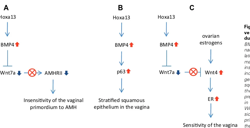
duct in Wnt7a mutant mice thus fails to regress, but rather persists (Parr and McMahon, 1998). As discussed, BMP4 inhibits Wnt7a signaling in the vaginal primordium. It is able to explain the fact that AMHRII expression in the caudal part of the Müllerian duct is less than the intermediate part (Xavier and Allard, 2003). Therefore, BMP4-induced conversion makes the vaginal primordium insensi-tive to AMH (Fig. 1A). Taken together, BMP4 convert the interme-diate mesoderm nature of the vaginal primordium, though it is derived from the Müllerian duct.
BMP4 reshapes the Müllerian vagina
BMP4 in the vaginal primordium not only converts the
interme-diate mesoderm nature, but also exerts effects on the vaginal differentiation. For instance, the Müllerian vagina becomes distinct from the uterus during organ differentiation, though both are derived from the Müllerian duct exhibit a similar epithelial pheno-type at birth. Both the cranial part and caudal part of the vagina eventually possess the same histologic features.
Generation of stratified squamous epithelium
It is first found in Xenopus that BMP4 induces differentiation of
epidermis or stratified squamous epithelium (Wilson and Hemmati-Brivanlou, 1995). BMP4 induces p63 expression via Smad4 and Smad5 signaling pathway in zebrafish and Xenopus (Bakkers et al., 2002, Tribulo et al., 2007). In rodents, BMP4 is also able to
induce stratified squamous epithelial differentiation of embryonic stem cells; p63 is activated and plays a key role in this process (Aberdam et al., 2007). p63 is a basal cell marker, and plays a
crucial role in regulation of stem cell commitment in squamous epithelium (Yang and McKeon, 2000, Pellegrini et al., 2001). It is
also essential for squamous epithelial differentiation in the vagina. p63-null vaginal epithelium reverts to the epithelial phenotype similar to the uterus (Kurita and Cunha, 2001, Kurita et al., 2004,
Kurita et al., 2005). Moreover, in the vagina, both BMP4 and p63
expression patterns have remarkably similar temporospatial char-acteristics. Therefore, BMP4-induced conversion generates the stratified squamous epithelial cells, which replace the columnar epithelium initially exhibited in the Müllerian vagina (Fig. 1B).
Sensitivity to estrogen
Wnt4 controls the expression of the genes encoding estrogen receptors ((ERs); the estrogen receptor α (ERα) is downregulated in Wnt4-null mice (Heikkila et al., 2005). Because Wnt7a inhibits
Wnt4 signaling (Miller and Sassoon, 1998), an inverse association is found between Wnt7a and ERα (Li et al., 2001). Thus, Wnt4
signaling is enhanced in the caudal Müllerian tissue because of suppression of Wnt7a signaling by BMP4. 1) The ER signaling is consequently enhanced in vaginal primordium. Nuclei of the epi-thelial and stromal cells in the vagina and of the stromal cells in the uterus show strong ER expression on the day of birth (Sato et al.,
Fig. 1. BMP4 induces the con-version of the Müllerian duct-derived vagina. (A) BMP4 represses Wnt7a sig-naling, and in turn down-regu-lates AMHRII expression. It makes the vaginal primordium insensitive to AMH. (B) BMP4 induces p63 expression, and generation of the stratified squamous epithelial cells in the vagina. (C) BMP4 re-presses Wnt7a signaling, and in turn up-regulates Wnt4. Wnt4 enhances ER expres-sion, which makes the vaginal primordium more sensitive to the estrogen than the uterine primordium.
Hoxa13
BMP4
Strafied squamous epithelium in the vagina
p63
ER
Sensivity of the vagina to the estrogen Hoxa13
BMP4
Wnt7a Wnt4
ovarian estrogens Hoxa13
BMP4
Wnt7a AMHRII
Insensivity of the vaginal primordium to AMH
B
C
1996), whereas the uterine epithelium of the neonatal animal is devoid of estrogen-binding activity (Bigsby and Cunha, 1986, Bigsby et al., 1990, Sato et al., 1996). The ER exhibition pattern is
consistent with the expression pattern of Wnt4, which is expressed in the stroma subjacent to the luminal epithelium of the uterus and the vagina (Miller et al., 1998). But it is opposed to the expression
pattern of Wnt7a, which is expressed only in the uterine epithelium but not uterine stroma and vagina (Miller and Sassoon, 1998). 2) It is also supported by a phenomenon that the vagina is more sensitive to estrogenic stimulation than the uterus. Under estro-genic stimulation, almost all ovariectomized mice exhibit stratifica-tion of vaginal epithelium, while few take place in the uterus (Mori
et al., 1992). ERα is required for E2-induced cornification and normal epithelial stratification (Buchanan et al., 1998).
On the other hand, under BMP4 stimulation, Smad4 and Smad1 physically interact with the ER. BMP4 and estrogens act through overlapping intracellular signaling mechanisms. They have addi-tive effects at low concentrations, mediating proliferation (Paez-Pereda et al., 2003). Therefore, BMP4-induced conversion makes
the vaginal primordium more sensitive to the estrogen than the uterine primordium (Fig. 1C).
Müllerian origin of the “sinus” vagina
In the mouse, the Müllerian duct begins to form as an invagina-tion of the surface epithelium of the mesonephros at E11.5 and continues to elongate until it reaches the urogenital sinus at E13.5 in both male and female embryos (Kobayashi and Behringer, 2003). During the rest of gestation, the vaginal primordium contin-ues its down-growth to the vestibule.
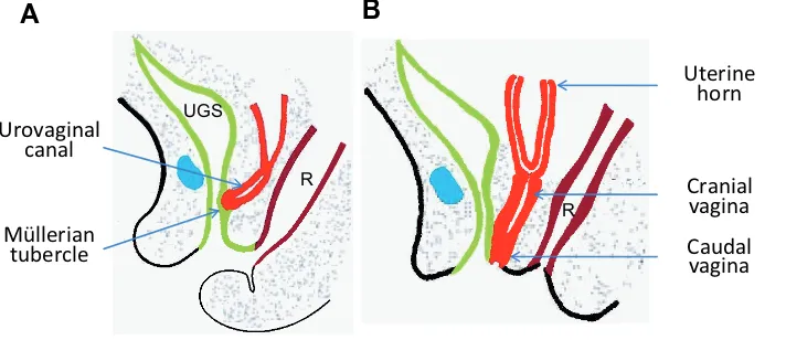
At birth the cranial portion of the vagina has a lumen lined with pseudostratified columnar epithelium indicating the Müllerian ori-gin, while the caudal portion of the vagina has a solid epithelial cord. With time the caudal portion of the vagina canalizes and the cranial portion stratifies, so that eventually the entire vagina is lined by a stratified epithelium. According to the morphological observa-tions, Forsberg (Forsberg, 1965, Forsberg, 1973) believed that the rodent vagina consisted of a cranial Müllerian part and a caudal part derived from the urogenital sinus: the “sinus” vagina.
The “sinus” vagina originates from the Müllerian duct
Do the different epithelial phenotypes between the vagina and uterus mean different origins other than the Müllerian duct origin?
In contrast to Forsberg’s deduction, the following provide evi-dence that the urogenital sinus epithelium is unable to replace the vaginal epithelium. Actually, Forsberg (Forsberg and Olivecrona, 1965) himself found no signs of invasion of sinus epithelium into the Müllerian vagina. Additionally, later studies confirmed that the growth rate of urogenital sinus epithelial cells was lower than both caudal and cranial epithelial cells in the vagina (Boutin and Cunha, 1996). Later, Forsberg also did a transplantation experiment to prove that the epithelium in the Müllerian vagina performed the phenotypic transformation by itself. At E14.5, the Müllerian vagina analogue was removed and transplanted into the thigh muscle of newborn mice. After 12-day growing, stratification transformation of the Müllerian epithelium (including a superficial zone and a basal zone) occurs (Forsberg and Norell, 1966).
In addition, the stratified squamous phenotype does not support roles of the Wolffian duct in the vaginal development. For instance,
the normal vagina is lined by stratified squamous epithelium, while persistent Gartner’s duct, a vestigial Wolffian duct, is lined by columnar epithelium.
Finally, the stratified squamous epithelial phenotype is unique to the Müllerian duct-derived vagina. 1) The stratified squamous epithelial phenotype is determined by the vaginal stroma (Cooke et al., 1987, Boutin and Cunha, 1997). The urogenital sinus
mesen-chyma fails to induce it on either the urogenital sinus epithelium or the vaginal epithelium under estrogenic conditions (Cunha and Young, 1992). 2) As discussed, under the induction of BMP4, the Müllerian duct-derived vaginal primordium itself generates the squamous epithelial phenotype. The epithelium in the vaginal primordium did not need other sources, but spontaneously achieved the conversion from uterus-like columnar to stratified squamous epithelium. Thus the stratified squamous phenotype is unable to support the possibility of other origins than the Müllerian duct.
Does the vagina in Tfm mice originate from different origins other than the Müllerian duct?
The conventional concept on the “sinus” vagina was previously supported by the presence of a shortened ‘‘sinus” vagina in androgen-insensitive Tfm mice. The vagina in Tfm mice was believed to be derived from the other tissues than the Müllerian ducts because it is resistant to the anti-Müllerian effect of AMH. It was previously plausible because at that time the Müllerian duct was commonly believed to regress in the male due to the inhibition by AMH. However, this view contradicts the following fact. Normal vaginal epithelium exhibits cyclical changes in both the morphol-ogy and the histolmorphol-ogy during the estrus cycle. These changes are variously caused by several steroidal hormones (Jones and Edgren, 1973). If the “urogenital sinus-derived” vagina (or “sinus” vagina) could display hormonal responses, other urogenital sinus deriva-tives should also have this feature. The answer is negative as the bladder and the urethra (the urogenital sinus derivatives) do not have a menstrual cycle like the uterus and the vagina.
Then what about the vagina in Tfm mice? The adult Tfm vaginal epithelium responds steroid stimulations like Müllerian-derived and not urogenital sinus-derived epithelium, indicating its nature of the Müllerian duct (Boutin and Cunha, 1996). Moreover, recon-struction analysis of the vagina in Tfm mice further confirms that the caudal Müllerian duct participates in vaginal formation (Drews et al., 2002).
In fact, under BMP4 induction, the vaginal primordium is insen-sitive to AMH induction as discussed. The caudal Müllerian tissue consequently persists in the male and retains the ability to form the vaginal structure. A question thus arises why no vagina forms in the normal male. 1) The caudal Müllerian derivatives including the uterus and vagina are androgen target organs. The androgen receptor (AR) is constitutively expressed in both the mesenchyma surrounding the caudal Müllerian duct (Drews et al., 2001) and its
mature derivatives (Pelletier et al., 2004). Strikingly, animal studies
prostate development (Cai, 2008). 2) In rats, the window of sensitivity to AMH is E14-15 days. After that, exposure to AMH does not cause Müllerian duct regression (Tsuji et al., 1992).
However, the fetal testes produce the androgen after that time. As discussed, the androgen represses BMP4 effects and thus sup-presses vaginal organogenesis in the male. Therefore, under androgenic induction, the caudal Müllerian tissue fails to form the vagina, while it in turn participates in prostate development in the normal male. If the androgen signaling is disrupted, such as in Tfm mice, it forms the vagina rather than the prostate. Taken together, the “sinus” vagina is not derived from the urogenital sinus. On the contrary, the whole vagina is derived from the Müllerian duct.
The “sinus” vagina forms due to BMP4-mediated caudal extension of the Müllerian duct
The Müllerian duct extends downwards to the vestibule
Early view held that the vagina developed in a caudal-cranial direction, deduced from morphological observation of the sinovaginal bulbs (Koff, 1933). It seemed plausible because it seemed to be in agreement with Grunwald’s observations on early development of the Müllerian duct. In early embryonic stage, the Wolffian duct first reaches the urogenital sinus. Under its guidance, the Müllerian duct reaches the urogenital sinus too (Grunwald, 1941). However, this is the case for early development of the Müllerian duct, but not for the formation of the caudal vagina. According to the position relationship between vaginal bud and orifice of major vestibular gland, Witschi (Witschi, 1970) observed that the vagina progresses down the dorsal wall of the urogenital sinus on its way to a separate opening in the vestibule. It is confirmed by Drews (Drews, 2007), who generated intersex mice composed of androgen insensitive XTfm and normal X+ cells. In
nial part of the vagina is still lined by the columnar epithelium at birth. The Müllerian duct differentiates cranial-caudally, forming the oviduct, uterus, cervix and cranial part of vagina, which is completed by two weeks after birth (Yin and Ma, 2005). Thus, the cranial part of the vagina is not yet converted by BMP4 at birth, indicating that Hoxa13-induced BMP4 functions after birth and is not responsible for solidification of the caudal vagina before birth. Then, what causes the solidification in the caudal vaginal primor-dium?
The Müllerian duct reaches the urogenital sinus at E13.5. It is observed in normal mice (Bok and Drews, 1983) and rats (Sanchez-Ferrer et al., 2006) that the caudal bifurcated Müllerian duct
reaches the urogenital sinus and forms the Müllerian tubercle, on which the “sinus” vagina is formed. Actually, in the developing mouse BMP4 is abundantly expressed from E11.5-E13.5 in mes-enchyma surrounding urogenital sinus (Brenner-Anantharam et al., 2007). At birth, BMP4 still remains high in the urogenital sinus
(Lamm et al., 2001). Therefore, in the embryonic stage, extrinsic
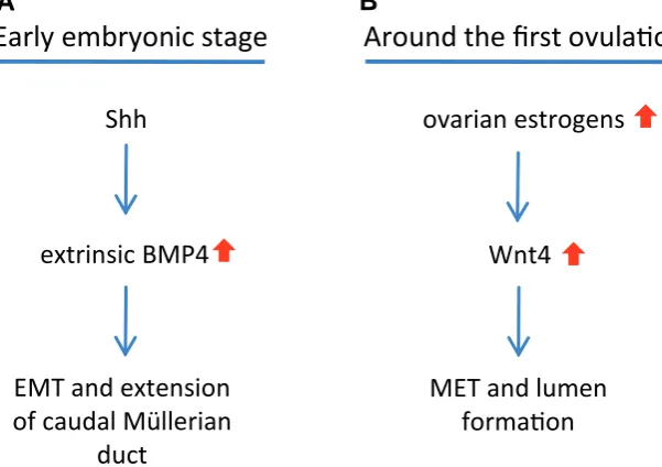
BMP4 produced from the urogenital sinus mesenchyma induces the EMT in the inserted caudal end of the Müllerian duct (Fig. 2A). Next step, BMP4 induces c-myc expression (Paez-Pereda et al., 2003), which is an important mediator for cell proliferation and
migration (Biro et al., 1993, Koster et al., 1996). It also induces Rho
GTPase activation (Theriault et al., 2007), which plays an
impor-tant role in cell migration (Raftopoulou and Hall, 2004). Therefore, BMP4 is involved in tissue migration. It is in agreement with the fact that the EMT plays a role in the disaggregation of epithelial cells and the reshaping of these cells for movement (Hay, 1995). For examples, 1) BMP4 is required for early mesoderm migration such as gastrulation (Winnier et al., 1995). Most mouse embryos
ho-mozygous for a null mutation in BMP4 die around gastrulation
Fig. 2. Development of the caudal part of the vagina.(A) The caudal end of the Müllerian duct inserts in the urogenital sinus wall and forms the Müllerian tubercle. During early embryonic stage, Shh induces BMP4 in the urogenita sinus mesenchyma. The extrinsic BMP4 induces the epithelial-mesenchymal transition (EMT) in the caudal end of the Müllerian duct to form the caudal vaginal primordium, and also guides it to extend caudally along the urogenital sinus wall. (B) Around the first ovulation, the ovarian estrogens enhance Wnt4 signaling, which induces the mesenchymal-epithelial transition (MET) and lumen formation in the caudal vagina.
feminised intersexes the AR defective cells formed a vagina and the androgen responsive cells male sex organs. Similar to the Tfm/Y mice, the AR-mutated vagina descends and enters the penile bulb (an analogue of female vestibule). Strikingly, the AR+/+ Wolffian ducts, which normally open on the urethra in wild type mice, also descend with the vagina and enters the penile bulb. Therefore, the Müllerian duct constitutively possesses the ability to extend down-wards and it can even drive the Wolffian duct to extend caudally together.
Extrinsic BMP4-induced caudal extension of the Müllerian duct
At birth, the caudal part of the mouse vagina is solid. Thus, a question arises why the caudal part is solid, but cranial part remains patent at birth. BMP4 can induce the epithelial-mesenchymal transition (EMT) (Endo et al., 2002, Theriault et al., 2007,
Molloy et al., 2008). It induces expression of Snail
(Chiba et al., 2005, Cazillis et al., 2006, Theriault et al., 2007), which represses E-cadherin expression
and triggers the EMT (Davidson and Sukumar, 2005). Therefore, solidification of the caudal part of the uterovaginal canal may be a consequence of BMP4-induced EMT.
Although Hoxa13-induced BMP4 reshapes the Müllerian duct into the vaginal primordium, the
cra-Wnt4
MET and lumen
formaon
ovarian estrogens
Shh
extrinsic BMP4
EMT and extension
of caudal Müllerian
duct
Early embryonic stage
Around the first ovulaon
(Winnier et al., 1995). 2) BMP4 also plays an essential role in
elongation of the ureter bud (Miyazaki et al., 2000, Cain and
Bertram, 2006). Thus, BMP4 not only induces the EMT, but also leads to the caudal extension of the caudal Müllerian duct.
Then what direction does the caudal Müllerian duct follow to migrate along the urogenital sinus wall? In fact, BMP4 can serve as a chemoattractant for periureteral mesenchymal cells and induce locally the smooth muscle layer of the ureter at BMP4-expressing sites (Miyazaki et al., 2003). Data from human samples show that
BMP4 expression in the urogenital sinus mesenchyma proceeds in a cranial-to-caudal sequence (Jenkins et al., 2007). In mice, diffuse
BMP4 expression persists in the female urogenital sinus on embry-onic day 18 and at birth (Lamm et al., 2001). Thus, extrinsic BMP4
expressed in wall of the urogenital sinus guides the caudal end of the vaginal primordium to extend caudally (Fig. 3A and 3B).
The concept that the urogenital sinus guides caudal extension of the vaginal primordium via BMP4 signaling is supported by Sonic hedgehog (Shh) genetic mutation studies. The onset of the parti-tion of the cloaca into the ventral urogenital sinus and dorsal anorectum is observed clearly by E10.5 (Mo et al., 2001). Shh is
essential for development of the urogenital organs (urethra and bladder) (Mo et al., 2001, Haraguchi et al., 2007). Shh induces
BMP4 expression in the surrounding mesenchyma (Bitgood and McMahon, 1995). Under Shh induction, an urorectal septum forms in a cranial-to-caudal pattern. In the normal female, the vagina runs in the urorectal septum down to the perineum. In Shh-null animals, which exhibit improper urorectal separation (Mo et al., 2001,
Perriton et al., 2002), the vagina never passes over the septum,
although Hoxa13 can induce BMP4 in the caudal vaginal primor-dium. Therefore, Shh induces BMP4 in the urogenital sinus mes-enchyma and BMP4 guides mesmes-enchymal vaginal primordium to extend caudally.
The androgen antagonizes BMP4-mediated caudal extension of the Müllerian duct
The androgen down-regulates BMP4 signaling (Kuslak and Marker, 2007, Pu et al., 2007). Extension of the caudal Müllerian
mesenchyma is severely inhibited if the embryos are exposed to the androgen in early stage. The vagina thus fails to reach the perineum and can only open into the urethra. It is supported by animal studies, in which prenatal introduction of androgens leads to varied vaginal opening from the urethra at the level of the Wolffian duct opening to separate opening at the perineum. The inhibition effects of the androgen are dependent on timing and
dosage of the androgen exposure (Lillie, 1917, Greene and Ivy, 1937, Yucel et al., 2003). Therefore, BMP4 mediates caudal
extension of the Müllerian duct to form the vagina.
Lumen formation in the caudal vagina
Generation of vaginal epithelium
The caudal vaginal primordium remains solid until the onset of sexual maturity. The origin of vaginal epithelium thus becomes a major concern. As discussed, solid evidence refutes the conven-tional concept that the urogenital sinus epithelium ascends to participate in vaginal organogenesis. Thus, a question arises if the solid vaginal primordium is able to generate the epithelium by itself. The EMT can revert to generate the epithelium through the mesenchymal-epithelial transition (MET), which is crucial for ver-tebrate organogenesis. BMP4 expression is reduced in the uro-genital sinus with gestation (Kuslak and Marker, 2007), indicating that its ability of EMT induction declines. On the other hand, Wnt4 is expressed in the vaginal primordium, and can be up-regulated by the estrogen (Hou et al., 2004). One of key roles of Wnt4 in
organogenesis is induction of the MET. Wnt4 positively regulates Rac1 and JNK pathways, which are involved in the MET (Osafune
et al., 2006). It is shown that Wnt4 is involved in epithelial
genera-tion in some mesoderm-derived organs, such as the kidney and the Müllerian duct derivatives. 1) Wnt4 is absolutely required for epithelialization in kidney development (Stark et al., 1994, Kispert et al., 1998). In Wnt4-deficient mice, the metanephric
mesen-chyma never enters the MET (Stark et al., 1994). 2) Wnt4 is also
essential for formation of the Müllerian duct. Wnt4-null animals lack the Müllerian duct (Vainio et al., 1999). Adult vagina expresses
Wnt4 in the epithelium only (Miller et al., 1998). Therefore, vaginal
epithelium can be generated through Wnt4-induced MET (Fig. 2B).
Lumen formation
The vagina of the mouse is closed at birth and does not open until the animal is 24 to 28 days of age, around the first ovulation representing the onset of puberty in female mice (Green, 1975). Vaginal opening is caused by the rise of estrogen levels, while the first ovulation caused by ovarian cyclic activity due to a gonadotro-pin surge. With estrogenic stimulation, the first vaginal cornification also occurs 24 to 120 hours after establishment of the vaginal opening (Green, 1966). Additionally, both prenantal exposure (Honma et al., 2002) and postnatal introduction (Takasugi and
Bern, 1962) of estrogens are able to induce precocious vaginal opening. Therefore, the ovarian estrogens around the first
ovula-Fig. 3. The caudal vagina forms due to BMP4-mediated caudal extension of the Müllerian duct.(A) The caudal end of the Müllerian duct inserts into the urogenital sinus (UGS) wall, in which BMP4 is strongly expressed. The caudal end of the Müllerian duct is solidified to form the Müllerian tubercle (the caudal vaginal pri-mordium) via the extrinsic BMP-induced EMT. Furthermore, BMP4 expressed in a cranial-to-caudal sequence in the UGS wall guides the solid caudal vaginal primordium to extend cau-dally. (B) The solid caudal vaginal primordium reaches the perineum. R, the rectum; UGS, the urogenital sinus.
Urovaginal
canal
Müllerian
tubercle
Uterine
horn
Cranial
vagina
Caudal
vagina
UGSR
R
tion induce vaginal canalization.
In cavitation, the lumen is generated by apoptosis of cells in the middle of the structure (Lubarsky and Krasnow, 2003). Animal studies show that the estrogen induces lumen-generated apopto-sis in vaginal epithelial cells (Rao et al., 1998). Two molecules,
Wnt4 and β-catenin, are involved in vaginal lumen formation. 1) Wnt4 binds both canonical and noncanonical Frizzled receptors (Lyons et al., 2004), which activate JNK-dependent pathway
(Lisovsky et al., 2002). Additionally, Wnt4 activates p38
mitogen-activated protein kinase (MAPK) in a novel noncanonical signaling pathway (Chang et al., 2007). p38 MAPK has also been shown to
play a role in tubulogenesis (Karihaloo et al., 2005, Montesano et al., 2007). Both JNK (Lei and Davis, 2003) and p38 (Choi et al.,
2005) are involved in induction of apoptosis. Consequently, Wnt4 is required for tubulogenesis in the kidney (Kispert et al., 1998,
Saulnier et al., 2002, Kobayashi et al., 2005, Itaranta et al., 2006)
and breast (Brisken et al., 2000). 2) Nuclear β-catenin accumula-tion can induce activaaccumula-tion of the p53-p21WAF1 pathway (Saegusa
et al., 2004, Chandar et al., 2005, Buslei et al., 2007) and
overex-pression of cyclin D1 (Shtutman et al., 1999, Saegusa et al., 2004),
leading to suppression of cell proliferation or induction of cell senescence. It is consistent with apoptosis induced by overexpres-sion of β-catenin (Kim et al., 2000, Olmeda et al., 2003). Both
estrogen (Cardona-Gomez et al., 2004, Chen et al., 2005) and
Wnt4 (Lin et al., 2006) inhibit glycogen synthase kinase 3, leading
to stabilization of β-catenin. It is shown that estrogen treatment causes an increase in cytosolic β-catenin followed by the accumu-lation of β-catenin in the nucleus (Chandar et al., 2005, Rider et al.,
2006). Accumulation of β-catenin in the cytoplasm is involved in the formation of adherens junctions of mammalian epithelia where it links α-catenin to E-cadherin, while translocation of β-catenin to the nucleus is implicated in lumen formation and squamous differen-tiation.
Conclusions
An intense controversy persists on the vaginal development since 1830 when Johannes Müller traced and recognized develop-ment of the Müllerian duct. Previous morphological studies cannot resolve the controversy. Advances in molecular biology and genet-ics provide insights into the old topgenet-ics on the vagina. The present review summarizes evidence from all morphological observations and genetic and molecular studies. Firstly, it shows that BMP4 reshapes the intermediate mesoderm-derived Müllerian duct into the vaginal primordium. This conversion makes the vagina acquire different features from the uterus, including stratified squamous epithelium and insensitivity to AMH. Secondly, the solid evidence shows that the “sinus” vagina is derived from the Müllerian duct rather than other tissues. The “sinus” vagina exhibits biological features that are unique to the Müllerian duct-derived vagina. Tfm mouse vagina forms because the vaginal primordium is insensitive to AMH induction and the androgen fails to repress vaginal mor-phogenesis. Lastly, extrinsic BMP4 from the urogenital sinus mesenchyma solidifies the caudal end of the Müllerian duct via the EMT, and further guides it (or the “sinus” vagina) to extend caudally along the urogenital sinus wall. Under estrogenic stimulation, the vaginal primordium reverts to generate the epithelium through the MET and eventually attains canalization. Therefore, the present review solves a century of controversy over the origin and
organo-genesis of the vagina. The knowledge in the organoorgano-genesis determines understanding of clinical diseases, particularly con-genital malformations and tumors (Bissell and Radisky, 2001). So, the present review will provide new insight in studies on diseases of the female reproductive tract.
References
ABERDAM, D., GAMBARO, K., ROSTAGNO, P., ABERDAM, E., DE LA FOREST DIVONNE, S. and ROULEAU, M. (2007). Key role of p63 in BMP-4-induced epidermal commitment of embryonic stem cells. Cell Cycle 6: 291-294.
BAKKERS, J., HILD, M., KRAMER, C., FURUTANI-SEIKI, M. and HAMMERSCHMIDT, M. (2002). Zebrafish DeltaNp63 is a direct target of Bmp signaling and encodes a transcriptional repressor blocking neural specification in the ventral ectoderm.
Dev Cell 2: 617-627.
BEHRINGER, R.R., CATE, R.L., FROELICK, G.J., PALMITER, R.D. and BRINSTER, R.L. (1990). Abnormal sexual development in transgenic mice chronically ex-pressing mullerian inhibiting substance. Nature 345: 167-170.
BIGSBY, R.M. and CUNHA, G.R. (1986). Estrogen stimulation of deoxyribonucleic acid synthesis in uterine epithelial cells which lack estrogen receptors. Endocrinol-ogy 119: 390-396.
BIGSBY, R.M., LI, A.X., LUO, K. and CUNHA, G.R. (1990). Strain differences in the ontogeny of estrogen receptors in murine uterine epithelium. Endocrinology 126:
2592-2596.
BIRO, S., FU, Y.M., YU, Z.X. and EPSTEIN, S.E. (1993). Inhibitory effects of antisense oligodeoxynucleotides targeting c-myc mRNA on smooth muscle cell proliferation and migration. Proc Natl Acad Sci USA 90: 654-658.
BISSELL, M.J. and RADISKY, D. (2001). Putting tumours in context. Nat Rev Cancer
1: 46-54.
BITGOOD, M.J. and MCMAHON, A.P. (1995). Hedgehog and Bmp genes are coexpressed at many diverse sites of cell-cell interaction in the mouse embryo.
Dev Biol 172: 126-138.
BOK, G. and DREWS, U. (1983). The role of the Wolffian ducts in the formation of the sinus vagina: an organ culture study. J Embryol Exp Morphol 73: 275-295.
BOUTIN, E.L. and CUNHA, G.R. (1996). Does sinus vaginal epithelium persist in the adult mouse vagina? Dev Dyn 206: 403-411.
BOUTIN, E.L. and CUNHA, G.R. (1997). Estrogen-induced epithelial proliferation and cornification are uncoupled in sinus vaginal epithelium associated with uterine stroma. Differentiation 62: 171-178.
BRENNER-ANANTHARAM, A., CEBRIAN, C., GUILLAUME, R., HURTADO, R., SUN, T.T. and HERZLINGER, D. (2007). Tailbud-derived mesenchyme promotes urinary tract segmentation via BMP4 signaling. Development 134: 1967-1975.
BRISKEN, C., HEINEMAN, A., CHAVARRIA, T., ELENBAAS, B., TAN, J., DEY, S.K., MCMAHON, J.A., MCMAHON, A.P. and WEINBERG, R.A. (2000). Essential function of Wnt-4 in mammary gland development downstream of progesterone signaling. Genes Dev 14: 650-654.
BUCHANAN, D.L., KURITA, T., TAYLOR, J.A., LUBAHN, D.B., CUNHA, G.R. and COOKE, P.S. (1998). Role of stromal and epithelial estrogen receptors in vaginal epithelial proliferation, stratification, and cornification. Endocrinology 139:
4345-4352.
BUSLEI, R., HOLSKEN, A., HOFMANN, B., KREUTZER, J., SIEBZEHNRUBL, F., HANS, V., OPPEL, F., BUCHFELDER, M., FAHLBUSCH, R. and BLUMCKE, I. (2007). Nuclear beta-catenin accumulation associates with epithelial morphogen-esis in craniopharyngiomas. Acta Neuropathol 113: 585-590.
CAI, Y. (2008). Participation of caudal mullerian mesenchyma in prostate develop-ment. J Urol 180: 1898-1903.
CAIN, J.E. and BERTRAM, J.F. (2006). Ureteric branching morphogenesis in BMP4 heterozygous mutant mice. J Anat 209: 745-755.
CAPEL, B. (1998). Sex in the 90s: SRY and the switch to the male pathway. Annu Rev Physiol 60: 497-523.
CARROLL, T.J., PARK, J.S., HAYASHI, S., MAJUMDAR, A. and MCMAHON, A.P. (2005). Wnt9b plays a central role in the regulation of mesenchymal to epithelial transitions underlying organogenesis of the mammalian urogenital system. Dev Cell 9: 283-292.
CAZILLIS, M., RASIKA, S., MANI, S., GRESSENS, P. and LELIEVRE, V. (2006). In vitro induction of neural differentiation of embryonic stem (ES) cells closely mimics
molecular mechanisms of embryonic brain development. Pediatr Res 59:
48R-53R.
CHANDAR, N., SALUJA, R., LAMAR, P.C., KOLMAN, K. and PROZIALECK, W.C. (2005). P53 and beta-catenin activity during estrogen treatment of osteoblasts.
Cancer Cell Int 5: 24.
CHANG, J., SONOYAMA, W., WANG, Z., JIN, Q., ZHANG, C., KREBSBACH, P.H., GIANNOBILE, W., SHI, S. and WANG, C.Y. (2007). Noncanonical Wnt-4 signaling enhances bone regeneration of mesenchymal stem cells in craniofacial defects through activation of p38 MAPK. J Biol Chem 282: 30938-30948.
CHEN, B., PAN, H., ZHU, L., DENG, Y. and POLLARD, J.W. (2005). Progesterone inhibits the estrogen-induced phosphoinositide 3-kinase—>AKT—>GSK-3beta— >cyclin D1—>pRB pathway to block uterine epithelial cell proliferation. Mol Endocrinol 19: 1978-1990.
CHIBA, S., KUROKAWA, M.S., YOSHIKAWA, H., IKEDA, R., TAKENO, M., TADOKORO, M., SEKINO, H., HASHIMOTO, T. and SUZUKI, N. (2005). Noggin and basic FGF were implicated in forebrain fate and caudal fate, respectively, of the neural tube-like structures emerging in mouse ES cell culture. Exp Brain Res
163: 86-99.
CHOI, B.M., YOO, K.H., BAE, I.S., OH, M.H., HONG, Y.S., LEE, J.W. and KIM, S.K. (2005). Angiotensin-converting enzyme inhibition modulates mitogen-activated protein kinase family expressions in the neonatal rat kidney. Pediatr Res 57:
115-123.
COOKE, P.S., FUJII, D.K. and CUNHA, G.R. (1987). Vaginal and uterine stroma maintain their inductive properties following primary culture. In vitro Cell Dev Biol
23: 159-166.
COSSU, G. and BORELLO, U. (1999). Wnt signaling and the activation of myogenesis in mammals. Embo J 18: 6867-6872.
CUNHA, G.R. and YOUNG, P. (1992). Role of stroma in oestrogen-induced epithelial proliferation. Epithelial Cell Biol 1: 18-31.
DAVIDSON, N.E. and SUKUMAR, S. (2005). Of Snail, mice, and women. Cancer Cell
8: 173-174.
DREWS, U. (2007). Helper Function of the Wolffian Ducts and Role of Androgens in the Development of the Vagina. Sex Dev 1: 100-110.
DREWS, U., SULAK, O. and OPPITZ, M. (2001). Immunohistochemical localisation of androgen receptor during sex-specific morphogenesis in the fetal mouse.
Histochem. Cell Biol 116: 427-439.
DREWS, U., SULAK, O. and SCHENCK, P.A. (2002). Androgens and the develop-ment of the vagina. Biol Reprod 67: 1353-1359.
ENDO, Y., OSUMI, N. and WAKAMATSU, Y. (2002). Bimodal functions of Notch-mediated signaling are involved in neural crest formation during avian ectoderm development. Development 129: 863-873.
FORSBERG, J.G. (1965). An experimental approach to the problem of the derivation of the vaginal epithelium. J Embryol Exp Morphol 14: 213-222.
FORSBERG, J.G. (1973). Cervicovaginal epithelium: its origin and development. Am J Obstet Gynecol 115: 1025-1043.
FORSBERG, J.G. and NORELL, K. (1966). Differentiation of the epithelium in early grafts of the mouse mullerian vaginal region. Experientia 22: 402-404.
FORSBERG, J.G. and OLIVECRONA, H. (1965). Further studies on the differentia-tion of the epithelium in the mouse vaginal anlage. Cell Tissue Res 66: 867-877.
GEORGE, F.W. (1993). Postnatal expression of high rates of 5 alpha-reductase in the female rat urogenital tract. J Dev Physiol 19: 187-191.
GREEN, E.L. (1966). Biology of the Laboratory Mouse. Blakiston Division,
McGraw-Hill, New York.
GREEN, E.L. (1975). Biology of the Laboratory Mouse. Dover Publications, New York.
GREENE, R.R. and IVY, A.C. (1937). The Experimental Production of Intersexuality in the Female Rat with Testosterone. Science 86: 200-201.
GRUNWALD, P. (1941). The relation of the growing Mullerian duct to the Wolffan duct and its importance for the genesis of malformations. Anat Rec 81: 1-19.
HARAGUCHI, R., MOTOYAMA, J., SASAKI, H., SATOH, Y., MIYAGAWA, S., NAKAGATA, N., MOON, A. and YAMADA, G. (2007). Molecular analysis of coordinated bladder and urogenital organ formation by Hedgehog signaling.
Development 134: 525-533.
HAY, E.D. (1995). An overview of epithelio-mesenchymal transformation. Acta Anat (Basel) 154: 8-20.
HEIKKILA, M., PRUNSKAITE, R., NAILLAT, F., ITARANTA, P., VUORISTO, J., LEPPALUOTO, J., PELTOKETO, H. and VAINIO, S. (2005). The partial female to male sex reversal in Wnt-4-deficient females involves induced expression of testosterone biosynthetic genes and testosterone production, and depends on androgen action. Endocrinology 146: 4016-4023.
HIRSINGER, E., DUPREZ, D., JOUVE, C., MALAPERT, P., COOKE, J. and POURQUIE, O. (1997). Noggin acts downstream of Wnt and Sonic Hedgehog to antagonize BMP4 in avian somite patterning. Development 124: 4605-4614.
HONMA, S., SUZUKI, A., BUCHANAN, D.L., KATSU, Y., WATANABE, H. and IGUCHI, T. (2002). Low dose effect of in utero exposure to bisphenol A and diethylstilbestrol on female mouse reproduction. Reprod Toxicol 16: 117-122.
HOU, X., TAN, Y., LI, M., DEY, S.K. and DAS, S.K. (2004). Canonical Wnt signaling is critical to estrogen-mediated uterine growth. Mol Endocrinol 18: 3035-3049.
ITARANTA, P., CHI, L., SEPPANEN, T., NIKU, M., TUUKKANEN, J., PELTOKETO, H. and VAINIO, S. (2006). Wnt-4 signaling is involved in the control of smooth muscle cell fate via Bmp-4 in the medullary stroma of the developing kidney. Dev Biol 293: 473-483.
JENKINS, D., WINYARD, P.J. and WOOLF, A.S. (2007). Immunohistochemical analysis of Sonic hedgehog signalling in normal human urinary tract development.
J Anat 211: 620-629.
JONES, C.M., LYONS, K.M. and HOGAN, B.L. (1991). Involvement of Bone Morpho-genetic Protein-4 (BMP-4) and Vgr-1 in morphogenesis and neurogenesis in the mouse. Development 111: 531-542.
JONES, R.C. and EDGREN, R.A. (1973). The effects of various steroids on the vaginal histology in the rat. Fertil Steril 24: 284-291.
KARIHALOO, A., NICKEL, C. and CANTLEY, L.G. (2005). Signals which build a tubule. Nephron Exp Nephrol 100: e40-e45.
KENGAKU, M., CAPDEVILA, J., RODRIGUEZ-ESTEBAN, C., DE LA PENA, J., JOHNSON, R.L., BELMONTE, J.C. and TABIN, C.J. (1998). Distinct WNT pathways regulating AER formation and dorsoventral polarity in the chick limb bud.
Science 280: 1274-1277.
KIM, D. and DRESSLER, G.R. (2005). Nephrogenic factors promote differentiation of mouse embryonic stem cells into renal epithelia. J Am Soc Nephrol 16: 3527-3534.
KIM, K., PANG, K.M., EVANS, M. and HAY, E.D. (2000). Overexpression of beta-catenin induces apoptosis independent of its transactivation function with LEF-1 or the involvement of major G1 cell cycle regulators. Mol Biol Cell 11: 3509-3523.
KISPERT, A., VAINIO, S. and MCMAHON, A.P. (1998). Wnt-4 is a mesenchymal signal for epithelial transformation of metanephric mesenchyme in the developing kidney. Development 125: 4225-4234.
KOBAYASHI, A. and BEHRINGER, R.R. (2003). Developmental genetics of the female reproductive tract in mammals. Nat Rev Genet 4: 969-980.
KOBAYASHI, A., SHAWLOT, W., KANIA, A. and BEHRINGER, R.R. (2004). Require-ment of Lim1 for female reproductive tract developRequire-ment. Development 131:
539-549.
KOBAYASHI, T., TANAKA, H., KUWANA, H., INOSHITA, S., TERAOKA, H., SASAKI, S. and TERADA, Y. (2005). Wnt4-transformed mouse embryonic stem cells differentiate into renal tubular cells. Biochem Biophys Res Commun 336: 585-595.
KOFF, A.K. (1933). Development of the vagina in the human fetus. Contrib Embryol
24: 61-90.
KOSTER, R., BLATT, L.M., STREUBERT, M., ZIETZ, C., HERMEKING, H., BRYSCH, W. and STURZL, M. (1996). Consensus-interferon and platelet-derived growth factor adversely regulate proliferation and migration of Kaposi’s sarcoma cells by control of c-myc expression. Am J Pathol 149: 1871-1885.
KURITA, T. and CUNHA, G.R. (2001). Roles of p63 in differentiation of Mullerian duct epithelial cells. Ann N Y Acad Sci 948: 9-12.
KURITA, T., CUNHA, G.R., ROBBOY, S.J., MILLS, A.A. and MEDINA, R.T. (2005). Differential expression of p63 isoforms in female reproductive organs. Mech Dev
122: 1043-1055.
diethylstil-bestrol-induced cervicovaginal adenosis. Development 131: 1639-1649.
KUSLAK, S.L. and MARKER, P.C. (2007). Fibroblast growth factor receptor signaling through MEK-ERK is required for prostate bud induction. Differentiation 75:
638-651.
LAMM, M.L., PODLASEK, C.A., BARNETT, D.H., LEE, J., CLEMENS, J.Q., HEBNER, C.M. and BUSHMAN, W. (2001). Mesenchymal factor bone morphogenetic protein 4 restricts ductal budding and branching morphogenesis in the developing prostate. Dev Biol 232: 301-314.
LEI, K. and DAVIS, R.J. (2003). JNK phosphorylation of Bim-related members of the Bcl2 family induces Bax-dependent apoptosis. Proc Natl Acad Sci USA 100:
2432-2437.
LI, S., CHIANG, T.C., DAVIS, G.R., WILLIAMS, R.M., WILSON, V.P. and MCLACHLAN, J.A. (2001). Decreased expression of Wnt7a mRNA is inversely associated with the expression of estrogen receptor-alpha in human uterine leiomyoma. J Clin Endocrinol Metab 86: 454-457.
LILLIE, F.R. (1917). The Free-Martin, a study of the action of sex hormones in foetal life of cattle. J Exp Zool 23: 371-452.
LIN, C.L., WANG, J.Y., HUANG, Y.T., KUO, Y.H., SURENDRAN, K. and WANG, F.S. (2006). Wnt/beta-catenin signaling modulates survival of high glucose-stressed mesangial cells. J Am Soc Nephrol 17: 2812-2820.
LISOVSKY, M., ITOH, K. and SOKOL, S.Y. (2002). Frizzled receptors activate a novel JNK-dependent pathway that may lead to apoptosis. Curr Biol 12: 53-58.
LUBARSKY, B. and KRASNOW, M.A. (2003). Tube morphogenesis: making and shaping biological tubes. Cell 112: 19-28.
LYONS, J.P., MUELLER, U.W., JI, H., EVERETT, C., FANG, X., HSIEH, J.C., BARTH, A.M. and MCCREA, P.D. (2004). Wnt-4 activates the canonical beta-catenin-mediated Wnt pathway and binds Frizzled-6 CRD: functional implications of Wnt/beta-catenin activity in kidney epithelial cells. Exp Cell Res 298: 369-387.
MARCELLE, C., STARK, M.R. and BRONNER-FRASER, M. (1997). Coordinate actions of BMPs, Wnts, Shh and noggin mediate patterning of the dorsal somite.
Development 124: 3955-3963.
MERICSKAY, M., KITAJEWSKI, J. and SASSOON, D. (2004). Wnt5a is required for proper epithelial-mesenchymal interactions in the uterus. Development 131:
2061-2072.
MILLER, C., PAVLOVA, A. and SASSOON, D.A. (1998). Differential expression patterns of Wnt genes in the murine female reproductive tract during development and the estrous cycle. Mech Dev 76: 91-99.
MILLER, C. and SASSOON, D.A. (1998). Wnt-7a maintains appropriate uterine patterning during the development of the mouse female reproductive tract.
Development 125: 3201-3211.
MISHINA, Y., REY, R., FINEGOLD, M.J., MATZUK, M.M., JOSSO, N., CATE, R.L. and BEHRINGER, R.R. (1996). Genetic analysis of the Mullerian-inhibiting substance signal transduction pathway in mammalian sexual differentiation.
Genes Dev 10: 2577-2587.
MIYAZAKI, Y., OSHIMA, K., FOGO, A., HOGAN, B.L. and ICHIKAWA, I. (2000). Bone morphogenetic protein 4 regulates the budding site and elongation of the mouse ureter. J Clin Invest 105: 863-873.
MIYAZAKI, Y., OSHIMA, K., FOGO, A. and ICHIKAWA, I. (2003). Evidence that bone morphogenetic protein 4 has multiple biological functions during kidney and urinary tract development. Kidney Int 63: 835-844.
MO, R., KIM, J.H., ZHANG, J., CHIANG, C., HUI, C.C. and KIM, P.C. (2001). Anorectal malformations caused by defects in sonic hedgehog signaling. Am J Pathol 159:
765-774.
MOLLOY, E.L., ADAMS, A., MOORE, J.B., MASTERSON, J.C., MADRIGAL-ESTEBAS, L., MAHON, B.P. and O’DEA, S. (2008). BMP4 induces an epithelial-mesenchymal transition-like response in adult airway epithelial cells. Growth Factors 26: 12-22.
MONTESANO, R., CARROZZINO, F. and SOULIE, P. (2007). Low concentrations of transforming growth factor-beta-1 induce tubulogenesis in cultured mammary epithelial cells. BMC Dev Biol 7: 7.
MORI, T., MILLS, K.T. and BERN, H.A. (1992). Sensitivity of the vagina and uterus of mice neonatally exposed to estrogen or androgen to postnatal treatment with estrogen or androgen. Proc Soc Exp Biol Med 199: 466-469.
OLMEDA, D., CASTEL, S., VILARO, S. and CANO, A. (2003). Beta-catenin regulation during the cell cycle: implications in G2/M and apoptosis. Mol Biol Cell 14:
2844-2860.
OSAFUNE, K., TAKASATO, M., KISPERT, A., ASASHIMA, M. and NISHINAKAMURA, R. (2006). Identification of multipotent progenitors in the embryonic mouse kidney by a novel colony-forming assay. Development 133: 151-161.
PAEZ-PEREDA, M., GIACOMINI, D., REFOJO, D., NAGASHIMA, A.C., HOPFNER, U., GRUBLER, Y., CHERVIN, A., GOLDBERG, V., GOYA, R., HENTGES, S.T. et al. (2003). Involvement of bone morphogenetic protein 4 (BMP-4) in pituitary
prolactinoma pathogenesis through a Smad/estrogen receptor crosstalk. Proc Natl Acad Sci USA 100: 1034-1039.
PARR, B.A. and MCMAHON, A.P. (1995). Dorsalizing signal Wnt-7a required for normal polarity of D-V and A-P axes of mouse limb. Nature 374: 350-353.
PARR, B.A. and MCMAHON, A.P. (1998). Sexually dimorphic development of the mammalian reproductive tract requires Wnt-7a. Nature 395: 707-710.
PATEL, S.R. and DRESSLER, G.R. (2004). Expression of Pax2 in the intermediate mesoderm is regulated by YY1. Dev Biol 267: 505-516.
PELLEGRINI, G., DELLAMBRA, E., GOLISANO, O., MARTINELLI, E., FANTOZZI, I., BONDANZA, S., PONZIN, D., MCKEON, F. and DE LUCA, M. (2001). p63 identifies keratinocyte stem cells. Proc Natl Acad Sci USA 98: 3156-3161.
PELLETIER, G., LUU-THE, V., LI, S. and LABRIE, F. (2004). Localization and estrogenic regulation of androgen receptor mRNA expression in the mouse uterus and vagina. J Endocrinol 180: 77-85.
PERRITON, C.L., POWLES, N., CHIANG, C., MACONOCHIE, M.K. and COHN, M.J. (2002). Sonic hedgehog signaling from the urethral epithelium controls external genital development. Dev Biol 247: 26-46.
POST, L.C. and INNIS, J.W. (1999). Infertility in adult hypodactyly mice is associated with hypoplasia of distal reproductive structures. Biol Reprod 61: 1402-1408.
PU, Y., HUANG, L., BIRCH, L. and PRINS, G.S. (2007). Androgen regulation of prostate morphoregulatory gene expression: Fgf10-dependent and -independent pathways. Endocrinology 148: 1697-1706.
RAFTOPOULOU, M. and HALL, A. (2004). Cell migration: Rho GTPases lead the way. Dev Biol 265: 23-32.
RAO, K.S., ZANOTTI, S., REDDY, A.G., RAUCH, F., MANNHERZ, H.G. and GUPTA, P.D. (1998). Oestradiol regulated programmed cell death in rat vagina: terminal differentiation or apoptosis? Cell Biol Int 22: 105-113.
RIDER, V., ISUZUGAWA, K., TWAROG, M., JONES, S., CAMERON, B., IMAKAWA, K. and FANG, J. (2006). Progesterone initiates Wnt-beta-catenin signaling but estradiol is required for nuclear activation and synchronous proliferation of rat uterine stromal cells. J Endocrinol 191: 537-548.
SAEGUSA, M., HASHIMURA, M., KUWATA, T., HAMANO, M. and OKAYASU, I. (2004). Beta-catenin simultaneously induces activation of the p53-p21WAF1 pathway and overexpression of cyclin D1 during squamous differentiation of endometrial carcinoma cells. Am J Pathol 164: 1739-1749.
SANCHEZ-FERRER, M.L., ACIEN, M.I., SANCHEZ DEL CAMPO, F., MAYOL-BELDA, M.J. and ACIEN, P. (2006). Experimental contributions to the study of the embryology of the vagina. Hum Reprod 21: 1623-1628.
SATO, T. and IGUCHI, T. (2004). Differential expression of BMP2, BMP4 and Homeobox genes in mouse uterus and vagina. Zoolog Sci 21: 1343 (Abstr.)
SATO, T., OHTA, Y., OKAMURA, H., HAYASHI, S. and IGUCHI, T. (1996). Estrogen receptor (ER) and its messenger ribonucleic acid expression in the genital tract of female mice exposed neonatally to tamoxifen and diethylstilbestrol. Anat Rec 244:
374-385.
SAULNIER, D.M., GHANBARI, H. and BRANDLI, A.W. (2002). Essential function of Wnt-4 for tubulogenesis in the Xenopus pronephric kidney. Dev Biol 248: 13-28.
SHTUTMAN, M., ZHURINSKY, J., SIMCHA, I., ALBANESE, C., D’AMICO, M., PESTELL, R. and BEN-ZE’EV, A. (1999). The cyclin D1 gene is a target of the beta-catenin/LEF-1 pathway. Proc Natl Acad Sci USA 96: 5522-5527.
STARK, K., VAINIO, S., VASSILEVA, G. and MCMAHON, A.P. (1994). Epithelial transformation of metanephric mesenchyme in the developing kidney regulated by Wnt-4. Nature 372: 679-683.
SUZUKI, M., UENO, N. and KUROIWA, A. (2003). Hox proteins functionally cooperate with the GC box-binding protein system through distinct domains. J Biol Chem
278: 30148-30156.
TAKASUGI, N. and BERN, H.A. (1962). Crystals and concretions in the vaginae of persistent-estrous mice. Proc Soc Exp Biol Med 109: 622-624.
axis in the mouse and human female reproductive system: late establishment and persistent adult expression of the Hoxa cluster genes. Biol Reprod 57: 1338-1345.
THERIAULT, B.L., SHEPHERD, T.G., MUJOOMDAR, M.L. and NACHTIGAL, M.W. (2007). BMP4 induces EMT and Rho GTPase activation in human ovarian cancer cells. Carcinogenesis 28: 1153-1162.
TORRES, M., GOMEZ-PARDO, E., DRESSLER, G.R. and GRUSS, P. (1995). Pax-2 controls multiple steps of urogenital development. Development 121:
4057-4065.
TRIBULO, C., AYBAR, M.J. and SANCHEZ, S.S. (2007). A dominant negative form of p63 is regulated by BMP4 and participates in Xenopus epidermis development. Dev Biol 306: 368 (Abstr.)
TSUJI, M., SHIMA, H., YONEMURA, C.Y., BRODY, J., DONAHOE, P.K. and CUNHA, G.R. (1992). Effect of human recombinant mullerian inhibiting substance on isolated epithelial and mesenchymal cells during mullerian duct regression in the rat. Endocrinology 131: 1481-1488.
VAINIO, S., HEIKKILA, M., KISPERT, A., CHIN, N. and MCMAHON, A.P. (1999). Female development in mammals is regulated by Wnt-4 signalling. Nature 397:
405-409.
VITI, J., GULACSI, A. and LILLIEN, L. (2003). Wnt regulation of progenitor maturation in the cortex depends on Shh or fibroblast growth factor 2. J Neurosci 23:
5919-5927.
WAROT, X., FROMENTAL-RAMAIN, C., FRAULOB, V., CHAMBON, P. and DOLLE, P. (1997). Gene dosage-dependent effects of the Hoxa-13 and Hoxd-13 mutations on morphogenesis of the terminal parts of the digestive and urogenital tracts.
Development 124: 4781-4791.
WILSON, P.A. and HEMMATI-BRIVANLOU, A. (1995). Induction of epidermis and inhibition of neural fate by Bmp-4. Nature 376: 331-333.
WINNIER, G., BLESSING, M., LABOSKY, P.A. and HOGAN, B.L. (1995). Bone morphogenetic protein-4 is required for mesoderm formation and patterning in the mouse. Genes Dev 9: 2105-2116.
WITSCHI, E. (1970). Development and differentiation of the uterus. In Prenatal Life,
(Ed. H.C. MACK). Wayne State University, Detroit, MI, pp.11-35.
XAVIER, F. and ALLARD, S. (2003). Anti-Mullerian hormone, beta-catenin and Mullerian duct regression. Mol Cell Endocrinol 211: 115-121.
YANG, A. and MCKEON, F. (2000). P63 and P73: P53 mimics, menaces and more.
Nat Rev Mol Cell Biol 1: 199-207.
YIN, Y. and MA, L. (2005). Development of the mammalian female reproductive tract.
J Biochem 137: 677-683.
YUCEL, S., CAVALCANTI, A.G., WANG, Z. and BASKIN, L.S. (2003). The impact of prenatal androgens on vaginal and urogenital sinus development in the female mouse. J Urol 170: 1432-1436.
Further Related Reading, published previously in the Int. J. Dev. Biol.
See our recent Special Issue Epigenetics & Development edited by Saadi Khochbin and Stefan Nonchev at: http://www.ijdb.ehu.es/web/contents.php?vol=53&issue=2-3
Proprotein convertases modulate budding and branching morphogenesis of rat ventral prostate
Katsunori Uchida, Masahiro Kanai, Shigenori Yonemura, Kenichiro Ishii, Yoshifumi Hirokawa and Yoshiki Sugimura Int. J. Dev. Biol. (2007) 51: 229-233
Cadherin-mediated cell-cell adhesion and tissue segregation in relation to malignancy
Ramsey A. Foty and Malcolm S. Steinberg Int. J. Dev. Biol. (2004) 48: 397-409
Abnormal sex-duct development in female moles: the role of anti-Müllerian hormone and testosterone
Federico Zurita, Francisco J Barrionuevo, Philippe Berta, Esperanza Ortega, Miguel Burgos and Rafael Jiménez
Int. J. Dev. Biol. (2003) 47: 451-458
BMPs and BMP receptors in mouse metanephric development: in vivo and in vitro studies
Gemma Martinez, Yuji Mishina and John F Bertram Int. J. Dev. Biol. (2002) 46: 525-533
Tenascin expression and postnatal development of the human prostate
T Shiraishi, H Kato, S Komada, H Imai, Y Hirokawa, I Kusano, R Yatani and T Sakakura
Int. J. Dev. Biol. (1994) 38: 391-395