Comparative epigenetic evaluation of human embryonic stem
and induced pluripotent cells
RAHA FAVAEDI
1, MARYAM SHAHHOSEINI*
,1, MAHAMMAD PAKZAD
2, SEPIDEH MOLLAMOHAMMADI
2and HOSSEIN BAHARVAND
21Department of Genetics, Reproductive Biomedicine Research Center, Royan Institute for Reproductive Biomedicine,
ACECR, Tehran, Iran and 2Department of Stem Cells and Developmental Biology, Cell Science Research Center,
Royan Institute for Stem Cell Biology and Technology, ACECR, Tehran, Iran.
ABSTRACT Histone H3 lysine 9 methylation has been shown to be a critical barrier to efficient cell reprogramming. This discovery allows the assessment of the cell pluripotency state by consider-ing the extent of H3K9 methylation vs. acetylation at the same position. A set of pluripotent and differentiated human cells including embryonic stem cells, their differentiated and reprogrammed counterparts, along with human fibroblasts and their derived reprogrammed cells, were used to evaluate the ratio of total H3K9 methylation over acetylation using a quantitative ELISA-based approach. Also, the occurrence of the H3K4me3 and H3K27me3 bivalent marks was evaluated. Additionally, using ChIP-qPCR the occurrence of these histone marks on the regulatory regions of stemness genes (Nanog, Oct4 and Sox2) as well as on genes indicating fibroblast differentiation (Vim, COL1A1 and THY1) was evaluated. We evidence remarkably high ratios of H3K9ac/K9me2 in ES and iPS cells vs. differentiated cells. In iPSCs, a direct relationship between the ratios of total H3K9ac/H3K9me2 and the ratios of these marks on pluripotency gene regulatory regions and their expression was observed. In differentiated cells, in contrast, the ratios of global H3K9ac/K9me2 is low but the active genes escape this general situation and bear higher amounts of H3K9ac vs. H3K9me. Total H3K4me3/K27me3 ratios presented the same trends, but with reduced amplitudes. We propose that the rapid quantitative measurements of relative amounts of H3K9ac and K9me2 in iPS cells compared to the parental differentiated cells constitute a reliable and convenient cri-terion to rapidly assess the cell pluripotency potentials and the efficiency of cell reprogramming.
KEY WORDS:
ESC, iPSC, epigenetic, histone modification
Introduction
Development of technologies for production of induced pluripo-tent stem cells (iPSCs) have opened new opportunities to study dynamics of the epigenetic events that underlay pluripotency, dif-ferentiation and reprogramming. Re-establishment of the properties of embryonic stem (ES) cell through making iPS cell generation opens numerous applications in regenerative medicine (Takahashi et al., 2007) . Although iPSCs appear as potential alternative to ES cells in various applications, different studies show that they present both genetic and epigenetic abnormalities (Chin et al., 2009, Chin et al., 2010). It appears therefore necessary to define criteria to reliably assess the potentials and quality of iPS cells. Histone post-translational modifications (PTMs) are at the heart
www.intjdevbiol.com
*Address correspondence to: Maryam Shahhoseini. Royan Institute, No. 2, Hafez St., Banihashem St., Resalat Ave., Tehran-Iran. P. O. Box: 19395-4644.
Tel: +98 21 23562737. Fax: +98 21 22310406. E-mail: [email protected]
Supplementary Material (four data charts) for this paper is available at: http://dx.doi.org/10.1387/ijdb.140332ms
Accepted: 26 May 2016.
ISSN: Online 1696-3547, Print 0214-6282 © 2016 UPV/EHU Press
Printed in Spain
Abbreviations used in this paper: dif-ESC, differentiated embryonic stem cell; ESC,
embryonic stem cell; hESC, human embryonic stem cell; H3K9ac, acetylation of lysine number 9 on histone H3; H3K9me2, di-methylation of lysine 9 on histone H3; H3K4me3,tri-methylation of lysine 4 on histone H3 ; H3K27me3,tri-methylation of lysine 27 on histone H3; iPSC, induced pluripotent stem cell; PTM, post translational modification.
2006). Furthermore, the importance of this histone mark during development and mostly in ESC biology has been well substanti-ated (Hezroni et al., 2011, Wen et al., 2009).
In addition, embryonic stem cells possess bivalent domains that contain coexisting active and repressive histone marks at promoters of developmentally important genes. These bivalent marks refer to chromatin regions bearing two concurrent modifi-cations: tri-methylated lysine 4 and 27 on histone H3 (H3K4me3 and H3K27me3), which are normally associated with gene tran-scription and gene silencing respectively. Since bivalent marks show reduced levels in differentiated cells compared to ESCs, they appear as important to maintain cell pluripotency (Bernstein et al., 2006, Zhao et al., 2007).
Another critical histone modification is the methylaion of histone H3 at lysine 9 (i. e., H3K9me2), which is associated with gene repression in ESCs. This histone mark is found at low levels in ESCs but its occurrence increases during cell differentiation (Wen et al., 2009). Many studies support a critical role for histone H3 lysine 9 (H3K9) methylation in promoting /inhibiting of cell repro-gramming and the pluripotency states during iPSC generation (Ang et al., 2011, Singhal et al., 2010).
Interestingly more recent studies point to histone H3 lysine 9 (H3K9) methylation as primary epigenetic determinant acting as a barrier to efficient cell reprogramming both following induced pluripotent stem cell generation and somatic cell nuclear transfer (Chen et al., 2013).
These discoveries allow for the development of specific ap-proaches to assess the pluripotency states of the cells by con-sidering the extent of H3K9 methylation.
In the present study a set of pluripotent and differentiated human cells representing cells going from embryonic state (ESC) into commitment (diff-ESC) and coming back to pluripotency (iPS1), along with human fibroblasts and their reprogrammed counterpart (iPS2), were used to evaluate the ratio of total amounts of H3K9 methylation and acetylation using a quantitative ELISA-based technique. We have also measured following the same method the occurrence of the so-called bivalent H3 modifications: H3K4me and H3K27me. Moreover mRNA expression of several stemness genes (Nanog, Oct4, Sox2) as well as genes indicating fibroblast differentiation (Vim, COL1A1 and THY1) were measured quan-titatively by qRT-PCR. Using a ChIP-qPCR approach we also tested these four critical histone marks on the regulatory regions of mentioned genes. These investigations allowed us to propose a method to rapidly and reliably assess the cell pluripotency poten-tials based on the quantitative measurement of H3K9ac/K9me2.
Results
ESC differentiation towards fibroblast cells
Fibroblast like cells were derived from ES cells by changing medium and performing passage methods. Morphology of these cells showed the typical spindly shape as normal fibroblast mor-phology. Immunofluorescence staining depicts the expression of fibroblast marker-Vimentin. The cells were negative for ALP (Alkaline phosphatase), Oct4 and TRA-1-81 (Fig. 1).
Characterization of iPSC line derived from differentiated ESC
Induced pluripotent stem cell line (iPS1) derived from
ESC-Fig. 1. Characterization of fibroblast cells differentiated from ESC cells.(A) Phase contrast image demonstrates typical morphology of fibroblast cells, (B) Immunostaining for Vimentin antigen as an important fibroblast marker, (C) Cells were negative for ALP (Alkaline phosphatase), (D-I) Immu-nostaining for two stemness markers, Oct-3/4 and TRA-1-81 shows no expression in these cells.
G
B
C
D
E
F
H
I
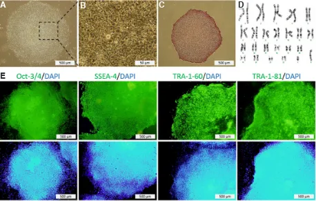
fibroblast (dif-ESC) showed normal morphology of hESC colonies as round colonies with definite edge cells with obvious nuclei and thin cytoplasm (Fig. 2A-D). The cells were ALP positive and maintained a normal karyotype with diploid 46 XY karyotype. In addition, immunofluorescent staining showed positive expression of the nuclear marker Oct4 and the surface markers SSEA-4, TRA-1-60 and TRA-1-81 (Fig. 2E).
Differential expression of stemness and fibroblast genes in iPSC, ESC and differentiated cells
The relative expression of three stemness genes (Nanog, Oct4 and Sox2) and three genes indicating fibroblast differentiation
(Vim, COL1A1 and THY1) in all cell lines were quantitatively determined by real-time PCR, using GAPDH gene for normal-ization. As expected, the expression levels of Nanog, Oct4 and Sox2 were significantly higher in embryonic stem cell (ESC) and in both induced pluripotent stem cells (iPS1 and iPS2) than in diff-ESC and in human fibroblasts (Fig. 3). In contrast, the expression of fibroblast genes (Vim, COL1A1 and THY1), in pluripotent cells have showed significantly lower levels than in differentiated cells (Fig. 4) (supplemental data S2). In the case of COL1A1, it seemed that in iPS cell lines, especially iPS1, this gene has escaped the global reprogramming and has remained transcribed. In these iPS cells, Vim and THY1 showed a better Fig. 2. Characterization of iPS1 which re-reprogramed from differentiated ESC.(A)Morphology of iPS1. (B) Higher magnification of iPS1. (C) Expression of ALP (Alkaline phosphatase). (D) Normal Karyotype of cells at passage 12. (E) Expression of Oct-3/4, SSEA-4, TRA-1-60 and TRA-1-81.
Fig. 3. Relative mRNA expression levels of stemness marker genes in all studied cell lines. The results are expressed as 2^ΔΔCT (mean ± SEM). Means labeled with vastly different let-ters are significantly different in p≤0.05 (see supplemental data S2).
B
C
A
D
reprogramming and present a transcription rate which is closer to that observed in ESCs (Fig. 4).
Different levels of histone 3 K9 acetylation and methylation in iPSC, ESC and differentiated cells
Total chromatin extracted from iPS cell lines, ES cells and differentiated ES cell line were used to evaluate the occurrence H3K9ac and H3K9me2. For this purpose, the chromatin-ELISA method, which allows for a relative quantitative assessment of these histone marks was used. As shown in Fig. 5A, the levels of H3K9ac, in pluripotent cells lines were drastically higher than its level in dif-ESC and fibroblast cells. The same approach was used to measure the relative amounts of H3K9me2 in the same cells. In this case, compared to differentiated cells, we observed low levels of this mark in pluripotent cells (Fig. 5A) (supplemental data S3). We observed that similarly to H3K9ac, H3K4me3 is higher in pluripotent cells compared to the differentiated cells, while
regions of marker genes of stemness: Nanog, Oct4 and Sox2. Likewise, significant high levels of tri-methylated H3K4 were also observed on these regions (see also supplemental data S4).
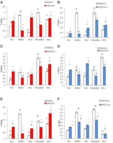
However, regulatory region of marker genes for fibroblast differ-entiation (Vim, COL1A1 and THY1), were epigenetically marked by hypermethylation of H3K9 and H3K27 in pluripotent cells (Fig. 7). In fibroblast and differentiated ESC (dif-ESC) the repressive marks of H3K9me2 and H3K27me3 showed significantly higher incorporation than H3K9ac and H3K4me3 at up-stream regions of Nanog, Oct4 and Sox2 genes (Fig. 6). However, this condition at upstream regions of Vim, COL1A1 and THY1 was reversed (Fig. 7) (see supplemental data S4).
Discussion
Embryonic stem cells have specific epigenetic characteristics, which distinguish them from terminally differentiated cells. A re-Fig. 4. Relative mRNA expression levels of fibroblast marker genes in all studied cell lines.
The results are expressed as 2^ΔΔCT (mean ± SEM). Means labeled with vastly different letters are significantly different in p≤0.05 (see supplemental data S2).
H3K27me3 showed a reverse situation with higher levels in differentiated cells (Fig. 5B, supplemental data S3).
Localized post translational modifications on gene regulatory regions
After evaluation of total levels of H3K9ac/ me2 and H3K4/K27 me3 PTMs on chroma-tin of cells, chromachroma-tin immunoprecipitation (ChIP) was used to detect the presence of the mentioned histone modifications on the regulatory regions of specific marker genes indicating stemness states and fibroblast dif-ferentiation states respectively in plurippotent and committed cells.
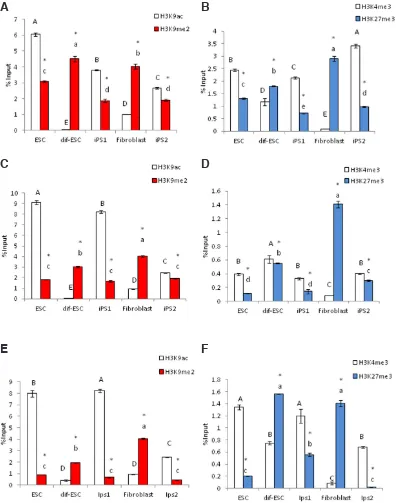
To this aim, after immunoprecipitation of chromatin with H3K9ac, H3K9me2, H3K4me3 and H3K27me3 antibodies, enriched DNA were amplified by using designed primers corresponding to the regulatory regions of the mentioned genes respectively in the considered cell lines. As shown in Fig 6, in all pluripotent cells acetylated H3K9 (H3K9ac) was the dominant modification on regulatory
Fig. 5. Total levels of post translational modifications (PTMs) by ELISA method in all studied cell lines. Relative content of (A) H3K9ac/me2 and (B) bivalent marks of H3K4/27me3 in pluripotent cells of ES, iPS1 and iPS2 as well as dif-ESC and fibroblast cell lines after adjusting for chromatin loading based on H2A reactivity by ELISA. Means labeled with vastly different letters are significantly different in p≤0.05 (see supplemental data S3). Asterisk sign shows significant difference of (A) H3K9ac vs. H3K9me2 and (B) H3K4me3 vs. H3K27me3 in p≤0.05.
markable specificity of these cells is their hyper-dynamic chromatin, which has been associated with high levels of histone acetylation (Apostolou and Hochedlinger, 2013, Ikegami et al., 2009, Meshorer et al., 2006). With this respect, histone H3K9 represents a key position. Indeed, H3K9 is a site where competing chemical modi-fications mediate different functional outputs. Acetylation of H3K9 is associated with both dynamic chromatin and gene transcriptional activity. In contrast, methylation of H3K9 not only blocks further modifications of this specific lysine but also underlays the estab-lishment of heterochromatin and gene transcriptional repression.
Another specificity of ES cells’ chromatin is that in these cells, the developmentally important genes are epigenetically distinguished by a unique ‘bivalent’ mark signature consisting of histone H3
K4 and K27 tri-methylation (Pan et al., 2007). Individually, these histone marks indicate active and repressive genes respectively (Pan et al., 2007).
Since post transcriptional modifications have an undoubted es-sential role in chromatin dynamicity of cells, it could be a worthwhile way to evaluate the chromatin state of the cells in pluripotency, differentiation and fate determination of ESCs. Here we report an ELISA-based quantitative measurement of total chromatin H3K9ac, H3K9me2 as well as H3K27me3 and H3K4me3 in a specific set of cell lines that has been carefully chosen to reflect different global epigenetic states. Human embryonic stem cells were used as a reference of pluripotent cells and were compared with iPS cells generated from two different sources: differentiated ES cells and
Fig. 6. Chromatin immunoprecipi-tation (ChIP) analysis of histone modifications on regulatory re-gions of stemness marker genes. Incorporated PTMs on regulatory regions of (A,B)Nonog, (C,D)Oct4
and (E,F)Sox2 in all cell lines (see supplemental data S4).
fibroblasts.
Our specific simple and quantitative histone PTM mea-surement approach performed on these cells allowed us to demonstrate a direct relation-ship between the pluripotency state of the cells and the in-vestigated histone PTMs. More specifically, in pluripotent cells the ratio of total H3K9ac and H3K9me2 was found to indicate the presence of these marks on pluripotency gene promot-ers as well as the extent of their expression. In contrast to pluripotent cells, in differenti-ated cells, the ratio of whole chromatin H3K9ac/H3K9me2 is low but interestingly, the active gene promoters are epigeneti-cally distinguished and do not follow the general situation of H3K9 modifications. More pre-cisely, in pluripotent cells, the global amounts of H3K9ac and H3K9me2 mirror the presence of these marks on active gene regulatory regions: the ratio of total H3 K9ac/K9me2 is high, so is this ratio on the pluripotency gene promoters (Nanog, OCT4 and SOX2). This is not the case of repressed gene promoters (Vim, COL1A1 and THY1) in the same cells.
In remarkable contrast, in
B
C
D
E
F
differentiated cells (fibroblast and dif-ES cells), the ratio of global chromatin K9ac/K9me2 on histone H3 is low, however, promoters of active genes escape this general situation and bear, as expected, higher amounts of H3K9ac compared to H3K9me2.
In agreement with the data presented here, previous studies have demonstrated that H3K9ac is abundant on chromatin of ES cells and is one of the characteristic features of these cells (Meshorer and Misteli, 2006). Additionally, inhibition of histone-deacetylases (HDACs) can enhance gene reactivation in induced pluripotency experiments and promote the reprogramming process (Huangfu et al., 2008).
Histone H3 lysine 9 methylation has been shown in contrast to be an efficient barrier to cell reprogramming both following induced
pluripotent stem cell generation and somatic cell nuclear transfer (Becker et al., 2016).
It is therefore expected that an efficient cell reprogramming of differentiated cells restores high levels of H3K9ac with a correspond-ing significant reduction in H3K9 methylation. The comparison of the total amount of H3K9ac with that observed in the parental cells and in a reference ES cell line, could therefore be used as a good indicator of the efficiency of cell reprogramming.
In parallel to H3K9ac/H3K9me2, we have also analysed H3K-4me3/H3K27me3 at global and loci-specific levels. Our chromatin ELISA tests showed that compared to differentiated cells, higher levels of H3K4me3 could be detected in all three pluripotent cell lines. These results could also be correlated with significant levels
Fig. 7. Chromatin immunopre-cipitation (ChIP) analysis of his-tone modifications on regulatory regions of fibroblast marker genes. Incorporated PTMs on regulatory regions of (A,B) THY1, (C,D) Vim
and (E,F)COL1A1 in all cell lines (see supplemental data S4).
of H3K4me3 upstream of stem-ness marker genes in pluripotent cells compared to fibroblast and dif-ES cells. A reverse situation was found for H3K27me, which is more abundant in differentiated cells and marks repressed genes in both ES cells and differenti-ated cells.
Based on the data presented here, it appears that in pluripotent cells, the promoters of pluripoten-cy genes follow the general situa-tion of whole chromatin H3K9ac/ K9me2 as well as total H3K4me3/ H3K27me3. This “PTM signature” of chromatin in cells therefore clearly reflects their pluripotency or differentiation state.
Based in these data, we propose that a quantitative mea-surement of relative amounts of H3K9ac and H3K4me3 compared to H3K9me2 and H3K27me3 in iPS cells with respect to both the parental differentiated cells and a reference ES cell line could con-stitute a convenient approach to assess the efficiency and success of cell epigenetic reprogramming.
Materials and Methods
Cell cultureIn this experiment, five human cell lines were studied including an ES cell line (Baharvand et al., 2006, Baharvand et al., 2004) before and
B
C
D
E
F
after differentiation to fibroblast (ESC and dif-ESC), and an iPS cell line reprogrammed from these fibroblast cell (iPS1). Accordingly, a human dermal fibroblast cell line as a completely and naturaly differentiated cell line was used in this study as well as an iPS line originated from it (iPS2) (Table 1) (Mollamohammadi et al., 2009, Totonchi et al., 2010).
Differentiation process of ESC towards dif-ESC was simply preformed by single cell dissociating of ESCs with TrypLE enzyme (Life Technologies: Cat. No. 12605-028) and seeding single cells on 0.1% gelatin (Sigma-Aldrich: Cat. No. G2500)-coated tissue culture dishes in DMEM medium (Life Technologies: Cat. No. 12800-082) supplemented with 10% FBS (Life Technologies: Cat. No. 16140-071). Passage of the cell line was performed every 3-4 days. Characterization of cells was conducted according to the following parameters: checking of morphology as well as checking of expression of Vimentin (Santa Cruz Biotechnology: Cat. No. sc-32322) as positive marker and Alkaline phosphatase (ALP) (Sigma-Aldrich: Cat. No.86R), Oct-3/4 (Santa Cruz Biotechnology: Cat. No. sc-5279) and TRA-1-81 (Life Technologies: Cat. No. 41-1100) as negative markers. Approved dif-ESC was re-reprogrammed in iPS1(Totonchi et al., 2010) and standard charactrization including morphology, ALP, karyotyping, and expressing Oct-3/4, SSEA-4 (Life Technologies: Cat. No. 41-4000), TRA-1-60 (Life Technologies: Cat. No. 41-1000) and TRA-1-81 were performed. Aforesaid cell lines except fibroblast and dif-ESC were cultured in hESC medium supplemented with 20% knockout serum replacement (Life Technologies: Cat. No. 10828-028), and passage was performed every seven days (Mol-lamohammadi et al., 2009).
Karyotype analysis
For karyotype analysis, at day 5 after culture the cells were treated with 0.66 mM thymidine (Sigma-Aldrich: Cat. No. T1895) for 16 h at 37°C in
5% CO2. After being washed, the cells were left for 5 h and then treated with colcemid (Life Technologies: Cat. No. 15212-012) 0.15 mg/ml for 30 min. Isolated human pluripotent stem cells were exposed to 0.075 M KCl at 37°C for 16 min and then were fixed in three consecutive immersions in
ice-cold 3:1 methanol to glacial acetic acid and later on dropped onto pre-cleaned chilled slides. Chromosomes were visualized by using standard G-band staining. At least 20 metaphase spreads were screened, and 10 of them were evaluated for chromosomal re-arrangements.
RNA isolation and quantitative real-time PCR
RNA isolation and RT-PCR was performed on three biological replicates as described before(Shahhoseini et al., 2010). mRNA quantification was performed in duplicates by quantitative real-time PCR (qRT-PCR) on a StepOnePlus Real-Time PCR System (AB Applied Biosystems) using SYBR Green master mix (AB Applied Biosystems), with designed prim-ers listed in Table 2. qRT-PCR condition was 95°C for 10 minutes, and 40
cycles of 95°C for 15 seconds and 60°C for 60 seconds. Gene expression
data were analyzed using ΔΔCt quantitative method to estimate relative fold change values.
Chromatin shearing and soluble chromatin preparation
To gain nucleosomes as antigens, chromatin was sheared by using a
histone ChIP kit according to manufactured instructions (Diagenode: Cat. No. kch-orgHIS-012). Briefly, each 1×10⁶ cells were suspended in PBS then 37% formaldehyde (w/v) was added to cells to reach 1% final con-centration of formaldehyde and mixed immediately and incubated gently on a shaking platform for 10 minutes at room temperature. To quench the cross-linking reaction of formaldehyde, glycine was added to reach final concentration of 125 mM. Upon being washed with PBS, lysis buffer was added to the pellet of cells and then cells were sonicated for 10 minutes (30”on/30”off, UCD200- Bioruptor sonication system, Diagenode) to get soluble sheared chromatin.
Chromatin- ELISA analysis
The chromatin ELISA technique was performed according to Dai and Rasmussen’s research (2007) (Dai and Rasmussen, 2007). Briefly, diluted sheared chromatin was coated into 96 well plates (Nunc: 96FMaxisorp, Cat. No. 456537) using coating solution (KPL: Cat. No.50-84-00). After overnight incubation at 4°C, each well was washed four times with 200 mL wash solution (KPL Cat. No.50-63-00), then blocked by adding 100 mL blocking solution (KPL: Cat. No. 50-84-00) and incubated 1 hour at
room temperature (RT). Blocking buffer was aspirated and 50 mL of first
antibodies (diluted in blocking solution) (see supplemental data S1) were added to each well and incubated at RT for 1-2 hours, then washed as described above. Then, 100 mL HRP-conjugated antibodies (supplemental
data S1) as secondary antibody (diluted in blocking solution) were added and incubated at RT for 1 hour. After washing, 50 mL of 3,3-5,5-tetra methyl
benzidine (TMB) peroxidase substrate (KPL: Cat. No. 50-76-00) was added to each well at RT for 10 minutes. Finally, 50 mL, 2 N H2SO4 was added to each well to stop reaction. Optical absorption of plates was read at 450 nm by ELISA-reader (Biotech -ELX 800).
In current study three independent cultures of each cell line were exam-ined as cellular replicates and the soluble chromatin of each replicate was loaded in two or three separate wells for two or three times as technical
References Definition
Cell lines
(Baharvand et al., 2006,
Baharvand et al., 2004b)
Human Embryonic Stem Cell ESC Induced pluripotent stem cell reprogrammed from
dif-ESC iPS1
(Mollamohammadi et al., 2009)
Human fibroblast Fibroblast
(Mollamohammadi et al., 2009)
Induced pluripotent stem cell reprogrammed from fibroblast
iPS2
TABLE 1
DEFINITION OF THE CELL LINES USED IN THIS STUDY
Gene Primers (5´→ 3´) temperature(°C) Annealing size (bp) Product qRT-PCR primers
NANOG F: AAAGTCTTAAAGCTGCCTTAAC
R: CAGTCGGATGCTTCAAAG 60 130
OCT4 F: GTTCTTCATTCACTAAGGAAGG
R: CAAGAGCATCATTGAACTTCAC 60 101
SOX2 F:GGGAAATGGGAGGGGTGCAAAAGAGG
R:TTGCGTGAGTGTGGATGGGATTGGTG 60 151
VIM F:GGCTCGTCACCTTCGTGAAT
R:GAGAAATCCTGCTCTCCTCGC 60 110
THY1 F:TAGTGAAGGCGGATAAGTAGAGG
R: ACCCGTGAGACAAAGAAGC 60 124
COL1A1 F:CCTGTCTGCTTCCTGTAAAC
R:ATGTTCGGTTGGTCAAAGATAAA 60 211
ChIP Real-time PCR Primer
NANOG F: AATTCACAAGGGTGGGTCAG
R:TAACATGAGGCAACCAGCTC 60 133
OCT4 F: GTTGGGGAGCAGGAAGCA
R: GGGGCAGCTCTAACCCTAAA 60 83
SOX2 F: TCGCTAGAAACCCATTTATTCC
R: CTGCCTTGACAACTCCTG 60 93
VIM F: CGAGTTTCCTCTTTCACCC
R: ACTTCTGCAGCCTTTGGA 60 187
THY1 F: GGCTTTAACCCTTTCTTCTGAC
R: AAGGAAATGTGGAGGCGT 60 106
COL1A1 F: GGGCCCCTTTTATACTGTCC
R: TCTCCATTCCAACTCCCAAA 60 132
TABLE 2
PRIMER PAIRS USED FOR CHIP REAL-TIME PCR
NANOG (Nanog homeobox), OCT4 (POU class 5 homeobox 1), SOX2 (SRY box 2), VIM (Vimentin),
replicates. After subtracting of background from each well, values were normalized by dividing them into loading-controlled values. At least, values were expressed as means±SEM of 3 separate experiments.
Chromatin immunoprecipitation (ChIP)-real time PCR analysis
Chromatin immunoprecipitation (ChIP) experiments were conducted as previously described on three biological replicates (Favaedi et al., 2012). For each immunoprecipitation reaction 1× 10⁵ cells was used. Recovered DNA from percipiation fractions and total chromatin input were applied for quantitative real-time PCR with designed primers for regulatory regions of 6 genes involved in stemness and fibroblast differentiation (Table 2). Real-time PCR was performed on a step one plus Real-Time PCR System (Applied Biosystems) by using SYBR Green PCR master mix (Applied Biosystems: Cat. No. 4367659). PCR condition was 95°C for 10 min; and 40 cycles of 95°C for 15 s, 60°C for 45 s. The results are expressed as
%input which means percentage of enriched DNA associated with given immunoprecipitated histone modifications relative to a 1/100 dilution of input chromatin (mean ± SEM; triplicate experiment).
Statistical analysis
Values are expressed as means±SEM of three separate experiments. All data was analyzed by using One-way ANOVA and paired samples T-test, and differences were considered statistically significant at p≤0.05 (see supplemental data S2 and S3).
Acknowledgments
We wish to thank Dr. Saadi.Khochbin for his worthwhile guidance and Farideh Moeinvaziri for her kind help. The authors would like to dedicate this paper to the memory of Dr. Saeid Kazemi Ashtiani, the late founder of Royan Institute. This project was financially supported by the Royan Institute (Grant No. 173).
References
ANG, Y.-S., TSAI, S.-Y., LEE, D.-F., MONK, J., SU, J., RATNAKUMAR, K., DING, J., GE, Y., DARR, H. AND CHANG, B. (2011). WDR5 MEDIATES SELF-RENEWAL AND REPROGRAMMING VIA THE EMBRYONIC STEM CELL CORE TRAN-SCRIPTIONAL NETWORK. Cell 145: 183-197.
APOSTOLOU, E. AND HOCHEDLINGER, K. (2013). CHROMATIN DYNAMICS DURING CELLULAR REPROGRAMMING. Nature 502: 462-471.
BAHARVAND, H., ASHTIANI, S.K., TAEE, A., MASSUMI, M., VALOJERDI, M.R., YAZDI, P.E., MORADI, S.Z. AND FARROKHI, A. (2006). GENERATION OF NEW HUMAN EMBRYONIC STEM CELL LINES WITH DIPLOID AND TRIPLOID KARYOTYPES. Dev Growth Differ 48: 117-128.
BAHARVAND, H., ASHTIANI, S.K., VALOJERDI, M.R., SHAHVERDI, A., TAEE, A. AND SABOUR, D. (2004). ESTABLISHMENT AND IN VITRO DIFFERENTIATION OF A NEW EMBRYONIC STEM CELL LINE FROM HUMAN BLASTOCYST. Differ 72: 224-229.
BECKER, J.S., NICETTO, D., ZARET, K.S. (2016). H3K9me3-Dependent Hetero-chromatin: Barrier to Cell Fate Changes. Trends Genet. 32: 29-41.
BERNSTEIN, B.E., MIKKELSEN, T.S., XIE, X., KAMAL, M., HUEBERT, D.J., CUFF, J., FRY, B., MEISSNER, A., WERNIG, M. and PLATH, K. (2006). A bivalent chromatin structure marks key developmental genes in embryonic stem cells. Cell 125: 315-326.
CHEN, J., LIU, H., LIU, J., QI, J., WEI, B., YANG, J., LIANG, H., CHEN, Y., CHEN, J. and WU, Y. (2013). H3K9 methylation is a barrier during somatic cell reprogram-ming into iPSCs. Nat Genet 45: 34-42.
CHIN, M.H., MASON, M.J., XIE, W., VOLINIA, S., SINGER, M., PETERSON, C., AMBARTSUMYAN, G., AIMIUWU, O., RICHTER, L. and ZHANG, J. (2009). Induced pluripotent stem cells and embryonic stem cells are distinguished by gene expression signatures. Cell Stem Cell 5: 111-123.
CHIN, M.H., PELLEGRINI, M., PLATH, K. and LOWRY, W.E. (2010). Molecular analyses of human induced pluripotent stem cells and embryonic stem cells. Cell Stem Cell 7: 263-269.
DAI, B. and RASMUSSEN, T.P. (2007). Global epiproteomic signatures distinguish embryonic stem cells from differentiated cells. Stem Cells 25: 2567-2574. FAVAEDI, R., SHAHHOSEINI, M., AKHOOND, M.R.(2012).Comparative epigenetic
analysis of Oct4 regulatory region in RA-induced differentiated NT2 cells under adherent and non-adherent culture conditions. Mol Cell Biochem 363: 129-134. HEZRONI, H., TZCHORI, I., DAVIDI, A., MATTOUT, A., BIRAN, A., NISSIM-RAFINIA,
M., WESTPHAL, H. and MESHORER, E. (2011). H3K9 histone acetylation pre-dicts pluripotency and reprogramming capacity of ES cells. Nucleus 2: 300-309. HUANGFU, D., MAEHR, R., GUO, W., EIJKELENBOOM, A., SNITOW, M., CHEN, A.E.
and MELTON, D.A. (2008). Induction of pluripotent stem cells by defined factors is greatly improved by small-molecule compounds. Nat Biotechnol 26: 795-797. IKEGAMI, K., OHGANE, J., TANAKA, S., YAGI, S. and SHIOTA, K. (2009). Interplay
between DNA methylation, histone modification and chromatin remodeling in stem cells and during development. Int J Dev Biol 53: 203-214.
KOUZARIDES, T. (2007). Chromatin modifications and their function. Cell 128: 693-705. MESHORER, E. and MISTELI, T. (2006). Chromatin in pluripotent embryonic stem
cells and differentiation. Nat Rev Mol Cell Biol 7: 540-546.
MESHORER, E., YELLAJOSHULA, D., GEORGE, E., SCAMBLER, P.J., BROWN, D.T. and MISTELI, T. (2006). Hyperdynamic plasticity of chromatin proteins in pluripotent embryonic stem cells. Dev Cell 10: 105-116.
MOLLAMOHAMMADI, S., TAEI, A., PAKZAD, M., TOTONCHI, M., SEIFINEJAD, A., MASOUDI, N. and BAHARVAND, H. (2009). A simple and efficient cryopreserva-tion method for feeder-free dissociated human induced pluripotent stem cells and human embryonic stem cells. Hum Reprod 24: 2468-2476.
PAN, G., TIAN, S., NIE, J., YANG, C., RUOTTI, V., WEI, H., JONSDOTTIR, G.A., STEWART, R., THOMSON, J.A.(2007). Whole-genome analysis of histone H3lysine 4 and lysine 27 methylation in human embryonic stem cells. Cell Stem Cell 1:299–312.
SINGHAL, N., GRAUMANN, J., WU, G., ARAÚZO-BRAVO, M.J., HAN, D.W., GREBER, B., GENTILE, L., MANN, M. and SCHÖLER, H.R. (2010). Chromatin-remodeling components of the BAF complex facilitate reprogramming. Cell 141: 943-955. TAKAHASHI, K., TANABE, K., OHNUKI, M., NARITA, M., ICHISAKA, T., TOMODA, K.
and YAMANAKA, S. (2007). Induction of pluripotent stem cells from adult human fibroblasts by defined factors. Cell 131: 861-872.
TOTONCHI, M., TAEI, A., SEIFINEJAD, A., TABEBORDBAR, M., RASSOULI, H., FARROKHI, A., GOURABI, H., AGHDAMI, N., HOSSEINI-SALEKDEH, G. and BAHARVAND, H. (2010). Feeder-and serum-free establishment and expansion of human induced pluripotent stem cells. Int J Dev Biol 54: 877.
WEN, B., WU, H., SHINKAI, Y., IRIZARRY, R.A. and FEINBERG, A.P. (2009). Large histone H3 lysine 9 dimethylated chromatin blocks distinguish differentiated from embryonic stem cells. Nat Genet 41: 246-250.
Bone morphogenetic protein 4 promotes craniofacial neural crest induction from human pluripotent stem cells
Sumiyo Mimura, Mika Suga, Kaori Okada, Masaki Kinehara, Hiroki Nikawa and Miho K. Furue Int. J. Dev. Biol. (2016) 60: 21-28
http://dx.doi.org/10.1387/ijdb.160040mk
5 yr ISI Impact Factor (2013) = 2.879
Two decades of reproductive biomedicine and stem cell biology in Iran: the Royan Institute
Sophie Rousseaux
Int. J. Dev. Biol. (2014) 58: 643-647 http://dx.doi.org/10.1387/ijdb.140245sr
Generation of pluripotent stem cells via protein transduction
Xia Li, Pengfei Zhang, Chao Wei and Yunhai Zhang Int. J. Dev. Biol. (2014) 58: 21-27
http://dx.doi.org/10.1387/ijdb.140007XL
Epigenetic features of testicular germ cell tumours in relation to epigenetic character-istics of foetal germ cells
Dina G. Kristensen, Niels E. Skakkebæk, Ewa Rajpert-De Meyts and Kristian Almstrup Int. J. Dev. Biol. (2013) 57: 309-317
http://dx.doi.org/10.1387/ijdb.130142ka
Signaling pathways during maintenance and definitive endoderm differentiation of embryonic stem cells
Lina Sui, Luc Bouwens and Josué K. Mfopou Int. J. Dev. Biol. (2013) 57: 1-12