R E S E A R C H
Open Access
Functional properties of the HIV-1 long
terminal repeat containing single-nucleotide
polymorphisms in Sp site III and CCAAT/enhancer
binding protein site I
Sonia Shah, Aikaterini Alexaki, Vanessa Pirrone, Satinder Dahiya, Michael R Nonnemacher and Brian Wigdahl
*Abstract
Background:HIV-1 gene expression is driven by the long terminal repeat (LTR), which contains many binding sites shown to interact with an array of host and viral factors. Selective pressures within the host as well as the low fidelity of reverse transcriptase lead to changes in the relative prevalence of genetic variants within the HIV-1 genome, including the LTR, resulting in viral quasispecies that can be differentially regulated and can potentially establish niches within specific cell types and tissues.
Methods:Utilizing flow cytometry and electromobility shift assays, specific single-nucleotide sequence polymorphisms (SNPs) were shown to alter both the phenotype of LTR-driven transcription and reactivation. Additional studies also demonstrated differential loading of transcription factors to probes derived from the double-variant LTR as compared to probes from the wild type.
Results:This study has identified specific SNPs within CCAAT/enhancer binding protein (C/EBP) site I and Sp site III (3 T, C-to-T change at position 3, and 5 T, C-to-T change at position 5 of the binding site, respectively) that alter LTR-driven gene transcription and may alter the course of viral latency and reactivation. The HIV-1 LAI LTRs
containing the SNPs of interest were coupled to a plasmid encoding green fluorescent protein (GFP), and polyclonal HIV-1 LTR-GFP stable cell lines utilizing bone marrow progenitor, T, and monocytic cell lines were constructed and utilized to explore the LTR phenotype associated with these genotypic changes.
Conclusions:Although the 3 T and 5 T SNPs have been shown to be low-affinity binding sites, the fact that they can still result in effective HIV-1 LTR-driven gene expression, particularly within the TF-1 cell line, has suggested that the low binding site affinities associated with the 3 T C/EBP site I and 5 T Sp site III are potentially compensated for by the interaction of nuclear factor-κB with its corresponding binding sites under selected physiological and cellular conditions. Additionally, tumor necrosis factor-αand Tat can enhance basal transcription of each SNP-specific
HIV-1 LTR; however, differential regulation of the LTR is both SNP- and cell type-specific.
Keywords:HIV-1 genetics, Viral transcription, Integration, Viral latency, Single-nucleotide polymorphisms, C/EBP, Sp
* Correspondence:[email protected]
Department of Microbiology and Immunology, and Center for Molecular Virology and Translational Neuroscience, Institute for Molecular Medicine and Infectious Disease, Drexel University College of Medicine, Philadelphia, Pennsylvania 19102, USA
Background
HIV-1-associated immunologic and neurologic disease is dependent on the ability of the virus to infect subsets of resident immune and central nervous system (CNS) cell populations. In vitro and in vivo investigations have shown that HIV-1 infection of active CD4+T lymphocytes initiates a highly productive infection [1-7]. In contrast, HIV-1-infected monocytic cell populations produce only limited quantities of virus due to several host-cell replica-tion blocks including barriers that limit the reverse tran-scription process [8,9] and nuclear import [10]. These barriers result in a more chronic infection because this cell type is more resistant to the cytopathic effects of HIV-1 gene products [11-13] and has a longer lifespan in vivo. The chronic nature of HIV-1 replication in cells of the monocyte-macrophage lineage is likely a contributor to the central importance of these cells in evasion of HIV-1 detection and elimination by the immune system and the maintenance of viral reservoirs. The virus can utilize cells of this lineage as a vehicle facilitating its transport across the blood–brain barrier (BBB) and its entry into the CNS [14-16], thereby promoting HIV-1-associated neuropatho-genesis and the development of minor neurocognitive impairment, as well as the more severe CNS disease, HIV-1-associated dementia (HIVD). HIV-1 infection of the CNS occurs soon after infection; however, under most cir-cumstances, prolonged productive viral replication, char-acterized by the formation of multinucleated giant cells with progressive loss of cognitive, behavioral, and motor deficits, is likely to occur only after severe immunosup-pression and breakdown of the BBB. The pathological events that eventually lead to the development of HIVD may be initiated outside the CNS and involve the process of monocyte activation and many important events associ-ated with passage of activassoci-ated cells across the BBB. Peri-vascular macrophages likely play a critical role in the pathogenesis of HIVD as they are located on the paren-chymal side of the BBB and the pool is continuously renewed through bone marrow-derived macrophages, par-ticularly during CNS inflammation [14]. Thus, the bone marrow may serve as a source of HIV-1-infected macro-phages and may play a critical role in neuroinvasion and progression of CNS disease.
Genetic variation within the HIV-1 viral genome is a naturally occurring process driven by the low fidelity of reverse transcriptase, coupled with the selective pressures brought about within the host such as antiretroviral ther-apy, recreational drug use, immunological pressures, viral recombinatory events, host-cell phenotype, and rates of virus production [17-19]. These events result in single nu-cleotide polymorphisms (SNPs) throughout the genome including the promoter region, designated the long ter-minal repeat (LTR). Genetic variation occurs within LTR binding sites where host transcription factors and viral
regulatory proteins bind, altering the way the LTR drives viral transcription. The resultant viral quasispecies are likely shaped by the selective pressures operative within a variety of cellular and tissue niches that ultimately maintain specific sets of quasispecies to form viral reser-voirs in susceptible cell types and end-organ tissues [20-27]. The accumulation of specific LTR sequence configurations over time may also result from accumu-lation of poorly replicating viruses or latent proviruses in long-lived cell subsets in circulation or within viral reservoirs, such as the resting memory CD4+ T-cells, monocytes and macrophages, and hematopoietic pro-genitor cells (HPCs) [28].
Results
Parental (wild-type) and 3T5T LTR results in cell type–specific phenotypes within different promoter backbones
The DREXELMED HIV/AIDS Genetic Analysis Cohort is currently comprised of approximately 504 patients, 78.26% of whom are on continuous highly active anti-retroviral therapy (HAART). Within this patient cohort, 457 patients and 1,113 longitudinal samples have been genotyped and the HIV-1 LTRs contain many SNPs, in-cluding the previously identified C-to-T SNP at position 5 (5 T) in Sp site III, that occurs in approximately 24.9% of the LTRs analyzed to date compared to the conB se-quence (GAGGCGTGGC) derived from the Los Alamos National Laboratory, which occurs at in about 67.8% of the LTRs analyzed. The 5 T SNP results in dramatically reduced binding of Sp1, Sp3, and the truncated form of Sp3 [23]. Approximately 5.1% of the patients within this cohort have a SNP within the LTR at C/EBP site I (AGCTTTCTACAA), where there is a C-to-T change at position 3 (3 T) that also results in a knockout binding phenotype [23,25-27,37,38]. Finally, a small number of the patients (1.0%) in the DREXELMED HIV/AIDS
Genetic Analysis Cohort have been found to have the 3T5T double variant.
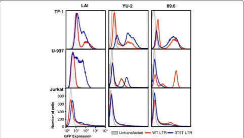
As one approach to understand the effects of the 3 T and 5 T variations together on LTR-driven gene expres-sion within several backbones, the WT and 3T5T varia-tions were introduced into the LAI, YU-2, and 89.6 HIV-1 LTR backbones and then ligated into the pEGFP-N1 vec-tor where the LTR can drive the expression of the green fluorescent protein (GFP). The constructs were then sta-bly transfected into TF-1, U-937, and Jurkat cells. These cells were selected because they were all representative of cellular phenotypes that have been identified as potential or confirmed viral reservoirs where HIV-1 could poten-tially avoid recognition from immune cells and thera-peutic agents, thus enabling the virus to survive for long periods within the host and to continue to replicate when the intra- and extracellular environment was con-ducive to virus production [2,13,28,33,45]. Within TF-1 cells, the 3T5T YU-2 and 89.6 LTRs resulted in both a nonexpressing population and a high-expressing popu-lation (Figure 1), whereas the 3T5T LAI LTR also re-sulted in an intermediate-expressing population. The intermediate-expressing population of cells was also
YU-2 89.6
800
600
400
200
0
100 101 102 103 104
Number of cells
GFP Expression TF-1
U-937
Jurkat
LAI
Untransfected WT LTR 3T5T LTR
observed within U-937 cells containing the 3T5T LAI LTR, whereas it was absent from U-937 cells containing the 3T5T YU-2 or 89.6 LTRs (Figure 1). In contrast, no phenotypic differences were found between the WT and 3T5T LAI, YU-2, or 89.6 LTRs within Jurkat cells (Figure 1), indicating that this cell phenotype may play in differentially controlling expression of LTRs containing genotypic alterations. The LAI backbone was selected for further study due to the presence of the large intermediate expressing cell population within cells containing the 3T5T LAI backbone that was mainly absent in both the YU-2 and 89.6 backbones (Figure 1). Additionally, it has been commonly used in other scientific studies, shares se-quence similarity with the conB sese-quence except that it contains a 6G change within C/EBP site I, and the HIV-1 LAI virus has the ability to infect all three cell line models used in this study.
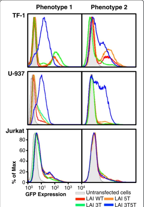
Double-variant LTR results in two different phenotypes in hematopoietic progenitor, promonocytic, and Jurkat T cells Transfected cells were selected under G418 selective pressure for over 4 weeks to develop stably transfected cell lines. Those cell lines were then frozen and preserved for future experimental use. As one approach to under-standing the basic effects of genetic variation on LTR function, basal LTR-driven GFP expression was assessed using flow cytometry to examine the LTR-driven GFP expression patterns obtained from a number of cell pop-ulations independently recovered from low-temperature storage.
Interestingly, each recovered cell population resulted in different LTR-driven expression patterns. This was particularly evident within the cell populations containing the 3T5T double-variant LTR, which could be grouped into two general phenotypic expression patterns. Stored frozen cells were independently recovered and grown in culture conditions more than six times each; representa-tive histograms are shown in Figure 2. With TF-1 cells, generally phenotype 1 was characterized by a very small nonexpressing population of cells, in addition to a larger population of cells that express intermediate levels of LTR-driven GFP expression (Figure 2, top left). Within TF-1 cells, phenotype 2 was generally characterized by a large population of non-GFP-expressing cells as well as a population of cells that expressed both intermediate and high levels of GFP (Figure 2, top right). The TF-1 cell lines containing the HIV-1 parental LAI LTR as well as the 3 T and 5 T variant LTR generally exhibited the same two LTR-driven GFP expression phenotypes in that they exhibited large non-GFP-expressing popula-tions of cells and a smaller high-GFP-expressing popu-lation of cells (Figure 2, top panels). The 5 T genotype generally resulted in higher amounts of LTR-driven GFP expression (Figure 2, top panels) when compared with
GFP expression driven by the WT and 3 T-containing LAI LTR.
Similar to the stably transfected TF-1 cells, basal LTR-driven GFP expression from stably transfected U-937 cell populations independently recovered from low-temperature storage also resulted in two distinct pheno-types specifically visible with the 3T5T-containing LTR cell populations (Figure 2, middle panels). In the U-937
% of Max
GFP Expression
Phenotype 1 Phenotype 2
Untransfected cells LAI WT
LAI 3T
LAI 5T LAI 3T5T
TF-1
U-937
Jurkat
80
60
40
20
0
100 101 102 103 104
cells, phenotype 1 was observed as a small non-GFP-expressing population with a larger intermediate-GFP-expressing population (Figure 2, middle left), whereas the 3T5T U-937 phenotype 2 cell population included a large non-GFP-expressing cell population and large popu-lations of intermediate- and high-GFP-expressing cells (Figure 2, middle right). With respect to the U-937 cell populations carrying other LTR genotypes (WT, 3 T, and 5 T), the expression profiles consisted of mainly non/low levels of GFP expression and smaller populations of intermediate-GFP-expressing cells (Figure 2, middle panel). When the stably transfected Jurkat cells were recovered from low-temperature storage, they too displayed two dis-tinct phenotypes. Phenotype 1 was characterized by a large low-expressing population of GFP-expressing cells within all Jurkat cell populations carrying each genotype and smaller populations of intermediate/high-GFP-expressing cells (Figure 1, bottom left). Within phenotype 2, there was a large population of nonexpressing cells that was similar to the WT Jurkat cell population, and a smaller population of cells that exhibited low/intermediate levels of GFP ex-pression (Figure 2, bottom right).
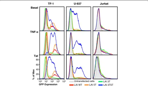
Double-variant LTR results in altered gene expression in basal and stimulated stably transfected cell lines
To better understand the kinetics behind LTR-driven gene transcription within LTRs that contained the SNPs of interest, the genotypic changes were analyzed in the presence of LTR activators such as tumor necrosis factor (TNF)-α and the viral transactivating protein Tat. For the following studies, TF-1, U-937, and Jurkat cell popu-lations displaying phenotype 2 were selected for further study because of their large populations of non-GFP-expressing cells, which could represent a model cell line population representative of known cellular HIV-1 reser-voirs within the body containing an integrated LTR struc-ture in a non-expressing or silent mode that could be activated under the selected physiological conditions.
Inflammatory cytokines such as TNF-αare elevated in patients with AIDS, and these particular cytokines are also involved in monocyte-macrophage differentiation as well as activation of HIV-1 gene transcription [46-53]. To study the effects of inflammatory cytokines on genet-ically altered LTR-driven gene expression in TF-1, U-937, and Jurkat cells, cells were treated with TNF-α(20 ng/mL) for 24 hours, after which time GFP expression was analyzed using flow cytometry. TNF-α stimulated cells of the monocytic and T-cell lineage through the binding of TNF-αto the TNF receptor, which causes the phosphorylation and release of IκB from nuclear
factor-κB (NF-κB), in turn allowing NF-κB to translocate from the cytoplasm into the nucleus of the cell. NF-κB has then been shown to be able to activate many host and viral genes through the initial recruitment of the positive
transcription elongation factor, as previously reviewed [45]. In addition, in the absence of functional Sp sites,
NF-κB binding to the enhancer region of the LTR can result in alteration in viral replication in T cells [43-45,54]. As expected, the cells were stimulated by exposure to TNF-α, and as a result of the stimulation, LTR-driven gene ex-pression was increased in each of the cell populations containing the selected LTR genotypes (Figure 3, middle row). In cells expressing the WT, 3 T, and 5 T LTRs, there was still a large population of cells that remained nonexpressers, while there was also a shift in the low-intermediate-expressing cells to a higher level of GFP expression (Figure 3). Interestingly, the 5 T LTR resulted in a higher level of change in LTR-driven GFP expression than the WT LTR, indicating that this genotype (which occurs frequently in the DREXELMED HIV/AIDS Genetic Analysis Cohort) could be involved in evading immune activation within the host under basal conditions, but under stimulatory conditions the 5 T genotype could be activated to produce a higher level of gene expression and virus production. Additionally, the stimulated 3T5T LTR exhibited a shift of the low-intermediate-GFP-expressing cell population to a majority of cells expressing a high level of GFP, and the majority of the nonexpressing cell population shifted to an intermediate level of GFP expres-sion (Figure 3).
Unexpectedly, the U-937 cells expressing GFP driven by the WT, 3 T, and 5 T LTRs were not stimulated to any great extent by TNF-αtreatment. Normally, TNF-α would cause U-937 cells to differentiate from their promo-nocytic state into monocyte-macrophages, which would lead to an alteration in general cellular gene expression (Figure 3). In contrast, as a result of the stimulation, the 3T5T LTR-driven gene transcription and gene expression as modeled by GFP expression were elevated (Figure 3). In these cells, the population of nonexpressing cells showed no change in GFP expression; however, the lower-GFP-expressing cell population decreased in prevalence dra-matically, resulting in a separate population of cells expressing intermediate/high levels of GFP as driven by the 3T5T LTR.
that can model what occurs in inactivated T cells in an HIV-1-infected patient. To study the activity of the 3 T, 5 T, and 3T5T LTRs within the Jurkat cell line, stably transfected cells stimulated with TNF-α(20 ng/mL), and as a result the WT, 3 T, 5 T, and 3T5T LTRs displayed an increase in LTR-driven gene transcription (Figure 3). The LAI 3 T and 5 T LTRs exhibited a slightly higher level of gene transcription when stimulated, indicating that there might be a cell type-specific LTR activation with a specific genotype.
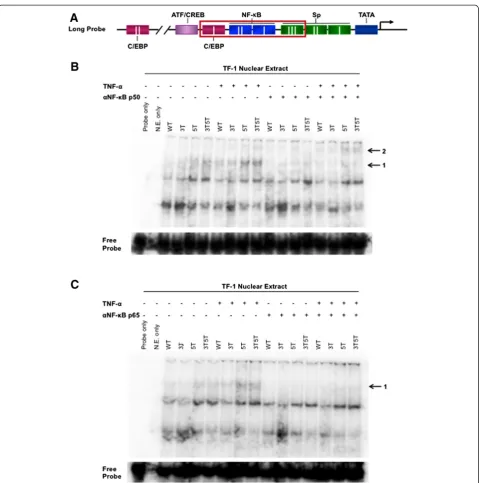
Double-variant LTR results in more efficient binding of NF-κB complexes to the LTR
As seen in Figures 2 and 3, the 5 T and 3T5T SNPs re-sulted in altered LTR-driven GFP expression. To deter-mine whether the 5 T and 3T5T SNPs altered binding of transcription factors to their cognate binding sites on the LTR, and whether the 3T5T SNP could potentially lead to altered and more efficient binding of NF-κB
complexes to NF-κB site I and site II, electromobility shift assays were performed. Nuclear extract was isolated from WT untransfected TF-1 cells that were untreated or treated with 20 ng/mL TNF-αfor 24 hours and then incubated with the long probes containing the WT, 3 T, 5 T, and 3T5T variants depicted in Figure 4A. Extracts were also incubated in the absence or presence of gel shift antibodies against NF-κB p50 and p65. When ex-tracts were incubated with the 5 T and 3T5T long probe, in both activated and nonactivated conditions, a large molecular weight complex (arrow 1 in Figure 4B) formed more prominently with those two probes in comparison to the others. Additionally, incubation with the NF-κB p50 antibody resulted in abrogation of binding of that complex to the long probe, and a shift in activated extracts incubated with the 5 T and 3T5T probes (arrow 2 in Figure 4B). This observation indicates that 5 T and 3T5T configurations allow for better binding of NF-κB p50 to its cognate binding site, potentially due to the lack of steric Basal
TNF-Tat
TF-1 U-937 Jurkat
Untransfected cells LAI WT
LAI 3T LAI 5T LAI 3T5T
% of Max
GFP Expression
80
60
40
20
0
100 101 102 103 104
hindrance from both C/EBP and Sp factors. When ex-tracts were incubated with long probes and the NF-κB p65 antibody, again, the same high molecular weight com-plex was more abundant with the 5 T and 3T5T probes, and binding of that complex to all probes was abrogated
loading to the 5 T- and 3T5T-containing LTRs and that it is comprised of NF-κB p50 and p65, suggesting that under normal conditions, the loading is not as effective. This could explain the intermediate levels of expression observed in Figure 3, and under activated conditions the much higher level of activation that occurs with those genotypes.
Stably transfected cell clones from TF-1 and U-937 cell lines reflect the GFP expression profiles of the total populations used to select the cell clones
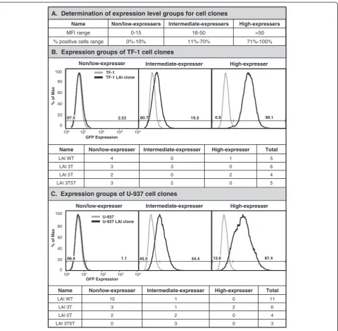
In order to effectively study how the LTR functions to drive GFP expression from the standpoint of a single cell, sets of clones were developed from each stably transfected cell line (TF-1 and U-937) using serial dilution and then propagating a population from a single cell. Once each single cell gave rise to a population of clones, their basal level of LTR-driven GFP expression was assessed by flow cytometry. Each clonal population was designated into one of three categories: nonexpresser, intermediate ex-presser, or high expresser based on arbitrary designations from the geometric mean fluorescence intensity (MFI) and GFP-positive cell percentage (Figure 5A). Figure 5B displays representative histograms for stably transfected TF-1 clonal populations containing the WT, 3 T, 5 T, or 3T5T LTR that fall into each category. There are a total of four nonexpresser and one high-expresser TF-1 LAI LTR WT clones; three nonexpressing and three intermediate-expressing TF-1 LAI LTR 3 T clones; two nonintermediate-expressing and two high-expressing TF-1 LAI LTR 5 T clones; and three nonexpressing and two intermediate-expressing TF-1 LAI LTR 3T5T clones (Figure 5B). When these clones were compared with the populations from which they were derived (Figure 3, top panel), their expression pro-files are representative of the variant cell line from the ex-pression pattern observed under basal conditions prior to cell cloning. Figure 5C displays representative histograms for stably transfected U-937 promonocytic clonal popula-tions containing the WT, 3 T, 5 T, or 3T5T LTR that fall into each category. There are a total of 10 nonexpressing and one intermediate-expressing U-937 LAI LTR WT clones; three nonexpressing, one intermediate expressing, and two high-expressing U-937 LAI LTR 3 T clones; two nonexpressing and two intermediate-expressing U-937 LAI LTR 5 T clones; and three intermediate-expressing U-937 LAI LTR 3T5T clones (Figure 5C). Interestingly, the Jurkat T-cell clones were difficult to propagate be-cause as single cells, they would become quiescent and had a propensity to die quickly instead of multiplying. Therefore it was difficult to obtain a large number of Jurkat T-cell clones to study. Additionally, because of the low LTR-driven GFP activation within the Jurkat T-cell populations, they were no longer used for the pur-poses of this study.
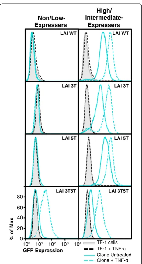
Nonexpressing 3T5T LAI LTR within TF-1 cells can be induced to drive GFP expression
Because of the expression phenotypes observed with the nonexpressers and intermediate expressers, it was of inter-est to determine whether stimulation of the LTR with in-flammatory cytokines could induce these LTRs to drive GFP expression. If so, this might represent a model for
“promoter latency and reactivation”within the bone mar-row progenitor cell type with LTRs containing naturally occurring SNPs. TF-1 cell clones were treated with TNF-α (at 20, 50, 100, 200, or 300 ng/mL) to determine whether inflammatory cytokine induction of the LTR could drive GFP expression in these cells. Clones that had higher levels of GFP expression were used as controls to show that the LTR was inducible through treatment. The titra-tion of TNF-α concentrations resulted in very similar levels of LTR-induced GFP expression (data not shown). Therefore, in this and subsequent experiments, only repre-sentative data showing the results of the TNF-αtreatment at 20-ng/mL were shown (Figure 6). Under stimulated condi-tions, the high-expressing TF-1 clones containing the LAI WT or 5 T LTR drove a higher level of GFP expression than the same unstimulated LTRs (Figure 6). In contrast, under the same conditions, the TF-1 nonexpressing clones contain-ing the LAI WT or 5 T LTRs were not stimulated to drive GFP expression. Stimulated nonexpressing TF-1 clones con-taining the LAI 3 T LTR were not induced into driving higher levels of GFP expression; however, the intermediate-expressing clone used as a control was induced and was thereby able to drive a higher level of GFP expression. Inter-estingly, the difference in MFI values between stimulated and unstimulated GFP levels of expression was greater with the clone containing the 3 T variant than the difference between stimulated and unstimulated MFI observed with the WT and 5 T LTRs. As shown in Figure 5B, the 3T5T LAI LTR re-sulted in both a population and clones that had intermediate levels of LTR-driven GFP expression. Therefore, for this experiment, a low-expressing clone and an intermediate-expressing clone were selected to represent non- and high-expressing clones, respectively. Under stimulated conditions, the low- and intermediate-expressing LTRs drove a higher level of GFP expression than in unstimulated conditions within the TF-1 cell background. However, the low-expressing 3T5T cell clone had a larger difference in MFI between unstimulated and stimulated conditions when com-pared with the intermediate-expressing cell clone (Figure 6).
U-937 clones containing nonexpressing LTR phenotypes cannot be induced into driving GFP expression
GFP expression using flow cytometry. Figure 7 shows representative histograms from both nonexpressing and expressing phenotypes within each backbone expressed within the stably transfected U-937 cell clones. With
TNF-αstimulation, the high-expressing cell clone containing the
LAI 3 T LTR drove a higher level of GFP expression than the same unstimulated LTR (Figure 7). In contrast, under the same conditions, the nonexpressing clone containing the LAI 3 T LTR was not stimulated to drive GFP ex-pression. Stimulated nonexpressing clones containing
Name Non/low-expresser Intermediate-expresser High-expresser Total
LAI WT 4 0 1 5
LAI 3T 3 3 0 6
LAI 5T 2 0 2 4
LAI 3T5T 3 2 0 5
100
High-expresser
Name Non/low-expresser Intermediate-expresser High-expresser Total
LAI WT 10 1 0 11
LAI 3T 3 1 2 6
LAI 5T 2 2 0 4
LAI 3T5T 0 3 0 3
87.4
High-expresser
54.4
Intermediate-expresser
100
80
60
40
20
% of Max
100 101 102 103 104
GFP Expression 1.1
Non/low-expresser
U-937 U-937 LAI clone
80
60
40
20
0
100 104
2.53
101 103
Non/low-expresser
TF-1 TF-1 LAI clone
102
GFP Expression
Intermediate-expresser
% of Max
A. Determination of expression level groups for cell clones
B. Expression groups of TF-1 cell clones
C. Expression groups of U-937 cell clones
Name Non/low-expressers Intermediate-expressers High-expressers
MFI range 0-15 16-50 >50
% positive cells range 0%-10% 11%-70% 71%-100%
0
97.5 80.7 19.3 0.9 99.1
98.9 45.5 12.6
TF-1 cells TF-1 + TNF-Clone Untreated Clone +
TNF-80
60
40
20
0
100 101 102 103 104
% of Max
GFP Expression Non/Low- Expressers
High/ Intermediate-
Expressers LAI WT
LAI 3T
LAI 5T
LAI WT
LAI 3T
LAI 5T
LAI 3T5T LAI 3T5T
Figure 6Nonexpressing TF-1 cell clones containing the 3T5T LAI long terminal repeat (LTR) could be induced into
LTR-driven green fluorescent protein (GFP) expression.TF-1 cells stably transfected with the HIV-1 LAI LTR (wild type [WT], 3 T, 5 T, and 3T5T) were serially diluted in order to obtain 1 cell in 1 mL of media (approximately 1 cell in 10 wells of a 96-well plate). Cell clone populations were propagated from the single cell and then were analyzed using flow cytometry for their basal GFP expression. The clonal populations were then designated in one of three categories (nonexpresser, intermediate expresser, and high expresser) based on their geometric mean fluorescence intensity (MFI) and their percent cell positive values (Figure 5A). Non/low-expressing and high-expressing LAI WT and LAI 5 T LTR containing clones were treated with a range of tumor necrosis factor-α(TNF-α) concentrations (20–300 ng/mL). Representative histograms (at a TNF-αconcentration of 20 ng/mL) showing levels of GFP expression obtained with the untreated, stably transfected cell clone (solid turquoise line) compared with the treated, stably transfected cell clone (dashed turquoise line), untreated WT TF-1 cells (solid black line), and treated WT TF-1 cells (dashed black line). Each clone shown is a representative of the group.
Non/Low- Expressers
% of Max
GFP Expression
U-937 cells U-937 + TNF-Clone Untreated Clone +
TNF-High/ Intermediate-
Expressers
80
60
40
20
0
100 101 102 103 100
LAI WT
LAI 3T
LAI 5T
LAI WT
LAI 3T
LAI 5T
LAI 3T5T
the LAI WT and 5 T LTRs were also not induced into driving higher levels of GFP expression; however, the intermediate-expressing clones that were used as con-trols for each backbone were able to be induced and drive higher levels of GFP expression. Interestingly, the low-intermediate-expressing phenotype within the U-937 LAI WT cell clone had the largest difference in unstimu-lated versus stimuunstimu-lated MFIs (33.4 and 80.7, respectively) and percent positive GFP-expressing cell number (9.67% and 98.5%, respectively). As shown in Figure 5C and as one might expect, the 3T5T LAI LTR resulted in clones that had varying intermediate levels of LTR-driven GFP expres-sion. Therefore, for this experiment, only one intermediate-expressing clone was selected to represent a high-intermediate-expressing phenotype (Figure 7, bottom panels). Under stimulated con-ditions, the intermediate clone exhibited a transition into high-GFP-expressing cells, in comparison to the stimulated levels of the expressing clones containing the WT, 3 T, or 5 T LTRs.
Discussion
Genetic variation within the HIV-1 genome is a naturally occurring phenomenon because of the low fidelity of re-verse transcriptase as well as selective pressures present within the host [17,18]. These viral quasispecies are po-tentially able to establish niches and reservoirs within HPCs, cells of the monocyte-macrophage lineage, as well as T cells that can reseed infection into the periphery and CNS of infected patients and possibly other end organs as well [20-28]. Nucleotide changes within the NF-κ B-proximal C/EBP and Sp transcription factor binding sites within the viral promoter have been observed in both the pre-HAART and HAART eras and have been shown to guide differential regulation of viral expression by the LTR [20-22,55-57]. Interestingly, the prevalence of the 3 T, 5 T, and 3T5T LTR variants in the HAART era, at least in the DREXELMED HIV/AIDS Genetic Analysis Cohort is lower when compared to the levels observed from published studies in the pre-HARRT era [23-27,37]. However, the DREXELMED HIV/AIDS Genetic Analysis Cohort is over-all much healthier because of the prolonged use of combin-ation antiretroviral therapy. However, we hypothesize that the prevalence of the 3 T, 5 T, and 3T5T LTR variants will become more prevalent with the development of greater disease severity across this HIV-1-infected patient popula-tion. In fact, one of the patients that has shown severe CD4 decline and neurologic impairment carries both of these variations as we have previously published [19].
The TF-1, U-937, and Jurkat T-cell lines were used as models of HPCs, monocytes, and CD4+ T cells, respect-ively, in order to study the effects of the previously de-scribed SNPs on LTR-driven gene expression within a chromatin-based microenvironment. Each cell type was stably transfected with a specific LTR variant of interest
and then stored at low temperature for prolonged pe-riods prior to experimentation. Flow cytometric analysis showed that subsequent recovery of the stored cell pop-ulations containing the HIV-1 variant LTR resulted in two different LTR-driven GFP expression phenotypes with each model cell line (TF-1, U-937, and Jurkat) and parental or variant LTR (3 T, 5 T, and 3T5T) (Figure 2). We theorized that once multiple cell samples were stored at low temperature for prolonged periods, the same ex-pression phenotype would be maintained for each sample that was recovered from storage and passaged to approxi-mately the same level. However, as shown in Figure 2, this was not the case; several phenotypes were associated with the specific genotypic changes within the LTRs stably transfected across all model cell lines. Alterations in gene expression from cells recovered from low-temperature storage could be caused by differences in the stably trans-fected cells that survive the thaw and reculturing process. Cell death is a major problem when the cell line popula-tions are thawed and recultured, as cells with different phenotypes may be the ones surviving each time this cycle occurs. The cell clones that have very distinct expression profiles, and maintain their expression profiles during subsequent thaws and reculture from low-temperature storage, provide further evidence of this phenomenon. In another sense, a recent study showed that when rheuma-toid arthritis synovial fibroblast (RASF) cells were in cul-ture, as the culture passages increased from 2 to 8, up to 10% of the genes within this cell line are differentially expressed [58]. In contrast to our observations, however, after long-term storage, gene expression within the RASF after passage 2 was comparable to gene expression prior to freezing [58]. This is the first study to show that long-term storage of stably transfected cells of the HPC, mono-cytic, and T-cell lineages can result in alterations in gene expression patterns.
HIV/AIDS Genetic Analysis Cohort) could be involved in evading immune activation within the host under basal conditions, under stimulatory conditions it might be acti-vated to produce a higher level of gene expression and virus production. Additionally, TNF-α stimulation of the stably transfected TF-1 and U-937 cells (Figure 3) sug-gested that a 3T5T LTR-containing proviral DNA could remain quiescent under certain metabolic conditions or in a basal viral gene expression mode. Only minimal intracel-lular stimulatory conditions would be required to drive low levels of viral gene expression and virus production within these cell populations, allowing evasion of HAART elimination of the infecting cell. The viral genome con-taining the 3T5T LTR could promote the evasion of ef-fective HAART elimination, potentially contributing to the development of a niched viral reservoir for virus with specific genetic signatures that favor the quiescent or low-level gene expression mode. Subsequently, when an in-fected patient has a proinflammatory response to either the HIV-1 infection or an opportunistic infection, the 3T5T double-variant LTR-containing viral genotype could again result in an activation of the viral genome from a la-tent viral reservoir. This conclusion has been based on the increase in NF-κB binding to the 3T5T double variant and enhanced operation of these low-affinity C/EBP and Sp binding sites in the presence of elevated levels of the corresponding activation factors and a repopulation of cir-culating virus within the patient’s peripheral blood and lymphoid tissues and within specialized reservoirs. These changes would thereby make HAART less effective in re-moving and reducing virus from within the immune and nervous systems as well as other end organs.
The cell clones from each stably transfected cell line containing the WT, 3 T, 5 T, or 3T5T LTRs were devel-oped to facilitate studies to explain the observable differ-ences between each of the LTRs within each cell line. The low-GFP-expressing 3T5T LTR-containing TF-1 cell clone exhibited a larger difference in MFI between un-stimulated and un-stimulated conditions compared with the intermediate-GFP-expressing cell clone. This observation may suggest that virus containing the 3T5T LTR genotype with a low enough expression profile in the body to facili-tate evasion of HAART and the immune response, could, under stimulated conditions, produce a large amount of virus, reseeding infection into the body, possibly with viral strains that may be more resistant to treatment. These ob-servations suggest that across LTRs containing each of the SNPs of interest (3 T, 5 T, and 3T5T), there is a differen-tial expression within individual GFP-expressing clones. These results also show that within an LTR, in this case the 3T5T-containing LAI LTR, stimulation by cytokines naturally occurring during inflammation and infection can lead to differential regulation of the LTR between different cell lines. In addition, non-GFP-expressing cell clones,
regardless of the LTR or cell type, could not be induced into expression with TNF-α stimulation, whereas the intermediate- and high-GFP-expressing cell clones con-taining each SNP of interest could be induced. This important result shows that differences in regulation of LTR-driven expression may also be attributable to the lo-cation of integration within the human genome, proviral DNA copy number, or possibly differences in epigenetic controls operative on the LTRs.
Conclusions
The experimental approach used in this study has pro-vided a means for understanding SNP-specific LTR func-tion within models of HIV-1-susceptible cells. However, this approach has not been without its limitations. In the model system used in this study, the function of the LTR is only observed within the context of an integrated plas-mid. In addition, only the LAI backbone is used, which has been derived from an X4-tropic virus. The LAI back-bone was used because (1) it has been commonly used in other scientific studies, (2) across the 12 binding sites within the LTR, it shares sequence similarity with the conB sequence except that it contains a 6G change within C/EBP site I, and (3) full-length HIV-1 LAI virus has the ability to infect all three cell line models used in this study. It would be interesting to determine the effects of the other LTR backbones, such as YU-2, an R5 virus, and 89.6, which has been defined to be an X4/R5 virus, on SNP-specific LTR driven gene expression within stably transfected TF-1, U-937, and Jurkat cells. It would also be important to determine whether the effects of these SNPs on LTR-driven gene expression observed within the stably transfected cells used in this study would remain if full-length pseudotype virus developed from molecular clones using the different HIV-1 backbones were used to infect the HPCs, monocytes, and T cells.
of virus and spectrum of viral quasispecies in the per-iphery which could potentially cross the BBB reseeding virus in the CNS.
Methods Cell culture
The TF-1 CD34+erythromyeloid leukemia cell line (ATCC, Manassas, VA) was grown in RPMI 1640 medium with L-glutamine (Cellgro, Herndon, VA) supplemented with 10% heat-inactivated fetal bovine serum (FBS; GemCell, West Sacramento, CA), antibiotics (penicillin and strepto-mycin, at a concentration of 0.04 mg/mL each; Cellgro), glucose (4.5 g/mL; Cellgro), sodium pyruvate (1 mM; Cellgro) and HEPES (10 mM; Cellgro), and recombinant human granulocyte-macrophage colony stimulating factor (2 ng/mL; eBioscience, San Diego, CA). The cells were maintained at a density between 1–5 × 10⁵cells/mL.
The U-937 human promonocytic cell line (ATCC, CR1593.2) was grown in RPMI 1640 medium with L-glutamine supplemented with 10% heat-inactivated FBS, antibiotics (penicillin and streptomycin, at a concentra-tion of 0.04 mg/mL each), glucose (4.5 g/mL), sodium pyruvate (1 mM), and HEPES (10 mM). The cells were maintained at a density between 1–5 × 10⁵cells/mL.
The human T-cell line Jurkat (ATCC, TIB-152) was grown in RPMI 1640 medium with L-glutamine supple-mented with 10% heat-inactivated FBS, sodium bicar-bonate (0.05%), and antibiotics (penicillin, streptomycin, and kanamycin at 0.04 mg/mL each). The cells were main-tained at a density between 1 × 10⁵ and 1 × 10⁶cells/mL as recommended by ATCC. All cells were maintained at 37°C in 5% CO2at 90% relative humidity.
Plasmid construction and site-directed mutagenesis The HIV-1 LAI LTR sequence was amplified by PCR from previously published LAI-LTR-Luc reporter con-structs [59,60]. The following primers were used for the amplification of LAI LTR: 5′
-GGC-CGG-ATT-AAT-TGG-AAG -GGC-TAA-TTC-ACT-CC-3′ and 5′
-GAC-CGG-TTG-CTA-GAG-ATT-TTC-CAC-AC T-G-3′ (Integrated
DNA Technologies). Following amplification, the HIV-1 LAI LTR was cloned into the pEGFP-N1 vector (Clontech, Mountain View, CA), which contains the eGFP gene. The PCR product and the pEGFP-N1 vector were digested with theVspI (AseI) and theBshTI (AgeI) restriction en-zymes and the digested PCR product was ligated into the digested pEGFP-N1 vector, generating the LAI LTR-GFP reporter construct.
Site-directed mutagenesis (SDM) was performed using the QuickChange mutagenesis procedure (Stratagene, La Jolla, CA) on the HIV-1 LAI LTR-GFP reporter construct to incorporate a C-to-T mutation at position 5 of the Sp site III (5 T). The following primers were used: CTT TCC
AGG GAG GTG TGG CCT GGG CG and CGC CCA
GGC CAC ACC TCC CTG GAA AG. The mutagenized
nucleotide is underlined. This process generated the LAI LTR-5 T-GFP reporter construct. SDM was also per-formed on the HIV-1 LAI LTR-GFP reporter construct to incorporate a C-to-T mutation at position 3 of the C/EBP site I (3 T). The following primers were used: GCT GAC
ATC GAGTTT GCT ACA AGG G and CCC TTG TAG
CAAACT CGA TGT CAG C. The mutagenized
nucleo-tide is underlined. This process generated the LAI LTR-3 T-GFP, which was used for an additional round of SDM to incorporate a C-to-T mutation at position 5 of the Sp site III (5 T) as indicated previously. This process gener-ated the LAI LTR-3T5T-GFP reporter constructs. For both the parental and mutated HIV-1 LTR constructs au-thenticity was confirmed by DNA sequencing (Genewiz, South Plainfield, NJ) and sequence analyses using Laser-gene software (DNASTAR, Inc., Madison, WI).
Stable transfection of TF-1, U-937, and Jurkat cells and stably transfected cell clone development
TF-1, U-937, and Jurkat cells (ATCC) were transfected with the constructs using the AMAXA Nucleofector System and AMAXA procedure V, as described by the manufacturer (Lonza, Basel, Switzerland). Transfected cells were passaged under G418 (800 ng/mL; Mediatech, Manassas, VA) selection beginning 24 hours after trans-fection and passaged under G418 selection for at least 2 months to develop stably transfected populations. The TF-1 and U-937 cell populations were each serially diluted to a final concentration of 1 cell/mL media and plated into 96-well plates, where they were marked and tracked for growth. Once clonal cells became a confluent clonal population within the well, they were moved to a larger well with more media until they could be transferred to flasks and propagated normally. Basal levels of LTR-driven GFP expression within each cell population and clonal population were measured using flow cytometry.
Assessment of GFP expression utilizing flow cytometry TF-1, U-937, and Jurkat cells were washed with flow cytometry buffer [Hanks balanced saline solution (Med-iatech), FBS (3%), and NaN3 (0.02%)] and aliquots of
1 × 10⁶ cells were fixed with paraformaldehyde (1%). Flow cytometry analysis was performed utilizing a Calibur Flow Cytometer (BD Biosciences, San Diego, CA) and an-alyzed the results using Flowjo version 8.8.7 software (Tree Star, Ashland, OR).
Oligonucleotide synthesis and radiolabeling
temperature as previously described [37]. Blunt-ended double-stranded oligonucleotides were end-labeled using
32
P-ATP and T4 polynucleotide kinase as described (Pro-mega, Madison, WI). The sequences of the probes used in this study are as follows: LAI LTR WT: TCG AGC TTG CTA CAA GGG ACT TTC CGC TGG GGA CTT TCC
AGG GAG GCG TGG CC; LAI LTR 3 T: TCG AGT
TTG CTA CAA GGG ACT TTC CGC TGG GGA CTT TCC AGG GAG GCG TGG CC; LAI LTR 5 T: TCG AGC TTG CTA CAA GGG ACT TTC CGC TGG GGA
CTT TCC AGG GAG GTG TGG CC; LAI LTR 3T5T:
TCG AGTTTG CTA CAA GGG ACT TTC CGC TGG
GGA CTT TCC AGG GAG GTG TGG CC. The SNPs
are indicated as underlined nucleotides.
Electromobility shift analysis
Electrophoretic mobility shift assay binding reactions were performed as previously described [37]. The radiolabeled LAI LTR probes were incubated with activated [TNF-α (20 ng/mL) for 24 hours] or normal nuclear extract from TF-1 cells for 30 minutes at 4°C in the presence of with poly[dI-dC)] (1μg), bovine serum albumin (BSA), and 1× binding buffer. Reactions (20μL, final volume) were sup-plemented with BSA (15 μg). The following antibodies (Santa Cruz Biotechnology, Santa Cruz, CA) were used to determine complex formation: NF-κB p50 (H-119X),
NF-κB p65 (C-20X). DNA-protein complexes were resolved at 4°C by electrophoresis for 3 hours at 200 V in a 4.5% non-denaturing polyacrylamide gel, which was then dried at 80°C for 2.5 hours and used for autoradiography.
Nuclear protein extraction from TF-1, U-937, and Jurkat cells Nuclear protein extraction was performed as previously described [61]. Briefly, 3 × 10⁷ cells were harvested for each nuclear extraction and washed once with 1× phosphate-buffered saline. The cells were incubated for 15 minutes with lysis buffer on ice and then 2 μL 10% NP-40 detergent was added and cells were inverted sev-eral times. Cells were centrifuged at 1100 RPM for 5 mi-nutes and then resuspended, through inverting, in lysis buffer and then centrifuged again. The nuclei pellets were then resuspended in nuclear extract buffer and then incubated for at 4°C for 30 minutes on a vortexer set to a speed of 3. The nuclear lysate was centrifuged at 14,000 RPM for 10 minutes at 4°C, after which time the supernatant was removed and quantitated with respect to protein concentration as previously described [62]. Nuclear extracts were then stored at−80°C.
Abbreviations
BBB:Blood–brain barrier; BSA: Bovine serum albumin; C/EBP: CCAAT/ enhancer binding protein; CNS: Central nervous system; FBS: Fetal bovine serum; GFP: Green fluorescent protein; HAART: Highly active antiretroviral therapy; HIVD: HIV-associated dementia; HPC: Hematopoietic progenitor cell; LTR: Long terminal repeat; MFI: Mean fluorescent intensity; NF-κB: Nuclear factor-κB; RASF: Rheumatoid arthritis synovial fibroblast; SNP:
Single-nucleotide polymorphism; TNF-α: Tumor necrosis factor-α; WT: Wild type.
Competing interests
The authors declare that they have no competing interests.
Authors’contributions
SS contributed to the development of stably transfected cell lines, developed the stably transfected cell clones, completed all flow cytometric acquisition and analysis, contributed to study conception and design, drafted figures, and drafted the manuscript. KA constructed the LTR-plasmid constructs and contributed to the development of stably transfected populations. SD contributed to the design and completion of the electrophoretic mobility shift assays. VP helped draft the manuscript. SS, MN and BW conceived of the study and participated in its design and coordination and helped to draft the manuscript. All authors read and approved the final manuscript.
Acknowledgments
These studies were funded in part by the Public Health Service, National Institutes of Health, through grants from the National Institute of Neurological Disorders and Stroke, NS32092 and NS46263, the National Institute of Drug Abuse, DA19807 (Dr. Brian Wigdahl, Principal Investigator), and under the Ruth L. Kirschstein National Research Service Award 5T32MH079785 (Jay Rappaport, PI, Brian Wigdahl PI of the Drexel subcontract). The contents of the paper are solely the responsibility of the authors and do not necessarily represent the official views of the NIH.
Received: 8 August 2013 Accepted: 25 April 2014 Published: 16 May 2014
References
1. Han Y, Wind-Rotolo M, Yang HC, Siliciano JD, Siliciano RF:Experimental approaches to the study of HIV-1 latency.Nat Rev Microbiol2007,5:95–106. 2. Marcello A:Latency: the hidden HIV-1 challenge.Retrovirology2006,3:7. 3. Ho DD, Neumann AU, Perelson AS, Chen W, Leonard JM, Markowitz M:
Rapid turnover of plasma virions and CD4 lymphocytes in HIV-1 infection.Nature1995,373:123–126.
4. Zack JA, Arrigo SJ, Weitsman SR, Go AS, Haislip A, Chen IS:HIV-1 entry into quiescent primary lymphocytes: molecular analysis reveals a labile, latent viral structure.Cell1990,61:213–222.
5. McElrath MJ, Pruett JE, Cohn ZA:Mononuclear phagocytes of blood and bone marrow: comparative roles as viral reservoirs in human immunodeficiency virus type 1 infections.Proc Natl Acad Sci U S A1989, 86:675–679.
6. Weissman D, Daucher J, Barker T, Adelsberger J, Baseler M, Fauci AS:Cytokine regulation of HIV replication induced by dendritic cell-CD4-positive T cell interactions.AIDS Res Hum Retroviruses1996,12:759–767.
7. Bukrinsky MI, Sharova N, Dempsey MP, Stanwick TL, Bukrinskaya AG, Haggerty S, Stevenson M:Active nuclear import of human
immunodeficiency virus type 1 preintegration complexes.Proc Natl Acad Sci U S A1992,89:6580–6584.
8. Folks TM, Justement J, Kinter A, Schnittman S, Orenstein J, Poli G, Fauci AS: Characterization of a promonocyte clone chronically infected with HIV and inducible by 13-phorbol-12-myristate acetate.J Immunol1988, 140:1117–1122.
9. O'Brien TR:HIV-2 transmission: implications for spread of HIV-1.JAMA
1994,271:903–904.
10. Diamond TL, Roshal M, Jamburuthugoda VK, Reynolds HM, Merriam AR, Lee KY, Balakrishnan M, Bambara RA, Planelles V, Dewhurst S, Kim B: Macrophage tropism of HIV-1 depends on efficient cellular dNTP utilization by reverse transcriptase.J Biol Chem2004,279:51545–51553. 11. Nicholson JK, Cross GD, Callaway CS, McDougal JS:In vitro infection of
human monocytes with human T lymphotropic virus type III/ lymphadenopathy-associated virus (HTLV-III/LAV).J Immunol1986, 137:323–329.
13. Shah S, Nonnemacher MR, Pirrone V, Wigdahl B:Innate and adaptive factors regulating human immunodeficiency virus type 1 genomic activation.J Neuroimmune Pharmacol2010,5:278–293.
14. Strazza M, Pirrone V, Wigdahl B, Nonnemacher MR:Breaking down the barrier: the effects of HIV-1 on the blood–brain barrier.Brain Res2011, 1399:96–115.
15. Fischer-Smith T, Croul S, Adeniyi A, Rybicka K, Morgello S, Khalili K, Rappaport J: Macrophage/microglial accumulation and proliferating cell nuclear antigen expression in the central nervous system in human immunodeficiency virus encephalopathy.Am J Pathol2004,164:2089–2099.
16. Glass JD, Fedor H, Wesselingh SL, McArthur JC:Immunocytochemical quantitation of human immunodeficiency virus in the brain: correlations with dementia.Ann Neurol1995,38:755–762.
17. Coffin JM:HIV population dynamics in vivo: implications for genetic variation, pathogenesis, and therapy.Science1995,267:483–489. 18. Gao F, Robertson DL, Morrison SG, Hui H, Craig S, Decker J, Fultz PN,
Girard M, Shaw GM, Hahn BH, Sharp PM:The heterosexual human immunodeficiency virus type 1 epidemic in Thailand is caused by an intersubtype (A/E) recombinant of African origin.J Virol1996, 70:7013–7029.
19. Li L, Aiamkitsumrit B, Pirrone V, Nonnemacher MR, Wojno A, Passic S, Flaig K, Kilareski E, Blakey B, Ku J, Parikh N, Shah R, Martin-Garcia J, Moldover B, Servance L, Downie D, Lewis S, Jacobson JM, Kolson D, Wigdahl B:Development of co-selected single nucleotide polymorphisms in the viral promoter precedes the onset of human immunodeficiency virus type 1-associated neurocognitive impairment.Journal of neurovirology2011,17:92–109.
20. Berkhout B, Jeang KT:Functional roles for the TATA promoter and enhancers in basal and Tat-induced expression of the human immunodeficiency virus type 1 long terminal repeat.J Virol1992,66:139–149.
21. Quinones-Mateu ME, Mas A:Lain de Lera T, Soriano V, Alcami J, Lederman MM, Domingo E: LTR and tat variability of HIV-1 isolates from patients with divergent rates of disease progression.Virus Res1998,57:11–20.
22. Zhang S, Pointer D, Singer G, Feng Y, Park K, Zhao LJ:Direct binding to nucleic acids by Vpr of human immunodeficiency virus type 1.
Gene1998,212:157–166.
23. Nonnemacher MR, Irish BP, Liu Y, Mauger D, Wigdahl B:Specific sequence configurations of HIV-1 LTR G/C box array result in altered recruitment of Sp isoforms and correlate with disease progression.J Neuroimmunol
2004,157:39–47.
24. Burdo TH, Gartner S, Mauger D, Wigdahl B:Region-specific distribution of human immunodeficiency virus type 1 long terminal repeats containing specific configurations of CCAAT/enhancer-binding protein site II in brains derived from demented and nondemented patients.J Neurovirol
2004,10(Suppl 1):7–14.
25. Burdo TH, Nonnemacher M, Irish BP, Choi CH, Krebs FC, Gartner S, Wigdahl B:High-affinity interaction between HIV-1 Vpr and specific sequences that span the C/EBP and adjacent NF-kappaB sites within the HIV-1 LTR correlate with HIV-1-associated dementia.DNA Cell Biol
2004,23:261–269.
26. Ross HL, Gartner S, McArthur JC, Corboy JR, McAllister JJ, Millhouse S, Wigdahl B:HIV-1 LTR C/EBP binding site sequence configurations preferentially encountered in brain lead to enhanced C/EBP factor binding and increased LTR-specific activity.J Neurovirol2001,7:235–249. 27. Ross HL, Nonnemacher MR, Hogan TH, Quiterio SJ, Henderson A,
McAllister JJ, Krebs FC, Wigdahl B:Interaction between CCAAT/enhancer binding protein and cyclic AMP response element binding protein 1 regulates human immunodeficiency virus type 1 transcription in cells of the monocyte/macrophage lineage.J Virol2001,75:1842–1856. 28. Carter CC, Onafuwa-Nuga A, McNamara LA:Riddell Jt, Bixby D, Savona MR,
Collins KL: HIV-1 infects multipotent progenitor cells causing cell death and establishing latent cellular reservoirs.Nat Med2010,16:446–451. 29. Burnett JC, Miller-Jensen K, Shah PS, Arkin AP, Schaffer DV:Control of
stochastic gene expression by host factors at the HIV promoter.
PLoS Pathog2009,5:e1000260.
30. Jordan A, Defechereux P, Verdin E:The site of HIV-1 integration in the human genome determines basal transcriptional activity and response to Tat transactivation.EMBO J2001,20:1726–1738.
31. Mitchell RS, Beitzel BF, Schroder AR, Shinn P, Chen H, Berry CC, Ecker JR, Bushman FD:Retroviral DNA integration: ASLV, HIV, and MLV show distinct target site preferences.PLoS Biol2004,2:E234.
32. Weinberger LS, Burnett JC, Toettcher JE, Arkin AP, Schaffer DV:Stochastic gene expression in a lentiviral positive-feedback loop: HIV-1 Tat fluctuations drive phenotypic diversity.Cell2005,122:169–182. 33. Schroder AR, Shinn P, Chen H, Berry C, Ecker JR, Bushman F:HIV-1
integration in the human genome favors active genes and local hotspots.Cell2002,110:521–529.
34. Koot M, Keet IP, Vos AH, De Goede RE, Roos MT, Coutinho RA, Miedema F, Schellekens PT, Tersmette M:Prognostic value of HIV-1 syncytium-inducing phenotype for rate of CD4+ cell depletion and progression to AIDS.
Ann Intern Med1993,118:681–688.
35. Nielsen C, Pedersen C, Lundgren JD, Gerstoft J:Biological properties of HIV isolates in primary HIV infection: consequences for the subsequent course of infection.AIDS1993,7:1035–1040.
36. Corboy JR, Buzy JM, Zink MC, Clements JE:Expression directed from HIV long terminal repeats in the central nervous system of transgenic mice.
Science1992,258:1804–1808.
37. Hogan TH, Stauff DL, Krebs FC, Gartner S, Quiterio SJ, Wigdahl B:Structural and functional evolution of human immunodeficiency virus type 1 long terminal repeat CCAAT/enhancer binding protein sites and their use as molecular markers for central nervous system disease progression.
J Neurovirol2003,9:55–68.
38. Hogan TH, Nonnemacher MR, Krebs FC, Henderson A, Wigdahl B:HIV-1 Vpr binding to HIV-1 LTR C/EBP cis-acting elements and adjacent regions is sequence-specific.Biomed Pharmacothe2003,57:41–48.
39. McAllister JJ, Phillips D, Millhouse S, Conner J, Hogan T, Ross HL, Wigdahl B: Analysis of the HIV-1 LTR NF-kappaB-proximal Sp site III: evidence for cell type-specific gene regulation and viral replication.Virology2000,274:262–277. 40. Berkhout B, Gatignol A, Rabson AB, Jeang KT:TAR-independent activation
of the HIV-1 LTR: evidence that tat requires specific regions of the promoter.Cell1990,62:757–767.
41. Harrich D, Garcia J, Mitsuyasu R, Gaynor R:TAR independent activation of the human immunodeficiency virus in phorbol ester stimulated T lymphocytes.EMBO J1990,9:4417–4423.
42. Gomez-Gonzalo M, Carretero M, Rullas J, Lara-Pezzi E, Aramburu J, Berkhout B, Alcami J, Lopez-Cabrera M:The hepatitis B virus X protein induces HIV-1 replication and transcription in synergy with T-cell activation signals: functional roles of NF-kappaB/NF-AT and SP1-binding sites in the HIV-1 long terminal repeat promoter.J Biol Chem2001,276:35435–35443. 43. Leonard J, Parrott C, Buckler-White AJ, Turner W, Ross EK, Martin MA, Rabson AB:
The NF-kappa B binding sites in the human immunodeficiency virus type 1 long terminal repeat are not required for virus infectivity.J Virol1989, 63:4919–4924.
44. Ross EK, Buckler-White AJ, Rabson AB, Englund G, Martin MA:Contribution of NF-kappa B and Sp1 binding motifs to the replicative capacity of human immunodeficiency virus type 1: distinct patterns of viral growth are determined by T-cell types.J Virol1991,65:4350–4358.
45. Kilareski EM, Shah S, Nonnemacher MR, Wigdahl B:Regulation of HIV-1 transcription in cells of the monocyte-macrophage lineage.Retrovirology
2009,6:118.
46. Choe H, Farzan M, Sun Y, Sullivan N, Rollins B, Ponath PD, Wu L, Mackay CR, LaRosa G, Newman W, Gerard N, Gerard C, Sodroski J:The beta-chemokine receptors CCR3 and CCR5 facilitate infection by primary HIV-1 isolates.
Cell1996,85:1135–1148.
47. Fouchier RA, Groenink M, Kootstra NA, Tersmette M, Huisman HG, Miedema F, Schuitemaker H:Phenotype-associated sequence variation in the third variable domain of the human immunodeficiency virus type 1 gp120 molecule.J Virol1992,66:3183–3187.
48. Zhu T, Mo H, Wang N, Nam DS, Cao Y, Koup RA, Ho DD:Genotypic and phenotypic characterization of HIV-1 patients with primary infection.
Science1993,261:1179–1181.
49. Griffin WS, Stanley LC, Ling C, White L, MacLeod V, Perrot LJ, White CL 3rd, Araoz C:Brain interleukin 1 and S-100 immunoreactivity are elevated in Down syndrome and Alzheimer disease.Proc Natl Acad Sci U S A1989, 86:7611–7615.
50. Akira S, Isshiki H, Nakajima T, Kinoshita S, Nishio Y, Natsuka S, Kishimoto T: Regulation of expression of the interleukin 6 gene: structure and function of the transcription factor NF-IL6.Ciba Found Symp1992, 167:47–62. discussion 62–47.
52. Henderson AJ, Zou X, Calame KL:C/EBP proteins activate transcription from the human immunodeficiency virus type 1 long terminal repeat in macrophages/monocytes.J Virol1995,69:5337–5344.
53. Connor RI, Mohri H, Cao Y, Ho DD:Increased viral burden and
cytopathicity correlate temporally with CD4+ T-lymphocyte decline and clinical progression in human immunodeficiency virus type 1-infected individuals.J Virol1993,67:1772–1777.
54. Parrott C, Seidner T, Duh E, Leonard J, Theodore TS, Buckler-White A, Martin MA, Rabson AB:Variable role of the long terminal repeat Sp1-binding sites in human immunodeficiency virus replication in T lymphocytes.J Virol1991,65:1414–1419.
55. Michael NL, D'Arcy L, Ehrenberg PK, Redfield RR:Naturally occurring genotypes of the human immunodeficiency virus type 1 long terminal repeat display a wide range of basal and Tat-induced transcriptional activities.J Virol1994,68:3163–3174.
56. Estable MC, Bell B, Merzouki A, Montaner JS, O'Shaughnessy MV, Sadowski IJ: Human immunodeficiency virus type 1 long terminal repeat variants from 42 patients representing all stages of infection display a wide range of sequence polymorphism and transcription activity.J Virol1996, 70:4053–4062.
57. Kirchhoff F, Greenough TC, Hamacher M, Sullivan JL, Desrosiers RC:Activity of human immunodeficiency virus type 1 promoter/TAR regions and tat1 genes derived from individuals with different rates of disease progression.Virology1997,232:319–331.
58. Neumann E, Riepl B, Knedla A, Lefevre S, Tarner IH, Grifka J, Steinmeyer J, Scholmerich J, Gay S, Muller-Ladner U:Cell culture and passaging alters gene expression pattern and proliferation rate in rheumatoid arthritis synovial fibroblasts.Arthritis Res Ther2010,12:R83.
59. Alexaki A, Quiterio SJ, Liu Y, Irish B, Kilareski E, Nonnemacher MR, Wigdahl B: PMA-induced differentiation of a bone marrow progenitor cell line activates HIV-1 LTR-driven transcription.DNA Cell Biol2007,26:387–394. 60. Krebs FC, Hogan TH, Quiterio S, Gartner S, Wigdahl B:Lentiviral LTR-directed
expression, sequence variation, and disease pathogenesis.Los Alamos National Laboratory HIV Sequence: Compendium; 2002.
61. Dignam JD, Lebovitz RM, Roeder RG:Accurate transcription initiation by RNA polymerase II in a soluble extract from isolated mammalian nuclei.
Nucleic Acids Res1983,11:1475–1489.
62. Bradford MM:A rapid and sensitive method for the quantitation of microgram quantities of protein utilizing the principle of protein-dye binding.Anal Biochem1976,72:248–254.
doi:10.1186/1743-422X-11-92
Cite this article as:Shahet al.:Functional properties of the HIV-1 long terminal repeat containing single-nucleotide polymorphisms in Sp site III and CCAAT/enhancer binding protein site I.Virology Journal201411:92.
Submit your next manuscript to BioMed Central and take full advantage of:
• Convenient online submission
• Thorough peer review
• No space constraints or color figure charges
• Immediate publication on acceptance
• Inclusion in PubMed, CAS, Scopus and Google Scholar
• Research which is freely available for redistribution