R E S E A R C H
Open Access
Impact of sustained RNAi-mediated suppression
of cellular cofactor Tat-SF1 on HIV-1 replication in
CD4+ T cells
Victoria A Green
1, Patrick Arbuthnot
1and Marc S Weinberg
1,2*Abstract
Background:Conventional anti-HIV drug regimens targeting viral enzymes are plagued by the emergence of drug resistance. There is interest in targeting HIV-dependency factors (HDFs), host proteins that the virus requires for replication, as drugs targeting their function may prove protective. Reporter cell lines provide a rapid and
convenient method of identifying putative HDFs, but this approach may lead to misleading results and a failure to detect subtle detrimental effects on cells that result from HDF suppression. Thus, alternative methods for HDF validation are required. Cellular Tat-SF1 has long been ascribed a cofactor role in Tat-dependent transactivation of viral transcription elongation. Here we employ sustained RNAi-mediated suppression of Tat-SF1 to validate its requirement for HIV-1 replication in a CD4+ T cell-derived line and its potential as a therapeutic target.
Results:shRNA-mediated suppression of Tat-SF1 reduced HIV-1 replication and infectious particle production from TZM-bl reporter cells. This effect was not a result of increased apoptosis, loss of cell viability or an immune
response. To validate its requirement for HIV-1 replication in a more relevant cell line, CD4+ SupT1 cell populations were generated that stably expressed shRNAs. HIV-1 replication was significantly reduced for two weeks (~65%) in cells with depleted Tat-SF1, although the inhibition of viral replication was moderate when compared to SupT1 cells expressing a shRNA targeting the integration cofactor LEDGF/p75. Tat-SF1 suppression was attenuated over time, resulting from decreased shRNA guide strand expression, suggesting that there is a selective pressure to restore Tat-SF1 levels.
Conclusions:This study validates Tat-SF1 as an HDF in CD4+ T cell-derived SupT1 cells. However, our findings also suggest that Tat-SF1 is not a critical cofactor required for virus replication and its suppression may affect cell growth. Therefore, this study demonstrates the importance of examining HIV-1 replication kinetics and cytotoxicity in cells with sustained HDF suppression to validate their therapeutic potential as targets.
Background
Current anti-HIV drug regimens target several viral enzymes simultaneously, with the aim of preventing the emergence of drug resistance. However, efficacy of these drugs is limited by the problems of emergence of drug resistance that results from viral diversity and mutability. Host factors required by the virus for replication, so-called HIV-dependency factors (HDFs), represent at-tractive therapeutic targets since their coding sequences
remain constant relative to the sequence variability of viral targets within a patient and across the pandemic.
Support for the notion that HDFs may be suitable therapeutic targets comes from a genome association study showing that single nucleotide polymorphisms in ZNRD1 are associated with slowed disease progression [1], and that a naturally occurring deletion in the CCR5 gene renders individuals resistant to an R5-tropic virus infection without associated physiological problems [2,3]. There have been several clinical trials showing the positive impact CCR5 deletion from CD4+ T cells has on T cell longevity, viral suppression and patient health (reviewed in [4]). This was most emphatically demon-strated by the apparent cure of the‘Berlin patient’[5-7].
* Correspondence:[email protected]
1Antiviral Gene Therapy Research Unit, Health Sciences Faculty, University of
the Witwatersrand, Johannesburg, South Africa
2Department of Molecular and Experimental Medicine, The Scripps Research
Institute, La Jolla, CA, USA
There is therefore interest in identifying other HDFs that modulate HIV infection since drugs inhibiting their function may prove protective.
A number of reporter cell lines have been developed as convenient laboratory tools for the quantification of HIV replication. When coupled with RNA interference (RNAi)-mediated gene silencing, these models provide a rapid method for the identification of putative HDFs. This approach has been employed in genome-wide stud-ies [8,9]. However, most putative HDFs identified by such approaches have yet to be validated in cells that are nat-urally infected by HIV. This is necessary as reporter cell lines may be misleading with respect to HDF importance, as exemplified in a study where only half of putative HDFs were validated as such in a T cell-derived line [10].
HIV-1 Tat-specific factor 1 (Tat-SF1) [NCBI RefSeq_ peptide: NP_055315] has long been a candidate HDF since its identification as a cofactor for Tat-dependent transactivation of viral transcription elongation [11-14]. Tat-SF1 is an RNA-binding protein [12] that functions as a transcription elongation and splicing factor of cellu-lar transcripts [15-17]. Most of the previous work on Tat-SF1 has focused on in vitro immunodepletion experiments of nuclear extracts. Other studies have demonstrated that RNAi-mediated suppression of Tat-SF1 inhibited HIV-1 replication in the HeLa-derived TZM-bl reporter cell line [8,18], mediated by a disrup-tion to splicing of viral transcripts [18]. However, it was unknown whether this protein functions as an HDF in cells that are a natural target of HIV and, if so, whether the long-term impact of suppressing Tat-SF1 adversely affects these cells.
In this study we examined the impact of Tat-SF1 sup-pression, mediated by anti-Tat-SF1 short hairpin RNAs (shRNAs), in both TZM-bl reporter cells and CD4+ T cell-derived SupT1 cell lines. Inhibition of Tat-SF1 ex-pression resulted in a significant inhibition of HIV-1 repli-cation, although this was less pronounced than when suppressing the known lentiviral integration cofactor LEDGF/p75 [19,20]. In addition, Tat-SF1 suppression was attenuated during serial passage of transduced SupT1 cell lines, suggesting that Tat-SF1 suppression may confer a growth disadvantage to cells and therefore preclude its utility as a therapeutic target. The approach used here demonstrates that thorough analysis is required for HDF validation and detection of subtle changes to cell physi-ology that may result from HDF inhibition.
Results
RNAi-mediated suppression of Tat-SF1 without cytotoxicity
RNAi effectors, such as shRNAs, may be exploited to validate roles of HDFs. To suppress expression of en-dogenous Tat-SF1, which is encoded by the HTATSF1
gene, three U6 RNA Polymerase (Pol) III shRNA expres-sion cassettes, shhtatsf1-a, shhtatsf1-b and shhtatsf1-c, were generated (Additional file 1A). The shRNA loop sequences were derived from micro RNA- (miR-) 31. Through the introduction of mismatches in the anti-guide strand, G:U wobbles were created to enhance the thermodynamic asymmetry of the shRNA stems and fa-cilitate intended mature guide strand bias [21-23].
Initial assessment of the ability of shRNAs to knock-down their cognate target sequences was made using a dual luciferase reporter assay. The three Tat-SF1 mRNA (htatsf1) target sites were inserted downstream of the
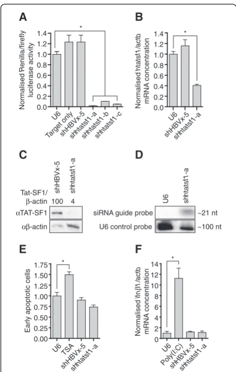
Renillaluciferase ORF within a psiCheck dual-luciferase plasmid. Ratios ofRenillato constitutively expressed fire-fly luciferase activities were used to assess efficiency of shRNA-mediated target knockdown. Allhtatsf1-targeted shRNAs significantly reduced Renilla/firefly luciferase activity ratios compared to controls ie cells receiving the U6 plasmid, a construct with shRNA expression targeting hepatitis B virus X protein (shHBVx-5) [24] or the psiCheck target construct only (>90% knock-down; Figure 1A). Greatest knockdown was observed with shhtatsf1-a, which effectively inhibited expression of the endogenous mRNA target in TZM-bl cells, as determined by quantitative reverse transcription PCR (qRT-PCR) (~60% knockdown; Figure 1B). Western blot analysis demonstrated that shhtatsf1-a expression also mediated a significant reduction in Tat-SF1 (4% of shHBVx-5 control; Figure 1C). Small RNA Northern blot detected the ~21 nt shhtatsf1-a guide strand (Figure 1D), confirming that the exogenous shRNA was processed as intended and that the observed suppres-sion of Tat-SF1 expressuppres-sion was mediated by an RNAi mechanism.
expression cassettes may be used to transiently silence Tat-SF1 expression without inducing apoptosis or an interferon response in TZM-bl cells.
Suppression of Tat-SF1 inhibits HIV-1 replication in reporter cells
The effects of Tat-SF1 silencing on HIV-1 replication were initially assessed in TZM-bl cells. HeLa-derived TZM-bl cells may be infected with HIV-1 to a similar extent to human peripheral blood mononuclear cells (PBMCs) because they express transgenic HIV receptor CD4 and coreceptor CCR5 [26-28]. Furthermore, TZM-bl cells permit relatively simple assessment of HIV-1 replication as they contain an integrated Tat-dependent luciferase reporter [26-28].
HIV-1 replication was quantified both by measure-ment of capsid protein p24 levels in culture supernatant and Tat-induced reporter gene activity (Figure 2A). Cells were transfected with the shhtatsf1-a expression con-struct, or controls, and infected 48 h later with virus derived from the HIV-1 subtype B molecular clone p81A-4 (HIV-1p81A-4) [29,30]. Tat-induced luciferase ac-tivity in cells with suppressed Tat-SF1 expression was ~20% of controls at 48 h after infection (Figure 2B). This effect was similar to that observed in cells expressing shTAT and shLTR-U5, previously developed shRNA ex-pression cassettes that directly target sequences within the Tat open reading frame (ORF) and U5 region of the viral leader transcripts, respectively [31,32]. Tat-SF1 sup-pression also reduced infectious particle production by ~70% (Figure 2C). Collectively these results confirm previ-ous reports that Tat-SF1 functions as an HDF in TZM-bl cells [8,18]. Given the limitations associated with transient host factor suppression for HDF validation, and the poten-tial bias of reporter output, the impact of sustained Tat-SF1 suppression on HIV-1 replication kinetics over a time course was investigated.
Stable expression ofhtatsf1-targeting shRNAs in SupT1
cells inhibits HIV-1 replication
The impact of sustained Tat-SF1 suppression on HIV-1 replication kinetics was assessed in CD4+ T cell-derived SupT1 cells [33], a model that more closely simulates natural HIV-1 infection than TZM-bl cells. An add-itional control shRNA was used, shpsip1-a, targeting the known HIV-1 cofactor LEDGF/p75 [20], which is encoded by thePSIP1 gene. U6 RNA Pol III shRNA ex-pression cassettes were incorporated into second-generation lentiviral vectors that also included a GFP reporter cassette. The dual luciferase reporter assay con-firmed that the shRNAs remained capable of target si-lencing within the context of the lentivector (Additional file 3A). Recombinant lentiviruses were then generated and used to transduce SupT1 cells at a multiplicity of
C
Target onlyshHBVx-5shhtatsf1 -a
Figure 1shRNAs suppress Tat-SF1 expression without
infection (MOI) of 0.15. After fluorescence activated cell sorting (FACS), a population of transduced SupT1 cells was propagated (Additional file 3B and C).
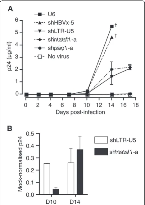
SupT1 cells with stable shRNA expression were infected with HIVp81A-4. HIV-1 p24 concentrations in culture supernatant were measured regularly during a period of 17 days to assess HIV-1 replication kinetics (Figure 3A). The concentration of p24 rose to ~5μg/ml on day 14 in the culture supernatant of control cells with no shRNA, or shHBVx-5, expression. No p24 meas-urement was made in these control cells on day 17 as a result of cell death from the high levels of virus replica-tion. In contrast, p24 levels in culture supernatant of cells expressing shpsip1-a were only detected on day 4, and never reached more than 0.1μg/ml during the time course, in accordance with the importance of LEDGF/ p75 in HIV-1 replication [20]. Culture supernatant of cells with shhtatsf1-a expression exhibited p24 levels of ~2 μg/ml on day 14 (Figure 3A), a reduction of ~65% compared with the U6 mock, which was similar to that observed with shLTR-U5 expression (Figure 3B). These data show that sustained Tat-SF1 suppression inhibits HIV-1 subtype B replication in a T cell-derived line, al-beit to a lesser extent than silencing of LEDGF/p75.
Tat-SF1 expression increases following serial passage of shhtatsf1-a-expressing SupT1 cells
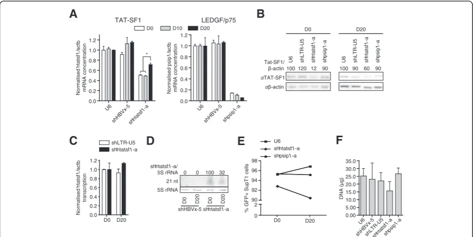
Close inspection of HIVp81A-4replication kinetics reveals that on day 14, p24 levels in shhtatfs1-a-‐expressing SupT1 cells, relative to the U6 control, were increased
compared with day 10 (~95% versus ~65% knockdown; Figure 3B). In contrast, the suppression of p24 levels in shLTR-U5-‐expressing cells was maintained at ~75%. The apparent attenuation of HIV-1 replication inhibition may result from adaptation of the virus to another cofac-tor, or may be a result of increased Tat-SF1 expression. However, cofactor adaptation is unlikely considering the duration of the assay. To determine whether there was increasing Tat-SF1 expression over the time course, SupT1 cell lines were raised and cultured for periods equivalent to the HIVp81A-4replication assay. The level of
htatsf1mRNA was suppressed throughout, compared to the U6 control, although htatsf1 mRNA concentration increased significantly from day 10 (~49%) to day 20 (~70%; Figure 4A). These results were corroborated by Western blot analysis of Tat-SF1 expression (Figure 4B). In contrast, the degree of suppression of psip1 mRNA was sustained in the shpsip1-a-expressing cell line throughout the time course (Figure 4A), demonstrating that the increase in shRNA target expression was specific to the shhtatsf1-a-expressing SupT1 cell line.
Several mechanisms, which are not mutually exclusive, may account for the observed increase in Tat-SF1 ex-pression during serial passage of SupT1 cells expressing shhtatsf1-a. These are: (1) increasedHTATSF1 transcrip-tion; (2) reduced shhtatsf1-a expression; and, (3) positive selection for untransduced cells in the population where there is no Tat-SF1 suppression. Nuclear run-on analysis revealed no alteration in HTATSF1 transcription rates, relative to transcription of ACTB, at day 20 compared with day 0 in SupT1 cells expressing shhtatsf1-a
A
B
C
Figure 2Tat-SF1 suppression inhibits HIV-1 infectious particle production from TZM-bl cells.2A. Schematic of the HIV-1 infection
protocol. 2B. TZM-bl cell lysates were analysed for luciferase activity 72 h post-transfection and 48 h post-infection with HIV-1p81A-4at a TCID 50of
(Figure 4C). Northern blot analysis showed that expres-sion of the shhtatsf1-a-derived guide strand was ~30% at day 20 of that detected on day 0 (Figure 4D), whereas the reduction in shpsip1-a-derived guide strand was less pronounced (~87% at day 20; Additional file 4). Flow cytometry on SupT1 cell lines over the time course showed that the GFP+ cells slightly diminished in the population transduced with shhtatsf1-a-expressing lenti-virus, in contrast to SupT1 populations with no shRNA, or shpsip1-a, expression (Figure 4E). The size of the population transduced with shhtatsf1-a-expressing len-tivirus was less than controls, as indicated by quanti-fication of extracted DNA, although not significant (Figure 4F). Collectively, these data demonstrate that the inhibition of HIV-1 replication on Tat-SF1 suppression is attenuated over time as a result of an increase in
Tat-SF1 expression. Such an increase is predominantly a re-sult of a decrease in shhtatsf1-a guide strand expression.
Discussion
Here we demonstrate that suppression of Tat-SF1 inhi-bits HIV-1 replication in both TZM-bl reporter cells and CD4+ T cell-derived SupT1 cells. Tat-SF1 has previously been shown to function as an HDF in TZM-bl cells [18], although we further demonstrated that the inhibitory effect on HIV-1 following RNAi-mediated Tat-SF1 sup-pression was not a result of cellular toxicity or induction of an immune response (Figure 1E and F) and includes inhibition of infectous particle production (Figure 2C). This study also examined the effect of sustained Tat-SF1 suppression on HIV-1 replication in T cell-derived SupT1 cells, a model that more closely mimics natural HIV-1 cellular targets. This approach had the added benefit of permitting quantification of HIV-1 replication kinetics for over two weeks. Tat-SF1 suppression inhib-ited HIV-1 replication in SupT1 cells throughout the time course (Figure 3A). Nevertheless, the inhibition of HIV-1 replication was modest compared with SupT1 cells with sustained suppression of the integration cofac-tor LEDGF/p75 (Figure 3A), suggesting that Tat-SF1 is a less critical HIV-1 cofactor than LEDGF/p75. This may be because Tat-SF1 is involved in increasing the effi-ciency of viral processes that still occur in its absence. This is consistent with its proposed function as one of a set of non-redundant RNA Pol II elongation factors that act cooperatively to facilitate efficient transcription elongation [16]. This is also consistent with observations that Tat-SF1 suppression results in a shift in the ratio of unspliced to spliced HIV-1 transcripts, but not complete loss of the spliced class [18]. These effects may be mediated by Tat-SF1 stabilising the large, multi-protein transcription elongation and splicing complexes [14], whilst not being critical for their activities. In contrast, our results confirm previous reports that LEDGF/p75 is a critical integration cofactor [20], and suggest that it is a good therapeutic target, as its suppression resul-ted in almost complete ablation of HIV-1 replication (Figure 3A). Indeed, there has been considerable pro-gress in developing LEDGF/p75-HIV-1 integrase inter-action inhibitors (reviewed in [34]).
Along with the limited inhibition of HIV-1 replication in SupT1 cells, other observations suggest that Tat-SF1 may not constitute a viable anti-HIV-1 therapeutic tar-get. Tat-SF1 suppression was attenuated over time in SupT1 cells (Figure 4A and B) as a result, at least in part, of decreased shhtatsf1-a guide strand expression (Figure 4D). This may arise from epigenetic silencing of the shRNA expression cassette, or untransduced cells (or transduced cells with low or no shhtatsf1-a expres-sion) within the population proliferating at a faster rate 0
1 2 3 4 5 6
p24 (µg/ml)
Days post-infection U6
shHBVx-5 shLTR-U5 shhtatsf1-a shpsip1-a No virus
0 2 4 6 8 10 12 14 16 18
†
†
shhtatsf1-a shLTR-U5
Mock-normalised p24
0.0 0.1 0.2 0.3 0.4 0.5
D10 D14
A
B
Figure 3Sustained Tat-SF1 suppression inhibits HIV-1
replication in CD4+ T cell-derived SupT1 cells.SupT1 cell lines, with stable shRNA expression generated by lentiviral transduction, were infected with HIV-1p81A-4at a TCID
50of 50/ml, in triplicate. 3A.
than those with shhtatsf1-a expression. Although these mechanisms are not mutually exclusive, our data favours the former as the primary mechanism for the reduction in guide strand expression, since the decrease in the per-centage of GFP+ SupT1 cells is less than the reduction in shhtatsf1-a guide strand expression (Figure 4D and E). Regardless, when compared to the other SupT1 populations, the reduction in guide strand and percent of GFP+ cells was specific to the shhtatsf1-a population (Figure 4D and E, and Additional file 4), implying there is a selective pressure on cells to restore Tat-SF1 expres-sion levels. Such a growth disadvantage on Tat-SF1 sup-pression would account for the small reduction in cell number within the population after serial passage (Figure 4F). This was not a significant difference, pos-sibly because of adaptation to increase Tat-SF1 levels (Figure 4B).
Reduced Tat-SF1 expression may confer a growth dis-advantage by disrupting expression of Tat-SF1 transcrip-tion and splicing targets, which have recently been shown to include genes involved in the cell cycle and nucleic acid metabolism [15]. Of course, reduced cell proliferation is not a desirable side effect, particularly in
immune cells, which may preclude Tat-SF1 inhibition as an anti-HIV therapeutic strategy. However, it has been demonstrated that cells with greater resistance to HIV-‐1 replication undergo preferential expansion in vivo [35]. Therefore, any growth disadvantage associated with Tat-SF1 suppression may be outweighedin vivoby a selective advantage in the context of an HIV-1 infection. Further experiments are needed to verify whether this is the case, but the observations reported here would certainly ex-clude prophylactic targeting of Tat-SF1. Nonetheless, as an HDF, Tat-SF1 expression heterogeneity should be con-sidered a possible HIV-1 susceptibility factor.
More generally, this study highlights the limitations associated with HDF validation in a reporter cell line. Although convenient, there may be bias toward host fac-tors with a more direct influence on reporter gene ex-pression. In addition, the expression levels of host factors differ between cell types, which may alter HIV-1 replication kinetics [36], particularly in reporter cell lines that are not derived from natural targets of HIV-1. Fur-thermore, measurement of TZM-bl reporter gene activ-ity requires cell lysis, preventing serial monitoring of HIV-1 replication and, as such, are most useful for
D0 D10 D20
Figure 4Attenuation of shRNA-mediated Tat-SF1 suppression over time.Samples were isolated from SupT1 cells with stable shRNA
expression at time points equivalent to those of the HIV-1p81A-4replication assay. 4A. Total SupT1 RNA was analysed by qRT-PCR, in triplicate.
Target mRNA levels are given relative toβ-actin mRNA (actb) normalised to the U6 cell line. Left panel: Tat-SF1 mRNA (htatsf1). Right panel: LEDGF/p75 mRNA (psip1). 4B. SupT1 cell lysates were subject to PAGE and Western blot. Day 20 samples were prepared in duplicate and representative blots are shown. Mean Tat-SF1 expression is given relative toβ-actin and normalised to the U6 control at each time point. 4C. Nuclei isolated from SupT1 cells were subject to nuclear run-on analysis to quantifyhtatsf1transcription, in triplicate. Samples from both shLTR-U5- and shhtatsf1-a-expressing cells were normalised to those isolated at a time point equivalent to day 0 of the HIV-1p81A-4replication
assay. 4D. Total SupT1 RNA was subject to small RNA PAGE and Northern blot to assess shhtatsf1-a guide strand expression relative to 5S rRNAs. 4E. Proportion of GFP+ SupT1 cells. SupT1 cell populations were analysed by flow cytometry with 5 × 10³events acquired per sample. 4F.
transient suppression experiments, which may lead to overlooking HDFs with long half-lives and not detect subtle detrimental effects on cell physiology resulting from HDF suppression. Thus the limitations of bias, cell type and transient suppression that are associated with reporter cell lines may cause a distortion in the relative importance and therapeutic potential of an HDF. This was observed in this study, where transient suppression-experiments in TZM-bl cells suggested that Tat-SF1 was a critical HIV-1 cofactor, in contrast to the findings from sustained suppression-experiments in SupT1 cells, an approach which is less subject to distortion from bias and cell type. Furthermore, this study reveals that Tat-SF1 suppression may confer a growth disadvantage only apparent on serial passage of cells. In contrast, previous reports that LEDGF/p75 comprises a promising thera-peutic target were confirmed. Overall this study provides an experimental template for the approach required to validate HDFs and the therapeutic potential of their tar-geting, and should be extended to putative HDFs identi-fied by genome-wide screens.
Conclusions
HDFs represent potential therapeutic targets and, as such, putative HDFs require validation. Focusing on the HIV-1 cofactor Tat-SF1, this study highlights the limita-tions associated with HDF validation in the TZM-bl re-porter cell line. We demonstrate an alternative approach for determining the impact that host factor suppression has on HIV-1 replication and cell physiology, which employs sustained RNAi-mediated host factor suppres-sion in a cell line derived from a physiological substrate of HIV-1. This approach was used to validate Tat-SF1 as an HDF in CD4+ T cell-derived SupT1 cells: sustained RNAi-mediated Tat-SF1 suppression inhibits HIV-1 rep-lication in SupT1 cells. However, the inhibitory effect was modest compared to cells with sustained suppres-sion of the integration cofactor LEDGF/p75, suggesting that Tat-SF1 is not a critical HIV-1 cofactor. Further-more, Tat-SF1 suppression is attenuated over time, sug-gesting that reduced Tat-SF1 levels confer a growth disadvantage to cells. Thus, whilst this study reveals that Tat-SF1 functions as an HDF in SupT1 cells, further studies are required to determine whether variants might modulate HIV-1 infection and its suppression would have a long-term inhibitory effect on HIV-1 repli-cationin vivo.
Methods
shRNA constructs
shRNAs shhtatsf1-a, shhtatsf1-b and shhtatsf1-c were designed to targethtatsf1transcript [NCBI RefSeq_RNA: NM_014500.3] at the sequences GCT ACA TAT CAG GCC AAT TAT, GCG CAT CTA GTT CTA CCG CAA
and CTG CAA CTG GAA TGG CGT T, respectively (Additional file 1). These target sites were selected from sequences suggested by The RNAi Consortium (www. broad.mit.edu/genome_bio/trc/rnai.html). All shRNAs were designed to contain a loop sequence derived from miR-31. G:U mismatches were incorporated at the 3’end of the anti-guide strand of some shRNAs to decrease thermo-dynamic stability of this end of the hairpin stem and favour selection of the intended guide strand. RNA Pol III U6 shRNA expression cassettes were generated by a two-step PCR approach described previously [37]. These were cloned into pTZ57R/T (Fermentas). Construct sequence was confirmed by automated cycle sequencing.
Several previously developed constructs were used as controls in experiments: a mock pTZU6+1 (U6) con-struct with no shRNA sequence [38]; a shRNA negative control, shHBVx-5, which targets an irrelevant site in hepatitis B virus (HBV) X protein [24]; and, two positive controls, shLTR-U5 and shTAT, which are named after the location of their target sequences within HIV-1 tran-scripts and were initially developed based on subtype B molecular clone HXB2 [GenBank: K03455] [31,32]. shRNA shpsip1-a was adapted from a guide strand pre-viously shown to inhibit LEDGF/p75 expression [20] that targets the p75 isoform of psip1 transcript [NCBI RefSeq_RNA: NM_033222.2] at the sequence GAC AGC ATG AGG AAG CGA A.
Cell culture and transfections
HeLa-derived TZM-bl cells (NIH AIDS Research and Reference Reagent Program), which express the HIV re-ceptor CD4 and corere-ceptor CCR5 and contain a lucifer-ase reporter driven by a Tat-inducible LTR promoter derived from pSG3.1 [GenBank: L02317] [26-28], were maintained in Dulbecco’s Modified Eagle’s Medium (DMEM), supplemented with 10% heat-inactivated fetal calf serum (FCS), at 37°C and 5% CO2. HEK293T, HeLa and SupT1 cells (NIH AIDS Research and Reference Reagent Program), the latter a non-Hodgkin’s T cell lymphoma suspension cell line expressing high levels of surface CD4 [33], were maintained in the same media.
Transfections were carried out using 1 μl Lipofecta-mine2000 (Invitrogen) to 1 μg DNA, according to manufacturer’s instructions. Medium was changed 5 h post-transfection. Where appropriate, a plasmid with constitutive eGFP expression (pCI-eGFP) was cotrans-fected followed by fluorescence microscopy 48 h later to verify equivalent transfection efficiencies [39].
Dual luciferase reporter assay
directly into theXhoI-NotI sites of psiCheck2. AnEcoRV site was inserted within each annealed dsDNA insert to facilitate screening. The oligonucleotides used for psi-Check htatsf1 were: TCG AGA TAT CGC TAC ATA TCA GGC CAA TTA TGC GCA TCT AGT TCT ACC GCA AAC TGC AAC TGG AAT GGC GTT GC; and, CTA GAT GCG CAT AAT TGG CCT GAT ATG TAG CGA TAT CGG CCG CAA CGC CAT TCC AGT TGC AGT TTG CGG TAG AA; and, for psiCheckpsip1were: TCG AGA TAT CAG ACA GCA TGA GGA AGC GAA GCA GCT ACA GAA GTC AAG ATT GC; and, GGC CGC AAT CTT GAC TTC TGT AGC TGC TTC GCT TCC TCA TGC TGT CTG ATA TC. Target constructs psiCheck HBVx [40] and psiCheck LTR [31] have been described previously.
HeLa or HEK293T cells were seeded at 5.0 × 10⁴and 1.2 × 10⁵cells per well, respectively, in a 24-well culture plate and transfected 24 h later with 500 ng shRNA expression construct, 100 ng of psiCheck target reporter construct and 10 ng pCI-eGFP, in triplicate. Firefly and
Renilla luciferase activities were determined 48 h later using the Dual Luciferase Reporter Assay System (Pro-mega) and a Veritas dual-injection luminometer (Turner Biosystems), according to manufacturer’s instructions.
Renilla: firefly luciferase activity ratios were normalised to the U6 control mean.
Quantitative RT-PCR of cellular factor mRNAs
TZM-bl cells were seeded at 5.0 × 10⁴cells per well in a 24-well culture plate and transfected 24 h later with 500 ng shRNA expression construct and 10 ng pCI-eGFP, in triplicate. Total TZM-bl cellular RNA was extracted using TriReagent (Sigma-Aldrich) 48 h later, or from stably transduced SupT1 cells cultured for periods equivalent to days 0, 10 and 20 of the HIV-1 replication assay (see below). Total RNA was subjected to DNase treatment (Promega) and random-primed reverse-transcription using the SuperScript III reverse transcriptase (RT) (Invitrogen). cDNA was analysed for target mRNA expression relative to β-actin mRNA (actb) transcript NM_01101.2 using the SensiMix Lite Kit (Quantace) with the following pri-mers:htatsf1forward AGTGGGACCTGGACAAAAAGG;
htatsf1 reverse GTT CCG GGG CTT TTT CTT GTG;
psip1 forward GCT GAA CAA AGA CAG CAT GAG
GA;psip1 reverse ATT GCT CTC CCC GTT ATG TTG
TG; actbforward AGG TCA TCA CCA TTG GCA ATG AG; and, actb reverse TCT TTG CGG ATG TCC ACG TCA. The qPCR was performed in a Carousel-based Light-cycler V.2 System (Roche) with the following parameters: denaturation at 95°C for 10 min, 50 cycles of denaturation at 95°C, annealing at 60°C and extension at 72°C, each for 10 s. Amplification cycles were followed by melting curve analysis to verify the specificity of the PCR products. No RT controls were included for each sample and no cDNA
controls for each primer set. Target mRNA: actb ratios were normalised to the mean expression ratio of U6-transfected samples.
Western blot
TZM-bl cells were seeded at 1.5 × 10⁵cells per well in a 6-well culture plate and transfected 24 h later with 2μg shRNA expression construct and 10 ng pCI-eGFP. Cells were harvested 72 h post-transfection and lysed with RIPA buffer. Total protein was quantified using the BCA Protein Assay Kit (Pierce). A ladder composed of IgG-binding proteins ranging from 22 to 120 kDa in size and 80μg of samples were resolved on a 12% polyacrylamide gel. Protein was transferred to a PVDF membrane (Milli-pore) and probed with rabbit polyclonal antibodies to Tat-SF1 (a gift from M. Garcia-Blanco) at 1:100 and
β-actin (GenWay Biotech) at 1:1,000. The latter was used to quantify loading of samples. HRP-conjugated donkey anti-rabbit IgG secondary antibody (GenWay Biotech) was used at a dilution of 1:25,000 and proteins were detected with SuperSignal West Pico Chemilumin-escent Substrate (Pierce). Images were acquired with a G-BOX (Syngene). Levels of target protein are reported relative to levels of β-actin and normalised to the shHBVx-5 control.
SupT1 cells were similarly analysed by Western blot, with the exception that cells were harvested after culture periods equivalent to days 0 and 20 of the HIV-1 replica-tion assay (see below). Day 20 samples were prepared in duplicate. Mean target protein expression relative to levels ofβ-actin are reported normalised to the U6 mock at each time point.
Northern blot analysis of shRNA guide strand processing
TZM-bl cells were seeded at 2 × 10⁶cells in a 60 cm2 culture dish and transfected with 20 μg shRNA expres-sion plasmid 24 h later. Total cellular RNA was isolated from TZM-bl cells 48 h post-transfection, or SupT1 cells, using TriReagent (Sigma-Aldrich). Thirty micro-grams of RNA was resolved on urea denaturing 15% polyacrylamide gels and blotted onto nylon membranes. RNA molecular weight markers were run alongside the cellular RNA. Blots were hybridised to DNA oligo-nucleotide probes of complementary sequence to hairpin-derived guide strands and, therefore, of the same sequence as the shRNA target sequences (see above).
exposed to an imaging plate and viewed on a FLA-7000 phosphorimager (Fujifilm), stripped and reprobed.
For SupT1 RNA analysis, levels of 5S rRNAs on the ethidium bromide-stained polyacrylamide gel verified equal loading of the samples. The RNA ladder and DNA probes were labelled at their 3’ends with the DIG Oligo-nucleotide 3’-end Labelling Kit according to manufac-turer’s instructions (Roche). Following hybridisation, chemiluminescence detection of bound probes was enabled by incubation of the membranes with alkaline phosphatase-conjugated anti-DIG antibody, incubation with CDP-Star (Roche) and image acquisition with a G-BOX (Syngene).
Apoptosis quantification
TZM-bl cells were seeded at 3 × 10⁴ cells per well on CELLocate microgrid coverslips (Eppendorf ) in a 24-well culture plate. Cells were transfected with 500 ng shRNA expression constructs 24 h later, in duplicate. Another subset of cells was treated with 500 nM trichostatin-A 80 h post-seeding as a positive control. Seventy-two hours transfection, or 16 h post-trichostatin-A treatment, apoptosis was quantified using the TACS Annexin V-FITC Apoptosis Detection Kit (R&D Systems). Fluorescence images were acquired for two fields of view per well on an Axiovert 100 M micro-scope with image capture by AxioVision 2.0.5 software (Carl Zeiss Microimaging). Fluorescence was quantified using ImageJ 1.40 g (developed by W. Rasband, NIH) and reported normalised to the U6 mock.
MTT assay for cell viability
TZM-bl cells were seeded at 1 × 10⁴cells per well in a 96-well culture plate. Cells were either transfected with 100 ng shRNA expression construct, or treated with 10, 100 or 500 nM trichostatin-A, 24 h later, in triplicate. A further 48 h later, 0.1 mg of 3-(4,5-dimethylthiazol-2-yl)-2,5-diphenyltretrazolium bromide (MTT) was added to each well. Cells were incubated at 37°C for 1 h, media removed and formazan precipitates resuspended in 200μl DMSO. Absorbance at 570 nm, with a reference wavelength of 655 nm, was determined in a Model 680 microplate reader (BioRad) and reported normalised to the cell control, which was not transfected or treated with TSA.
Immune response
TZM-bl cells were seeded at 3 × 10⁴cells per well in a 24-well culture plate and transfected with 500 ng shRNA expression construct or 1 μg of the double-stranded RNA polyinosinic:polycytidylic acid (poly(I:C) (Sigma-Aldrich) as a positive control, in triplicate. Total RNA was extracted using TriReagent (Sigma-Sldrich) 48 h post-transfection and subject to DNase treatment, re-verse transcription and qPCR, as described above.
Primers used to amplify interferon-β mRNA (ifnb1) were: forward TCC AAA TTG CTC TCC TGT TGT GCT; and, reverse CCA CAG GAG CTT CTG ACA CTG AAA A.ifnb1:actbexpression ratios were normal-ised to the mean expression ratio of U6-transfected samples.
Virus preparation and propagation
1.2 × 10⁶ HEK293T cells were seeded in a 25 cm2 culture flask and transfected 24 h later, using PolyFect transfection reagent (Qiagen), with 4 μg of subtype B molecular clone p81A-4 (HIV-1p81A-4) (NIH AIDS Re-search & Reference Reagent Program) [29,30]. Media was replaced 24 h later. A further 24 h later, media was col-lected, filtered, made up to 20% FCS, aliquoted and stored at−80°C.
Median tissue culture infectious dose (TCID50) was determined using the Spearman-Karber method [41,42]. TZM-bl and SupT1 cells were seeded at 1 × 10⁴ cells per well in a 96-well culture plate and infected with vari-ous dilutions of virus, in triplicate, 24 h later. For TZM-bl cells, infections were carried out in the presence of 15 μg/ml DEAE-D. Cells were washed with PBS 24 h post-infection, referred to as day 0. For TZM-bl cells, luciferase activities were determined in cell lysates 48 h post-infection using the Bright-Glo Luciferase Assay Sys-tem (Promega). Samples were considered luciferase posi-tive if the luminescence signal was greater than that of the mean of the no virus samples plus two standard deviations. SupT1 cells were incubated for 7 days post-washing and both day 0 and day 7 culture supernatant samples were analysed for the HIV-1 antigen p24 by ELISA using the HIV antigen mAb Kit (Murex Biotech). Samples were classed as positive if the A450was greater than the absorbance of the kit’s negative control + 0.50.
HIV-1 replication in TZM-bl reporter cells
Luciferase Assay System (Promega). Data are reported nor-malised to the U6 mock.
Generation of shRNA-expressing SupT1 cell lines
shRNA expression cassettes were excised from pTZ plasmids by digestion with EcoRI and AccI and cloned into theEcoRI andClaI sites of second generation lenti-vector pLVTH (Addgene plasmid 12262, deposited by D. Trono) [43], which encodes a GFP reporter. Lenti-viruses were generated from the shRNA-expressing len-tivectors by transfecting 3.6 × 10⁶HEK293T cells in a 60 cm2culture dish 24 h later with 5μg shRNA-expressing lentivector, 3.8 μg psPAX2 and 2.5 μg pMD2.G (Addgene plasmids 12260 and 12259, respectively, both deposited by D. Trono). Culture media collected 24 and 48 h post-transfection was pooled, filtered and stored at
−80°C. Lentiviruses were titred based on non-linear re-gression of the number of GFP+ SupT1 cells following transduction with various dilutions of lentivirus. This was determined using a FACSCalibur flow cytometer (BD Biosciences) to acquire 5 × 10³ events per sample with analysis by FlowJo 9.1 (Tree Star). SupT1 cells were gated based on forward and side scatter characteristics and GFP+ cells determined from that subset by compari-son of transduced with untransduced cells.
SupT1 cells were seeded at 3 × 10⁵ cells per 75 cm2 culture flask and incubated with lentivirus at a MOI of 0.15. Cells were cultured for 5 days prior to harvest and fluorescence activated cell sorting (FACS) on a FACSCa-libur. Sorted GFP+ cells were concentrated by centrifu-gation and cultured in DMEM with 20% FCS, 100 U/ml penicillin, 100μg.ml streptomycin, 50μg/ml tetracycline, 100 μg/ml ampicillin, 170 μg/ml chloramphenicol, 50 μg/ml kanamycin and 100 μg/ml ciprofloxacin for 1 week. Sorted cell lines were cultured for a further week without antibiotics and stocks made. The proportion of GFP+ SupT1 cells in each cell line was determined by flow cytometry and FlowJo analysis (Tree Star) based on the acquisition of 5 × 10³ events immediately prior to sorting (pre-sort) and freezing (post-sort). Thawed SupT1 cell lines were cultured for 5 days prior to seed-ing in all subsequent experiments.
HIV-1p81A-4replication in SupT1 cell lines
SupT1 cell lines with shRNA expression were seeded at 2 × 10⁴cells per well in a round-bottomed 96-well culture plate and immediately infected with HIV-1p81A-4 at a TCID50 of 50/ml in duplicate. Mock SupT1 cells with the U6 promoter but no shRNA ex-pression were cultured both with and without infec-tion as controls. Twenty-four hours post-infecinfec-tion, cells were washed with PBS, resuspended in 350 μl media and pelleted prior to removal of 150 μl media as day 0 samples. Cells were resuspended with
replacement of the media removed. Cells were pel-leted and another 150 μl media sample removed seventy-two hours post-infection (day 2). Samples were removed in the same fashion on days 4, 7, 10, 14 and 17. All samples were stored at−80°C prior to analysis of p24 content by ELISA (Murex Biotech). Dilutions of the kit positive control were used to generate a standard curve of p24 levels from which absolute levels of p24 in the experi-mental samples were determined.
Nuclear run-on analysis ofhtatsf1transcription
SupT1 cell lines expressing either shhtatsf1-a or shLTR-U5 were cultured for periods equivalent to days 0 and 20 of the HIV-1p81A-4replication assay before harvesting of cell nuclei, in triplicate. Nuclear run-on was per-formed as previously described [44], with modification to use biotin-tagged transcripts [45]. Biotinylated RNA was isolated using Dynabeads MyOne Streptavidin C1 beads (Invitrogen), prior to reverse transcription and qPCR. htatsf1:actb transcription ratios were normalised to the mean expression ratio of day 0 samples.
Proliferation of SupT1 cell lines
SupT1 cell lines were analysed by flow cytometry after culture for periods equivalent to days 0 and 20 of the HIV-1p81A-4 replication assay (see below). The propor-tion of GFP+ SupT1 cells in each populapropor-tion was deter-mined following acquisition of 5 × 10³ events on a FACSCalibur (BD Biosciences) and analysis using FlowJo 9.1 (Tree Star). SupT1 cell lines with shRNA expression were also seeded at 5 × 10⁴cells per well in a 12-well plate in quadruplicate. After 20 days culture, cellular DNA was extracted and quantified by NanoDrop (Thermo Fisher Scientific), in duplicate.
Statistics
Data are expressed as the mean ± the standard error of the mean (SEM). Statistical difference was considered signifi-cant (*) whenp<0.05. Data were analysed using non-linear regression, unpaired t-test, one-way ANOVA, followed by Dunnett’s multiple comparison post-tests, and two-way ANOVA, followed by Bonferroni post-tests, where appro-priate, using Prism 4.0c (GraphPad Software).
Additional files
Additional file 1:Tat-SF1-targeting shRNAs.Schematic of predicted
structures of shRNAs targeting Tat-SF1 mRNA (htatsf1).G:U wobble base-pairs, through the introduction of mismatches in the anti-guide strand, are indicated by black triangles.
Additional file 2:shRNA expression does not alter cell viability.
untreated with TSA. Data are expressed as the mean ± SEM. *,p<0.05, one-way ANOVA with Dunnett post-tests relative to mock construct, U6.
Additional file 3:Generation of shRNA-expressing SupT1 cell lines.
S3A. Dual luciferase activities were assessed in HEK293T cell lysates 48 h post-transfection with lentivector shRNA expression cassettes and cognate psiCheck reporter constructs, in triplicate. TargetRenillaluciferase levels are given relative to firefly luciferase and normalised to the U6 mock construct for each psiCheck reporter. Data are expressed as the mean ± SEM. *,p<0.05, two-way ANOVA with Bonferroni post-tests. S6B. Representative flow cytometry plots of the SupT1 cell sorting strategy. SupT1 cells were transduced with lentivirus carrying shRNA expression constructs and a GFP reporter at a MOI of 0.15. These populations were sorted to generate a population with >90% GFP expression for use in all subsequent experiments. S6C. Proportion of GFP+ SupT1 cells in each population pre- and post-sort based on acquisition of 5 × 10³events by
flow cytometry.
Additional file 4:Time course of shpsip1-a guide strand expression
in SupT1 cells.Total SupT1 RNA was subject to small RNA PAGE and Northern blot to assess shpsip1-a guide strand expression relative to 5S rRNAs. Samples were isolated at time points equivalent to days 0 and 20 of the HIV-1p81A-4replication assay.
Competing interests
The authors declare that no competing interests exist.
Authors' contributions
VAG conceived, designed and performed the experiments, analysed the data and wrote the paper. PA and MSW conceived experiments and wrote the paper. All authors read and approved the final manuscript.
Acknowledgements
We thank Samantha Barichievy for early technical assistance relating to working with HIV-1, Mariano A. Garcia-Blanco for anti-Tat-SF1 antibodies, and Heather B. Miller for a Tat-SF1 Western blot protocol. Financial support for this work from the South African National Research Foundation, Poliomyelitis Research Foundation and Medical Research Council is gratefully
acknowledged. In addition, VAG was supported by bursaries from the Poliomyelitis Research Foundation, Mellon Foundation and AIDS Research Initiative. The funders had no role in study design, data collection and analysis, decision to publish, or preparation of the manuscript.
Received: 22 May 2012 Accepted: 18 October 2012 Published: 15 November 2012
References
1. Fellay J, Shianna KV, Ge D, Colombo S, Ledergerber B, Weale M, Zhang K, Gumbs C, Castagna A, Cossarizza A, Cozzi-Lepri A, De Luca A, Easterbrook P, Francioli P, Mallal S, Martinez-Picado J, Miro JM, Obel N, Smith JP, Wyniger J, Descombes P, Antonarakis SE, Letvin NL, McMichael AJ, Haynes BF, Telenti A, Goldstein DB:A whole-genome association study of major determinants for host control of HIV-1.Science2007,317:944–947. 2. Huang Y, Paxton WA, Wolinsky SM, Neumann AU, Zhang L, He T, Kang S,
Ceradini D, Jin Z, Yazdanbakhsh K, Kunstman K, Erickson D, Dragon E, Landau NR, Phair J, Ho DD, Koup RA:The role of a mutant CCR5 allele in HIV-1 transmission and disease progression.Nat Med1996,2:1240–1243. 3. Liu R, Paxton WA, Choe S, Ceradini D, Martin SR, Horuk R, MacDonald ME,
Stuhlmann H, Koup RA, Landau NR:Homozygous defect in HIV-1 coreceptor accounts for resistance of some multiply-exposed individuals to HIV-1 infection.Cell1996,86:367–377.
4. Cohen J:The emerging race to cure HIV infections.Science2011,
332:784–785. 787–789.
5. Allers K, Hutter G, Hofmann J, Loddenkemper C, Rieger K, Thiel E, Schneider T:Evidence for the cure of HIV infection by CCR5{Delta}32/{Delta}32 stem cell transplantation.Blood2011,117(10):2791–2799.
6. Hutter G, Ganepola S:Eradication of HIV by transplantation of CCR5-deficient hematopoietic stem cells.ScientificWorldJournal2011,
11:1068–1076.
7. Hutter G, Nowak D, Mossner M, Ganepola S, Mussig A, Allers K, Schneider T, Hofmann J, Kucherer C, Blau O, Blau IW, Hofmann WK, Thiel E:Long-term
control of HIV by CCR5 Delta32/Delta32 stem-cell transplantation.N Engl J Med2009,360:692–698.
8. Brass AL, Dykxhoorn DM, Benita Y, Yan N, Engelman A, Xavier RJ, Lieberman J, Elledge SJ:Identification of host proteins required for HIV infection through a functional genomic screen.Science2008,319:921–926. 9. Zhou H, Xu M, Huang Q, Gates AT, Zhang XD, Castle JC, Stec E, Ferrer M,
Strulovici B, Hazuda DJ, Espeseth AS:Genome-scale RNAi screen for host factors required for HIV replication.Cell Host Microbe2008,4:495–504. 10. Eekels JJ, Geerts D, Jeeninga RE, Berkhout B:Long-term inhibition of HIV-1
replication with RNA interference against cellular co-factors.Antiviral Res 2011,89:43–53.
11. Parada CA, Roeder RG:A novel RNA polymerase II-containing complex potentiates Tat-enhanced HIV-1 transcription.EMBO J1999,18:3688–3701. 12. Zhou Q, Sharp PA:Tat-SF1: cofactor for stimulation of transcriptional
elongation by HIV-1 Tat.Science1996,274:605–610.
13. Zhou M, Deng L, Lacoste V, Park HU, Pumfery A, Kashanchi F, Brady JN, Kumar A:
Coordination of transcription factor phosphorylation and histone methylation by the P-TEFb kinase during human immunodeficiency virus type 1 transcription.J Virol2004,78:13522–13533.
14. Kim JB, Yamaguchi Y, Wada T, Handa H, Sharp PA:Tat-SF1 protein associates with RAP30 and human SPT5 proteins.Mol Cell Biol1999,
19:5960–5968.
15. Miller HB, Robinson TJ, Gordan R, Hartemink AJ, Garcia-Blanco MA:
Identification of Tat-SF1 cellular targets by exon array analysis reveals dual roles in transcription and splicing.RNA2011,17:665–674. 16. Chen Y, Yamaguchi Y, Tsugeno Y, Yamamoto J, Yamada T, Nakamura M,
Hisatake K, Handa H:DSIF, the Paf1 complex, and Tat-SF1 have nonredundant, cooperative roles in RNA polymerase II elongation.
Genes Dev2009,23:2765–2777.
17. Li XY, Green MR:The HIV-1 Tat cellular coactivator Tat-SF1 is a general transcription elongation factor.Genes Dev1998,12:2992–2996. 18. Miller HB, Saunders KO, Tomaras GD, Garcia-Blanco MA:Tat-SF1 is not
required for Tat transactivation but does regulate the relative levels of unspliced and spliced HIV-1 RNAs.PLoS One2009,4:e5710.
19. Llano M, Vanegas M, Fregoso O, Saenz D, Chung S, Peretz M, Poeschla EM:
LEDGF/p75 determines cellular trafficking of diverse lentiviral but not murine oncoretroviral integrase proteins and is a component of functional lentiviral preintegration complexes.J Virol2004,
78:9524–9537.
20. Llano M, Saenz DT, Meehan A, Wongthida P, Peretz M, Walker WH, Teo W, Poeschla EM:An essential role for LEDGF/p75 in HIV integration.Science 2006,314:461–464.
21. Ma JB, Yuan YR, Meister G, Pei Y, Tuschl T, Patel DJ:Structural basis for 5'-end-specific recognition of guide RNA by the A. fulgidus Piwi protein.
Nature2005,434:666–670.
22. Parker JS, Roe SM, Barford D:Structural insights into mRNA recognition from a PIWI domain-siRNA guide complex.Nature2005,434:663–666. 23. Schwarz DS, Hutvagner G, Du T, Xu Z, Aronin N, Zamore PD:Asymmetry in
the assembly of the RNAi enzyme complex.Cell2003,115:199–208. 24. Carmona S, Ely A, Crowther C, Moolla N, Salazar FH, Marion PL, Ferry N,
Weinberg MS, Arbuthnot P:Effective inhibition of HBV replicationin vivo by anti-HBx short hairpin RNAs.Mol Ther2006,13:411–421.
25. Karpala AJ, Doran TJ, Bean AG:Immune responses to dsRNA: implications for gene silencing technologies.Immunol Cell Biol2005,83:211–216. 26. Derdeyn CA, Decker JM, Sfakianos JN, Wu X, O'Brien WA, Ratner L, Kappes
JC, Shaw GM, Hunter E:Sensitivity of human immunodeficiency virus type 1 to the fusion inhibitor T-20 is modulated by coreceptor specificity defined by the V3 loop of gp120.J Virol2000,74:8358–8367.
27. Platt EJ, Wehrly K, Kuhmann SE, Chesebro B, Kabat D:Effects of CCR5 and CD4 cell surface concentrations on infections by macrophagetropic isolates of human immunodeficiency virus type 1.J Virol1998,
72:2855–2864.
28. Wei X, Decker JM, Liu H, Zhang Z, Arani RB, Kilby JM, Saag MS, Wu X, Shaw GM, Kappes JC:Emergence of resistant human immunodeficiency virus type 1 in patients receiving fusion inhibitor (T-20) monotherapy.
Antimicrob Agents Chemother2002,46:1896–1905.
30. Walter BL, Wehrly K, Swanstrom R, Platt E, Kabat D, Chesebro B:Role of low CD4 levels in the influence of human immunodeficiency virus type 1 envelope V1 and V2 regions on entry and spread in macrophages.J Virol 2005,79:4828–4837.
31. Barichievy S, Saayman S, von Eije KJ, Morris KV, Arbuthnot P, Weinberg MS:
The inhibitory efficacy of RNA POL III-expressed long hairpin RNAs targeted to untranslated regions of the HIV-1 5' long terminal repeat.
Oligonucleotides2007,17:419–431.
32. Saayman S, Barichievy S, Capovilla A, Morris KV, Arbuthnot P, Weinberg MS:
The efficacy of generating three independent anti-HIV-1 siRNAs from a single U6 RNA Pol III-expressed long hairpin RNA.PLoS One2008,3:e2602. 33. Smith SD, Shatsky M, Cohen PS, Warnke R, Link MP, Glader BE:Monoclonal
antibody and enzymatic profiles of human malignant T-lymphoid cells and derived cell lines.Cancer Res1984,44:5657–5660.
34. De Luca L, Ferro S, Morreale F, Chimirri A:Inhibition of the interaction between HIV-1 integrase and its cofactor LEDGF/p75: a promising approach in anti-retroviral therapy.Mini Rev Med Chem2011,11:714–727. 35. Swan CH, Buhler B, Steinberger P, Tschan MP, Barbas CF 3rd, Torbett BE:
T-cell protection and enrichment through lentiviral CCR5 intrabody gene delivery.Gene Ther2006,13:1480–1492.
36. Li J, Liu Y, Kim T, Min R, Zhang Z:Gene expression variability within and between human populations and implications toward disease susceptibility.PLoS Comput Biol2010,26(8):6.
37. Castanotto D, Li H, Rossi JJ:Functional siRNA expression from transfected PCR products.RNA2002,8:1454–1460.
38. Bertrand E, Castanotto D, Zhou C, Carbonnelle C, Lee NS, Good P, Chatterjee S, Grange T, Pictet R, Kohn D, Engelke D, Rossi JJ:The expression cassette determines the functional activity of ribozymes in mammalian cells by controlling their intracellular localization.RNA1997,3:75–88. 39. Passman M, Weinberg M, Kew M, Arbuthnot P:In situ demonstration of
inhibitory effects of hammerhead ribozymes that are targeted to the hepatitis Bx sequence in cultured cells.Biochem Biophys Res Commun 2000,268:728–733.
40. Weinberg MS, Ely A, Barichievy S, Crowther C, Mufamadi S, Carmona S, Arbuthnot P:Specific inhibition of HBV replicationin vitroandin vivo with expressed long hairpin RNA.Mol Ther2007,15:534–541.
41. Chou TC, Talalay P:Quantitative analysis of dose-effect relationships: the combined effects of multiple drugs or enzyme inhibitors.Adv Enzyme Regul1984,22:27–55.
42. Kahan BD, Gibbons S, Tejpal N, Chou TC, Stepkowski S:Synergistic effect of the rapamycin-cyclosporine combination: median effect analysis of
in vitroimmune performances by human T lymphocytes in PHA, CD3, and MLR proliferative and cytotoxicity assays.Transplant Proc1991,
23:1090–1091.
43. Wiznerowicz M, Trono D:Conditional suppression of cellular genes: lentivirus vector-mediated drug-inducible RNA interference.J Virol2003,
77:8957–8961.
44. Morris KV, Chan SW, Jacobsen SE, Looney DJ:Small interfering RNA-induced transcriptional gene silencing in human cells.Science2004,
305:1289–1292.
45. Zhang MX, Ou H, Shen YH, Wang J, Coselli J, Wang XL:Regulation of endothelial nitric oxide synthase by small RNA.Proc Natl Acad Sci U S A 2005,102:16967–16972.
doi:10.1186/1743-422X-9-272
Cite this article as:Greenet al.:Impact of sustained RNAi-mediated suppression of cellular cofactor Tat-SF1 on HIV-1 replication in CD4+ T cells.Virology Journal20129:272.
Submit your next manuscript to BioMed Central and take full advantage of:
• Convenient online submission
• Thorough peer review
• No space constraints or color figure charges
• Immediate publication on acceptance
• Inclusion in PubMed, CAS, Scopus and Google Scholar
• Research which is freely available for redistribution