S H O R T R E P O R T
Open Access
Comparative analysis of the genome sequences
and replication profiles of chikungunya virus
isolates within the East, Central and South African
(ECSA) lineage
Karen Caiyun Chen
1, Yiu-Wing Kam
2, Raymond Tzer Pin Lin
3, Mary Mah-Lee Ng
4, Lisa FP Ng
2and Justin Jang Hann Chu
1*Abstract
Background:A comparative analysis of the genomic and replication profiles of different geographical chikungunya
virus (CHIKV) isolates of the East, Central and South African (ECSA) lineage was performed.
Findings:Analysis of the data revealed the different growth kinetics for the different isolates. Deep genome sequencing analysis further revealed specific amino acid mutations in the viral nsP1, nsP3, nsP4, E1 and E2 proteins in the different isolates. Despite the difference in viral genomic profiles, the virus-induced ultrastructural changes within infected cells remained highly conserved among the different chikungunya virus isolates.
Conclusions:These findings provide insights into the genomic and replication profiles of the re-emerging chikungunya virus isolates of the ECSA lineage.
Keywords:Chikungunya virus, Genome sequencing, Virus morphogenesis, Virus replication kinetics
Findings
Chikungunya virus (CHIKV) was first isolated following an outbreak in Tanzania in the 1950s [1]. CHIKV infection typically induced an acute onset of painful syndrome as characterised by polyarthralgia, fever, asthenia, headache, myalgia and skin rashes [2,3].
CHIKV is an enveloped virus that possesses a single-stranded, positive-sense RNA genome. The 12Kb viral genome begins with an UTR at the 5’-terminal and followed by two-third of the coding regions for the non-structural proteins (nsP1-4), and the remaining one-third of the coding regions encodes for the structural proteins (Capsid-E3-E2-6K-E1), and a 3’-terminal poly-A-tail [4]. The non-structural proteins are processed into four nsPs and responsible for different roles in virus replication. nsP1 is important in viral RNA synthesis initiation and viral RNA
capping [5]. nsP2 possesses protease and RNA helicase ac-tivities [6,7]. nsP3 is composed of three domains and this viral protein is required for the formation and localization of replication complexes [7,8]. nsP4 possesses RNA-dependent RNA polymerase (RdRp) activity that is important for repli-cation and synthesis of the viral genome [8,9].
The urban transmission of the CHIKV involves the naive human host and the Aedes mosquito, with Aedes aegypti and Aedes albopictus as the main vectors [10]. Although the mortality rate is rather low, the epidemic potential of the virus is rather fearful due to the prolonged morbidity and the rapid spread of the virus. In 2004, the virus managed to catch fresh attention when it re-emerged and set its foot around the world. The first wave started in Kenya, the re-emergence of the virus was then reported in countries around the Indian Ocean [11,12]. From 2006 onwards, more countries including India and Sri Lanka have also reported severe outbreaks of the CHIKV infections [13]. In addition, out-breaks of CHIKV were also documented in South East * Correspondence:[email protected]
1
Department of Microbiology, Laboratory of Molecular RNA Virology and Antiviral Strategies, Yong Loo Lin School of Medicine, National University Health System, National University of Singapore, MD4, 5 Science Drive 2, Singapore 117597, Singapore
Full list of author information is available at the end of the article
Asia, with countries such as Malaysia, Thailand and Singapore being affected [11].
Phylogenetic analysis of CHIKV genome revealed three lineages namely: Asian, West African (WA) and East, Central and South African (ECSA) [14]. Viral genome sequence analysis revealed that the CHIKV isolates from the recent outbreaks in the India Ocean and South East Asia were predominantly attributed by the ECSA lineage [12,15,16]. In addition, detailed phylogenetic analysis further identified two distinct sub-lineages in the ECSA clades between the outbreaks in Indian Ocean island and India [11]. The CHIKV outbreak in Indian Ocean, in particularly island of La Reunion, carries a distinct substitution of alanine for valine at the 226 position of the CHIKV E1 protein. This E1:A226V substitution has been suggested to allows a better adaptation of the virus in the Aedes albopictus as compared to classical vector, Aedes aegypti, which leads to a rapid dissemination of the virus in La Reunion [17]. In addition, the re-emergence CHIKV outbreak occurring in the South East Asia was determined to be more related to the Indian sub-lineage of ECSA as compared to the Indian Ocean [12].
Singapore had it first outbreak with CHIKV in the year of 2008 [18,19]. In 2008 alone, several outbreaks have occurred in Singapore with the first outbreak occurring in January 2008 and genome sequencing of the CHIKV isolate revealed that the virus does not harbour the distinct E1:A226V mutation. The subsequent episodes of outbreaks in Singapore were caused by the CHIKV with the distinct E1:A226V mutation [18]. Here, in this study, we are interested in the comparative analysis of the gen-omic sequences and replication profiles of the different Singapore CHIKV isolates and the La Reunion (IMT) CHIKV isolate that are belonging to ECSA lineage. The first CHIKV isolate, SGP007 is a wild type E1:226A CHIKV isolated from patient in Singapore in 2008. The second CHIKV isolate SGP011, contains the E1:A226V mutation and was also isolated from patient during the Singapore outbreak in 2008 [20]. The third isolate, La Reunion (IMT) was isolated from the outbreak in La Reunion that contains the E1:A226V mutation [20]. All three isolates used were of low passage numbers upon isolation from patients.
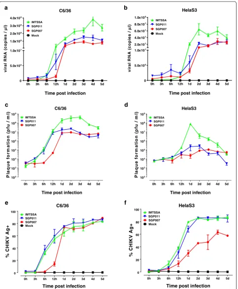
In the first part of the study, the virus infection was performed at a multiplicity of infection (MOI) of 10 and the replication kinetics of the three CHIKV isolates (SGP007, SGP011 and IMT) was analysed by negative-strand viral RNA production via quantitative RT-PCR (qRT-PCR) within Aedes albopictus (C6/36) cells and HeLa cells. Viral RNA was extracted from CHIKV-infected cell cultures using QIAamp viral RNA MINI kit (Qiagen, Hilden, Germany) and the qRT-PCR was performed using QuantiTect® Probe RT-PCR Kit (Qiagen, Hilden, Germany) with Applied Biosystems
(ABI) 7900HT Fast Real-Time PCR System, using a TaqMan assay modified from Plaskon and colleagues [21]. In the C6/36 cells, we observed three distinct rate of growth kinetics for the three different CHIKV isolates. The E1:A226V substitution in the E1 glycoprotein of the La Reunion isolate (IMT) allowed better adaptation in the C6/36 cells and resulted in a rapid increase in viral RNA copies as early as 6 hours post infection. The Singapore isolates (SGP007 and SGP011) started in-creasing in viral RNA copies from 12 hours post-infection. The SGP011 isolate, containing the E1:A226V substitution in the E1 glycoprotein, showed higher cop-ies of viral RNA when compared to the SGP007 isolate, but still showed a poorer adaptability compared to the IMT isolate (Figure 1a). We then compared the replica-tion kinetics in HeLa cells, a susceptible human cell line. Generally, we saw the same trends as for the C6/36 cells, with a sharp increase in viral load in the IMT isolate when compared to the two Singapore isolates (Figure 1b). Interestingly, it was noted that the infectious virus titres obtained by viral plaque assays further supported the no-tion that the IMT isolate produced a much higher infec-tious virus titre in both C6/36 and HeLa cells (Figure 1c and d) when compared to both SGP007 and SGP011 isolates. Notable, it was observed that SGP011 produced infectious viruses at an earlier time point of 12 hours post-infection when compared to SGP007 in C6/36 cells. We then proceeded to detect the presence of CHIKV antigens using flow cytometry in an effort to further define the virus replication kinetics. CHIKV-infected cells were fixed and permeabilised (BD Bioscience) be-fore being stained with an anti-alphavirus Ab (10μg/ml, Santa Cruz) for 30 min, followed by APC-conjugated goat anti-mouse IgG F(ab)2 Ab (10 μg/ml, Invitrogen) for 30 min. Data was acquired in BD FACS Canto II (BD Biosciences) using BD FACSDiva software. In both C6/36 and HeLa cell lines, IMT and SGP011 isolates demonstrated similar infectivity kinetics and both iso-lates showed a significantly higher viral antigen staining compared to SGP007 (Figure 1e and f ). This suggested that both IMT and SGP011 exhibited higher virus infec-tivity and viral antigen production in the infected cells when compared to SGP007 isolate.
HelaS3
assembler and bowtie enabled by Pipeline Pilot, with hash size = 31 and insert size = 150. All three sequences seemed to be missing 20–50 bp from the 3' and 5' ends when compared to the prototype isolate (S27 African prototype). In order to perform end-repair, 100 000 reads were mapped with bowtie to the prototype isolate, and some of the end sequences were recovered. The libraries were compared with the ERCC spike-in RNA controls (Invitrogen), and all three libraries had a > 0.99 correlation between normalised read counts and the control concen-trations in a log scale.
When compared to the CHIKV S27 African prototype isolate [22], we identified seven nucleotide (nt) substitu-tions within the genome of the SGP007 CHIKV isolate that resulted in amino acid (aa) changes (Additional file 1: Table S1). S27 African prototype and SGP007 CHIKV isolates had identical amino acid residues at various posi-tions (nsP3 aa524, nsP4 aa75 and E1 aa226). The E1 A226V mutation suggested a closer identity between these two isolates. However, SGP007 CHIKV obtained an extra unique mutation at various positions (nsP1 aa60, nsP3 aa137, nsP4 aa563 and E2 aa178). It remains unknown whether these mutations will support the unique feature of SGP007 CHIKV in terms of virus replication. We did not identify any unique mutations within the nsP2, Capsid, E3 or 6K protein sequences.
Till now, there is limited information available regard-ing the relationship between the unique amino acid resi-dues and the functions of alphavirus nsPs. Previous studies have suggested the role of E1 A226V mutation in the fitness of CHIKV in Aedes aegypti and Aedes albopictus mosquitoes [23]. Further studies have also suggested the role of E2 L210Q mutation in the sequen-tial adaptive mutations in CHIKV and enhancement of efficient vector switching in nature [24]. More amino acid positions within the CHIKV structural proteins have been demonstrated to show positive selection effect in virus circulation and persistence in endemic areas [25]. Here, we would like to postulate the role and effect of amino acid mutations in the nsPs on virus replication efficiency.
We first identified one unique mutation in SGP007 (nsP3 A137V) located within the macro domain region (Additional file 1: Table S1). The first 160 amino acids of the N-terminal region of nsP3 are known as the macro domain, which possesses ADP-ribose 1”-phosphate phosphatase activity. In addition, a study using Sindbis virus suggested an important role of the nsP3 macro domain in virus replication [26]. However, structural mapping suggested that this amino acid is not positioned within the enzymatic groove of the protein (data not shown). Therefore, it is unlikely that mutations at this position would alter the phosphatase activity and further studies would be needed to investigate the importance
of this mutation (nsP3 A137V) in CHIKV virus replication.
We further suggested a possible role for theopalstop codon that located at the C-terminal of the nsP3 proteins in enhancing CHIKV virus replication in vitro. Several alphaviruses express two different isoforms of nsP3, depending on the presence of anopalstop codon or an open reading frame upstream of nsP4 (7 amino acids before the C-terminal of the nsP3) [27,28]. Expression of CHIKV nsP3 in different isoforms with different proper-ties or functions remains to be further characterized. CHIKV S27 African prototype and SGP007 have an iden-tical amino acid residue at nsP3 position 524 (arginine), but in IMT and SGP011, this position encodes a stop codon instead. In vitro infection results showed that SGP007 resulted in lower virus replication efficiency when compared to IMT and SGP011 (Figure 1). It was shown previously that the Opal stop codon at this particular position improved O’Nyong-Nyong virus infectivity, rela-tive to viruses encoding an arginine at the corresponding position [29]. In addition, a study using Semliki Forest virus also showed improved nsP3 protein stability after removing the nsP3 C-terminal region [28]. Therefore, our results are consistent with previous findings from other alphaviruses that CHIKV isolates possessing an opal stop codon upstream of nsP4 could improve nsP3 protein sta-bility and virus infectivity. This in turn could enhance virus replication. In addition, the T75A amino acid residue at the nsP4 N-terminal region was found to correlate with the nsP3 C-terminal region in regulating CHIKV viral replication.
One last unique mutation identified from CHIKV SGP007 was the I563T amino acid residue at nsP4 located within the RdRp region. Within the structural region, one unique mutation identified from CHIKV SGP007 was the R178H amino acid residue at E2 glyco-protein and is located within the E2 domain B region. However, the precise role of the differences in these amino acids in the various CHIKV isolates in virus repli-cation remains undefined.
same MOI and processed for the ultrastructural analysis at different time points post-infection with the different isolates of CHIKV.
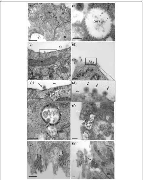
In the mocked-infected cells, normal morphology of the cells was observed and recorded (Figure 2a). The C6/36 cells were characterized by large nuclei, occupying two-thirds of the cells and the other cellular organelles were also observed. At higher magnification, stacks of Golgi apparatus, smooth and rough endoplasmic reticu-lar and mitochondria were clearly identified and docu-mented (Figure 2a).
Generally, upon infection with the different CHIKV iso-lates, the virus induced structures and morphogenesis observed were highly conserved. The first distinct viral-induced structures observed, were the presence of the cy-topathic vacuoles type I (CPV I) complexes. This occurred as early as 6 to 8 hours post-infection. These distinct vacu-oles measured to be approximately 800 nm to 2000 nm in diameter and are characterized by the presence of mem-branous spheres lining on the vacuoles (Figure 2b). These membranous spheres (Figure 2b, arrows) measured to be 60 nm to 80 nm in sizes and were seen to be neatly arranged on the vacuoles. These vacuoles were also docu-mented in cells infected with other alphaviruses and these vacuoles were proven to be the replication complexes where viral RNA synthesis occur and were seen in the early infection [30]. Other studies also showed the pres-ence of the viral replicases in CPV I complexes of other alphaviruses. The staining of the cellular markers for the cytoplasmic endosome and lysosome suggested the origin of these CPV I complexes [31,32]. As the infection progressed to 10 hours post-infection, the degenerated forms of CPV I can be observed. The membranous spheres within the CPV I complexes are no longer visible and are replaced by vacuoles containing bags of membra-nous whorls (data not shown).
The appearance of the CPV I complex at 8 hours post-infection was accompanied by the emergence of the mature virus particles budding at the plasma membrane (Figure 2c). The viral nucleocapsids of the CHIKV mea-sured approximately 30 nm to 40 nm in size can be seen to be lining in cytoplasm, underneath the plasma mem-brane (Figure 2ci). The viral nucleocapsids were seen to be expelled out of the cytoplasm through blebbing and pinching off the plasma membrane (Figure 2d), acquiring a portion of the membrane as its envelope (Figure 2di). The full mature virus was measured to be approximately 60 nm to 70 nm in size. The progression of the infection then saw an array of neatly arranged mature virus lining in rows on the peripheral of cells and in between the junction of two adjacent cells. This mode of virus release from the infected cells via budding through the plasma membrane occurred at early stages of infection and marked the first mode of viral release.
As the infection progressed, the dominant viral-induced structure was replaced by the cytopathic vacuoles type II (CPV II) complexes. This phenomenon occurred as early as 24 hours post-infection. Unlike the CPV I, these CPV II complexes were characterized by smaller vacuoles, measuring 300 nm to 400 nm in size (Figure 2e). These complexes were often observed in large number and clus-ter near the plasma membrane (Figure 2f). The cyto-plasmic side of the CPV II vacuoles was surrounded by neatly arranged electron-dense viral nucleocapsids meas-uring 30 nm to 40 nm in size. These nucleocapsids can be observed to be budding into the CPV II complexes, and were thought to mature in these CPV II vacuoles. Subse-quently, these CPV II vacuoles can be seen to contain full matured enveloped CHIKV viruses (Figure 2f, arrow), and the fusion of these vacuoles with the plasma membrane to release the matured virus out of the cell (Figure 2g). As such, it can be postulated that the CPV II may function as exocytosis vesicles or vacuoles for the release of the CHIKV particles at late infection. In addition, crystalline array of virus particles accumulating on the surface of the infected cells can be seen (Figure 2h). This mode of virus release through exocytosis of the CPV II complex with the fusion with the plasma membrane marked the second mode of viral release during the late stages of infection and may provide a means of rapid release of the mature viruses to the exterior of cell before cell death sets in.
Pm
CPV II
(f)
CPV II
CPV II
(h)
M M
ER CPV II
(e)
CPV II
(g)
(a)
M
N GA
ER M M
V
CPV I
(b)
(d)
i
Pm
(c)
i
MER
Pm
(c)
Pm
(d)
complexes were often present in cluster and located in close association to the plasma membrane. However, the number of CPV II complexes formed in HeLa cells was much more extensive than the infection in the C6/36 cells (Figure 3c). The rapid maturation and proliferation of the CHIKV virus might lead to the failure of proper exocytosis of the matured virus through fusion of the plasma membrane and resulted in the massive accumulation of matured viruses within the infected HeLa cells (Figure 3d).
This may ultimately lead to the cell death of the CHIKV-infected Hela cells. However, it is also important to note that Krejbich-Trotot and workers (2011) has recently illus-trated that CHIKV could also mobilize the apoptotic machinery by extrinsic and intrinsic pathways in Hela cells, a mechanism that could also be used to escape macrophages and viral host defences in vivo [34].
The re-emergence of CHIKV since 2004 has brought in much attention to this once neglected virus. This
M
GA
N
M
(a)
(b)
N
(c)
PmCPV II
(d)
Figure 3Replication of CHIKV (IMT isolate) in HeLa cells.(a) Ultrastructural analysis of mock-infected HeLa cells. Under high magnification, the cellular organelles such as the Golgi apparatus (GA) and mitochondria (M), can be seen. The bar corresponds to 0.2μm. (b) Infection of CHIKV in HeLa cells. Viral-induced membranous spheres are observed to associate with the plasma membrane [boxed]. The bar corresponds to 0.2μm. (c) CPV II complexes were detected at 12 hours post infection. The viral nucleocapsids (arrows) were observed to be budding in the CPV II complexes. The bar of c corresponds to 0.5μm. (d) Massive accumulation of mature CHIKV (arrows) in infected cells at late infection. The bar corresponds to 0.5μm.
(See figure on previous page.)
study has documented the differences in the replication kinetics of the different CHIKV isolates within the ECSA lineage. Despite the differences in the replication kinetics, the virus induced ultrastructural changes and virus morphogenesis remains highly conserved between the different CHIKV isolates of the ECSA lineage.
Additional file
Additional file 1: Table S1.Amino acid differences between the CHIKV S27 sequence (template) and three other CHIKV isolates (IMT, SGP007 and SGP011).
Competing interests
The authors declare that they have no competing interests.
Authors’contributions
KCC, YWK, RTPL, MMLN, LFPN and JJHC designed this study and revised the manuscript critically; KCC and YWK carried out this study and drafted the manuscript. All of the authors read and approved the final version of this manuscript.
Acknowledgements
We thank Diane Simarmata (SIgN) for technical assistance in the virus infections. We also, thank Josephine Lum and Francesca Zolezzi (SIgN) for their technical assistance in the deep sequence analysis, and Micheal Poidinger (SIgN) for bioinformatics data analysis.
This work is supported by grant from Biomedical Research Council, A*STAR, Grant No: 09/1/12/19/596 (R182-000-188-305) and National University of Singapore, Start-up grant (R182-000-165-133, R182-000-165-733).
Author details
1Department of Microbiology, Laboratory of Molecular RNA Virology and
Antiviral Strategies, Yong Loo Lin School of Medicine, National University Health System, National University of Singapore, MD4, 5 Science Drive 2, Singapore 117597, Singapore.2Singapore Immunology Network, Agency for
Science, Technology and Research (A* STAR), 8A Biomedical Grove, #04-06, Immunos, Singapore 138648, Singapore.3National Public Health Laboratory,
Communicable Disease Division, Ministry of Health, College of Medicine Building, 16 College Road, Singapore 169854, Singapore.4Department of
Microbiology, Flavivirology Laboratory, Yong Loo Lin School of Medicine, National University Health System, MD4, 5 Science Drive 2, National University of Singapore, Singapore, 117597, Singapore.
Received: 4 December 2012 Accepted: 28 May 2013 Published: 30 May 2013
References
1. Ross RW:The Newala epidemic. III. The virus: isolation, pathogenic properties and relationship to the epidemic.J Hyg (Lond)1956,54(2):177–191. 2. Gérardin P,et al:Estimating chikungunya prevalence in La réunion island
outbreak by serosurveys: two methods for two critical times of the epidemic.BMC Infect Dis2008,28(8):p99.
3. Sissoko D,et al:Seroprevalence and risk factors of chikungunya virus infection in Mayotte, Indian Ocean, 2005–2006: a population-based survey.PLoS One2008,3(8):e3066.
4. Hussain KM, Chu JJH:Insights into the interplay between chikungunya virus and its human host.Future Virol2011,6(10):1211–1223. 5. Ahola T,et al:Critical residues of semliki forest virus RNA capping
enzyme involved in methyltransferase and guanylyltransferase-like activities.J Virol1997,71(1):392–397.
6. Gomez de Cedron M:RNA helicase activity of Semliki Forest virus replicase protein NSP2.FEBS Lett1999,448(1):19–22.
7. Vasiljeva L,et al:Site-specific protease activity of the carboxyl-terminal domain of Semliki Forest virus replicase protein nsP2.J Biol Chem2001,
276(33):30786–30793.
8. Frolova E,et al:Formation of nsP3-specific protein complexes during sindbis virus replication.J Virol2006,80(8):4122–4134.
9. Tomar S,et al:Catalytic core of alphavirus nonstructural protein nsP4 possesses terminal adenylyltransferase activity.J Virol2006,80(20):9962–9969. 10. Sourisseau M,et al:Characterization of reemerging chikungunya virus.
PLoS Pathog2007,3(6):e89.
11. Ng LC, Hapuarachchi HC:Tracing the path of chikungunya virus– evolution and adaptation.Infect Genet Evol2010,10(7):876–885. 12. Hapuarachchi HC,et al:Re-emergence of chikungunya virus in south-east
asia: virological evidence from Sri lanka and singapore.J Gen Virol2010,
91(Pt 4):1067–1076.
13. Staples JE, Breiman RF, Powers AM:Chikungunya fever: an
epidemiological review of a re-emerging infectious disease.Clin Infect Dis
2009,49(6):942–948.
14. Powers AM,et al:Re-emergence of chikungunya and O'nyong-nyong viruses: evidence for distinct geographical lineages and distant evolutionary relationships.J Gen Virol2000,81(Pt 2):471–479. 15. Arankalle VA,et al:Genetic divergence of chikungunya viruses in India
(1963–2006) with special reference to the 2005–2006 explosive epidemic.J Gen Virol2007,88(Pt 7):1967–1976.
16. Schuffenecker I,et al:Genome microevolution of chikungunya viruses causing the Indian Ocean outbreak.PLoS Med2006,3(7):e263. 17. Vazeille M,et al:Two chikungunya isolates from the outbreak of La
reunion (Indian Ocean) exhibit different patterns of infection in the mosquito, aedes albopictus.PLoS One2007,2(11):e1168.
18. Ng LC,et al:Entomologic and virologic investigation of Chikungunya Singapore.Emerg Infect Dis2009,15(8):1243–1249.
19. Leo YS,et al:Chikungunya outbreak, Singapore, 2008.Emerg Infect Dis
2009,15(5):836–837.
20. Her Z,et al:Active infection of human blood monocytes by chikungunya virus triggers an innate immune response.J Immunol2010,
184(10):5903–5913.
21. Plaskon NE, Adelman ZN, Myles KM:Accurate strand-specific quantification of viral RNA.PLoS One2009,4(10):e7468.
22. Khan AH,et al:Complete nucleotide sequence of chikungunya virus and evidence for an internal polyadenylation site.J Gen Virol2002,
83(Pt 12):3075–3084.
23. Tsetsarkin KA,et al:A single mutation in chikungunya virus affects vector specificity and epidemic potential.PLoS Pathog2007,3(12):e201. 24. Tsetsarkin KA, Weaver SC:Sequential adaptive mutations enhance
efficient vector switching by chikungunya virus and its epidemic emergence.PLoS Pathog2011,7(12):e1002412.
25. Sumathy K, Ella KM:Genetic diversity of chikungunya virus, India 2006– 2010: evolutionary dynamics and serotype analyses.J Med Virol2012,
84(3):462–470.
26. Park E, Griffin DE:The nsP3 macro domain is important for sindbis virus replication in neurons and neurovirulence in mice.Virology2009,
388(2):305–314.
27. Strauss JH, Strauss EG:The alphaviruses - gene-expression, replication, and evolution (Vol 58, Pg 496, 1994).Microbiol Rev1994,58(4):806–806. 28. Varjak M, Zusinaite E, Merits A:Novel functions of the alphavirus
nonstructural protein nsP3 C-terminal region.J Virol2010,84(5):2352–2364. 29. Myles KM,et al:Effects of an opal termination codon preceding the nsP4
gene sequence in the O'Nyong-nyong virus genome on anopheles gambiae infectivity.J Virol2006,80(10):4992–4997.
30. Grimley PM, Berezesky IK, Friedman RM:Cytoplasmic structures associated with an arbovirus infection: loci of viral ribonucleic acid synthesis.J Virol
1968,2(11):1326–1338.
31. Froshauer S, Kartenbeck J, Helenius A:Alphavirus RNA replicase is located on the cytoplasmic surface of endosomes and lysosomes.J Cell Biol1988,
107(6 Pt 1):2075–2086.
32. Kujala P,et al:Biogenesis of the Semliki Forest virus RNA replication complex.J Virol2001,75(8):3873–3884.
33. Frolova EI,et al:Functional sindbis virus replicative complexes are formed at the plasma membrane.J Virol2010,84(22):11679–11695.
34. Krejbich-Trotot P,et al:Chikungunya virus mobilizes the apoptotic machinery to invade host cell defenses.FASEB J2011,25(1):314–325.
doi:10.1186/1743-422X-10-169
Cite this article as:Chenet al.:Comparative analysis of the genome
sequences and replication profiles of chikungunya virus isolates within the East, Central and South African (ECSA) lineage.Virology Journal2013
![Figure 3corresponds to 0.5complexes. The bar of c corresponds to 0.5(in HeLa cells. Viral-induced membranous spheres are observed to associate with the plasma membrane [boxed]](https://thumb-us.123doks.com/thumbv2/123dok_us/844418.1100624/7.595.58.540.380.664/figure-corresponds-complexes-corresponds-membranous-observed-associate-membrane.webp)