R E S E A R C H
Open Access
Prognostic role of hormone receptors in
endometrial cancer: a systematic review
and meta-analysis
Yanli Zhang
1†, Dong Zhao
1†, Changguo Gong
3†, Fengmei Zhang
3, Jing He
3, Wei Zhang
3, Yulan Zhao
2*and Jing Sun
1*Abstract
Background:
The aim of this study was to summarize the global predicting role of hormone receptors for survival
in endometrial cancer.
Methods:
Eligible studies were identified and assessed for quality through multiple search strategies. Data were
collected from studies comparing overall survival (OS), cancer-specific survival (CSS), or progression-free survival
(PFS) in patients with elevated levels of estrogen receptor (ER), progesterone receptor (PR), or human epidermal
growth factor receptor 2 (HER2) with those in patients with lower levels. The combined hazard ratios of ER, PR, and
HER2 for survival were calculated.
Results:
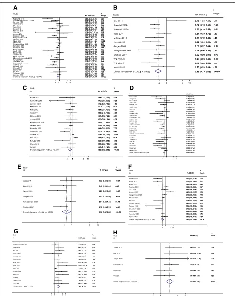
A total of 98 studies were included for meta-analysis (44 for ER, 38 for PR, and 16 for HER2). Higher
levels of either ER or PR could significantly indicate better survival. The pooled hazard ratios (HRs) of ER for
OS, CSS, and PFS were 0.75 (95 % CI, 0.68
–
0.83), 0.45 (95 % CI, 0.33
–
0.62), and 0.66 (95 % CI, 0.52
–
0.85),
respectively. The combined HRs of PR for OS, CSS, and PFS reached 0.63 (95 % CI, 0.56
–
0.71), 0.62 (95 % CI,
0.42
–
0.93), and 0.45 (95 % CI, 0.30
–
0.68), respectively. In contrast, elevated levels of HER2 could predict worse
outcome with a HR of 1.98 (95 % CI, 1.49
–
2.62) for OS, and a HR of 2.26 (95 % CI, 1.57
–
3.25) for PFS.
Conclusions:
In patients with endometrial cancer, higher level of ER and PR predicted favorable survival,
and increased level of HER2 was associated with poorer survival. All of the three hormone receptors had
prognostic value for survival.
Keywords:
Endometrial cancer, Estrogen receptor, Progesterone receptor, Human epidermal growth factor
receptor 2, Prognosis
Background
Endometrial cancer (EC) is the fourth most common
malignancy in women and the most common
gyneco-logic cancer [1], and in 2014, 52,630 new cases was
diag-nosed with an estimated 8590 deaths predicted in the
USA alone [2]. The incidence of EC is also increasing in
developing countries in the past decades [3, 4]. Overall,
the 5-year survival rates for EC are approximately
78
–
90 % for stage I, 74 % for stage II, 36
–
57 % for stage
III, and 20 % for stage IV [5]. Additionally, women with
metastatic disease have only a median survival of 7
–
12
months [6]. Such poor outcomes raise an urgent
require-ment that more accurate prognosis and predictive markers
should be applied for EC to guide the therapy and monitor
the disease progress for individual patients.
Endometrial cancer is the most common genital tract
malignancy in women and consists of two major
histo-logical types, endometrioid endometrial cancer, and
non-endometrioid endometrial cancer including
high-risk malignancies such as serous papillary and clear cell
carcinoma. Endometrioid endometrial carcinoma is the
* Correspondence:ylzhao@imet.ecnu.edu.cn;sunjing61867@126.com†Equal contributors
2School of Life Science, East China Normal University, North Zhongshan Road
#3663, Shanghai, People’s Republic of China
1Department of Minimally Invasive Gynecologic Surgery, Shanghai First
Maternity and Infant Hospital, Tongji University School of Medicine, Changle Road #536, Shanghai 200040, People’s Republic of China
Full list of author information is available at the end of the article
most common form, accountable for more than 75
–
90 %
of all cases of endometrial cancer [7].
Besides conventional clinical or pathological features,
some biological molecules have been proposed as
prog-nostic biomarkers in EC, such as P53, KRAS, PTEN,
EGFR, FGFR, estrogen receptors (ER), progesterone
re-ceptors (PR), human epidermal growth factor receptor 2
(HER2), and so on [8]. Among them, hormone receptors
are attractive because of their physiological functions.
Through binding to their receptors, estrogen drives
epi-thelial proliferation, and progesterone inhibits growth
and causes cell differentiation. Interestingly, women who
ovulate and produce progesterone almost never get
endometrial cancer. Oppositely, disruption of the
func-tions of hormone receptors can lead to several types of
malignancies [9]. Due to higher response rates reported
for hormone receptor-positive tumors, these receptors
are currently considered to be important therapeutic
tar-gets and markers for the choice of treatment [10]. HER2
is a member of the human epidermal growth factor
re-ceptor tyrosine kinase family, which regulates many
pro-cesses that can promote tumor cell proliferation and
survival [11]. HER2 pathway, which may interact with
ER, is one of the most important pathways that have
been implicated in the development of endocrine
resist-ance in breast cresist-ancer. With the development of
molecu-lar biology and immunologic method, all of the three
hormone receptors have been introduced to refine
out-come prediction of female cancers, such as breast
can-cer, ovarian cancan-cer, and endometrial cancer.
Our previous meta-analysis reported that higher level
of PR predicted favorable survival, and elevated level of
HER2 was associated with worse survival in ovarian
can-cer. Furthermore, ER-
β
may be a potentially strong
pre-dictor for better outcome [12]. A comparable situation
may also exist in research of EC, another malignant
tumor affected by the interaction between steroid
hor-mones and their respective receptors. Although a pile of
clinical studies on prognostic value of ER, PR, and HER2
expression levels in EC has also been done, no clear
con-clusion could be drawn to date. In 1985, Creasman et al.
reported that hormone receptor expression correlates
with disease-free survival in stages I and II endometrial
carcinoma [13]. However, inconsistent results were
obtained in the followed studies [14
–
17]. For example,
some studies showed that elevated levels of ER or PR
could significantly predict favorable outcome [18, 19],
whereas some other studies showed insignificant results
[20
–
22]. Moreover, some studies suggested that
ele-vated HER2 level was associated with poorer survival,
whereas other studies could not draw such significant
conclusion [20, 22].
Therefore, it is timely and necessary to analyze globally
the prognostic value of hormone receptors in a larger
population. In this study, we seek to conduct a
meta-analysis to evaluate the overall risk of hormone receptors
for endometrial cancer survival. We discussed endometrial
carcinoma and uterine papillary serous carcinoma in this
text.
Methods
We performed meta-analysis following the guidelines of
the Meta-analysis of Observational Studies in
Epidemi-ology group (MOOSE) [23].
Search strategy
We carefully searched online PubMed and EMBASE
from 1979 to May 2014 to identify relevant studies.
Three distinct sets of key words were used
simultan-eously in each set, namely,
“
estrogen receptor and
endo-metrial cancer prognosis,
” “
progesterone receptor and
endometrial cancer prognosis,
”
and
“
human epidermal
growth factor receptor 2 and endometrial cancer
prog-nosis.
”
Studies were considered eligible if they met the
following criteria: (1) they measured preoperative ER,
PR, or HER2 values; (2) they evaluated the potential
association between preoperative ER, PR, or HER2 levels
and the outcome of endometrial cancer; (3) their study
was retrospective or prospective in design; and (4) the
median period of follow-up was no shorter than 6
months. Articles were excluded based on the following
criteria: (1) review articles or letters, (2) non-English
articles, (3) laboratory studies, and (4) absence of key
information such as sample size, hazards ratio (HR),
95 %confidence interval (CI), and
P
value.
Titles, abstracts, full texts, and reference lists of all of
the identified reports were examined independently by
three reviewers (Zhang Y, Gong C, and Zhang F). These
extracted data have been double-checked by each other.
Disagreements were resolved by consensus between the
three readers or consultation with a fourth reviewer
(Zhao Y or Zhao D). In addition, a manual search was
conducted using references from the relevant literature,
including all of the identified studies, reviews, and
edito-rials. We e-mailed to the authors of studies for additional
information and the data needed for the meta-analytic
calculations. When duplicate studies were retrieved, we
included in our systematic review the study having
re-ported HRs or involving more patients (usually the latest).
This was performed to avoid overlapping between cohorts
and overestimation of the overall HR.
Quality assessment
study design; (3) clear definition of outcome assessment,
such as overall survival (OS), cancer-specific survival
(CSS), disease-specific survival (DSS), progression-free
survival (PFS), disease-free survival (DFS), or
recurrence-free survival (RFS); and (4) sufficient period of follow-up.
If a study does not mention all four points, it was excluded
so as not to compromise the quality of the meta-analysis.
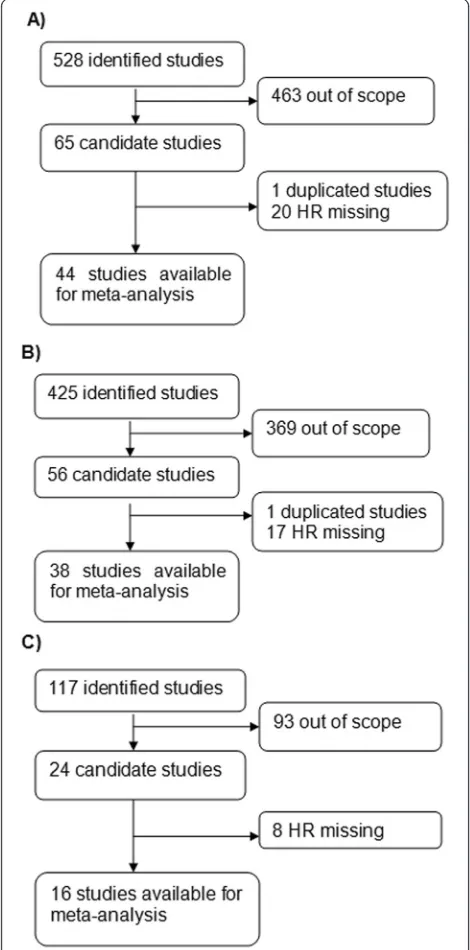
A flow diagram of the study selection process is presented
in Fig. 1.
Data extraction and conversion
The extracted data elements of this review included (1)
publication details: first author
’
s last name, publication
year, and origin of the studied population; (2) study
de-sign; (3) characteristics of the studied population: sample
size, age, stage of disease, or histological type; and (4)
HR of elevated ER, PR, and HER2 for OS, CSS
(includ-ing DSS), and PFS (includ(includ-ing DFS and RFS), as well as
their 95 % CI and
P
value. The simplest method
con-sisted in the direct collection of HR, odds ratio or risk
ratio, and their 95 % CI from the original article, with an
HR of less than 1 being associated with a better
out-come. If not available, the total numbers of observed
deaths/cancer recurrences and the numbers of patients
in each group were extracted to calculate HR .When
data were only available as Kaplan-Meier curves, data
were extracted from the graphical survival plots, and
estimation of the HR was then performed using the
described method.
Statistical analysis
A test of heterogeneity of combined HRs was conducted
using Cochran
Q
test and Higgins I-squared statistic. A
P
value of less than 0.05 was considered significant. A
random-effect model (Der Simonian and Laird method)
was used if heterogeneity was observed (
P
< 0.05),
whereas the fixed-effect model was applied in the
absence of between-study heterogeneity (
P
< 0.05).
Publi-cation bias was evaluated using the funnel plot with the
Egger bias indicator test [24]. All analyses were
con-ducted using Stata: Data Analysis and Statistical
Soft-ware V10.1 (http://www.stata.com/).
Results and discussion
A total of 528 records for ER were identified from a
pri-mary literature search in PubMed and EMBASE. After
manually screening the titles, abstracts, and key data,
463 studies were excluded because they were review
arti-cles, letters, non-English artiarti-cles, laboratory studies,
studies with important data missing, or studies irrelevant
to the current analysis. Of the 65 reports selected for
de-tailed evaluation, 1 study was excluded for being
dupli-cated; 20 others were excluded for lack of key data, such
as HR. The final meta-analysis was carried out for the
remaining 44 studies (
n
= 7119) for ER [13, 14, 16
–
18,
20
–
22, 25
–
61] (Fig. 1a). A similar identification process
was carried out in 425 studies for PR and 117 studies for
HER2. Finally, 38 studies recruiting 5502 patients for PR
[13, 14, 16
–
18, 20
–
22, 25, 26, 29, 31, 32, 34
–
43, 50
–
59,
61
–
65] (Fig. 1b) and 16 studies recruiting 1764 patients
for HER2 were included [20, 22, 33, 52, 54, 66
–
76]
(Fig. 1c). The main features of eligible studies are
sum-marized in Table 1. We collected data from Australia,
China, England, Finland, Germany, Greece, Italy, Japan,
Table 1
Summary table of the meta-analysis
A) ER
Country Study
design
Disease N Age (range) Survival analysis
Hazard ratios
Follow-up, months
Athanassiadou 1999 [39]
Greece R EC 80 62.7 (48–82) OS SC 140
Backe 1997 [54] German R EC 124 68 (30–94) OS Reported 57.6 (0.24–180)
Borazjani 1989 [40] USA R EC 44 66 (36–86) OS SC 120
Chambers 1988-1 [51]
USA R EC 168 – OS DE 24 (1–118.8)
Covens 2011 [45] USA P EC 67 – OS, PFS SC 36
Creasman 1985 [13] USA R EC 168 63 (30–92) DFS DE 25 (1–74)
Engelsen 2008 [34] Norway R EC 230 – OS SC 192
Felix 2012 [28] USA R EC 199 – OS, RFS SC 42 (0.8–144)
Fukuda 1998 [14] Japan R EEC 92 60.3 (31–86) DFS, OS SC, reported 61.2 (0–174)
Gates 2006 [52] USA R EC 108 64.2 (27–95) OS DE 60
Gonzalez-Rodilla 2013 [20]
Spain R EC 126 65.9 (43–88) OS Report 70
Gul 2010 [31] Turkey R EC 49 58.3 (30–81) OS DE 24
Huvila 2013 [61] Finland R EEC 182 67 (35–93) DFS Reported 62.8 (4.2–84.4)
Ito 2005 [57] Japan R EEC 103 57 OS Reported 60 (2–148)
Jongen 2009 [17] Netherlands R EEC 315 64.7 (32–89) DSS, RFS,
OS
SC 59.6 (0–258)
Kadar 1993 [16] USA R EC 137 – OS DE 60
Kamat 2009 [32] USA R EEC 139 63 (27–91) DSS Reported 24.9
Kalogiannidis 2008 [35]
Greece R EC 77 62.5 (35–80) OS, CSS, DFS DE 60 (9–120)
Kauppila 1986 [42] Finland R EC 153 – DFS, OS SC 42 (12–96)
Krakstad 2012-primary [27]
Norway P EC 182 – DSS SC 60
Krakstad
2012-prospective [27]
Norway P EC 474 – DSS SC 60
Lenhard 2013 [59] German P EC 292 65.1 (35.6–88) OS Reported 13.8 (13.1–14.5)
Liao 1986 [43] USA R EC 75 – OS SC 50
Lindahl 1992 [50] Sweden R EC 298 63 (36–87) OS DE 60
Martin 1983 [44] Australia P EC 87 (48–85) OS SC (8–68)
Merritt 2010 [55] USA R EEC 85 63.4 (39–91) DSS Reported 72
Mhawech-Fauceglia 2013 [48]
USA R EC 316 – OS DE 60
Mylonas 2010 [30] Germany R EEC 214 65.1 (35–88) PFS, CSS, OS SC 96.3 (0.03–176.8)
Palmer 1988 [41] Australia R EC 351 64.5 (31–89) OS SC 100
Pertschuk 1996 [47] Caucasian, Hispanic, Oriental
R EC 78 65.5 (38–89) OS SC 37.5 (13–161)
Pradhan 2012 [26] Norwegian P UPSC 52 72 (56–89) OS, PFS DE 60
Saito 2006 [56] Japan R EEC 103 57 DFS, OS Reported 60 (2–148)
Rahman 2013 [18] Japan R EEC 111 60 (26–85) PFS, OS Reported 52 (5–139)
Salvesen 1998 [58] Norway P EC 97 65 (37–92) OS DE 108 (60–180)
Shabani 2007 [36] Germany R EC 293 64.8 (35.5–88) PFS, CSS, OS SC 89.6 (3.2–135.5)
Table 1
Summary table of the meta-analysis
(Continued)
Singh 2007 [37] USA P EC 48 – OS Reported 19
Sivridis 2001 [38] Greece R EC 164 – OS SC 55 (19–167)
Song 2012 [29] Korea R EC 137 53.7 (30–82) OS Reported 60
Sun 2013 [46] China P EC 73 58 (30–78) DFS SC 43.4 (16–91)
Voss 2011 [22] England P EC 156 68.2 (37–89) DSS, RFS Reported 48.1 (0.1–141.5)
Wik 2013-R [49] Norway R EC 266 – DSS SC 300
Wik 2013-P [49] Norway P EC 153 – DSS SC 300
Zannoni 2013 [25] Italy P EEA 121 59 (35–88) DFS, OS Reported 38 (14–91)
Zhang 2013 [53] China R EC 239 54 (26–82) DFS, OS DE 67 (12–183)
Zou 2012 [21] China R EEC 60 51.3 (30–72) OS Reported 45.5 (3–69.5)
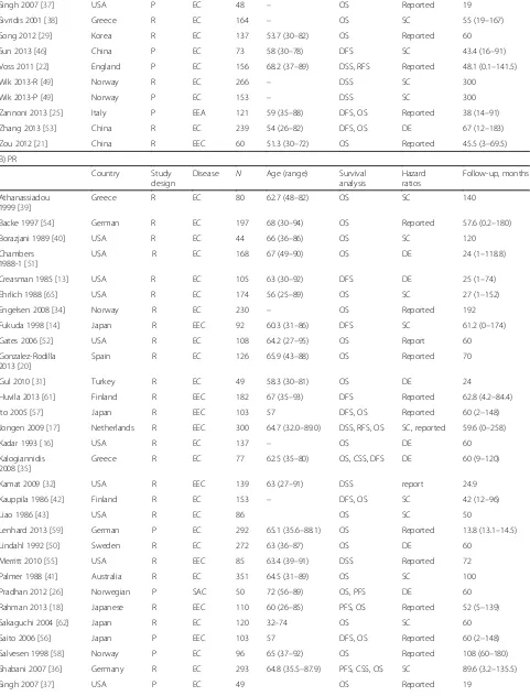
B) PR
Country Study
design
Disease N Age (range) Survival analysis
Hazard ratios
Follow-up, months
Athanassiadou 1999 [39]
Greece R EC 80 62.7 (48–82) OS SC 140
Backe 1997 [54] German R EC 197 68 (30–94) OS Reported 57.6 (0.2–180)
Borazjani 1989 [40] USA R EC 44 66 (36–86) OS SC 120
Chambers 1988-1 [51]
USA R EC 168 67 (49–90) OS DE 24 (1–118.8)
Creasman 1985 [13] USA R EC 105 63 (30–92) DFS DE 25 (1–74)
Ehrlich 1988 [65] USA R EC 174 56 (25–89) OS SC 27 (1–152)
Engelsen 2008 [34] Norway R EC 230 – OS Reported 192
Fukuda 1998 [14] Japan R EEC 92 60.3 (31–86) DFS SC 61.2 (0–174)
Gates 2006 [52] USA R EC 108 64.2 (27–95) OS Report 60
Gonzalez-Rodilla 2013 [20]
Spain R EC 126 65.9 (43–88) OS Reported 70
Gul 2010 [31] Turkey R EC 49 58.3 (30–81) OS DE 24
Huvila 2013 [61] Finland R EEC 182 67 (35–93) DFS Reported 62.8 (4.2–84.4)
Ito 2005 [57] Japan R EEC 103 57 DFS, OS Reported 60 (2–148)
Jongen 2009 [17] Netherlands R EEC 300 64.7 (32.0–89.0) DSS, RFS, OS SC, reported 59.6 (0–258)
Kadar 1993 [16] USA R EC 137 – OS DE 60
Kalogiannidis 2008 [35]
Greece R EC 77 62.5 (35–80) OS, CSS, DFS DE 60 (9–120)
Kamat 2009 [32] USA R EEC 139 63 (27–91) DSS report 24.9
Kauppila 1986 [42] Finland R EC 153 – DFS, OS SC 42 (12–96)
Liao 1986 [43] USA R EC 86 OS SC 50
Lenhard 2013 [59] German P EC 292 65.1 (35.6–88.1) OS Reported 13.8 (13.1–14.5)
Lindahl 1992 [50] Sweden R EC 272 63 (36–87) OS DE 60
Merritt 2010 [55] USA R EEC 85 63.4 (39–91) DSS Reported 72
Palmer 1988 [41] Australia R EC 351 64.5 (31–89) OS SC 100
Pradhan 2012 [26] Norwegian P SAC 50 72 (56–89) OS, PFS DE 60
Rahman 2013 [18] Japanese R EEC 110 60 (26–85) PFS, OS Reported 52 (5–139)
Sakaguchi 2004 [62] Japan R EC 120 32–74 OS SC 60
Saito 2006 [56] Japan P EEC 103 57 DFS, OS Reported 60 (2–148)
Salvesen 1998 [58] Norway P EC 96 65 (37–92) OS Reported 108 (60–180)
Shabani 2007 [36] Germany R EC 293 64.8 (35.5–87.9) PFS, CSS, OS SC 89.6 (3.2–135.5)
Korea, Netherlands, Norway, Spain, Sweden, Turkey,
and the USA.
A test of heterogeneity of combined HRs was conducted
using Cochran
Q
test and Higgins I-squared statistic. A
P
value of less than 0.05 was considered significant. A
random-effect model (Der Simonian and Laird method)
was used if heterogeneity was observed (
P
< 0.05), whereas
the fixed-effect model was applied in the absence of
between-study heterogeneity (
P
< 0.05). Publication bias
was evaluated using the funnel plot with the Egger bias
indicator test. For studies assessing EC, there mostly
appeared to have heterogeneity between studies for ER,
PR, and HER2 (
P
< 0.05). Hence, a random model was
applied to calculate a pooled HR and its 95 % CI. Higher
levels of either ER or PR could significantly indicate better
survival. The pooled HRs of ER for OS, CSS, and PFS
were 0.75 (95 % CI, 0.68
–
0.83), 0.45 (95 % CI, 0.33
–
0.62),
and 0.66 (95 % CI, 0.52
–
0.85), respectively (Fig. 2a
–
c).
The combined HRs of PR for OS, CSS, and PFS reached
0.63 (95 % CI, 0.56
–
0.71), 0.62 (95 % CI, 0.42
–
0.93), and
0.45 (95 % CI, 0.30
–
0.68), respectively (Fig. 2d
–
f ). In
contrast, elevated levels of HER2 could predict worse
outcome with a HR of 1.98 (95 % CI, 1.49
–
2.62) for OS,
and a HR of 2.26 (95 % CI, 1.57
–
3.25) for PFS (Fig. 2g, h).
Such results indicated that in patients with EC, higher
level of ER and PR predicted favorable survival, and
Table 1
Summary table of the meta-analysis
(Continued)
Sivridis 2001 [38] Greece R EC 164 – OS SC 55 (19–167)
Song 2012 [29] Korea R EC 137 53.7 (30–82) OS Reported 60
Steiner 2003 [63] Germany R EC 115 65 (38–81) OS, RFS SC 72 (36–156)
Sutton 1989 [64] USA R EC 139 61 (31–89) DFS SC, DE 28.9 (1–128)
Voss 2011 [22] England P EC 156 68.2 (37–89) DSS, RFS Reported 48.1 (0.1–141.5)
Zannoni 2013 [25] Italy P EEC 121 59 (35–88) DFS, OS Reported 38 (14–91)
Zhang 2013 [53] China R EC 239 54 (26–82) DFS, OS,
RFS
SC 67 (12–183)
Zou 2012 [21] China R EEC 60 51.3 (30–72) OS Reported 45.5 (3–69.5)
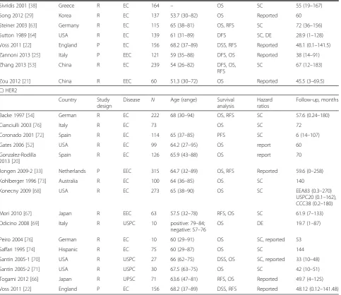
C) HER2
Country Study
design
Disease N Age (range) Survival analysis
Hazard ratios
Follow-up, months
Backe 1997 [54] German R EC 222 68 (30–94) OS, RFS SC 57.6 (0.24–180)
Cianciulli 2003 [76] Italy R EC 73 OS SC 72
Coronado 2001 [72] Spain R EC 114 65 (37–85) PFS SC 6 (14–107)
Gates 2006 [52] USA R EC 99 64.2 (27–95) OS report 60
Gonzalez-Rodilla 2013 [20]
Spain R EC 126 65.9 (43–88) OS report 70
Jongen 2009-2 [33] Netherlands P EEC 315 64.7 (32–89) OS, RFS Reported 59.6 (0–258)
Kohlberger 1996 [73] Australia R EC 100 64 (36–85) OS SC 140
Konecny 2009 [68] USA R EC 273 65 (38–90) OS SC EEA83 (0.3–270)
USPC20 (0.1–162), CCC38 (0.2–180)
Mori 2010 [67] Japan R EEC 63 57.5 (32–78) RFS, OS SC 61.9 (7–133)
Odicino 2008 [69] Italy R USPC 10 positive: 79–84;
negative: 57–76
OS DE 19.7 (1–87)
Peiro 2004 [76] German R EC 10 60 (29–91) OS SC, reported 53
Saffari 1995 [74] Hispanic R EC 75 60 (29–87) OS SC 144
Santin 2005-1 [70] USA R USPC 27 66 (62–75) DSS, OS SC, reported 33 (10–48)
Santin 2005-2 [71] USA R USPC 30 67.5 (63–75) OS SC 42 (10–51)
Togami 2012 [66] Japan R UPSC 71 63.6 (47–81) RFS, OS Reported 49.7 (4–125)
Voss 2011 [22] England P EC 156 68.2 (37–89) DSS, RFS Reported 48.12 (0.12–141.48)
Study design is described as prospective (P) or retrospective (R)
ECendometrial cancer,EECendometrioid endometrial cancer,UPSCuterine papillary serous carcinoma,OSoverall survival,CSScancer-specific survival,
DSSdisease-specific survival,PFSprogression-free survival,RFSrelapse-free survival,DFSdisease-free survival,DEdata-extrapolated,SCsurvival curve
−not reported
[ ]
increased level of HER2 was associated with poorer
sur-vival. All of the three hormone receptors had prognostic
value for survival. Then, publication bias of the ERs and
PRs studies were evaluated by funnel plots and Egger tests
as shown in Table 2.
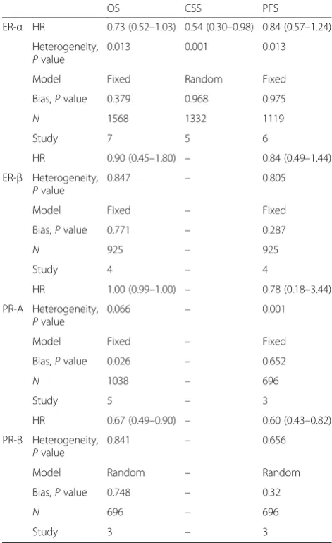
Previous studies reported that two distinct
recep-tors(ER-
α
and ER-
β
) may exert opposite effects on
cellular processes that include proliferation, apoptosis,
and migration, and their different effects may depend
on tumor type and disease stage [77]. Considering
that the different subtypes of ER and PR may have
different effects on cancer survival, we identified that
the studies focusing on ER-
α
, ER-
β
, PR-A, and PR-B
performed a meta-analysis. The pooled HRs of ER-
α
for OS, CSS, and PFS were 0.73 (95 % CI, 0.52
–
1.03),
0.54 (95 % CI, 0.30
–
0.98), 0.84 (95 % CI, 0.57
–
1.24),
respectively. The combined HRs of ER-
β
for OS and
PFS were 0.90 (95 % CI, 0.45
–
1.80) and 0.84 (95 %
CI, 0.49
–
1.44). The pooled HRs of PR-A for OS and
PFS were 1.00 (95 % CI, 0.99
–
1.00) and 0.78 (95 %
CI, 0.18
–
3.44). The combined HRs of PR-B for OS
and PFS were 0.67 (95 % CI, 0.49
–
0.90) and 0.60
(95 % CI, 0.43
–
0.82). The results are summarized in
Table 2.
The pathogenetic role and prognostic value of HER2
in EC, especially in uterine papillary serous carcinomas
[78], one of the most malignant histological types of EC,
have recently become the focus of several studies,
pro-viding the molecular basis for targeted immunotherapy
against the highly aggressive tumors [66, 69, 79
–
84].
Then we tried to identify the studies focusing on uterine
papillary serous carcinoma (UPSC) and performed a
meta-analysis. Although there were only four studies
(
n
= 138) that could be included in this subgroup
meta-analysis, the pooled HR was 2.41 with 95 % CI
from 1.54 to 3.76 (
P
< 0.05) for OS [66, 69
–
71]
(Fig. 3). The HR was significant, and it was
poten-tially strong as a HR of an empirical cutoff for strong
predictor [84].
Conclusions
This meta-analysis indicated that hormone receptors
may have value in predicting survival in patients with
endometrial cancer. The higher levels of ER and PR were
significantly associated with favorable survival, whereas
the increased level of HER2 predicted poorer survival.
All of the three hormone receptors had prognostic value
for survival. ER and PR expression are used to identify
endometrial cancer (EC) patients that could benefit of
hormone therapy, and there are many evidences
suggest-ing that they can be good biomarkers predictsuggest-ing
hormone therapy response, but further validation will be
required before they are incorporated in routine
man-agement of EC patients.
However, this meta-analysis has several limitations and
the conclusions should be tempered. First, marked
heterogeneity of subjects existed in distinct groups. The
heterogeneity of the population was probably due to the
difference in the baseline characteristics of patients (age,
tumor stage, race, methodology for assessing HRs
expression, or country), the cutoff value of markers, the
undergoing treatment, the duration of follow-up, and
others. To minimize the residual confounding effect
caused by the heterogeneity within these studies, a
random-effect model was applied. Furthermore, publication
Table 2
Comparison of the predicting value of ER-
α
, ER-
β
, PR-A,
and PR-B in EC patients
OS CSS PFS
Bias,Pvalue 0.379 0.968 0.975
N 1568 1332 1119
Bias,Pvalue 0.771 – 0.287
N 925 – 925
Bias,Pvalue 0.026 – 0.652
N 1038 – 696
A test of heterogeneity of combined HRs was conducted using CochranQtest and Higgins I-squared statistic. A random-effect model (Der Simonian and Laird method) was used if heterogeneity was observed (P< 0.05), whereas the fixed-effect model was applied in the absence of between-study heterogeneity (P< 0.05). Publication bias was evaluated using the funnel plot with the Egger bias indicator test
ECendometrial cancer,ER-αestrogen receptor-alpha,ER-βestrogen receptor-beta,PR-Aprogesterone receptor-A,PR-Bprogesterone receptor-B,HRhazards ratio,OSoverall survival,CSScancer-specific survival,DSSdisease-specific survival,PFSprogression-free survival,DFSdisease-free survival,
bias was detected in all the meta-analyses, and this
cannot be adequately overcome by currently available
statistical techniques. In addition, although the result of
UPSC subgroup about HER2 was promising, the
conclusion should be tempered for the relatively small
sample size.
Steroid hormones, including ovarian steroid
hor-mones progesterone and estrogen, play vital roles in
the development of benign endometrium and
endo-metrial cancer via their receptors [85]. Estrogens act
as a promoter of growth and proliferation of the
endometrium via estrogen receptors, while
progester-one acts as an estrogen antagonist in endometrial
maturation and inhibition of proliferation [86]. The
endometrium is very sensitive to sex hormones, and
thus a shift in the balance of estrogens and
progester-one can cause the development of endometrial cancer
[1]. The glandular epithelium from which the cancer
arises is hormone responsive, expressing both PRs
(PR-A and PR-B) and ERs (ER-
α
and ER-
β
) [87].
EC often develops from endometrial hyperplasia, which
is attributed to prolonged exposure to estrogen in the
ab-sence of (unopposed) sufficient progesterone [88], and is
often well differentiated and non-invasive or superficially
myoinvasive, rarely producing metastases and expressing
ER [89]. Whereas early-stage, well differentiated EC
usu-ally retain expression of both receptors, advanced stage,
poorly differentiated tumors often lack one or both of
these receptors, which has been correlated in many
stud-ies with a poor prognosis [19, 47]. In our meta-analysis,
both ER and PR tend to be linked with favorable outcome
of endometrial cancer and could be applied as a significant
predictor. Our results were consistent with most of the
previous basic studies that suggested the protective role of
PR in endometrial cancer.
Estrogens stimulate cell proliferation through the
classical estrogen receptors ER-
α
and ER-
β
. ER-
α
and
ER-
β
have a distinct pattern of expression in the
tis-sues [90], which varies during cellular proliferation
and differentiation [91]. Usually ER-
α
was the
domin-ant isoform in specimens of normal and diseased
endometrium [92, 93]. Some recent studies revealed that
ER-
α
was associated with aberrant proliferation,
inflam-mation, and the development of malignancy, whereas
ER-β
seemed to oppose ER-
α
actions on cell proliferation by
modulating the expression of many ER-
α
-regulated genes
and exhibits anti-migratory and anti-invasive properties in
cancer cells [77]. In large cohorts of EC patients, ER-
α
was related to early stage, lower-grade tumors [17, 33],
whereas ER-
β
was related to late stage EC [94]. Our study
also conducted a meta-analysis about different ERs, but
el-evated ER-
α
and ER-
β
levels alone had no significant value
in predicting favorable survival than non-distinguished
ER. Therefore, we suggested more studies on ER-
α
and
ER-
β
in the future to further clarify the distinct role of
ERs and PRs in the development of endometrial
carcin-oma and to also help identify diagnostic or therapeutic
markers.
The single-copy PR gene uses separate promoters and
translational start sites to produce two isoforms, PR-A
and PR-B [95], which are in fact two functionally distinct
transcription factors [96] and mediate their own
re-sponse genes [95, 97
–
99]. Studies in mice with selective
ablation of PR isoforms revealed that PR-A is necessary
for ovulation and modulates the anti-proliferative effects
of progesterone in the uterus and that PR-B is required
for normal mammary gland development and function
[100, 101]. To date, there is no evidence of such selective
roles of PR-A and PR-B in human tissues. Clinical data
in relation to the prevalence of steroid receptor isoforms
PR-A and PR-B are scarce, and the specific mechanism
is unclear. In our current meta-analysis, elevated ER-
α
,
ER-
β
, and PR-A levels did not reach significant level
ma-jorly due to the limited study number and sample size.
The polled HR of PR-B was associated with better
out-come, but there were only three studies that could be
in-cluded in this subgroup meta-analysis. Further analysis
in large scale study may contribute to the understanding
of ER and PR isoforms expression in EC.
In addition, HER2 plays a crucial role in the growth of
both normal tissue and malignant tumors [11]. HER2
amplification and overexpression have been shown to
play a key role in the pathogenesis of various different
cancer types, including breast, ovarian, gastric, and
esophageal carcinomas [102].
HER2 overexpression was also found to be associated
with endocrine therapy resistance, and HER2-positive
cancer might have a worse clinical outcome [103]. Our
study has demonstrated the predictive role of elevated
HER2 level for poorer survival. Such data may indicate
the harmful role of HER2 in endometrial cancer.
In summary, both elevated level of ER and PR
pre-dicted favorable survival, and elevated level of HER2 was
associated with worse survival in endometrial cancer.
The association between hormone receptor status and
survival raises the possibility of different subsets of
3patients with endometrial cancer with different biologic
behavior and different response to treatment but similar
histology or similar clinical performance. Conventional
histological examination alone may not be enough to
guide therapy and to refine the outcome prediction. We
suggest examining ER, PR, and HER2 levels to evaluate
endometrial cancer prognosis.
Abbreviations
EC:Endometrial cancer; ER: Estrogen receptor; HER2: Human epidermal growth factor receptor 2; OS: Overall survival; CSS: Cancer-specific survival; DSS: Disease-specific survival; PFS: Progression-free survival; DFS: Disease-free survival; RFS: Relapse-free survival; PR: Progesterone receptor; UPSC: Uterine papillary serous carcinoma.
Competing interests
The authors declare that they have no competing interests.
Authors’contributions
YZg, DZ, and CG participated in the data research and contributed equally. JS and YZo made central contributions to the conception and design of the meta-analysis and to the analysis and interpretation of data. YZg, DZ, FZ, JH, WZ and YZo were involved in drafting the manuscript. Each author has participated sufficiently in the work to take public responsibility for appropriate portions of the content. All authors gave final approval of the version to be published.
Acknowledgements
The analysis was supported by National Natural Science Foundation of China (NSFC81471436 and NSFC81402144) and SHDC12013125.
Author details
1Department of Minimally Invasive Gynecologic Surgery, Shanghai First
Maternity and Infant Hospital, Tongji University School of Medicine, Changle Road #536, Shanghai 200040, People’s Republic of China.2School of Life
Science, East China Normal University, North Zhongshan Road #3663, Shanghai, People’s Republic of China.3Institutes for Advanced
Interdisciplinary Research, East China Normal University, Shanghai, People’s Republic of China.
Received: 26 December 2014 Accepted: 10 June 2015
References
1. Yang S, Thiel KW, Leslie KK. Progesterone: the ultimate endometrial tumor suppressor. Trends Endocrinol Metab. 2011;22(4):145–52.
2. Siegel R, Ma J, Zou Z, Jemal A. Cancer statistics, 2014. CA Cancer J Clin. 2014;64(1):9–29.
3. Xiang YB, Zhang W, Gao LF, Liu ZW, Xu WH, Liu EJ, et al. Methods for time trend analysis of cancer incidence rates. Zhonghua Liu Xing Bing Xue Za Zhi. 2004;25(2):173–7.
4. Dan Yang HL-M. Year 1969–2003 study on evolution of endometrial cancer. Fudan Univ. J Med Sci. 2005;32(4):479–83.
5. Lewin SN, Herzog TJ, Barrena Medel NI, Deutsch I, Burke WM, Sun X, et al. Comparative performance of the 2009 international federation of gynecology and obstetrics’staging system for uterine corpus cancer. Obstet Gynecol. 2010;116(5):1141–9.
6. Oza AM, Elit L, Tsao MS, Kamel-Reid S, Biagi J, Provencher DM, et al. Phase II study of temsirolimus in women with recurrent or metastatic endometrial cancer: a trial of the NCIC Clinical Trials Group. J Clin Oncol.
2011;29(24):3278–85.
7. Carlson MJ, Thiel KW, Yang S, Leslie KK. Catch it before it kills: progesterone, obesity, and the prevention of endometrial cancer. Discovery Med. 2012;14(76):215–22.
8. Salvesen HB, Haldorsen IS, Trovik J. Markers for individualised therapy in endometrial carcinoma. Lancet Oncol. 2012;13(8):e353–61.
9. Moore RL, Dai Y, Faller DV. Sirtuin 1 (SIRT1) and steroid hormone receptor activity in cancer. J Endocrinol. 2012;213(1):37–48.
10. Kokka F, Brockbank E, Oram D, Gallagher C, Bryant A. Hormonal therapy in advanced or recurrent endometrial cancer. Cochrane Database Syst Rev. 2010;12:CD007926.
11. Saxena R, Dwivedi A. ErbB family receptor inhibitors as therapeutic agents in breast cancer: current status and future clinical perspective. Med Res Rev. 2012;32(1):166–215.
12. Zhao D, Zhang F, Zhang W, He J, Zhao Y, Sun J. Prognostic role of hormone receptors in ovarian cancer: a systematic review and meta-analysis. Int J Gynecol Cancer. 2013;23(1):25–33.
13. Creasman WT, Soper JT, McCarty Jr KS, McCarty Sr KS, Hinshaw W, Clarke-Pearson DL. Influence of cytoplasmic steroid receptor content on prognosis of early stage endometrial carcinoma. Am J Obstet Gynecol. 1985;151(7):922–32.
14. Fukuda K. Prognostic significance of progesterone receptor immunohistochemistry in endometrial carcinoma. Gynecol Oncol. 1998;69:220–5.
15. Kleine W, Maier T, Geyer H, Pfleiderer A. Estrogen and progesterone receptors in endometrial cancer and their prognostic relevance. Gynecol Oncol. 1990;38(1):59–65.
16. Kadar N, Malfetano JH, Homesley HD. Steroid receptor concentrations in endometrial carcinoma: effect on survival in surgically staged patients. Gynecol Oncol. 1993;50(3):281–6.
17. Jongen V, Briet J, de Jong R, ten Hoor K, Boezen M, van der Zee A, et al. Expression of estrogen receptor-alpha and -beta and progesterone receptor-A and -B in a large cohort of patients with endometrioid endometrial cancer. Gynecol Oncol. 2009;112(3):537–42.
18. Rahman MT, Nakayama K, Rahman M, Ishikawa M, Katagiri H, Katagiri A, et al. ESR1 gene amplification in endometrial carcinomas: a
clinicopathological analysis. Anticancer Res. 2013;33(9):3775–81. 19. Gehrig PA, Van Le L, Olatidoye B, Geradts J. Estrogen receptor status,
determined by immunohistochemistry, as a predictor of the recurrence of stage I endometrial carcinoma. Cancer. 1999;86(10):2083–9.
20. Gonzalez-Rodilla I, Aller L, Llorca J, Munoz AB, Verna V, Estevez J, et al. The E-Cadherin expression vs. tumor cell proliferation paradox in endometrial cancer. Anticancer Res. 2013;33(11):5091–5.
21. Zou J, Fan YJ, Meng YQ, Xu H, Fan J. An exploratory analysis of gamma-synuclein expression in endometrioid endometrial cancer. BMJ Open. 2012;2(2):e000611.
23. Stroup DF, Berlin JA, Morton SC, Olkin I, Williamson GD, Rennie D, et al. Meta-analysis of observational studies in epidemiology: a proposal for reporting. Meta-analysis Of Observational Studies in Epidemiology (MOOSE) group. JAMA. 2000;283(15):2008–12.
24. Egger M, Davey Smith G, Schneider M, Minder C. Bias in meta-analysis detected by a simple, graphical test. BMJ. 1997;315(7109):629–34. 25. Zannoni GF, Monterossi G, De Stefano I, Gargini A, Salerno MG, Farulla I,
et al. The expression ratios of estrogen receptorα(ERα) to estrogen receptorβ1 (ERβ1) and ERαto ERβ2 identify poor clinical outcome in endometrioid endometrial cancer. Hum Pathol. 2013;44(6):1047–54. 26. Pradhan M, Davidson B, Abeler VM, Danielsen HE, Tropé CG, Kristensen GB,
et al. DNA ploidy may be a prognostic marker in stage I and II serous adenocarcinoma of the endometrium. Virchows Arch.
2012;461(3):291–8.
27. Krakstad C, Trovik J, Wik E, Engelsen IB, Werner HMJ, Birkeland E, et al. Loss of GPER identifies new targets for therapy among a subgroup of ERα-positive endometrial cancer patients with poor outcome. Br J Cancer. 2012;106(10):1682–8.
28. Felix AS, Stone RA, Chivukula M, Bowser R, Parwani AV, Linkov F, et al. Survival outcomes in endometrial cancer patients are associated with CXCL12 and estrogen receptor expression. Int J Cancer.
2012;131(2):E114–21.
29. Song T, Lee JW, Choi CH, Kim TJ, Bae DS, Sung CO, et al. Ploidy and S-phase fraction are correlated with lymphovascular space invasion that is predictive of outcomes in endometrial cancer. Int J Clin Oncol. 2012;17(6):590–7. 30. Mylonas I. Prognostic significance and clinical importance of estrogen receptorαandβin human endometrioid adenocarcinomas. Oncol Rep. 2010;24(2):385–93.
31. Gul A, Keser S, Barisik N, Kandemir N, Cakır C, Sensu S, et al. The relationship of cerb B 2 expression with estrogen receptor and progesterone receptor and prognostic parameters in endometrial carcinomas. Diagn Pathol. 2010;5(1):13.
32. Kamat AA, Coffey D, Merritt WM, Nugent E, Urbauer D, Lin YG, et al. EphA2 overexpression is associated with lack of hormone receptor expression and poor outcome in endometrial cancer. Cancer. 2009;115(12):2684–92. 33. Jongen VHWM, Briët JM, de Jong RA, Joppe E, ten Hoor KA, Boezen HM,
et al. Aromatase, Cyclooxygenase 2, HER-2/neu, and P53 as prognostic factors in endometrioid endometrial cancer. Int J Gynecol Cancer. 2009;19(4):670–6.
34. Engelsen IB, Stefansson IM, Akslen LA, Salvesen HB. GATA3 expression in estrogen receptorα-negative endometrial carcinomas identifies aggressive tumors with high proliferation and poor patient survival. Am J Obstet Gynecol. 2008;199(5):543. e541–7.
35. Kalogiannidis I, Bobos M, Papanikolaou A, Makedos A, Amplianitis I, Vergote I, et al. Immunohistochemical bcl-2 expression, p53 overexpression, PR and ER status in endometrial carcinoma and survival outcomes. Eur J Gynaecol Oncol. 2008;29(1):19–25.
36. Shabani N, Kuhn C, Kunze S, Schulze S, Mayr D, Dian D, et al. Prognostic significance of oestrogen receptor alpha (ERα) and beta (ERβ), progesterone receptor A (PR-A) and B (PR-B) in endometrial carcinomas. Eur J Cancer. 2007;43(16):2434–44.
37. Singh M, Zaino RJ, Filiaci VJ, Leslie KK. Relationship of estrogen and progesterone receptors to clinical outcome in metastatic endometrial carcinoma: a Gynecologic Oncology Group Study. Gynecol Oncol. 2007;106(2):325–33.
38. Sivridis E, Giatromanolaki A, Koukourakis M, Anastasiadis P. Endometrial carcinoma: association of steroid hormone receptor expression with low angiogenesis and bcl-2 expression. Virchows Arch. 2001;438(5):470–7. 39. Athanassiadou P, Petrakakou E, Liossi A, Nakopoulou L, Zerva C, Dimopoulos
A, et al. Prognostic significance of p53, bcl-2 and EGFR in carcinoma of the endometrium. Acta Cytol. 1999;43(6):1039–44.
40. Borazjani G. Prognostic significance of steroid receptors measured in primary metastatic and recurrent endometrial carcinoma. Am J Obstet Gynecol. 1989;161:1253–7.
41. Palmer DC, Muir IM, Alexander AI, Cauchi M, Bennett RC, Quinn MA. The prognostic importance of steroid receptors in endometrial carcinoma. Obstet Gynecol. 1988;72(3 Pt 1):388–93.
42. Kauppila AJ, Isotalo HE, Kivinen ST, Vihko RK. Prediction of clinical outcome with estrogen and progestin receptor concentrations and their relationships to clinical and histopathological variables in endometrial cancer. Cancer Res. 1986;46(10):5380–4.
43. Liao BS, Twiggs LB, Leung BS, Yu WC, Potish RA, Prem KA. Cytoplasmic estrogen and progesterone receptors as prognostic parameters in primary endometrial carcinoma. Obstet Gynecol. 1986;67(4):463–7.
44. Martin JD, Hahnel R, McCartney AJ, Woodings TL. The effect of estrogen receptor status on survival in patients with endometrial cancer. Am J Obstet Gynecol. 1983;147(3):322–4.
45. Covens AL, Filiaci V, Gersell D, Lutman CV, Bonebrake A, Lee Y-C. Phase II study of fulvestrant in recurrent/metastatic endometrial carcinoma: a Gynecologic Oncology Group Study. Gynecol Oncol.
2011;120(2):185–8.
46. Sun L, Wang J, Zhang L, Li X, Shen D. Expression of ER-alpha36, a novel variant of estrogen receptor in endometrial carcinoma and its clinical significance. Gynecol Obstet Invest. 2013;75(1):68–72. 47. Pertschuk LP, Masood S, Simone J, Feldman JG, Fruchter RG, Axiotis CA,
et al. Estrogen receptor immunocytochemistry in endometrial carcinoma: a prognostic marker for survival. Gynecol Oncol. 1996;63(1):28–33. 48. Mhawech-Fauceglia P, Wang D, Samrao D, Liu S. duPont NC, Pejovic T.
Trefoil factor family 3 (TFF3) expression and its interaction with estrogen receptor (ER) in endometrial adenocarcinoma. Gynecol Oncol. 2013;130(1):174–80.
49. Wik E, Raeder MB, Krakstad C, Trovik J, Birkeland E, Hoivik EA, et al. Lack of estrogen receptor-αis associated with epithelial-mesenchymal transition and PI3K alterations in endometrial carcinoma. Clin Cancer Res. 2013;19(5):1094–105.
50. Lindahl B, Ferno M, Gullberg B, Norgren A, Willen R. 5-year survival rate in endometrial carcinoma stage I-II related to steroid receptor concentration, degree of differentiation, age and myometrial invasion. Anticancer Res. 1992;12(2):409–12.
51. Chambers JT, MacLusky N, Eisenfield A, Kohorn EI, Lawrence R, Schwartz PE. Estrogen and progestin receptor levels as prognosticators for survival in endometrial cancer. Gynecol Oncol. 1988;31(1):65–81.
52. Gates EJ, Hirschfield L, Matthews RP, Yap OW. Body mass index as a prognostic factor in endometrioid adenocarcinoma of the endometrium. J Natl Med Assoc. 2006;98(11):1814–22.
53. Zhang G-Y, Wu L-Y, Li B, Huang M-N, Zhang R, Li X-G. Retrospective analysis of prognostic variables and clinical outcomes in surgically staged intermediate risk endometrial carcinoma. Eur J Obstet Gynecol Reprod Biol. 2013;169(2):309–16.
54. Backe J, Gassel AM, Krebs S, Muller T, Caffier H. Immunohistochemically detected HER-2/neu-expression and prognosis in endometrial carcinoma. Arch Gynecol Obstet. 1997;259(4):189–95.
55. Merritt WM, Kamat AA, Hwang J-Y, Bottsford-Miller J, Lu C, Lin YG, et al. Clinical and biological impact of EphA2 overexpression and angiogenesis in endometrial cancer. Cancer Biol Ther. 2010;10(12):1306–14.
56. Saito S, Ito K, Nagase S, Suzuki T, Akahira J-I, Okamura K, et al. Progesterone receptor isoforms as a prognostic marker in human endometrial carcinoma. Cancer Sci. 2006;97(12):1308–14.
57. Ito K. 14-3-3 in endometrial cancer—a possible prognostic marker in early-stage cancer. Clin Cancer Res. 2005;11(20):7384–91.
58. Salvesen HB, Iversen OE, Akslen LA. Identification of high-risk patients by assessment of nuclear Ki-67 expression in a prospective study of endometrial carcinomas. Clin Cancer Res. 1998;4(11):2779–85.
59. Lenhard M, Heublein S, Kunert-Keil C, Vrekoussis T, Lomba I, Ditsch N, et al. Immunosuppressive Glycodelin A is an independent marker for poor prognosis in endometrial cancer. BMC Cancer. 2013;13:616.
60. Sho T, Hachisuga T, Nguyen TT, Urabe R, Kurita T, Kagami S, et al. Expression of estrogen receptor-αas a prognostic factor in patients with uterine serous carcinoma. Int J Gynecol Cancer. 2014;24(1):102–6.
61. Huvila J, Talve L, Carpen O, Edqvist PH, Ponten F, Grenman S, et al. Progesterone receptor negativity is an independent risk factor for relapse in patients with early stage endometrioid endometrial adenocarcinoma. Gynecol Oncol. 2013;130(3):463–9.
62. Sakaguchi H, Fujimoto J, Hong B, Nakagawa Y, Tamaya T. Drastic decrease of progesterone receptor form B but not A mRNA reflects poor patient prognosis in endometrial cancers. Gynecol Oncol.
2004;93(2):394–9.
64. Sutton GP, Geisler HE, Stehman FB, Young PC, Kimes TM, Ehrlich CE. Features associated with survival and disease-free survival in early endometrial cancer. Am J Obstet Gynecol. 1989;160(6):1385–91. discussion 1391–1383.
65. Ehrlich CE, Young PC, Stehman FB, Sutton GP, Alford WM. Steroid receptors and clinical outcome in patients with adenocarcinoma of the endometrium. Am J Obstet Gynecol. 1988;158(4):796–807.
66. Togami S, Sasajima Y, Oi T, Ishikawa M, Onda T, Ikeda S-I, et al. Clinicopathological and prognostic impact of human epidermal growth factor receptor type 2 (HER2) and hormone receptor expression in uterine papillary serous carcinoma. Cancer Sci. 2012;103(5):926–32.
67. Mori N, Kyo S, Nakamura M, Hashimoto M, Maida Y, Mizumoto Y, et al. Expression of HER-2 affects patient survival and paclitaxel sensitivity in endometrial cancer. Br J Cancer. 2010;103(6):889–98.
68. Konecny GE, Santos L, Winterhoff B, Hatmal M, Keeney GL, Mariani A, et al. HER2 gene amplification and EGFR expression in a large cohort of surgically staged patients with nonendometrioid (type II) endometrial cancer. Br J Cancer. 2009;100(1):89–95.
69. Odicino FE, Bignotti E, Rossi E, Pasinetti B, Tassi RA, Donzelli C, et al. HER-2/ neu overexpression and amplification in uterine serous papillary carcinoma: comparative analysis of immunohistochemistry, real-time reverse transcription-polymerase chain reaction, and fluorescence in situ hybridization. Int J Gynecol Cancer. 2008;18(1):14–21.
70. Santin AD, Bellone S, Van Stedum S, Bushen W, Palmieri M, Siegel ER, et al. Amplification of c-erbB2 oncogene. Cancer. 2005;104(7):1391–7. 71. Santin AD, Bellone S, Siegel ER, Palmieri M, Thomas M, Cannon MJ, et al.
Racial differences in the overexpression of epidermal growth factor type II receptor (HER2/neu): a major prognostic indicator in uterine serous papillary cancer. Am J Obstet Gynecol. 2005;192(3):813–8.
72. Coronado PJ, Vidart JA, Lopez-asenjo JA, Fasero M, Furio-bacete V, Magrina J, et al. P53 overexpression predicts endometrial carcinoma recurrence better than HER-2/neu overexpression. Eur J Obstet Gynecol Reprod Biol. 2001;98(1):103–8.
73. Kohlberger P, Loesch A, Koelbl H, Breitenecker G, Kainz C, Gitsch G. Prognostic value of immunohistochemically detected HER-2/neu oncoprotein in endometrial cancer. Cancer Lett.
1996;98(2):151–5.
74. Saffari B, Jones LA, el-Naggar A, Felix JC, George J, Press MF. Amplification and overexpression of HER-2/neu (c-erbB2) in endometrial cancers: correlation with overall survival. Cancer Res. 1995;55(23):5693–8. 75. Peiró G, Mayr D, Hillemanns P, Löhrs U, Diebold J. Analysis of HER-2/neu
amplification in endometrial carcinoma by chromogenic in situ
hybridization. Correlation with fluorescence in situ hybridization, HER-2/neu, p53 and Ki-67 protein expression, and outcome. Mod Pathol.
2004;17(3):227–87.
76. Cianciulli AM, Guadagni F, Marzano R, Benevolo M, Merola R, Giannarelli D, et al. HER-2/neu oncogene amplification and chromosome 17 aneusomy in endometrial carcinoma: correlation with oncoprotein expression and conventional pathological parameters. J Exp Clin Cancer Res. 2003;22(2):265–71.
77. Thomas C, Gustafsson JA. The different roles of ER subtypes in cancer biology and therapy. Nat Rev Cancer. 2011;11(8):597–608.
78. Brack S, Attinger-Toller I, Schade B, Mourlane F, Klupsch K, Woods R, et al. A bispecific HER2-targeting FynomAb with superior antitumor activity and novel mode of action. Mol Cancer Ther.
2014;13(8):2030–9.
79. Xu M, Schwartz P, Rutherford T, Azodi M, Santin A, Silasi D, et al. HER-2/neu receptor gene status in endometrial carcinomas: a tissue microarray study. Histopathology. 2010;56(2):269–73.
80. Rolitsky CD, Theil KS, McGaughy VR, Copeland LJ, Niemann TH. HER-2/neu amplification and overexpression in endometrial carcinoma. Int J Gynecol Pathol. 1999;18(2):138–43.
81. Santin AD, Bellone S, Van Stedum S, Bushen W, De Las Casas LE, Korourian S, et al. Determination of HER2/neu status in uterine serous papillary carcinoma: comparative analysis of immunohistochemistry and fluorescence in situ hybridization. Gynecol Oncol. 2005;98(1):24–30.
82. Slomovitz BM, Broaddus RR, Burke TW, Sneige N, Soliman PT, Wu W, et al. Her-2/neu overexpression and amplification in uterine papillary serous carcinoma. J Clin Oncol. 2004;22(15):3126–32.
83. Halperin R, Zehavi S, Habler L, Hadas E, Bukovsky I, Schneider D. Comparative immunohistochemical study of endometrioid and serous
papillary carcinoma of endometrium. Eur J Gynaecol Oncol. 2001;22(2):122–6.
84. Hayes DF, Isaacs C, Stearns V. Prognostic factors in breast cancer: current and new predictors of metastasis. J Mammary Gland Biol Neoplasia. 2001;6(4):375–92.
85. Liu R, Dong JT, Chen C. Role of KLF5 in hormonal signaling and breast cancer development. Vitam Horm. 2013;93:213–25.
86. Ito K, Utsunomiya H, Yaegashi N, Sasano H. Biological roles of estrogen and progesterone in human endometrial carcinoma—new developments in potential endocrine therapy for endometrial cancer. Endocr J. 2007;54(5):667–79.
87. Baylin SB, Herman JG. DNA hypermethylation in tumorigenesis: epigenetics joins genetics. Trends Genet. 2000;16(4):168–74.
88. Grady D, Gebretsadik T, Kerlikowske K, Ernster V, Petitti D. Hormone replacement therapy and endometrial cancer risk: a meta-analysis. Obstet Gynecol. 1995;85(2):304–13.
89. Bokhman JV. Two pathogenetic types of endometrial carcinoma. Gynecol Oncol. 1983;15(1):10–7.
90. Mueller SO, Korach KS. Estrogen receptors and endocrine diseases: lessons from estrogen receptor knockout mice. Curr Opin Pharmacol.
2001;1(6):613–9.
91. Yang P, Kriatchko A, Roy SK. Expression of ER-alpha and ER-beta in the hamster ovary: differential regulation by gonadotropins and ovarian steroid hormones. Endocrinology. 2002;143(6):2385–98.
92. Utsunomiya H, Suzuki T, Harada N, Ito K, Matsuzaki S, Konno R, et al. Analysis of estrogen receptor alpha and beta in endometrial carcinomas: correlation with ER beta and clinicopathologic findings in 45 cases. Int J Gynecol Pathol. 2000;19(4):335–41.
93. Sakaguchi H, Fujimoto J, Aoki I, Toyoki H, Khatun S, Tamaya T. Expression of oestrogen receptor alpha and beta in uterine endometrial and ovarian cancers. Eur J Cancer. 2002;38 Suppl 6:S74–5.
94. Jongen VH, Thijssen JH, Hollema H, Donker GH, Santema JG, Van der Zee AG, et al. Is aromatase cytochrome P450 involved in the pathogenesis of endometrioid endometrial cancer? Int J Gynecol Cancer. 2005;15(3):529–36.
95. Kastner P, Krust A, Turcotte B, Stropp U, Tora L, Gronemeyer H, et al. Two distinct estrogen-regulated promoters generate transcripts encoding the two functionally different human progesterone receptor forms A and B. EMBO J. 1990;9(5):1603–14.
96. Giangrande PH, Kimbrel EA, Edwards DP, McDonnell DP. The opposing transcriptional activities of the two isoforms of the human progesterone receptor are due to differential cofactor binding. Mol Cell Biol. 2000;20(9):3102–15.
97. Horwitz KB. The molecular biology of RU486. Is there a role for antiprogestins in the treatment of breast cancer? Endocr Rev. 1992;13(2):146–63.
98. Lessey BA, Alexander PS, Horwitz KB. The subunit structure of human breast cancer progesterone receptors: characterization by chromatography and photoaffinity labeling. Endocrinology. 1983;112(4):1267–74.
99. Gronemeyer H, Meyer ME, Bocquel MT, Kastner P, Turcotte B, Chambon P. Progestin receptors: isoforms and antihormone action. J Steroid Biochem Mol Biol. 1991;40(1–3):271–8.
100. Mulac-Jericevic B, Lydon JP, DeMayo FJ, Conneely OM. Defective mammary gland morphogenesis in mice lacking the progesterone receptor B isoform. Proc Natl Acad Sci U S A. 2003;100(17):9744–9.
101. Mulac-Jericevic B, Mullinax RA, DeMayo FJ, Lydon JP, Conneely OM. Subgroup of reproductive functions of progesterone mediated by progesterone receptor-B isoform. Science. 2000;289(5485):1751–4. 102. Borg A, Linell F, Idvall I, Johansson S, Sigurdsson H, Ferno M, et al. HER2/neu amplification and comedo type breast carcinoma. Lancet. 1989;1(8649):1268–9.