R
EPURPOSING
C
AS
9
AS A
P
ROGRAMMABLE
DNA-B
INDING
P
LATFORM FOR
G
ENOME
V
ISUALIZATION
,
T
ARGETED
E
PIGENETIC
M
ANIPULATION AND
E
XPLORATION OF
L
OCAL
C
HROMATIN
C
OMPOSITION
T
OBIASA
NTONErstgutachter: Prof. Dr. Heinrich Leonhardt Zweitgutachter: Prof. Dr. Dirk Eick
R
EPURPOSING
C
AS
9
AS A
P
ROGRAMMABLE
DNA-B
INDING
P
LATFORM FOR
G
ENOME
V
ISUALIZATION
,
T
ARGETED
E
PIGENETIC
M
ANIPULATION AND
E
XPLORATION OF
L
OCAL
C
HROMATIN
C
OMPOSITION
DISSERTATION
AN DER FAKULTÄT FÜR BIOLOGIE
DER LUDWIG-MAXIMILIANS-UNIVERSITÄT MÜNCHEN
VORGELEGT VON
TOBIAS ANTON AUS DACHAU
Content
SUMMARY ... 1
ZUSAMMENFASSUNG ... 3
1 INTRODUCTION ... 5
1.1 Epigenetic information ... 5
1.2 General genome architecture ... 6
1.2.1 Hierarchical genome organization ... 6
1.2.2 Eu- and heterochromatin ... 7
1.3 Epigenetic regulation of chromatin organization ... 10
1.3.1 DNA methylation dynamics ... 10
1.3.2 Histone posttranslational modifications (PTMs) ... 13
1.3.3 Crosstalk between epigenetic mechanisms: establishment of eu- and heterochromatin ... 17
1.4 3D-Organization of the genome ... 19
1.4.1 Chromosome territories ... 19
1.4.2 Subchromosomal compartments and chromosomal domains ... 20
1.5 Chromatin dynamics ... 23
1.5.1 Short- and long-range motility of chromatin ... 23
1.5.2 Visualization of chromatin dynamics ... 24
1.5.3 Modular DNA binding proteins ... 25
1.6 The CRISPR/Cas system ... 28
1.6.1 CRISPR/Cas mediated adaptive immunity ... 28
1.6.2 The Cas9 toolbox ... 33
1.8 Aims of this work ... 35
2 RESULTS ... 37
2.2 Visualization of genomic loci in living cells with a fluorescent CRISPR/Cas system ... 49
2.3 Site-specific recruitment of epigenetic factors with a modular CRISPR/Cas system ... 63
2.4 Determination of local chromatin composition by CasID ... 83
3 DISCUSSION ... 99
3.1 The CRISPR/Cas system as a tool to visualize chromatin ... 99
3.1.1 CRISPR imaging in comparison to modular DNA-binding proteins ... 100
3.1.2 Expanding the CRISPR imaging toolkit ... 104
3.2 Editing of epigenetic marks via the CRISPR/Cas system ... 107
3.3 Probing chromatin composition by CasID ... 108
4 ANNEX ... 115
4.1 References ... 115
4.2 Abbreviations ... 138
4.3 Statutory declaration and statement ... 141
4.4 Curriculum vitae ... 142
Summary
S
UMMARYIn eukaryotes, cellular identity is regulated by a complex interplay between epigenetic modifications, transcription factors, and the spatiotemporal organization of chromatin. Dissecting this network requires tools that precisely target desired functions to defined genomic sequences. So far, these tools were predominantly based on polydactyl zinc finger proteins (PZFs) and transcription activator-like effectors (TALEs). Yet, designing sequence-specific PZFs and TALEs for new target sequences remains challenging.
Since 2012, the emergence of the CRISPR/Cas system revolutionized the way of site-specific genome targeting. The endonuclease Cas9, the main component of the type II CRISPR/Cas system, can be recruited to defined sequences by a single guide RNA (sgRNA) and therefore has been extensively used for genome engineering approaches. Importantly, a mutated variant of Cas9 (dCas9) lacks enzymatic activity and can be guided to specific loci without inducing DNA double strand breaks at the underlying sequence.
To test the applicability of the CRISPR/Cas system as tool to visualize chromatin dynamics, we designed sgRNAs to recruit a fluorescently tagged dCas9 to major and minor satellite sequences, as well as telomeric repeats. We demonstrated that dCas9-eGFP labeled nuclear foci coincide with known target associated proteins and expanded this approach to reveal chromatin conformation via super resolution microscopy.
Moreover, we repurposed the CRISPR/Cas system to direct GFP-tagged epigenetic effector proteins to pericentric major satellites. Based on this approach, we showed that prevalent epigenetic modifications, such as cytosine methylation and hydroxymethylation can be erased or set de novo by recruiting the catalytic domains of TET1 and DNMT3A, respectively.
Finally, we developed a novel method to determine the protein composition of specific genomic elements. We combined the programmable DNA-recognition of dCas9 with the promiscuous biotin ligase activity of BirA* to biotinylate target-associated proteins, which were subsequently identified via tandem mass spectrometry. This approach allowed us to identify known and previously uncharacterized proteins, located at the close proximity of telomeres, major satellites, and minor satellites.
Summary
Z
USAMMENFASSUNGDie zelluläre Identität eukaryotischer Zellen wird über komplexe Wechselwirkungen zwischen epigenetischen Modifikationen, Transkriptionsfaktoren und dynamischer Chromatinorganisation reguliert. Die Entschlüsselung dieses Netzwerks erfordert präzise molekularbiologische Methoden, die die gezielte Rekrutierung bestimmter Funktionen zu definierten genomischen Sequenzen ermöglichen. Bis vor Kurzem basierten diese Methoden auf Zinkfingerproteinen (PZFs) oder transcription activator-like effectors (TALEs). Es ist jedoch sehr zeit- und kostenintensiv sequenzspezifische PZFs und TALEs für neue Zielsequenzen zu entwerfen und herzustellen.
Im Jahr 2012 revolutionierte die Veröffentlichung des CRISPR/Cas Systems die sequenzspezifische Modifikation des Genoms. Die Endonuklease Cas9, als die Hauptkomponente des Typ II CRISPR/Cas Systems, kann mit Hilfe einer single guide RNA (sgRNA) zu beinahe jeder benutzerdefinierten DNA-Sequenz rekrutiert werden und wurde deshalb exzessiv für gezieltes Genom-Engineering verwendet. Zudem kann eine katalytisch inaktive Form des Cas9-Proteins (dCas9) an definierte Sequenzen rekrutiert werden, ohne dabei DNA-Doppelstrangbrüche zu induzieren.
Um die gezielte Visualisierung bestimmter Chromatinabschnitte mit Hilfe des CRISPR/Cas Systems zu testen, haben wir sgRNAs entworfen, über die ein fluoreszierend markiertes dCas9-Protein zu Telomeren, sowie zu Minor- und Major Satellite Sequenzen rekrutiert werden kann. Wir konnten zeigen, dass mit dCas9-eGFP markierte Foci mit Proteinen kolokalisieren, die an den entsprechenden Sequenzen angereichert sind. Darüber hinaus haben wir diese Methode für hochauflösende Mikroskopie adaptiert und konnten somit die Ultrastruktur von Telomeren und Minor Satellites enthüllen.
Durch weitere Anpassungen des CRISPR/Cas Systems konnten wir GFP-markierte epigenetische Effektoren zu definierten Sequenzen rekrutieren. Hierbei konnten wir zeigen, dass durch die Rekrutierung der katalytischen Domänen von TET1 und DNMT3A zu perizentromerischen Major Satellite Sequenzen bestehende DNA-Modifikationen entfernt bzw. neu gesetzt werden können.
Introduction
1
I
NTRODUCTION1.1 Epigenetic information
Decades after Friedrich Miescher first isolated DNA from leukocytes in 1869 [Dahm, 2008], it was still widely thought that genetic information is stored and passed on to the next generation by the huge variety of proteins, present in all cells. However, the notion that a molecule that lacks the complexity of proteins cannot be responsible for heredity was proven to be false in the first half of the 20th century. With their groundbreaking experiments, Avery and colleagues [Avery et al., 1944] and later Hershey and Chase [Hershey & Chase, 1952] identified DNA as the “transforming principle” which accounts for the propagation of hereditary traits. Building up on the work of Chargaff, Wilkins and Franklin, Watson and Crick then provided the first model, which could adequately explain, how DNA was structured, duplicated and inherited by a daughter cell [Watson & Crick, 1953a; Watson & Crick, 1953b]. Taken together, these findings laid the foundation for the “central dogma of molecular biology”: genetic information is first transcribed from DNA into messenger RNA (mRNA), which in turn is translated into a sequence of amino acids that forms a protein [Crick, 1958; Crick, 1970].
functional outcome of the genetic code [Dobosy & Selker, 2001; Chow & Heard, 2009; Hawkins et al., 2010; Bannister & Kouzarides, 2011].
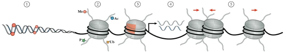
Figure 1: Overview of epigenetic modifications and mechanisms. Epigenetic modifications occur either on the DNA itself (DNA methylation (1)) or on histone tails (2). Individual canonical histones can also be exchanged by special histone variants (3). Non-coding RNAs are involved in chromatin modification and transcriptional gene silencing (4). Chromatin remodeling by rearranging nucleosomes can alter the general accessibility of chromatin (5).
1.2 General genome architecture
1.2.1 Hierarchical genome organization
Introduction
specialized chromatin loops like enhancers and insulators represent a tertiary chromatin structure. This higher-order chromatin organization is formed by long-distance contacts between secondary structure fibers but, similar to the 30-nm fiber, the exact organization and packaging of tertiary chromatin remains elusive [Woodcock & Dimitrov, 2001; Woodcock & Ghosh, 2010].
Figure 2: Domain architecture of histones and assembly of nucleosomes. A) The histone fold is a commonly shared motif of all eukaryotic histones. It comprises three α-helices, which are connected by flexible linkers. Additionally, each histone also harbors a largely unstructured N-terminal tail, which protrudes from the nucleosome. B) Heterodimerization of histones is mediated by their histone fold (only H3 (blue) and H4 (green) are shown as an example). C) Two heterodimers each of H2A-H2B and H3-H4, together with 147 bp of DNA form the barrel-shaped nucleosome particle (PDB 1AOI).
1.2.2 Eu- and heterochromatin
In addition to its hierarchical order of compaction, chromatin is also categorized in transcriptionally active eu- and inactive heterochromatin. Historically, these two forms of chromatin have been distinguished by their staining properties in interphase nuclei. Whereas euchromatin displays a more open and loosely packed conformation, heterochromatin is highly compacted [Heitz, 1928]. Since then, it became apparent that eu- and heterochromatin also differ in their classes of repetitive genomic elements, gene density and GC-content [Korenberg & Rykowski, 1988; Bickmore & Sumner, 1989; Gardiner, 1995].
2001; Ozsolak et al., 2007]. In particular, these SINEs belong to the Alu (human, ~ 300 bp) and B1 (mouse, ~140 bp) families and originated from a duplication and fusion of the 7SL RNA gene [Vassetzky et al., 2003; Tsirigos & Rigoutsos, 2009]. In metaphase chromosomes, euchromatin coincides with GC-rich R-bands, when reversely stained with Giemsa dye [Bickmore & Sumner, 1989]. Heterochromatin, on the other hand, denotes a transcriptionally inert state, which is characterized by regular nucleosomal arrays, low gene- and GC-content and the presence of long interspersed nuclear elements (LINEs) [Korenberg & Rykowski, 1988; Wallrath & Elgin, 1995]. Moreover, heterochromatin is commonly subdivided into facultative and constitutive heterochromatin (fHC and cHC, respectively). Despite being transcriptionally silent, fHC retains its capability to interconvert between eu- and heterochromatin by reshaping chromatin structure. Formation of fHC is essential in both silencing developmentally regulated homeobox (Hox) genes during the course of differentiation and ensuring dosage compensation in mammalian female cells by establishing an inactive X chromosome. In addition, fHC contains differentially expressed tissue specific genes [Trojer & Reinberg, 2007]. Whereas fHC is critical for regulatory aspects, cHC plays an important role in the structural integrity of chromatin and remains condensed throughout the cell cycle. It is mainly composed of tandemly arranged repeats and located at the pericentromeric, centromeric and telomeric regions (Figure 3) [Grewal & Jia, 2007; Saksouk et al., 2015].
Telomeres are specialized ribonucleoprotein complexes that form at the end of linear chromosomes and protect them from being recognized as DNA double-strand breaks (DSB) and thus subjected to the cellular repair machinery. Inappropriate repairing of chromosomal ends would result in chromosome fusions and unequal distribution of the genetic material during cell division [McClintock, 1938; Blasco et al., 1997]. Vertebrate telomeric DNA is composed of repeating units of a highly conserved motif (5´-TTAGGG3´), ranging from 9 – 15 kilobases (kb) in human and up to 100 kb in mice [Meyne et al., 1989; O'Sullivan & Karlseder, 2010]. These long tracts of telomeric DNA are recognized by a complex of six shelterin proteins (TRF1, TRF2, POP1, TPP1, TIN2 and RAP1), which have been suggested to promote a cap-like structure (T-loop) at chromosomal ends and thereby mask them from DNA damage response [Griffith et al., 1999; Stansel et al., 2001].
Centromeres appear as a constriction in metaphase chromosomes and act as nucleation site for kinetochore assembly during mitosis [Pluta et al., 1995]. Despite their conserved cellular function, centromeres display a varying composition between species. In human cells, centromeric DNA is based on tandemly repeated 171 bp monomers of α-satellites [Mitchell et
Introduction
reiterated so that centromeres span 0.2 – 5 megabases (Mb). Notably, the sequence composition between individual monomers as well as the organization of HORs is highly heterogeneous and chromosome-specific [Willard, 1985; Alexandrov et al., 1988; Aldrup-Macdonald & Sullivan, 2014]. Mouse centromeres, in contrast, are formed by ~120 bp minor satellite (MiS) units, which cluster in 0.6 Mb stretches and are located proximal to the telomere, resulting in acrocentric chromosomes [Pietras et al., 1983; Wong & Rattner, 1988; Kipling et al., 1991]. Similar to centromeric DNA, the pericentromeric region differs in sequence arrangement between species. Murine pericentromeric arrays are formed by tandemly arranged 234 bp major satellite (MaS) repeats and comprise up to ~ 6 Mb of DNA [Vissel & Choo, 1989; Choo, 1997]. Remarkably, MaS-repeats of several chromosomes cluster during interphase in so-called “chromocenters” (CC). MiS-sequences, however, are visible as single entities at the periphery of CCs, demonstrating the distinct three-dimensional organization of major and minor satellites [Guenatri et al., 2004]. Human pericentromeric regions, on the other hand, are predominantly composed of stretches of unordered monomeric α-satellites. In contrast to alphoid DNA of centromeres, pericentromeric sections
of chromosomes do not form higher order repeats and are frequently disrupted by LINEs, SINEs and simple repeats [Gosden et al., 1975; Manuelidis, 1978; Schueler et al., 2005; Saksouk et al., 2015].
B) Depiction of a human metacentric chromosome. The centromere (dark blue) is constituted by 171 bp α-satellite monomers, which are organized in higher order repeats (HOR). Sequence variations of α-satellites are indicated by different gray values. Pericentromeric DNA contains unordered α-satellites, which are disrupted by simple repeats (SR), LINEs and SINEs. Peri: pericentromere; cen: centromere.
1.3 Epigenetic regulation of chromatin organization
A complex and highly interconnected network of epigenetic mechanisms acts synergistically with transcription factors (TFs) and other non-histone proteins to establish and maintain a differential compaction of chromatin. As a result, chromatin accessibility by the transcription machinery is regulated, shaping the transcriptional activity of the genome and thus cellular identity [Dillon & Festenstein, 2002; Kouzarides, 2007; Lopes Novo & Rugg-Gunn, 2016].
1.3.1 DNA methylation dynamics
The postreplicative addition of a methyl-group at the C5 position of cytosine (5mC) is the first epigenetic modification, which has been shown to directly influence gene expression [McGhee & Ginder, 1979]. In mammals, 5mC is predominantly found within the context of CpG dinucleotides. In human embryonic stem cells (hESCs), however, nearly a quarter of global 5mC has been reported for non-CpG dinucleotides [Lister et al., 2009]. To a lower extent this phenomenon has also been observed in somatic cells, like neurons [Varley et al., 2013].
Introduction
transposable elements [Li et al., 1993; Woodcock et al., 1997; Gendrel et al., 2012; Barau et al., 2016].
While DNA methylation in promoter regions is generally associated with transcriptional silencing, methylation of the gene body correlates with gene expression [Wolf et al., 1984; Yang et al., 2014]. The exact functional consequences of this seemingly paradox situation are unknown. However, there is emerging evidence that methylation marks in gene bodies suppress cryptic transcriptional start sites and thereby minimize transcriptional noise [Bird, 1995; Suzuki et al., 2007]. Moreover, it has been shown that introns are less methylated than exons and excessive methylation of introns decreases polymerization speed of RNA polymerase II (RNAPII). As splicing is influenced by RNAPII kinetics, DNA methylation and cotranscriptional splicing might be linked [Laurent et al., 2010; Shukla et al., 2011].
A family of enzymes, termed DNA (cytosine C5) methyltransferases (DNMTs), catalyzes the covalent attachment of a methyl-group to the C5-position of cytosine. In mammals, the DNA methylation machinery is composed of four enzymes (DNMT1, DNMT3A, DNMT3B and DNMT3C) and one cofactor (DNMT3L) [Rottach et al., 2009; Barau et al., 2016]. The process of transferring the methyl-group is highly conserved among different species and has been described in detail for prokaryotic 5mC methyltransferases (MTases), such as M.HhaI [Klimasauskas et al., 1994]. Strikingly, the crystal structure of M.HhaI, bound to its target sequence, revealed that MTases form a covalent complex with their substrate, leading to an increased reactivity of the C5 atom of cytosine. Furthermore, the target base is “flipped” out of the DNA double helix (“base flipping”). The whole catalytic reaction involves the following steps: substrate recognition, base flipping, forming of a covalent bond between the C6-position of cytosine and the sulfhydryl-group of cysteine in a conserved proline-cysteine motif of the DNMT, transfer of the methyl-group from S-adenosyl-L-methionine (SAM) to the activated C5 atom and subsequent release of the enzyme via β-elimination [Cheng & Blumenthal, 2008].
studies provide evidence that de novo methylation during embryogenesis occurs by the combined activities of DNMT3 and DNMT1 enzymes. DNMT3 enzymes show selective binding preferences for CpG-flanking sequences. Moreover, DNMT3A binds to DNA in a tilted fashion, which allows methylation of two adjacent CpGs but prohibits enzymatic activity on the complementary strand. Sequence specificity, as well as the mode of action result in the accumulation of hemimethylated sites, which in turn represent an ideal substrate for DNMT1 [Fatemi et al., 2002; Kim et al., 2002; Handa & Jeltsch, 2005; Jia et al., 2007]. Furthermore, residual DNA methylation has been observed in DNMT3A/DNMT3B double-knockout mouse embryos, suggesting that DNMT1 possesses de novo methylation activity [Okano et al., 1999].
Introduction
by the BER pathway [Guo et al., 2011]. Furthermore, 5fC and 5caC are specifically recognized and excised by thymine DNA glycosylase (TDG) in vitro and in vivo [He et al., 2011; Pfaffeneder et al., 2011; Zhang et al., 2012a; Müller et al., 2014; Weber et al., 2016]. On the other hand, the presence of 5hmC itself may represent an epigenetic mark, as it has been detected in stable amounts in ESCs, which would contradict its role as a sole intermediate in DNA methylation [Dawlaty et al., 2011; Ficz et al., 2011; Pastor et al., 2011].
1.3.2 Histone posttranslational modifications (PTMs)
In addition to DNA methylation, epigenetic information is encoded in post-translational modifications (PTMs) of histones. As mentioned above, individual histones comprise a histone fold motif, as well as a largely unstructured 20 – 35 amino acids long N-terminal peptide, which protrudes from the nucleosome core particle [Luger et al., 1997]. These so-called histone-tails, and to a lesser extent the globular histone fold, are subject to a plethora of PTMs, including acetylation, phosphorylation, ubiquitination, sumoylation, ADP ribosylation, proline isomerization, deimination and methylation [Kouzarides, 2007]. Besides their function in other DNA-based processes such as repair, recombination and replication, histone PTMs play a crucial role in altering chromatin compaction and thereby regulate gene expression [Strahl & Allis, 2000; Taverna et al., 2007]. Whereas PTMs located at the histone fold domain directly affect histone-histone and histone-DNA interactions [Simon et al., 2011], post-translational modifications of the histone tail indirectly act on chromatin compaction by recruiting reader proteins. Notably, distribution and type of PTMs form a signature (often referred to as “histone code”), which is indicative for the chromatin state of a given locus. In the following, acetylation and methylation of histones are presented in more detail.
Writers of histone PTMs
Similar to PTMs themselves, the protein machineries that write, read and remove histone modifications have become central figures in studying chromatin organization (Figure 4). Histone PTMs are set by a group of enzymes, termed “writers”. Depending on the type of modification that is catalyzed, they are categorized as histone acetyltransferase (HAT) or methyltransferase (HMT).
HATs utilize the co-factor acetyl-CoA to catalyze the transfer of an acetyl-group to the ε -amino-group of lysine residues. Based on their subcellular localization, HATs are categorized into two major classes. The highly conserved type-B HATs are predominantly cytoplasmatic and acetylate newly synthesized histones, facilitating their assembly into nucleosomes [Richman et al., 1988; Parthun, 2007]. Type-A HATs, on the other hand, comprise the more diverse families of GNAT (Gcn5-related N-acetyltransferases), MYST (named after their founding members MOZ, Ybf2, Sas2 and Tip60) and p300/CBP (adenoviral E1A-associated protein of 300 kDa/CREB-binding protein) [Yang & Seto, 2007]. Type-A HATs mainly acetylate histone tails within nucleosomes and are often found associated in multiprotein complexes, which regulate substrate recognition and enzyme activity [Bannister & Kouzarides, 2011]. Interestingly, the interaction of p300/CBP HATs with the transcription factor CREB (cAMP response element-binding protein) demonstrates a direct link between histone acetylation and transcriptional activation [Shiama, 1997].
Although methylated glutamines and aspartates have been described [Biterge et al., 2014; Tessarz et al., 2014], histone methylation mainly occurs on lysine (mono-, di- or trimethylated) and arginine (mono-, symmetrically or asymmetrically dimethylated) residues. Depending on their substrate, HMTs are subdivided into histone lysine methyltransferases or histone arginine methyltransferases (HKMTs and PRMTs, respectively). Both classes contain several enzymes, which are characterized by both their substrate-specificity and the extent of methylation. For instance, the HKMT DIM5 specifically trimethylates H3K9 [Tamaru et al., 2003], whereas SET7/9 only monomethylates H3K4 [Xiao et al., 2003]. Similarly, type-I PRMTs generate monomethylated (Rme1) and asymmetrically dimethylated arginines, while type-II enzymes produce Rme1 and symmetrically dimethylated arginines. Yet, HKMTs, as well as PRMTs share SAM as a common methyl-group donor [Bannister & Kouzarides, 2011].
Erasers of histone PTMs
Introduction
(HDACs) catalyze the removal of acetyl-groups and are generally considered to be transcriptional repressors. Based on sequence homology to yeast Rpd3 (reduced potassium dependency 3), Hda1 (histone deacetylase 1) and Sir2 (silent information regulator 2), mammalian HDACs are grouped in three different classes [Yang & Seto, 2003]. Besides sequence divergences, HDACs differ in their reaction mechanisms. Rpd3- and Hda1-like deacetylases require Zn2+ for deacetylase activity, whereas Sir2-related HDACs depend on NAD+ as co-factor [Blander & Guarente, 2004; Lombardi et al., 2011]. In general, HDACs only demonstrate a low substrate specificity and even act on non-histone proteins [Sterner et al., 1979; Luo et al., 2000; Hubbert et al., 2002]. It has been shown, however, that human SIRT1 preferentially deacetylates H4K16 [Vaquero et al., 2004]. Studying substrate specificity is complicated by the fact that often multiple HDACs are part of diverse multiprotein complexes. For example, HDAC1 and HDAC2 are both subunits of CoREST, Sin3 and NuRD complexes [Yang & Seto, 2008].
Histone methylation has long been regarded as a stable modification [Bannister et al., 2002]. Indeed, comparable turnover rates between bulk histones and methylated lysine residues within them, suggested that histone methylation is not reversible [Byvoet et al., 1972; Duerre & Lee, 1974]. Moreover, H3K9me3 is necessary to form permanently silenced chromatin regions such as pericentric heterochromatin, suggesting that it is a static mark [Bannister et al., 2002]. However, with the discovery of the flavin-dependent KDM (lysine demethylase) LSD1 and later LSD2 (lysine-specific demethylase 1 and 2, respectively), this dogma has been changed [Shi et al., 2004; Karytinos et al., 2009]. Due to the formation of an imine intermediate in the oxidation reaction, these flavin-dependent KDMs are only capable to act on mono- or dimethylated lysine residues [Anand & Marmorstein, 2007]. Soon after the description of LSD1, a second class of KDMs has been discovered. This class is characterized by a Jumonji C (JmjC)-domain, catalyzes the removal of methyl-groups in a Fe2+ /2-oxoglutarate (2-OG) dependent manner and thus is capable to also target trimethylated lysines [Whetstine et al., 2006]. In contrast to lysine demethylation, mechanisms that result in arginine demethylation are still controversial [Böttger et al., 2015]. Yet, a recent study suggested that JmjC KDMs oxidize methylated arginines in vitro [Walport et al., 2016].
Readers of histone PTMs
the β-globin locus, as well as many enhancer and promoter elements throughout the genome reside within a hyperacetylated chromatin environment, which presumably facilitates a transcriptionally competent chromatin state [Kiefer et al., 2008; Wang et al., 2008]. Yet, the presence of multiple acetylated sites seems not to be a prerequisite for conformational change, as specific acetylation of H4K16 significantly perturbs the formation of compact higher order chromatin structures [Shogren-Knaak et al., 2006].
The majority ofevidence, however, suggests that PTMs act in trans by promoting or inhibiting the recruitment of a large variety of regulatory proteins to the nucleosomes. Those effector proteins are characterized by special domains, which confer substrate specificity.
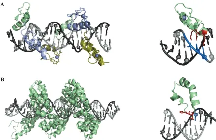
Methylated lysines are recognized by numerous domain types, including PHD (plant homeodomain) fingers and the so-called Tudor “royal” family of domains, which comprise chromodomains (chromatin organization modifier) (Figure 4B), Tudor, PWWP (Pro-Trp-Trp-Pro) and MBT (malignant brain tumor) domains [Maurer-Stroh et al., 2003; Kim et al., 2006; Champagne & Kutateladze, 2009]. Although these binding domains are sensitive to the degree of methylation, they share aromatic cage structures, typically formed by two to four aromatic amino acids, as a common recognition motif [Musselman et al., 2012]. Similar to writers of histone arginine methylation, insights into how this mark is recognized remain sparse. However, a recent study suggests that the Tudor domain of TDRD3 binds to H3R17 and H4R3, when the respective residues are asymmetrically dimethylated [Yang et al., 2010].
Introduction
Figure 4: Histone writers, readers and erasers. A) Histone PTMs are dynamic marks that are set up by writer enzymes (e.g. HATs and HMTs), interpreted by reader proteins (e.g. bromo- or chromodomain-containing proteins) and removed by eraser-proteins (e.g. HDACs and KDMs). B) Crystal structure of D. melanogaster HP1 chromodomain in complex with H3K9me3. Aromatic residues of tyrosine and tryptophan form a cage (blue), which is able to recognize K9me3 (green) (PDB 1KNE). C) Solution structure of human CBP bromodomain bound to H4K20ac. The bromodomain is composed of four α
-helices. Inter-helical loops form a hydrophobic pocket, which accommodates acetyl-lysine (green) (PDB 2RNY).
1.3.3 Crosstalk between epigenetic mechanisms: establishment of eu- and heterochromatin
Embryonic stem cells are characterized by an open and transcriptional permissive chromatin conformation. During differentiation, DNA methylation and histone PTMs act in an interconnected way to set up differentially compacted chromatin environments and thereby ensure cell type specific gene expression patterns [Meshorer & Misteli, 2006; Cedar & Bergman, 2009].
After implantation, embryos re-establish a bimodal DNA methylation pattern, that is, high methylation levels of interspersed CpG-sites and unmethylated CGIs. In this context, it has been suggested that RNAPII recruits H3K4-specific HKMTs of the trithorax family [Hughes et al., 2004; Guenther et al., 2007]. Since RNAPII is predominantly located at CGI-containing promoters, only these regions are marked by methylated H3K4. In turn, H3K4me3 impairs the binding of DNMT3L, which interacts with DNMT3A/B and is an essential co-factor for de novo methylation [Ooi et al., 2007]. Similarly, unmethylated CGIs are bound by CFP1 (CXXC finger protein 1), which in turn recruits H3K4 methyltransferases [Thomson et al., 2010]. These models are supported by the fact that DNA-methylation and H3K4-methylation generally show a strong anti-correlation [Meissner et al., 2008; Mohn et al., 2008].
During the course of embryonic development, targeted silencing of pluripotency-associated factors, such as Oct3/4 is an essential step in the establishment of tissue-specific cell lineages [Gidekel & Bergman, 2002]. Transcriptional silencing of the Oct3/4 gene is achieved by a multi-step process. First, transcription is reversibly repressed by GCNF (germ cell nuclear factor)-mediated recruitment of transcriptional co-repressors to the promoter [Fuhrmann et al., 2001]. This is followed by the binding of a complex, containing G9a and HDAC. Subsequently, the euchromatic mark H3K9ac is removed and replaced with H3K9me3. This modification, in turn, is recognized by the chromodomain of HP1. Local heterochromatinization is probably facilitated by dimerization of HP1, thus linking adjacent nucleosomes [Ruthenburg et al., 2007]. As a last step, G9a has been shown to recruit DNMT3A and DNMT3B, leading to de novo methylation and permanent silencing [Feldman et al., 2006; Epsztejn-Litman et al., 2008].
Formation of constitutive heterochromatin, such as pericentromeric satellite repeats, is dependent on the SET domain-containing HKMTs SUV39H1 and SUV39H2. These enzymes catalyze the formation of H3K9me3, which again is bound by HP1, leading to a further chromatin compaction. Additionally, SUV39 enzymes also recruit DNMT3A/B, which results in de novo methylation of these satellite sequences [Cedar & Bergman, 2009; Saksouk et al., 2015]. Interestingly, initial recruitment of HKMTs to satellite repeats seems to be mediated by non-coding RNA-duplexes, which naturally form at satellite sequences. These RNA-duplexes are processed by the RNA endonuclease Dicer, resulting in an RNA-induced silencing complex (RISC), which is targeted back to pericentromeric regions, where it is thought to interact with SUV39H1 and SUV39H2 [Fukagawa et al., 2004; Sugiyama et al., 2005]
Introduction
Xist, has been shown to recruit PRC2 (Polycomb repressive complex 2) to Xi in cis via its interaction with EZH2, the catalytic subunit of PRC2. EZH2 contains a SET domain and catalyzes trimethylation of H3K27 on surrounding nucleosomes [Zhao et al., 2008]. This mark is bound by HPC (heterochromatin-like chromodomain protein), which is a subunit of PRC1 (Polycomb repressive complex 1) and has been implicated in further heterochromatinization by catalyzing ubiquitination of H2AK119 [Plath et al., 2004; Schwartz & Pirrotta, 2008]. Finally, hypermethylation of X-linked promoter CGIs is thought to be achieved by recruitment of de novo methyltransferases by EZH2 [Norris et al., 1991; Vire et al., 2006].
Bivalent domains represent a special case in ESCs and are characterized by large repressing H3K27me3-enriched regions, harboring smaller activating H3K4me3 patches. In ES cells, these domains tend to coincide with promoters of developmentally regulated TF-genes, which are expressed at low levels. This bivalent modification pattern is predicted to keep TF-genes in a poised state that can be resolved to either an activated (H3K4me3) or fully repressed (H3K27me3) chromatin conformation [Azuara et al., 2006; Bernstein et al., 2006].
1.4 3D-Organization of the genome
Besides rather local eu- and heterochromatic regions, mammalian genomes in interphase nuclei are organized in a complex topological hierarchy. Historically, methods to decipher structural and topological properties of interphase chromosomes mainly relied on elaborate fluorescence in situ hybridization (FISH) techniques. More recently, chromosome conformation capture (3C) and its modifications (4C, 5C and Hi-C) provided local or genome-wide maps of intra- and inter-chromosomal contacts, expanding the understanding of nuclear architecture. These methods are based on mild formaldehyde crosslinking of chromatin, followed by fragmentation, ligation of DNA-fragments and subsequent detection by either PCR or next-generation sequencing [Dekker et al., 2013].
1.4.1 Chromosome territories
approaches. Most chromatin contacts are found within individual chromosomes and active loci predominantly form contacts among themselves [Simonis et al., 2006]. In addition, inactive genomic regions are spatially restricted to their own CT, whereas transcribed regions can also form inter-chromosomal contacts [Lieberman-Aiden et al., 2009; Kalhor et al., 2011; Zhang et al., 2012b]. Besides its purpose in functionally clustering transcribed loci, this intermingling between CTs is also thought to be a hot-spot for chromosomal translocations [Branco & Pombo, 2006; Zhang et al., 2012b].
Furthermore, chromosome painting and bleaching approaches demonstrate that the relative spatial distribution of individual CTs is confined during interphase but experiences a high variability between cell-types and from one interphase to the next [Walter et al., 2003; Mayer et al., 2005]. Yet, it has been shown that gene density and, to a lower extent, DNA content of chromosomes determine the spatial distribution of CTs. Whereas small, gene-rich chromosomes predominantly localize towards the nuclear interior, bigger and gene-poor chromosomes are found in vicinity of the nuclear periphery. This radial distribution of CTs is exemplified by human chromosomes 18 and 19. Although both chromosomes are similar in size, CTs of the gene-poor chromosome 18 were typically found at the nuclear periphery, whereas the gene-rich chromosome 19 preferentially localized at the nuclear interior [Croft et al., 1999; Cremer et al., 2001]. Similarly, individual CTs experience a polarized distribution of gene density. While gene-poor segments tend to localize on the surface of CTs, gene-rich segments, on the other hand, preferentially localize in the interior of CTs [Kupper et al., 2007].
1.4.2 Subchromosomal compartments and chromosomal domains
Introduction
and B compartments are constituted by groups of chromosomal domains (CDs), which mainly form intrachromosomal contacts. Yet, interchromosomal contacts have been reported, which probably reflect zones of chromosomal intermingling [Branco & Pombo, 2006; Dixon et al., 2012; Gibcus & Dekker, 2013]. The size of CDs varies drastically between species, ranging from tens of kbs to several Mbs (~100 kb in D. melanogaster, ~1 Mb in human and ~3 Mb in mouse) [Cavalli & Misteli, 2013; Gibcus & Dekker, 2013]. Notably, compartments are defined by groups of CDs with similar activities, resulting in blocks of alternate transcriptional activity (i.e. active or inactive) on mitotic chromosomes. Yet, these blocks do not represent simple on and off states, but rather a continuum of different transcriptional activities [Imakaev et al., 2012].
domains. In addition to the expected ribosomal DNA genes (rDNA), AT-rich, transcriptionally inactive sequences across all chromosomes have been described to associate with nucleoli in a domain-like pattern [Nemeth et al., 2010; van Koningsbruggen et al., 2010]. Interestingly, these sequences, termed nucleolar-associated domains (NADs), experience a substantial overlap with cLAD patterns. Hence, it has been suggested that NADs and cLADs comprise the same type of repressive chromatin, which is randomly distributed between the NL and the nucleolar periphery after mitosis. This model is supported by the observation that some CDs that associate with the nucleolus in a mother cell are repositioned to the NL in daughter cells [van Koningsbruggen et al., 2010].
Introduction
Figure 5: Hierarchical levels of chromatin organization in interphase nuclei. Top left) Chromosomes occupy distinct territories. Top right) Chromatin is organized in the two compartments A (light-gray) and B (dark-gray), which correspond to eu- and heterochromatin, respectively. Bottom) A and B compartments comprise transcriptionally active (green) and repressed (red) TADs, respectively. TADs are formed by local DNA contacts (e.g. enhancer/promoter loops).
1.5 Chromatin dynamics
1.5.1 Short- and long-range motility of chromatin
Although being restricted to distinct chromosome territories, chromatin does not represent a static, but rather dynamic structure [Lanctot et al., 2007; Soutoglou & Misteli, 2007]. Chromatin is subject to a plethora of epigenetic marks, which results in differentially compacted regions and thus provides a dynamic balance between genome packing and accessibility to the underlying DNA sequence. Therefore, TFs, in combination with adenosine triphosphate (ATP)-dependent chromatin remodeling complexes, have to induce local chromatin decompaction in order to bind heterochromatic regions [Luo & Dean, 1999; Clapier & Cairns, 2009]
confined to long-range movements, which occur less frequently and require passage through mitosis [Soutoglou & Misteli, 2007]. Except for early G1-phase, at which substantial rearrangements of subchromosomal domains can occur, chromatin motility is restricted to short-range movements during the cell cycle [Walter et al., 2003; Soutoglou & Misteli, 2007; Kind et al., 2013].
Notably, long-range chromatin movement correlates with transcriptional control. According to this model, loci move to either A or B compartments depending to their transcriptional status. Whereas in hematopoietic precursor cells, silent IgH- and Igκ-genes are located at the repressive nuclear periphery, these loci are internalized upon their activation in B-cells [Kosak et al., 2002]. Similarly, targeted transcriptional activation of a transgene results in a long-range movement to the nuclear interior in an actin/myosin dependent manner [Tumbar & Belmont, 2001; Chuang et al., 2006]. In addition, lineage specification of ESCs provides a striking example of large-scale chromatin rearrangement. During differentiation, a large subset of genes switch from A to B compartments and defined heterochromatic regions are formed, resulting in diminished genome plasticity [Hiratani et al., 2008; Dixon et al., 2015].
Yet, there is accumulating evidence, suggesting that radial positioning of genes lacks a direct causality to the transcriptional status. For example, many gene loci remain at the same radial position, even though their expression level changes [Hewitt et al., 2004; Zink et al., 2004; Meaburn & Misteli, 2008]. Vice versa, some genes are repositioned without detectable changes of their transcriptional output [Williams et al., 2005; Meaburn & Misteli, 2008].
1.5.2 Visualization of chromatin dynamics
Besides emerging 3C approaches, fluorescence microscopy-based methods are essential for deciphering the dynamics and spatiotemporal organization of chromatin. Direct imaging of nuclear proteins can be achieved by genetically fusing them to fluorescent proteins (FPs; e.g. GFP). Additionally, chromatin-associated proteins are indirectly visualized via FP-tagged camelid antibodies. These heavy-chain antibodies can be raised against virtually any epitope and fold readily in eukaryotic cells [Rothbauer et al., 2006; Romer et al., 2011]. Further, indirect visualization can also be accomplished by fusing the protein of interest to a SunTag (SUperNova tag). This short peptide tag is recognized by a co-expressed single-chain antibody (scFv), conjugated to a FP [Tanenbaum et al., 2014].
Introduction
(FRAP) or at a defined region around the ROI (FLIP) is measured over time. FCS, in contrast, is a single-molecule detection method, which measures the fluctuations around the mean of fluorescence within a confocal volume [Weiss, 2008]. These measurements provide information about the kinetics of protein diffusion that depend on protein size but also on protein-chromatin interactions [Voss & Hager, 2008].
Sequence-specific visualization of chromatin mainly relies on FISH-based approaches. Here, the target sequence is detected via complementary base pairing with an epitope- or fluorophore-labeled nucleic acid probe after the genomic DNA has been denatured. Using this technique on fixed cells, entire chromosomes, chromosome arms or single loci can be visualized. Moreover, by combining different fluorophores, simultaneous detection of several loci or even all chromosomes can be achieved [Bolzer et al., 2005].
In contrast to analyzing nuclear protein dynamics, in vivo visualization of chromatin is rather limited. Replication foci have been observed by incorporation of fluorescently labeled nucleotides, which revealed different replication times for eu- and heterochromatin [Bornfleth et al., 1999; Schermelleh et al., 2001]. Further, tandem insertion of the lac operator (LacO) at specific genomic sites and subsequent detection via FP-tagged lac repressor (LacI) has been used to study repositioning of chromatin domains and mobility of telomeres [Robinett et al., 1996; Tumbar & Belmont, 2001; Chuang et al., 2006; Jegou et al., 2009].
Specific chromatin regions can also be indirectly visualized via associated proteins. For instance, FP-tagged histone variant CENP-B binds to CENP-boxes and specifically localizes to centromeres [Shelby et al., 1996]. Similarly, a FP-fusion of the telomere-specific protein TRF1 has been utilized to study telomere dynamics in living cells [Krawczyk et al., 2012]. Although these methods allow for in vivo visualization of chromatin dynamics, target sites are limited.
1.5.3 Modular DNA binding proteins Zinc finger proteins
Recognition of user-defined sequences was first realized utilizing zinc finger (ZnF) proteins [Gersbach & Perez-Pinera, 2014]. This highly diverse group of proteins serves a large variety of biological functions, including transcriptional activation, protein folding, regulation of apoptosis and nucleic acid binding [Laity et al., 2001]. Notably, the classic Cys2His2 ZnF structural motif, which was first described in the transcription factor IIIA from Xenopus laevis, is conserved among higher eukaryotes and represents the predominant DNA-binding domain in humans [Miller et al., 1985; Rubin et al., 2000; Tupler et al., 2001].
predominantly facilitated by the α-helix, which establishes contact to three bases within the major groove of the DNA via amino acid residues at positions -1, 3 and 6 [Pavletich & Pabo, 1991; Elrod-Erickson et al., 1996]. Since each ZnF motif recognizes a distinct base triplet, tandem arrangement of up to six modules into a polydactyl ZnF protein (PZF) enables the recognition of unique genomic loci [Liu et al., 1997]. Yet, it has been demonstrated that individual zinc fingers display a preference for GC-rich substrates and that neighboring modules affect each other’s target specificity. Hence, target sequence prediction is limited and newly designed PZFs have to be subjected to a rigorous selection process, rendering this approach laborious and expensive [Segal et al., 1999; Ramirez et al., 2008; DeFrancesco, 2011].
Figure 6: Substrate recognition by modular DNA-binding proteins. A) Left: Crystal structure of two tandem ZnF proteins (Zif268) in complex with DNA. Individual ZnF modules are represented in different colors and the Zn2+ ion is
Introduction
Transcription activator-like effectors (TALEs)
Due to their inherent unfavorable features, PZFs have been widely replaced by designer transcription activator-like effectors (dTALEs).
TALEs represent the major class of virulence factors in the bacterial plant pathogen Xanthomonas spp., which is known to cause diseases in more than 200 plant families, among them many agriculturally important crops. Key to the pathogenicity of this bacterial genus is an Hrp-type III secretion system (T3S), enabling the injection of bacterial effector proteins into the plant cell [Buttner & Bonas, 2010]. Xanthomonas strains typically express and secrete a mixture of 20 – 40 effector proteins with functions ranging from suppressing the hosts defense mechanisms to modulation of the plants transcriptome. Interestingly, the largest family of effector proteins, termed transcription activator-like (TAL) effectors, was found to mimic plant specific TFs [Kay et al., 2007].
Due to its modular composition, the central repeat domain of TALEs can be rearranged to create dTALEs, which can be targeted to virtually any genomic sequence. TALEs have been demonstrated to be powerful tools in genome engineering [Miller et al., 2011; Mussolino et al., 2011], transcription modulation [Zhang et al., 2011; Bultmann et al., 2012; Mahfouz et al., 2012] and in vivo labeling of specific genomic sequences [Ma et al., 2013; Miyanari et al., 2013; Thanisch et al., 2014]. Yet, this highly repetitive structure also necessitates the use of elaborate and time-consuming cloning techniques [Morbitzer et al., 2011].
1.6 The CRISPR/Cas system
Prokaryotes employ a variety of innate defense mechanisms against foreign viral or plasmid DNA, including restriction/modification systems and blocking of phage adsorption [Samson et al., 2013]. In recent years, it became apparent that prokaryotes additionally possess means that confer adaptive immunity against invading genomic elements [Barrangou et al., 2007].
1.6.1 CRISPR/Cas mediated adaptive immunity
First observed in Escherichia coli, approximately half of bacteria (~ 45 %) and nearly all archaea (~ 84 %) are equipped with a sophisticated adaptive defense mechanisms called clustered regularly interspaced short palindromic repeats (CRISPR)/CRISPR-associated (Cas) [Ishino et al., 1987; Grissa et al., 2007; Wiedenheft et al., 2012]. Interestingly, these systems rely on small CRISPR-RNAs (crRNAs) to guide nucleases to foreign nucleic acids. All CRISPR/Cas systems comprise a set of cas genes, organized in operons, and a CRISPR-locus, harboring an array of genome-targeting sequences (termed spacers), which are derived from foreign DNA and are flanked by identical direct repeats [Ratner et al., 2016].
Introduction
Based on the presence of unique Cas proteins, the modes of crRNA maturation and RNA-guided interference, CRISPR/Cas systems are subdivided into three main types (type I, II and III). Type I systems utilize the endonucleases Cas6 or Cas5d to cleave the pre-crRNA within the repeat sequences and thereby facilitate the maturation of crRNA [Carte et al., 2008; Garside et al., 2012; Nam et al., 2012]. Subsequently, crRNA interacts with a complex of five Cas proteins (CasA – E) called Cascade (CRISPR-associated complex for antiviral defense) and mediates target recognition via complementary base pairing [Jore et al., 2011]. Upon target binding, conformational changes (R-loop formation) lead to the recruitment of the nuclease Cas3, which facilitates DNA degradation [Westra et al., 2012].
Similar to type I systems, type III systems also rely on Cas6 for crRNA maturation. However, in these systems, Cascade is replaced by a complex consisting of repeat units of Csm or Cmr proteins and target degradation requires Cas10 [Hrle et al., 2013; Staals et al., 2013; Staals et al., 2014; Samai et al., 2015]. Interestingly, type III systems are capable to target both DNA and RNA. Co-transcriptional RNA degradation is thought to ensure robust immunity against viral infections, when, due to mutations in protospacer sequences, DNA cleavage is abrogated [Jiang et al., 2016].
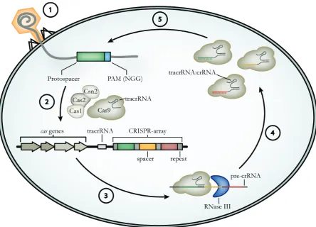
Figure 7: Type II CRISPR/Cas mediated adaptive immunity: Upon phage infection (1), Cas9 in complex with Cas1, Cas2, Csn2 and tracrRNA, scans the invasive genetic element and selects protospacers, which contain an appropriate PAM (5´-NGG-3´ for Staphylococcus pyogenes Cas9). The newly acquired spacer is then integrated into the CRISPR-array in a Cas1/Cas2-dependent manner (2). Following transcription of the CRISPR-array, the resulting pre-crRNA is bound by tracrRNA, by forming a repeat:anti-repeat duplex and processed into mature crRNA via RNase III in the presence of Cas9 (3). The mature crRNA stays in complex with tracrRNA and Cas9 (4) and mediates target recognition and degradation by complementarity between spacer and protospacer sequences (5).
Spacer-acquisition in type II CRISPR/Cas systems
Introduction
acquisition of new spacers. This indicates that Cas9 is solely required for selecting the PAM and binding the protospacer, whereas Cas1 cleaves the adjacent sequence [Heler et al., 2015]. Subsequently, Cas1 and Cas2 facilitate the integration of the newly acquired spacer sequence into the CRISPR-array by interacting with the secondary structure of the CRISPR repeat. This is then followed by nicking the repeat sequence at the 3´-end and ligation of the free hydroxyl-group to the spacer sequence. However, the exact mechanism of this process remains elusive [Nunez et al., 2015].
CRISPR-RNA maturation in type II systems
Following transcription of the CRISPR-array, the resulting pre-crRNA is processed, yielding mature crRNAs, each of them specific for one protospacer sequence. Processing of pre-crRNA requires the base pairing of every repeat sequence with a small non-coding trapre-crRNA, which is encoded in the vicinity of the cas genes and the CRISPR-array [Deltcheva et al., 2011]. Once formed, Cas9 interacts with these tracrRNA:pre-crRNA duplexes, probably protecting the spacer sequence from endonucleolytic cleavage by host RNases. Yet, this interaction is thought to be required for the recruitment of RNase III, which subsequently cleaves both strands of RNA within the double stranded repeat region, resulting in intermediate crRNAs composed of repeat-spacer-repeat sequences [Deltcheva et al., 2011; Chylinski et al., 2013]. In an elusive, second maturation event, these intermediate crRNAs are further trimmed, yielding spacer-repeat containing crRNAs. Notably, each mature crRNA remains in complex with the processed tracrRNA and the endonuclease Cas9, forming a ternary silencing complex [Jinek et al., 2012].
Target interference in type II systems
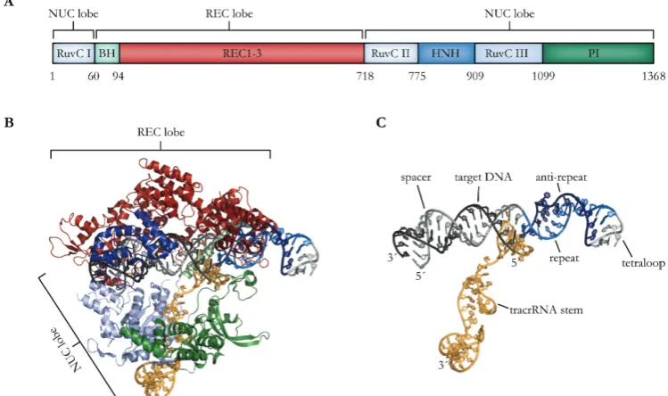
Due to extensive biochemical and crystallographic studies, the mode of target recognition and interference of Cas9 endonucleases is well established. Cas9 forms a bi-lobed structure with a larger recognition lobe (REC lobe) and a smaller nuclease lobe (NUC lobe), which are bridged by a α-helix (bridge helix; BH). Additionally, the C-terminus contains a PAM-interacting domain (PI) (Figure 8A, B). The two lobes form a clam-like shape with a positively charged central channel, which accommodates both the tracrRNA:crRNA duplex and the target DNA, eventually [Jinek et al., 2014; Nishimasu et al., 2014].
domains (RuvC and HNH) are positioned in a favorable way for subsequent target cleavage [Jinek et al., 2014; Nishimasu et al., 2014].
Finally, this ribonucleoprotein complex scans DNA to facilitate target cleavage. Notably, Cas9 constantly associates and disassociates from the DNA, until the PI domain encounters an appropriate PAM, which is directly associated with the target sequence [Sternberg et al., 2014; Knight et al., 2015]. In Staphylococcus pyogenes Cas9, the PI tightly binds to a GG dinucleotide within the PAM via two conserved tryptophan residues (W476 and W1126) [Jinek et al., 2014]. Interaction with the PAM is thought to lead to destabilization and local unwinding of the adjacent double-stranded DNA [Anders et al., 2014]. Subsequently, Cas9 is fully loaded onto the DNA, whereas the crRNA spacer displaces the non-complementary strand (R-loop) and forms a RNA:DNA heteroduplex. Interestingly, mismatches between crRNA and target DNA at the 5´-region of the spacer seem to be tolerated to some extent [Kabadi & Gersbach, 2014]. The separation of the DNA strands leads to their placement into the active sites of the two nuclease domains. The RuvC domain subsequently cleaves the non-complementary strand, whereas the HNH domain targets the complementary strand 3 bp upstream of the PAM [Jinek et al., 2014; Nishimasu et al., 2014; Sternberg et al., 2015].
Introduction
1.6.2 The Cas9 toolbox
Due to the fact that Cas9 recognizes its genomic target via Watson-Crick base pairing, this system can be reprogrammed to bind to user-defined sequences, by simply exchanging the spacer sequence of the crRNA. CRISPR/Cas9 genome targeting systems can be even further simplified, by replacing the two RNA components, which have to be expressed separately, by a synthetic fusion of tracrRNA and crRNA, named single guide RNA (sgRNA) (Figure 8C). By introducing a tetraloop-linker between the crRNA and tracrRNA, this chimeric sgRNA retains the double stranded repeat:anti-repeat duplex and structural features that are necessary for Cas9 interaction but can be transcribed from a single promoter [Jinek et al., 2012].
output of specific genes has been manipulated by fusing dCas9 to epigenetic effector proteins. For instance, dCas9 fused to the human HAT p300 is able to specifically target H3K27 acetylation to distal enhancers or promoter regions of the OCT4 gene, resulting in an increased mRNA level [Hilton et al., 2015]. Targeted de novo methylation via a dCas9-DNMT3A fusion, on the other hand, was used to silence the IL6ST gene in human cells [Vojta et al., 2016].
Aims
1.8 Aims of this work
Despite the fact that the genomes of many organisms have been sequenced, labeling and tracing of specific sequences in living cells has been a major challenge in studying the spatiotemporal dynamics of native chromatin. To date, tools to investigate nuclear organization either rely on prior fixation (FISH), or are based on DNA binding properties of fluorescently tagged proteins, such as PZFs or dTALEs. Designing sequence-specific PZFs and dTALEs for new target sequences, however, remains challenging.
Since 2012, the emergence of the CRISPR/Cas system opened new experimental possibilities for sequence-specific DNA recognition. The endonuclease Cas9 plays a vital role in the type II CRISPR/Cas system and can be recruited to endogenous loci by a sgRNA, whereas target specificity is mediated by complementarity between ~20 bp of the sgRNA and the corresponding DNA sequence. Importantly, a catalytic mutant of Cas9 (dCas9) that retains its DNA-binding capability without inducing DSBs at the target sequence, was described.
The first objective of this work was to harness dCas9 as a programmable DNA-binding platform for the visualization of distinct genomic loci in vivo. For this, we constructed a dCas9-eGFP protein and co-expressed it with sgRNAs, which were specific for endogenous repetitive sequences. To test the target-specificity of this new approach, we validated the signals, obtained by CRISPR imaging, with FISH and immunofluorescence microscopy.
Besides the spatiotemporal localization, the function of individual genomic loci is determined by their epigenetic status. To elucidate whether the CRISPR/Cas system is a suitable tool for epigenetic studies, we set out to target epigenetic modifications to specific sites. For this aim, we fused dCas9 to a binding protein (GBP). This setup enabled us to recruit GFP-tagged epigenetic effectors to a genomic region of interest and to investigate their functions in a defined environment.
Results
2
R
ESULTSPAPER TYPE
Nucleus 5:2, 163–172; March/April 2014; © 2014 Landes Bioscience
RESEARCH PAPER
Introduction
Almost one hundred and fifty years since the original description of chromosomes, many genomes have been fully sequenced. While our knowledge of DNA sequences has reached base pair (bp) resolution, detailed information on the positioning, nuclear arrangement and interactions of specific gene loci in living cells is still limited.
Despite the absence of internal membranes, the nucleus is a highly organized organelle. With fluorescent in situ hybridization (FISH) all chromosomes in an interphase nucleus were mapped and shown to occupy distinct territories.1 Additionally, gene rich
chromosomes were found to be preferably located in the center of the nucleus, whereas gene poor chromosomes reside mostly in proximity to the nuclear periphery reflecting a functional nuclear organization.2,3 Clearly, FISH represents an important tool to
label specific DNA sequences and to study nuclear architecture, but it is restricted to fixed specimens.
By now, several methods have been employed to label DNA in vivo4 and to investigate chromatin spatiotemporal dynamics,
for instance by the incorporation of fluorescently tagged chromatin proteins, like H2B-GFP.5 However, these methods do
not distinguish specific genomic sequences. To overcome these limitations, the lac operator and/or repressor recognition system has been developed.6 This system, however, relies on artificially
introduced sequences and does not provide information on endogenous genomic loci.
To date, targeting of specific endogenous genomic loci has been based on the sequence specific binding of Cys2His2 zinc finger modules (ZF),7-9 where individual ZFs bind to a distinct
trinucleotide sequence and are combined into polydactyl zinc finger proteins (PZF). It has been shown, however, that the target specificity of ZFs can be affected by their neighboring modules, which requires evaluation of every newly designed PZF.10,11 PZFs
have been widely replaced by designer transcription activator-like effectors (dTALEs), which have proven to be a powerful tool for genome engineering,12,13 influencing gene transcription14-16 and
were recently applied for labeling genomic sequences in vivo.17-19
Yet, the fact that DNA binding of dTALEs is mediated by tandemly arranged repeats, whereby every repeat only differs in two residues, necessitates the use of elaborate cloning techniques.20,21
*Correspondence to: Heinrich Leonhardt; Email: [email protected]; Yolanda Markaki; Email: [email protected] Submitted: 02/17/2014; Revised: 03/04/2014; Accepted: 03/10/2014; Published Online: 03/12/2014
http://dx.doi.org/10.4161/nucl.2848
Visualization of specific DNA sequences
in living mouse embryonic stem cells with a
programmable fluorescent CRISPR/Cas system
Tobias Anton, Sebastian Bultmann, Heinrich Leonhardt*, and Yolanda Markaki*
Department of Biology II; Center for Integrated Protein Science Munich (CIPSM); Ludwig Maximilians University Munich; Planegg-Martinsried, Germany
Keywords: CRISPR/Cas9, embryonic stem cells, DNA labeling, 3D-fluorescent in situ hybridization (3D-FISH), major satellite repeats, minor satellite repeats, telomeres, CENP-B, TRF2, 3D-SIM
Abbreviations: CRISPR, Clustered Regulatory Interspaced Short Palindromic Repeats; Cas, CRISPR-associated; eGFP, enhanced-green fluorescent protein; 3D-FISH, 3D-fluorescent in situ hybridization; gRNA, guide RNA; bp, base pair; MaS, major satellite
repeats; MiS, minor satellite repeats; CC, chromocenter; 3D-SIM, 3D-structured illumination microscopy
New experimental options became available with the discovery of the type II CRISPR/Cas system that is composed of clustered regularly interspaced short palindromic repeats (CRISPR) as well as CRISPR-associated (Cas) proteins and plays a vital role in prokaryotic adaptive immunity. Upon viral infection or plasmid uptake, short stretches (~30 bp) of foreign DNA (termed spacers) are incorporated between identical direct repeats into CRISPR arrays. Transcription of these arrays results in pre-CRISPR RNA (pre-crRNA), which subsequently interacts with a trans-activating crRNA (tracrRNA). This pre-crRNA/tracrRNA duplex forms a complex with the endonuclease Cas9, followed by further processing of pre-crRNA into crRNA. Endonuclease target-specificity is determined by the complementarity between spacer (crRNA) and protospacer (viral or plasmid) sequences.22
As an important step toward the applicability of this system, a crRNA:tracrRNA chimera, named guide RNA (gRNA), has been shown to be able to replace the two RNA components and to specifically target Cas9 to user-defined DNA sequences.23,24
The CRISPR/Cas system has recently been applied as a versatile tool for genome editing in a wide range of organisms.25-29
Here, we present an approach for labeling specific endogenous genomic loci in living murine embryonic stem cells based on a modified CRISPR/Cas system.
Results
Adapting the CRISPR/Cas system for tracing specific DNA sequences in living cells
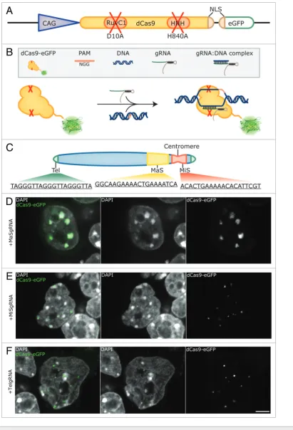
The CRISPR/Cas system features easily programmable sequence recognition, but combines it with an endonuclease activity. We, therefore, introduced mutations to inactivate the endonuclease activity of Cas930-32 and fused it to the enhanced
variant of GFP (eGFP) (Fig. 1A). By co-transfecting a plasmid encoding this eGFP-tagged, nuclease deficient Cas9 (dCas9-eGFP) together with a gRNA expression vector, we aimed to target specific genomic loci in mouse embryonic stem cells. In this way, we expected to achieve specific targeting of dCas9-eGFP without cleavage of the underlying sequences (Fig. 1B).
To test the feasibility of our method, we chose tandemly arranged repetitive DNA sequences, which enabled us to target dCas9-eGFP to extended genomic loci with a single gRNA construct. To this end, gRNAs directed to major (MaSgRNA) and minor satellite (MiSgRNA) repeats, as well as telomeres (TelgRNA) were designed. In mice, major satellite repeats consist of 234 bp repeat units that span within a 6 Mb region, whereas
minor satellite repeats range between 0.6 to 1.2 Mb and consist of 120 bp repeat units.33-36 Telomeric repeats vary in length
reflecting the cell’s replicative potential37 and in mouse amount
to approximately 20–30 kb with the 6 bp repeat sequence TTAGGG (Fig. 1C).
Effective tracing of repetitive DNA sequences using the CRISPR/Cas system
Mouse chromosomes are acrocentric as depicted in Figure 1C. Fluorescent in situ hybridization (FISH) experiments on metaphase chromosomes have shown that minor satellites are centromeric, whereas major satellite repeats occupy the subcentromeric part of the chromosome and are implicated in heterochromatin formation and sister chromatid cohesion.38-41
In interphase nuclei, centromeres cluster and form distinct chromocenters (CCs),41,42 which can be readily distinguished by
enhanced DAPI-staining intensity due to their AT-richness. J1 mouse embryonic stem cells were co-transfected with dCas9-eGFP and MaSgRNA encoding plasmids and imaged 48 h post-transfection. As depicted in Figure 1D, the CCs of MaSgRNA/dCas9-eGFP expressing cells show a bright eGFP signal, verifying the successful targeting of genomic DNA. The distribution exhibits remarkable specificity with very low background signals from freely diffusing dCas9-eGFP.
MiS repeats have been observed as individual focal entities at the periphery of CCs.41 We were able to observe this characteristic
distribution as eGFP fluorescent foci in the corresponding regions of MiSgRNA/dCas9-eGFP expressing cells (Fig. 1E).
Telomeres are capping the ends of chromosomes and have been found localized throughout the nucleus.43 Due to the
acrocentric nature of mouse chromosomes, telomeres located at the acrocentric end can also be detected in the direct vicinity of CCs apart from remote nuclear sites. By expressing TelgRNA/ dCas9-eGFP in J1 cells, small foci, often in the vicinity of CCs, were visible. The observed variable size of labeled telomeres is consistent with the fact that telomeres may form clusters44
(Fig. 1F). In some TelgRNA/dCas9-eGFP expressing cells, an elongated and fiber-like telomere-related eGFP signal was apparent, which was not observed in cells expressing MaS- or MiSgRNA/dCas9-eGFP. Taken together our results show that for all three targeted DNA sequences distinct labeling patterns were observed.
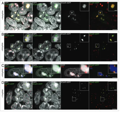
were generated and immuno-FISH experiments were performed. We used a protocol optimized for eGFP epitope preservation and
efficient probe hybridization,45 yielding robust and homogeneous
signals (Fig. 2). As it can readily be seen in Figure 2A, the probe