ABSTRACT
Funke, Melissa Marie. Characterizing a biomedical hydrogel device. (Under the direction of Dr. Christopher R. Daubert.)
E-MatrixTM, a hydrogel principally composed of gelatin and dextran, common food ingredients, is being manufactured with an amino acid formulation and considered as a new medical device. The compound purportedly accelerates the rate of healing once injected beneath a wound. The improved healing is believed to be caused by shifting the healing process from a slower healing adult inflammatory tissue stage to a quicker healing fetal regenerative tissue stage. In addition, gelatin and dextran are anticipated to interact within the medical device to form a stable hydrogel. The objectives of this study were to rheologically characterize E-MatrixTM, develop quality control protocols for evaluation of E-MatrixTM and gelatin, investigate the nature of the proposed relationship between gelatin and dextran, and examine rheological properties of E-MatrixTM components.
E-MatrixTM and the principle ingredient in the material, a 12% gelatin solution. The protocols evaluate specific rheological properties to compare either gelatin lots or manufactured E-MatrixTM batches with established standards. Incorporation of the rheological protocols into a quality control procedure would be a valuable tool for accessing the acceptability of gelatin lots and newly manufactured E-MatrixTM batches. To further understand and characterize E-MatrixTM, studies were performed to examine key physical components of the material. Specifically
solutions of 12% gelatin, 17% gelatin, 5% dextran, 12% gelatin-5% dextran, and the gelatin-rich domain of E-MatrixTM were rheologically examined and compared to rheological properties of E-MatrixTM. In addition the affect of ionic strength and salt valence was also examined through rheological analysis. To determine whether a protein-carbohydrate conjugation resulted from the Maillard reaction, a
spectrophotometric technique was performed to determine the degree of covalent conjugation by measuring the change in free amino groups.
E-MatrixTM was rheologically characterized at 37°C and 50°C as having
pseudoplastic and Newtonian material flow behaviors, respectively. Differential scanning calorimetry determined the calorimetric melt point of E-MatrixTM (23.9°C) and a 12% gelatin solution (26.0°C) to occur sooner than those determined
rheologically (33.7°C) and (32.7°C), respectively. Rheological protocols were
components of E-MatrixTM; individual components, salt type, and ionic strength concentration. Individual E-MatrixTM components were found to differ significantly in regard to rheological properties. However salt type; monovalent versus divalent, using NaCl and CaCl2 was not found to create significant differences for the properties examined in this study, but ionic strength concentration was found to produce rheological properties of significant difference. In addition, according to spectrophotometry, a hypothesized chemical interaction between gelatin and dextran was not likely occurring.
CHARACTERIZING A BIOMEDICAL HYDROGEL DEVICE
By
Melissa M. Funke
A Thesis submitted to the Graduate Faculty of North Carolina State University
in partial fulfillment of the requirements for the Degree of
Master of Science
Department of Food Science
Raleigh, North Carolina 2005
Approved by:
--- --- Dr. Christopher R. Daubert Dr. Brian Farkas
Chair of Advisory Committee
DEDICATION
BIOGRAPHY
Melissa Marie Funke, the second daughter of Ron and Judi Funke, was born on September 25, 1981. She grew up in Cincinnati, Ohio with her parents and older sister Betsy. Melissa was an avid swimmer, softball and soccer player. When Melissa was 12 her family moved to Wilmington, North Carolina and she immediately became a beach bunny. Melissa attended John T. Hoggard High School where she continued to swim competitively and play softball. In 1999, she moved to Wolfpack country, Raleigh, North Carolina, to pursue a degree in food science. As an undergraduate at NC State she continued to be very active and was a member of several organizations including an officer on the NCSU Club Swim Team, vice president of Delta Zeta sorority, and participated in a summer internship at TW Garner Foods working with Texas Pete hot sauce for the one summer she was able to pull herself away from the beach and pool, where she was a lifeguard and swim team coach. After graduating in May 2003, with a bachelor’s of science degree in food science, Melissa stuck around NC State for a few more years in order to earn a Master’s degree in food science and a minor in biotechnology. As a
graduate student Melissa continued to be an active student on campus by being a chairman of the food science club, representative for the Institute of Food
Technology, and an adviser to Delta Zeta sorority.
Upon completion of her master’s degree, Melissa is remaining in the land of sweet tea as she joins Encelle, Inc. in Greenville, North Carolina as a Product
ACKNOWLEDGEMENTS
In the process of getting to the point of completing a thesis and all that good stuff that comes with earning a master’s degree, some very important people have helped me along the way. I would not be here, nor would I be the person I am today,
without my family and friends and I would like to extend countless thanks to the following:
•My parents. I would not be where I am today without them. I thank them for all of
their love and for molding me into the person I am today. •My sister. For being my best friend, listening to me talk wayyyy too fast about a bunch of nothing, and for
being there for me. •My friends. You know who you are and there is no way this
experience would have been nearly as much fun, nor would I have survived without
you. •My adviser, Dr. Chris Daubert. I would like to thank him for his enthusiasm, time, involvement, availability, and desire to see me succeed, and for teaching me
how to make a drinkable cup of coffee. •Rheology lab mates. Thank you Jon, Jeff,
and Junhua for answering all of my mundane questions, Sankar for being a great mentor to me, Sharon, our lab manager for all of her answers and assistance, and
Deepti especially for letting me run SAS on her computer. •Debra Clare and Penny
Amato for their assistance with electrophoresis and DSC, respectively. •Fellow
dungeon/127 Withers dwellers. •The “cool kids” in the labs upstairs. •Last, but
•Thanks to everyone who helped make “the best years of my life” really and truly the
TABLE OF CONTENTS
LIST OF TABLES... xi
LIST OF FIGURES...xiii
CHAPTER 1. INTRODUCTION...1
1.1 RHEOLOGICAL CHARACTERIZATION OF E-MATRIXTM...2
1.2 RHEOLOGICAL PROTOCOL ...3
1.3 EXAMINATION OF E-MATRIXTM COMPONENTS ...3
1.4 SUMMARY...5
CHAPTER 2. LITERATURE REVIEW...6
2.1 INTRODUCTION...7
2.2 MEDICAL DEVICES...7
2.3 HYDROGELS...9
2.4 EXTRACELLULAR MATRIX ... 10
2.5 E-MATRIXTM... 11
2.6 COMPONENTS OF E-MATRIXTM... 12
2.6.1 GELATIN OVERVIEW... 12
2.6.1.1 COLLAGEN ... 13
2.6.1.2 GELATIN ... 14
2.6.1.3 GELATIN PRODUCTION ... 15
2.6.1.5 GELATIN AS A PHARMACEUTICAL
INGREDIENT... 22
2.6.2 DEXTRAN OVERVIEW ... 23
2.6.2.1 MOLECULAR AND PHYSICAL PROPERTIES OF DEXTRAN... 24
2.6.2.2 DEXTRAN AS A PHARMACEUTICAL INGREDIENT... 25
2.6.3 GELATIN-DEXTRAN COMPOUND... 26
2.6.3.1 PHASE SEPARATION... 27
2.6.3.2 EXPLANATION OF MAILARD REACTION ... 29
2.7 RHEOLOGICAL METHODS ... 31
2.7.1 SHEAR STRESS... 32
2.7.2 SHEAR STRAIN ... 32
2.7.3 SHEAR RATE... 33
2.7.4 VISCOSITY ... 33
2.7.5 SHEAR MODULUS ... 33
2.7.6 VISCOELASTICITY... 34
2.7.6.1 COMPLEX MODULUS ... 34
2.7.6.2 PHASE ANGLE ... 35
2.7.6.3 SHEAR STORAGE AND LOSS MODULI ... 35
2.7.7 SHEAR VISCOMETRY ... 36
2.8 CALORIMETRY ... 37
2.10 REFERENCES... 39
CHAPTER 3. RHEOLOGICAL CHARACTERIZATION OF E-MATRIXTM... 62
3.1 INTRODUCTION... 63
3.2 MATERIALS AND METHODS ... 64
3.2.1 SHEAR VISCOMETRY ... 65
3.2.1.1 STRESS SWEEPS ... 65
3.2.1.2 SHEAR RATE RAMPS ... 66
3.2.1.3 FREQUENCY SWEEPS ... 66
3.2.1.4 TEMPERATURE RAMPS ... 66
3.2.2 CALORIMETRY... 68
3.3 RESULTS AND DISCUSSION... 70
3.3.1 STRESS SWEEPS... 70
3.3.2 SHEAR RATE RAMPS... 71
3.3.3 FREQUENCY SWEEPS... 73
3.3.4 TEMPERATURE RAMPS... 75
3.3.5 CALORIMETRY... 77
3.4 CONCLUSIONS... 79
3.5 REFERENCES... 81
CHAPTER 4. A QUALITY CONTROL PROTOCOL TO CHARACTERIZE GELATIN, AN E-MATRIXTM COMPONENT... 94
4.2 MATERIALS AND METHODS ... 96
4.2.1 THE PROTOCOL ... 97
4.2.2 SHEAR VISCOMETRY ... 97
4.2.2.1 STRESS SWEEPS ... 98
4.2.2.2 SHEAR RATE RAMPS ... 98
4.2.2.3 FREQUENCY SWEEPS ... 98
4.2.2.4 TEMPERATURE RAMPS ... 99
4.2.3 PROTOCOL ANALYSIS... 101
4.3 RESULTS AND DISCUSSION... 102
4.3.1 SHEAR RATE RAMPS... 102
4.3.2 FREQUENCY SWEEPS... 105
4.3.3 TEMPERATURE RAMPS... 106
4.3.4 PROTOCOL IMPLEMENTATION... 106
4.4. CONCLUSIONS... 107
4.5 REFERENCES... 108
CHAPTER 5. EXAMINATION OF E-MATRIXTM COMPONENTS... 120
5.1 INTRODUCTION... 121
5.2 MATERIALS AND METHODS ... 124
5.2.1 O-PHTHALDIALDEHYDE ... 124
5.2.2 MONOVALENT AND DIVALENT SALTS ... 124
5.2.2.1 THE PROTOCOL... 125
5.2.3.1 THE PROTOCOL... 127
5.3 RESULTS AND DISCUSSION... 128
5.3.1 O-PHTHALDIALDEHYDE ... 128
5.3.2 MONOVALENT AND DIVALENT SALTS ... 129
5.3.3 E-MATRIXTM COMPONENTS ... 131
5.4 CONCLUSIONS... 134
LIST OF TABLES
CHAPTER 2
Table 2.1 Amino acid composition of gelatin and collagen-residues per 1000
residues ... 45
Table 2.2 Amino acid composition of gelatin ... 46
Table 2.3 Uses of gelatin and dextran ... 47
CHAPTER 3 Table 3.1 Composition of E-MatrixTM samples... 83
Table 3.2 Differential scanning calorimetry values for E-MatrixTM and a 12% gelatin solution ... 84
CHAPTER 4 Table 4.1 Composition of 12% gelatin solution... 110
Table 4.2 Rheological protocol for 12% gelatin solution ... 111
Table 4.3a Comparison of gelatin lots ... 112
Table 4.3b Comparison of gelatin lots ... 113
CHAPTER 5 Table 5.1 Rheological protocol for E-MatrixTM... 136
Table 5.2 Composition of gelatin-rich domain ... 137
Table 5.4 Absorbency of free amino groups present following the OPA
procedure... 139
Table 5.5 Comparison of salts and salt concentrations ... 140
Table 5.6 Comparison of materials... 141
LIST OF FIGURES
CHAPTER 2
Figure 2.1 Triple helix structure of collagen ... 48
Figure 2.2 Process flow diagram of Type A and B manufacturing... 49
Figure 2.3 pH discrepancies between gelatin produced by the acidic or basic method ... 50
Figure 2.4 Amino acid sequence in gelatin ... 51
Figure 2.5 Gelation of gelatin in hot water... 52
Figure 2.6 Mechanism of gelatin gelling... 53
Figure 2.7 Molecular diagram of dextran B512F ... 54
Figure 2.8 Molecular diagram of amylose ... 55
Figure 2.9 Phenomenon of phase separation occurring with the introduction of time and temperature into the system ... 56
Figure 2.10 Maillard reaction scheme ... 57
Figure 2.11 Molecular events in the initial stages of the Maillard reaction ... 58
Figure 2.12 Diagram of shear stress on a cube ... 59
Figure 2.13 Shear flow between parallel plates ... 60
Figure 2.14 Schematic of concentric cylinder... 61
Figure 3.1b Complex shear modulus (G*) for E-MatrixTM samples during stress
sweeps at 37°C... 86
Figure 3.1c Complex shear modulus (G*) for E-MatrixTM samples during stress sweeps at 50°C... 87
Figure 3.2a Stress for E-MatrixTM samples during shear rate ramps from 5-150s-1 at 37°C and 50°C ... 88
Figure 3.2b Viscosity for E-MatrixTM samples during shear rate ramps from 5-150s-1 at 37°C and 50°C ... 89
Figure 3.3 Triplicate averages of storage (G′) and loss (G″) moduli for E-MatrixTM samples during a frequency sweep from 0.01 to 10 Hz at 37°C and 50°C... 90
Figure 3.4 Phase angle and (G′) and loss (G″) moduli for E-MatrixTM samples during cooling (0°C to 25°C) at -1°C/min ... 91
Figure 3.5 Phase angle and (G′) and loss (G″) moduli for E-MatrixTM samples during heating (8°C to 35°C) at -1°C/min ... 92
Figure 3.6 Comparison of a 12% gelatin solution and E-MatrixTM heat flow versus temperature generated from DSC... 93
CHAPTER 4 Figure 4.1a 12% gelatin solution initial shear rate ramps at 50°C... 114
Figure 4.1b 12% gelatin solution initial shear rate ramps at 50°C... 115
Figure 4.2a 12% gelatin solution frequency sweeps at 39°C ... 116
Figure 4.3 12% gelatin solution cooling ramps from 50°C to 11°C... 118 Figure 4.4 12% gelatin solution heating ramps from 8°C to 39°C ... 119
CHAPTER 5
The chapters following the introduction provide a description of the experiments performed by Melissa Funke in the Food Rheology Laboratory at North Carolina State University from 2003 to 2005. Each chapter contains an introductory section with a summary of objectives, scientific approaches, and relevant literature to each particular study. A general overview for each subsequent chapter and the rationale for the project portrayed in the dissertation will be given in this section.
1.1 RHEOLOGICAL CHARACTERIZATION OF E-MATRIXTM
Rheological characterization was performed on E-MatrixTM to establish physical properties and to describe material flow behavior. To investigate the properties of the hydrogel, shear rate ramps, mechanical spectra, and temperature ramps were conducted. Shear rate ramps showed E-MatrixTM displayed pseudoplastic flow behavior at 37°C and Newtonian flow behavior at 50°C. As anticipated, shear rate ramps also showed the material was more viscous at lower temperatures.
Mechanical spectra were performed to investigate viscoelastic properties: shear
storage (G′) and loss moduli (G″) at 37°C and 50°C. The shear loss modulus
dominated across a frequency sweep at each temperature, indicating the material possessed more viscous than elastic behavior. Thermal transition points were also identified both rheologically and calorimetrically, using differential scanning
1.2 RHEOLOGICAL PROTOCOL
Rheological protocols were developed for both E-MatrixTM and the principle
ingredient in the material, a 12% gelatin solution. The protocols can be used as a quality control tool by the manufacturer of E-MatrixTM, Encelle, Inc. of Greenville, North Carolina. The protocols evaluate specific rheological properties to compare either gelatin lots or manufactured E-MatrixTM batches with established standards. These limits would be developed by using the protocol to evaluate numerous E-MatrixTM batches deemed to function successfully. A similar procedure would be used to evaluate gelatin lots used to formulate E-MatrixTM. Incorporation of the rheological protocols into a quality control procedure would be a valuable tool for accessing the acceptability of gelatin lots and newly manufactured E-MatrixTM batches.
1.3 EXAMINATION OF E-MATRIXTM COMPONENTS
concentrations present in E-MatrixTM. A monovalent salt, NaCl, and a divalent salt,
CaCl2, were added to 12% gelatin solutions to examine ionic strength effect at
concentrations below, equivalent, and greater than the ionic strength of E-MatrixTM. Thus the effect of a monovalent salt versus a divalent salt on the compound was investigated in addition to the effect of different salt concentrations. Because E-MatrixTM phase separates into a gelatin-rich domain and a dextran-rich domain at
37°C (body temperature), the gelatin-rich domain, hypothesized to be the functioning phase, was also studied. As anticipated, the different components of E-MatrixTM showed different rheological properties, and this knowledge is important to advance the understanding of E-MatrixTM functionality.
1.4 SUMMARY
To better understand E-MatrixTM functionality, physical and chemical techniques were used to characterize E-MatrixTM. Physical characterization was the focus of this project, specifically with respect to developing rheological techniques to characterize E-MatrixTM and its components. Chemical work was performed to
2.1 INTRODUCTION
Medical devices perform life sustaining functions and biological improvements and consist of an array of products ranging from pacemakers to liquid adhesives.
Gelatin and dextran are common components used for such devices. This literature review is relevant to the characterization and application of gelatin and dextran, specifically as ingredients in medical devices. In order to take a closer examination of a specific medical device, E-MatrixTM, the subsequent sections describe medical devices, E-MatrixTM, the principle components of E-MatrixTM, and rheological
properties. Methods and principles relevant to physically characterizing biopolymers will also be reviewed. Other analytical techniques were deployed throughout this project, and these methods are reviewed, again with respect to gelatin and dextran.
2.2 MEDICAL DEVICES
Medical devices are defined as products which function through a physical action as opposed to a chemical and metabolic action with the later being a drug (Sall 2004). The Food Drug & Cosmetic Act defines a medical device as "...an instrument, apparatus, implement, machine, contrivance, implant, in vitro reagent, or other similar or related article, including a component part, or accessory which is:
other animals, or intended to affect the structure or any function of the body of man or other animals…” Thus medical devices can appear to be both physically and functionally similar to drugs or be physical instruments used in the medical field. Medical devices are used for the purpose of diagnosis, cure, mitigation, treatment, or prevention of disease in humans or other animals or to affect the structure or any function of the body (Sall 2004). Hence there is an array of devices fitting into the medical device category.
To differentiate between various levels of medial devices, the FDA established three classes of medical devices. These device classes are based on the risk posed to the patient and/or the user of the device. Classes are also based on the level of control required to assure the safety and effectiveness of the device. Class I includes devices such as toothbrushes and irrigating syringes which pose the least risks and are subject to general controls (Sall 2004). Moderate risk devices subject to special controls are classified as Class II and include ultrasound imaging systems and pregnancy test kits. The third classification, Class III, are technologically
2.3 HYDROGELS
Many medical devices, such as syringes, sample bags, sample tubes, and artificial organs (blood vessels) are hydrogels (Watanabe and others 2004). A hydrogel is commonly defined as a colloidal gel in which the particles are dispersed in a liquid and the dispersion medium is water. Numerous medical devices are composed of hydrogels because these systems swell under conditions of excess water and retain large amounts of water while hydrated, making hydrogels able to function in the human body since they are able to absorb cell culture medium (Watanabe and others 2004). Hydrogels can act as drug delivery carriers, tissue engineering scaffolds, and biomedical devices (Watanabe and others 2004). A foreign body reaction causing an inflammatory or fibrotic reaction is a common problem following injection of materials into the body (Badylak 2004). However the likelihood of a foreign body reaction can be lowered with the use of hydrogels because the body is constructed of network structures (biomacromolecules) and a large percentage of water, similar to the structure of a hydrogel. Further, the surface free energies of the human body and a hydrogel are similar, also lowering the chance of a foreign body reaction (Watanabe and others 2004).
Balakrishnan and Jayakrishnan (2005) state hydrogels derived from natural proteins and polysaccharides mimic the extracellular matrix of the tissue comprised of
formed by hydrogels made from or cross-linked with unnatural components can produce detrimental effects if produced in excess amounts or leaked into the body (Balakrishnan and Jayakrishman 2005). Furthermore, hydrogel chemical properties are determined by the polymer backbone, functional side-chain in the monomer unit, and the cross-linking agent (Watanabe and others 2004). Using hydrogels to
transport drugs into the body allows stimuli-responsive drug release with stimuli being features such as temperature or pH (Watanabeand others 2004) or the presence of specific enzymes (Kosmala and others 1999). Hence a biomedical device made of a hydrogel can be designed to begin working only once a specific pH, such as that of the human body, is achieved. Because hydrogels are depending on composition can mimic human tissue extracellular matrices, hydrogels make ideal tissue engineering scaffolds (Balakrishnan and Jayakrishnan 2005).
2.4 EXTRACELLULAR MATRIX
and reconstruction after tissue or organ injury (Badylak 2004). These properties make the extracellular matrix an excellent scaffold for tissue engineering
applications, especially tissue repair and reconstruction (Badylak 2004). While attempting to create a man-made extracellular matrix, it is reasonable to incorporate collagen into man-made extracellular matrix because collagen is the most abundant protein within the natural extracellular matrix (Badylak 2004), so when attempting to reconstruct this matrix it is practical to use a collagen containing material; gelatin.
2.5 E-MATRIXTM
EncelleTM Inc. of Greenville, North Carolina developed a medical device, named
E-MatrixTM, which functions as an extracellular matrix. E-MatrixTM is a hydrogel principally composed of conventional food materials, gelatin (12% w/w) and dextran (5% w/w). Because hydrogels composed of natural polymers mimic the extracellular matrix scaffolding of skin (Balakrishnan and Jayakrishnan 2005) that naturally
regenerates and repairs tissue, it makes sense that E-MatrixTM is principally composed of a natural protein found in skin, collagen (gelatin), and a natural
polysaccharide (dextran). Hydrogels made of natural proteins and polysaccharides mimic tissue and extracellular matrices.
from a slower healing adult inflammatory tissue stage to a quicker healing fetal regenerative tissue stage. This switch is crucial as a developing fetus or newborn is able to heal wounds differently and generally better than adults and quicker and more effectively than older adults (Badylak 2004). Potential applications of E-MatrixTM include the following; dermal healing of diabetic foot ulcers and other cutaneous wounds, bone repair through dental surgery, spinal fusion, and non-union fracture repair, connective tissue repair of tendons, ligaments, and cartilage, and soft tissue augmentation in cosmetic surgery, gastroesophageal reflux disease, and urinary incontinence. E-MatrixTM is classified as a Class III medical device and hypothesized to form a matrix under the skin, mimicking an extracellular matrix found in fetal development and supplying a three dimensional scaffolding for tissue reconstruction. Hence, E-MatrixTM has great potential in a variety of areas.
2.6 COMPONENTS OF E-MATRIXTM
Since E-MatrixTM is principally composed of gelatin and dextran, importance was placed on examining these key elements as gelatin and dextran properties may directly and indirectly affect E-MatrixTM functionality.
2.6.1 GELATIN OVERVIEW
Being derived from collagen, gelatin is a protein possessing hydrocolloidal properties permitting functionality in assorted areas. The term hydrocolloid refers to a range of proteins and polysaccharides functioning as thickeners, gelling agents, stabilizing foams, emulsions, and dispersions (Williams and Phillips 2000). A closer
examination of collagen will first be discussed, followed by discussion of amino acid composition of gelatin, production, characterization/classification, physical
properties, and finally the many uses of gelatin.
2.6.1.1 COLLAGEN
Collagen, the backbone of gelatin, is a crucial component of the structural support system of vertebrates and invertebrates, because it is the major protein constituent of skin, tendon, cartilage, bone, and white fibrous connective tissue in mammals (Balian and Bowes 1977). Collagen is composed of three parallel tropocollagen strands which are chemically crosslinked to form a triple helix, giving collagen the triple helix formation (Figure 2.1). The tropocollagen rod is the fundamental molecular unit of collagen. When heated, the chemical cross-links that bind the tropocollagen monomers together break, and each tropocollagen strand becomes an individual gelatin molecule (Murano 2003). The total molecular weight of
tropocollagen is approximately 330,000, it has a 1.5 nm diameter, and a length of approximately 300 nm (Ledward 2000). Mild heating (40°C) causes the triple helix
to unfold, producing α-chains, β-chains, and γ-units. The β-chains consist of two
2000). Furthermore, collagen contains both intramolecular crosslinks and several intermolecular bonds (Ledward 2000).
The intra and intermolecular links cause animal tissue to have high tensile strength. As animals age, the number of stable linkages increase and these linkages begin to form trivalent (linking three instead of two collagen molecules together) linkages (Ledward 2000). This phenomenon is the cause of tougher collagen in older animals, and these stable linkages result in the need for severe processing of
collagen to create a soluble gel. These factors explain why commercial gelatins vary widely in molecular weights from 50,000 to 1,000,000 (Ledward 2000). Hence the initial state of the collagen and conversion from collagen to gelatin strongly dictates the final functional properties of gelatin.
2.6.1.2 GELATIN
Pure collagen cannot be used to produce gelatin, so converting collagen to gelatin and removing the greatest amount of non-collagenous material must occur (Johns 1977), a process crucial for the successful production of gelatin. Note gelatin is the derived protein of collagen, whereas gelatine is the total product produced from collagen- comprised of gelatin as the main component, plus various smaller
Though structurally the parent collagen is very similar to the resultant gelatin, some differences may exist, such as the amide content, conversion of arginine side
chains, and the overall balance of amino acid composition (Eastoe and Leach 1977). If the parent collagen is exposed to an alkaline treatment, the amide content may lower. In addition, prolonged exposure to alkali may increase the conversion of arginine side chains and the gelatins are often richer in the abundant amino acids and poorer in the rarer residues than parent collagens (Eastoe and Leach 1977). Also, when gelatin and collagen are placed in dilute acid or alkali, gelatin will
dissolve, but collagen will only swell or hydrate (Ledward 2000). Furthermore, when mildly heating (< 50°C) gelatin and collagen, gelatin again readily dissolves to form a viscous solution at all pHs, but collagen only shrinks and becomes unable to hold water (Ledward 2000). Hence the processing method used to manufacture gelatin plays an important role in the final material properties.
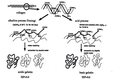
2.6.1.3 GELATIN PRODUCTION
Prior to a mild heat treatment to promote the irreversible break down of the fibrous collagen structure (Eastoe and Leach 1977), gelatin is trapped within the three-dimensional structure of collagen. To obtain gelatin, the secondary and higher structures (Ledward 2000) of collagen must be disrupted, which occurs during
presence of water. Acid and base processes produce gelatins commonly referred to as Type A and Type B, respectively. Figure 2.2 is a process flow diagram of these processes, and Figure 2.3 and Table 2.1 identify discrepancies which may result from the different processing method in regard to pH and amino acid composition, respectively. Following these extraction processes, gelatin is dried to form coarse granules, fine powders, or thin sheets, which are odorless, tasteless, and yellowish in color. Granules and powders are commonly produced in America, while sheets are more common in Europe.
Since gelatin is a protein, it is constructed of eighteen amino acids (Kirk-Othmer 1999). Glycine, proline, and hydroxyproline are the most abundant amino acids in gelatin and tend to form the most prevalent amino acid sequence present in collagen and gelatin; Gly-Pro-Hypro-Gly (deMan 1999). Overall, glycine is the most prevalent amino acid in gelatin. Table 2.2 lists the amino acid composition of gelatin, and Figure 2.4 is a common amino acid sequence within gelatin.
derived from all three α-chains which each differ (Eastoe and Leach 1977). Although a common structure is present, some unusual structures do exist.
Uncommon structures speculated to be present in gelatin include “esterlike” bonds,
aldehyde groups, and γ -glutamyl linkages (Eastoe and Leach 1977).
Because gelatin lacks tryptophan and contains only small amounts of some other essential amino acids, it is considered an incomplete protein, but is digestible by humans (Ensminger and others 1994). As a nutritionally incomplete protein, gelatin still has numerous applications. In fact, gelatin possesses several unique physical characteristics which compensate for its nutritional deficiency.
conditions, gelatin is capable to function as both a protective colloid and as a
flocculating agent (Wood 1977). In addition, the clarity of gelatin gels adds to the list of gelatin functionality (de Vries 2004). Furthermore, the unique molecular and physical properties of gelatin are a vital component in making this material functionally versatile.
Just as gelatins span an array of uses, individual gelatin molecules also differ greatly. Commercially desirable physical characteristics of gelatin include gel
strength, viscosity, adhesiveness, tack, color, and clarity, while related to the intrinsic chemical composition, molecular weight, and molecular configuration of gelatin is important (Veis 1964). Distinguishable physical characteristics of gelatin as previously noted, determine gelatin classifications such as quality grades. Manufacturing gelatin results in the production of various gelatin grades
distinguished by bloom strength, viscosity, and molecular weights which range from ~20,000 to 250,000 (Kirk-Othmer 1999).
homogenous by heating and stirring at 60°C. The gel is left at ambient conditions for 15 minutes, placed in a 10°C bath for 16-18 hours and finally tested with the Bloom gelometer (Ledward 2000). Unfortunately this test is very sensitive and difficult to repeat. A Texture Analyzer is a more modern instrument used to determine Bloom strength. A bloom value of 100 means a force of 100 g was required to plunge 4 mm into the gel. The higher the bloom value, the higher the grade and price.
The higher grade gelatins have gone through the fewest extractions during processing, and these extractions were performed at the lowest temperatures as higher temperatures cause proteolysis (Ledward 2000). In addition, gelatins of lighter color reflect higher grades (Ledward 2000). As previously mentioned, the strong inter and intra molecular links cause the heterogeneous composition of molecular weights within gelatin. Besides size variation, charge distribution also varies (Williams and Phillips 2000). In addition, moisture content varies and commonly ranges from 9-14% (Eastoe and Leach 1977). More specifically, food grade gelatins range in moisture from 8-12%, have less than 2% ash, and the remaining composition is protein (Ledward 2000). Therefore, there is a wide variability among gelatins.
2.6.1.4 PHYSICAL PROPERTIES OF GELATIN
product. Immersion of dry gelatin granules or sheets in hot water can lead to gelatin solubilising and the creation of a random coiled polypeptide solution (Murano 2003). At or above 40°C, gelatin is assumed to exist as a random coil and may still have some helical structure (Williams and Phillips 2000) from the collagen. With time and at the appropriate temperature, a gel will eventually form (Murano 2003). Figure 2.5 illustrates how adding dry gelatin to hot water can lead to the formation of a viscous gelatin solution. Upon cooling the solution, a thermo-reversible gel, can be formed. Transformation between solutions and gels is readily possible because physically, gelatin solutions increase viscosity with a decrease in temperature and can melt with a temperature increase (Murano 2003). This thermo-reversibility is due to gelatin possessing inter and intra-molecular hydrogen bonding (Chatterji 1990). Because junction zones are bound by weak hydrogen bonds, at temperatures of 35-40°C the hydrogen bonds will break causing the gel to melt (de Vries 2004; Abeles and others 1992).
The mechanism of gelation is simple, yet complex (Figure 2.6). At temperatures below 40°C gelatin molecules readily aggregate and gelation occurs to form a gel (Williams and Phillips 2000). As the temperature is lowered, the pyrolidine-rich regions of gelatin chains act as nucleation sites for the formation of potential junction zones (Ledward 2000). Junction zones are ordered structures of two or more
then formed (Guo and others 2003; Gekko 1993) when aggregation of three proline-L-proline II helix occurs (Ledward 2000; Gekko and Fukamizu 1991). The triple helix acts as gel junction points or zones, stabilized by inter chain hydrogen bonds. With time and accelerated by a decrease in temperature, additional junction zones form and existing junction zones grow. With junction zone growth and formation, the gel becomes more organized and stronger. In time, gel organization and strength increase. The growth of existing junction zones is believed to be the key contributing factor to increased gel strength.
In addition to time being a factor in increasing gel strength, concentration is also a factor. Increased concentration improves the chances that two or three different chains will form a junction. As the gel becomes more organized with time, the gel also becomes more thermally stable (Ledward 2000). Gekko and Fukamizu (1991) suggest gelatin gels result from large exothermic effects caused by the
intermolecular hydrogen bonds overcoming the entropy loss associated with the order of the polypeptide chains. Aggregation and growth of junction zones can continue to form a more mature and stronger gel over time.
factor within the pH range of 4-10 (Ledward 2000), but outside this range, gelation is greatly inhibited. Gelatin inhibition is most likely due to the high net positive or negative charge carried by the chains, so electrostatic forces prevent the formation of junction zones (Ledward 2000). Therefore at pH 1-3 and 11-14 gelatin will not readily gel.
Gelatin is insoluble in cold water; however, completely soluble in warm water.
Swelling of gelatin compares to the swelling of non-ionized crosslinked polymers, but the effects are complicated by charge interactions and the structure of the gelatin network (Wood 1977). Dry gelatin granules are capable of swelling and absorbing 5-10 times their weight in water to form a gel at 35-40°C (Ensminger 1994).
2.6.1.5 GELATIN AS A PHARMACEUTICAL INGREDIENT
Gelatin is not limited to the food, paper, or photographic industries. According to economic reports, gelatin use as a pharmaceutical ingredient is on the rise in the United States (Kirk-Othmer 1999). Gelatin is used most commonly as a drug encapsulating shell (Ledward 2000), permitting smooth oral drug delivery. Gelatin also binds pharmaceutical components in the manufacture of tablets. Furthermore, dentists use gelatin foam cubes to absorb blood during treatment, and in hospitals gelatin is used as blood substitutes to counteract high blood loss (Ledward 2000). In the current study, gelatin was used as a functional component and spacer in a
2.6.2 DEXTRAN OVERVIEW
Dextran is a polysaccharide composed of D-glucose subunits (Figure 2.7) connected by more than 50% α-1,6 glycosidic linkages with different additional branching
through α-1,2, α-1,3, and α-1,4 linkages (Monsan and Auriol 2004; Murano, 2003). Dextran is a non-charged D-glucosyl homopolysaccharide (Monsan and Auriol 2004), lacking strong viscosifying capabilities (Tromp and others 2004). Visually dextran is a white powder. Production of dextran is caused by lactic acid bacteria (Monsan and Auriol 2004) with Leuconostoc mesenteroides being used most prevalently for production of clinical grade dextran (de Belder 1996). At the industrial level, production is caused by partial hydrolysis, fractionation, and purification of native dextran produced using the bacterium Leuconostoc
mesenteroides to make a polymer consisting of approximately 95% α-1,6 linkages (Kennedy and others 1984), making it quite linear and having a small number of side chains. However physical properties of dextran, such as low viscosity, make dextran a beneficial, key ingredient in numerous ways in both the food and pharmaceutical industries. Furthermore, the fact that dextran is biodegradable and a natural
2.6.2.1 MOLECULAR AND PHYSICAL PROPERTIES OF DEXTRAN
This large and neutral polysaccharide comes in a range of molecular weights ranging from 500 to 6000 kDa (Monsan and Auriol 2004; Ebert and Schenk 1968; Groenwall and Ingelman 1948). Large physical size allows dextran to be used
widely in the medical field since it is too large to pass through uninjured vessel walls. The majority of dextran physical properties are dependent on the molecular weight distribution (de Belder 1996). For example, the viscosity of dextran is directly proportional to the molecular weight as Equation 1 illustrates (de Belder 1996) with [η] representing the intrinsic viscosity.
[η] = 2.43 x 10⁻³M
w0.42 (1)
Molecularly, dextrans are nearly identical to amylose, as amylose is a linear
sequence of glucose molecules linked together (Figure 2.8), but the slight branching of polymeric chains in dextran prevents stacking and causes most dextrans to be soluble in water (Walstra 2003) and electrolytes (de Belder 1996). Biologically, dextran can be degraded once in vivo by degrading enzymes present in animal and human tissues. However most hydrolysis products produced by dextran can be absorbed to rapidly raise the blood sugar and liver glycogen levels. If dextran is orally introduced into the body, it passes through the small intestine and metabolism or degradation occurs by colonic bacterial flora and fermented short-chain fatty acids in the small intestine (Monsan and Auriol 2004; de Belder 1996). However, a
solutions are thermally-stable, and consequently can be sterilized and used as injectable pharmaceutical components.
2.6.2.2 DEXTRAN AS A PHARMACEUTICAL INGREDIENT
Dextran plays an active role in the pharmaceutical industry. Specifically, unmodified dextran, with a molecular weight ranging from 75,000 ± 25,000, is used as a blood plasma substitute, as 6% solutions have a viscosity value similar to that of human blood plasma (Kennedy and others 1984). This polyglucose has been incorporated as a plasma expander and a drug deliverer. Additionally, iron dextran is injected into muscles for iron deficient patients. Infusing dextran into the patient intravenously is quite beneficial because its size is too large to pass through uninjured vessel walls and it is osmotically active. Furthermore, the emulsion polymerization of dextran with epichlorihydrin leads to insoluble cross-linked dextran beads which swell in water. This property has led to the ability of dextran to separate molecules by size and thus to be used in biological research. Dextran beads are used to treat skin wounds and lesions as they absorb wound exudates in secreting infected wounds and therefore accelerate the wound healing time (de Belder 1996). In the current project, dextran is used as a key component of E-MatrixTM directing cells toward a
2.6.3 GELATIN-DEXTRAN COMPOUND
Gelatin and dextran are natural, biodegradable polymers (Bigi and others 2004; Zhang and Chu 2000; Tabta and Ikada 1998). This feature is important because the gelatin-dextran compound is highly responsive to stimulus and has a good
biocompatibility with the body (Kurisawat and Yui 1998). Furthermore, Kosmala and others (2000) state combining gelatin and dextran can produce a selectively
enzymatically, but not hydrolytically, degradable hydrogel. Therefore incorporation of gelatin-dextran into drug delivery systems permits selective delivery of the drug only to sites containing the appropriate enzymes. Also hydrogels can aid in three dimensional tissue reconstruction scaffold (Watanabe and others 2004).
The addition of dextran to gelatin, even in small magnitudes, can powerfully increase the gelation rate of the solution and increase the elastic modulus of gelatin-dextran gels (Tolstoguzov 1993). It is thought this gelation rate increase occurs because junction zones leading to gelation form between polymeric chains and with the addition of dextran, the number of available chains forming junction zones increases (Tolstoguzov 1993). Kurisawa and Yui (1997) suggested that physical chain
2.6.3.1 PHASE SEPARATION
Like many polysaccharide and protein mixtures, gelatin and dextran are incompatible in aqueous solutions (Edelman and others 2001). Due to this
instability, phase separation, the phenomenon in which the formation of regions of different compositions form, (Walstra 2003) may take place with the introduction of time and temperature (Figure 2.9).
Two types of phase separation may occur within a polymer mixture such as the mixture of gelatin (2), and dextran (3), with water (1) as the solvent. One type is segregative phase separation and the second is associative phase separation (Walstra 2003). The two kinds of phase separation can be distinguished by a value called the second virial coefficient (B). Virial coefficient is a function of
physicochemical parameters and denotes the interaction between two polymers (gelatin and dextran) (Vivares and Bonnete 2004). Segregative separation occurs when the second virial coefficient B23>0 and the two components separate into a
phase rich in one polymer (2) and poor in the other (3) and a second phase rich in 3 and poor in 2 is formed (Walstra 2003). At w/w concentrations above 3% of both components, segregative phase separation occurs (Edelman and van der Linden 2001). Also, in this type of separation, the two polymers avoid each other and the polymers are named incompatible (Walstra 2003). If B23<0, associative phase
coacervate also occurs if the polymer concentration is large and B23~0 (Walstra
2003).
For a gelatin-dextran-water solution, at sufficient concentrations, usually above 3% (w/w) for each component, phase separation occurs forming a gelatin-rich region and a dextran-rich region. It is suggested the phase separation of gelatin and dextran is an entropy-driven process and the total polymer concentration at which phase separation occurs is nearly independent of temperature. Although heat is required for the biopolymer to separate into two domains, limited temperature dependence occurs between 40 and 80°C. As would be expected, above gelation temperature (~30°C), gelatin-dextran compounds exhibit normal liquid-liquid phase separation, but once the temperature is lowered below ~30°C and gelation occurs, separation is reduced (Edelman and others 2001). At the warmer temperatures (40-80°C), which are above the gelation temperature, gelation does not occur, and composition and hydrodynamic conditions determine the structure of the resulting two phase system (Packe and others 2003).
molecules throughout. Examination of E-MatrixTM under a microscope revealed
gelatin was the continuous phase of the multi phase system. Post phase separation, the dextran molecules may be trapped among the gelatin, causing dextran rich and gelatin rich domains with an amount of trapped dextran molecules remaining in the gelatin rich portion. Additionally, a viscosity difference exists between the gelatin-rich domain and the dextran-gelatin-rich domain (Edelman and others 2001).
2.6.3.2 EXPLANATION OF MAILLARD REACTION
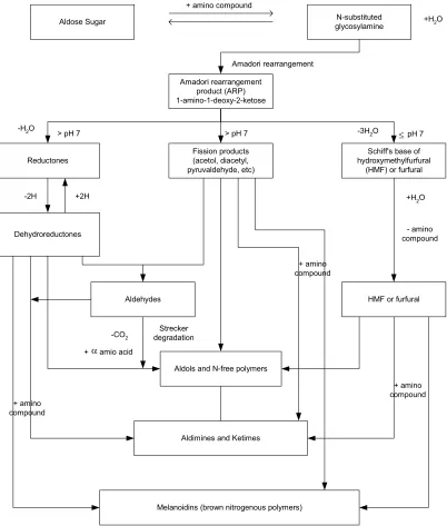
The Maillard reaction, also referred to as non-enzymatic browning, is a very
important chemical phenomena to the food industry. The reaction (Figures 2.10-11) affects food quality in processing and storage, as it may promote color change, aroma, and nutritional decline in foods. The reaction is a sequence of events beginning with the reaction of the amino group of amino acids, peptides, or proteins with a glycosidic hydroxyl group of sugars (Ellis 1959). This sequence terminates with the formation of brown nitrogenous polymers or melanoidins (Ellis 1959). Lysine is usually the most reactive amino acid due to its free ε-amino group (deMan 1999). The Maillard reaction is described by Hurst (1972) as a series of five
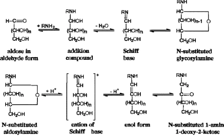
chemical steps. The first step involves an N-substituted glycosylamine being
produced from an aldose or ketose, reacting with a primary amino group of an amino acid, peptide, or protein (deMan 1999; Hurst 1972). This step is followed by the glycosylamine being rearranged by a reaction yielding an aldoseamine or
manipulated again with a second aldose to form a diketoseamine; or aldosamine reacts with a second amino acid to yield a diamion sugar. Amino or nonamino compounds are next produced by the degradation of amino sugars, losing one or more water molecules. Finally condensation of the amino or nonamino compounds, formed in the previous step, with each other or amino compounds, form brown pigments and polymers (deMan 1999). The brown pigments are a characteristic mark of the Maillard reaction having occurred and can be seen in browning of baked cookies.
A number of variables affect the Maillard reaction, including time, temperature, water content, water activity, pH, oxygen, metals, phosphates, sulfur dioxide, concentration of reactants, and the type of reactants and solvents (Miao and Roos 2004; deMan 1999). In addition, the viscosity of the matrix material affects the Maillard reaction rate (Miao and Roos 2004). Glass transition, water content, and water activity also affect the reaction rate (Craig and others 2001; Le Meste 1995; Karmas and others 1992).
Studies have deduced the Maillard reaction was responsible for covalently
chromatography to confirm the covalent attachment of dextran to ε-polylysine
through a Maillard reaction with the ε-polyllysine amino groups and the reducing-end carbonyl group of dextran. Another study also claims the Maillard reaction is
responsible for the covalent bond of a protein-carbohydrate substance with dried egg white being the protein and the polysaccharide being galactomannan with the bond occurring between the ε-amino groups in the protein and the reducing-end carbonyl residue in the polysaccharide (Kato and others 1993). Even though the Maillard reaction occurs more readily and rapidly at higher temperatures, it is noted that even at room temperature the reaction between polylysine and dextran takes place (Ho and others 1999). Thus lower temperatures, such as body temperature (37˚C), may be adequate for the Maillard reaction to occur.
2.7 RHEOLOGICAL METHODS
2.7.1 SHEAR STRESS
Stress (σ) is a measurement of force and is defined as a force per unit area (Equation 2). Stress is commonly expressed in Pascals (N/m2), and Figure 2.12 demonstrates the application of a shear stress onto an object.
A F =
σ (2)
2.7.2 SHEAR STRAIN
Strain is a dimensionless quantity representing the deformation of a physical body under the action of applied forces. The direction of the applied stress to the material surface determines the type of strain; normal strain or shear strain. Normal strain occurs when stress is applied to the surface of the material in a perpendicular
(normal) direction which occurs during compression (Daubert and Foegeding 1998). The second type of strain, shear strain, occurs when a stress is applied parallel to the surface of the material (Equation 3). In Equation 3 the shear strain (γ) is the
inverse tangent of the change in distance divided by the initial height of the material.
δ = γ − h d 1
2.7.3 SHEAR RATE
For fluids, a shear stress can induce a unique type of flow called shear flow (Figure 2.13). The differential change of strain with respect to time is known as shear (strain) rate (γ&), calculated as Equation 4, with units of sec-1.
dt dγ =
γ& (4)
2.7.4 VISCOSITY
Apparent viscosity (η) is the measurement of the resistance to flow or the fluidity of a material and is mathematically calculated as the ratio of shear stress to shear rate.
γ σ = η
& (5)
2.7.5 SHEAR MODULUS
Shear modulus (G) is the constant of proportionality used to relate shear stress with shear strain (Daubert and Foegeding 1998).
γ σ =
2.7.6 VISCOELASTICITY
Viscoelastic materials exhibit both viscous and elastic properties. Viscous properties possess fluid-like behavior, while elastic properties represent the solid properties of a material. Measurement of viscoelastic properties can be done using dynamic oscillation of shear stress or strain. Oscillation studies are the most common dynamic method for examining viscoelastic behavior (Steffe 1996). Harmonic oscillation of shear stress involves a material being oscillated sinusoidally with varying stress while the resulting strain is measured (Zhong 2003). When
performing dynamic tests, four assumptions are assumed: 1.) a constant stress or strain throughout the sample; 2.) no slip of the sample; 3.) sample homogeneity; and 4.) that measurements are performed within the linear viscoelastic region.
Furthermore, key functions describe viscoelastic behavior of a material, such as complex moduli, phase angle, storage modulus, and loss modulus.
2.7.6.1 COMPLEX MODULUS
The complex shear modulus (G*) is the ratio of stress and strain amplitudes during the oscillation and gives an indication of the strength of a gel as defined in Equation 7.
2 2
o o
* =σ = (G) +(G )
G ′ ′′
γ (7)
γ0 = shear strain amplitude
G′ = storage modulus
G″ = loss modulus
2.7.6.2 PHASE ANGLE
Phase angle (δ) is also related to the viscoelasticity of a material and is directly
related to the energy lost per cycle divided by the energy stored per cycle
(Steffe1996). Phase angles can vary from 0 to 90°, with 0° indicating an ideal solid material (Hookean solid) and 90° indicating an ideal viscous material (Newtonian fluid). Equation 8 describes the phase angle calculation, and in the current research, phase angle was used to designate gel and melt points.
G G = ) tan(
′ ′′
δ (8)
2.7.6.3 SHEAR STORAGE AND LOSS MODULI
The shear storage modulus (G′) indicates the degree of elastic behavior in a
material. Shear storage modulus is the component (in phase) with the stress, or the elastic behavior defined by Equation 9.
2.7.7 SHEAR VISCOMETRY
Rotational viscometry is a key approach for obtaining rheological measurements of viscoelastic fluid materials. Rotational viscometry involves an attachment with known geometry placed in contact with a sample, followed by mechanical rotation to shear the sample (Daubert and Foegeding 1998). Rheological attachments include concentric cylinders, cone and plate, and parallel plate geometries and mixers. The concentric cylinder is commonly referred to as the cup and bob because this
apparatus consists of a cup with radius Rc, and a bob with a slightly smaller radius
Rb that is placed inside the cup. Figure 2.14 displays the concentric cylinder.
2.8 CALORIMETRY
Differential scanning calorimetry (DSC) is a thermal analysis technique in which the difference in energy inputs into a substance and a reference material is measured as a function of temperature while the substance and reference are subjected to a controlled temperature program (Hohne and others 2003; Schenz and Davis 1999). Usually the sample is sealed inside a metal or ceramic pan and the reference is an empty pan of the same composition. The DSC technique can be used to detect phase transitions and other thermodynamic reactions within a sample (Resch 2004). The output from a DSC is the combination of all endothermic reactions (occurring when a material absorbs heat) and exothermic reactions (when heat is given off) that occur in the substance as it is heated (Schenz and Davis 1999). Common examples of endothermic reactions are melting, protein denaturation, and starch gelatinization, while common examples of exothermic reactions are crystallization (sugar, ice) and curing (crosslinking due to heat, chemical additives, or UV light). In this project DSC was used to determine a thermal transition point indicative of melting E-MatrixTM and a 12% gelatin solution.
2.9 SUMMARY
2.10 REFERENCES
Abeles RH, Frey PA, Jencks WP. 1992. Biochemistry. Boston: Jones and Bartlett Publishers. 212-217.
Badylak SF. 2004. Extracellular Matrix Scaffolds. In: Wnek GE and Bowlin GL. Encyclopedia of Biomaterials and Biomedical Engineering. NY: Marcel Dekker Inc. 561-567.
Balian G, Bowes JH. 1977. The Structure and Properties of Collagen. In: Ward, AG and Courts, A. The Science and Technology of Gelatin. New York: Academic Press. 1-30.
Balakrishnan B, Jayakrishnan A. 2005. Self-cross-linking biopolymers as
injectable in situ forming biodegradable scaffolds. Biomaterials 26(18):3941-3951.
Bigi A, Panzavolta S, and Rubini K. 2004. Relationship between triple-helix content and mechanical properties of gelatin films. Biomaterials 25(25):5675-5680. Chatterji PR. 1990. Interpenetrating Hydrogel Networks. I. The Gelatin-
Polyacrylamide System. J Appl Polym Sci 40(3-4):401-410.
Craig ID, Parker R, Rigby NM, Crairns P, Ring SG. 2001. Maillard reaction kinetics in model preservation system in the vicinity of glass transition: experiment and theory. J Agric Food Chem 49(10):4706-12.
Daubert CR, Foegeding EA. 1998. Rheological Principles for Food Analysis. In: Neilsen, SS editor. Food Analysis 2nd edition. Gaithersburg, MD: Aspen Publishers. 551-569.
de Belder AN. 1996. Medical Application of Dextran and Its Derivatives. In: Dumitriu,S. Polysaccharides in Medicinal Applications. New York: Mercel Dekker, Inc. 505-523.
Dellacherie E. 1996. Polysaccharides in Oxygen-Carrier Blood Substitutes. In: Dumitriu S ed. Polysaccharides in Medical Applications. NY: Marcel Dekker, Inc. 525-544.
deMan JM. 1999. Principles of Food Chemistry. Aspen Publishers, Inc.: Gaithersburg, MD. 120-172.
Cambridge, UK: The Royal Society of Chemistry. 23-31.
Diftis, N, Kiosseoglou V. 2004. Competitive adsorption between a dry-heated soy protein-dextran mixture and surface-active materials in oil-in-water emulsions. Food Hydrocolloids 18:639-646.
Eastoe JE, Leach AA. 1977. Chemical and Constitution of Gelatin. In: Ward AG and Courts A. The Science and Technology of Gelatin. New York: Academic Press. 73-107.
Ebert KH, Schenk G. 1968. Mechanism of biopolymer growth: The formation of dextran and levan. Adv Enzymol 30:179-210.
Edelman MW, van der Linden E, de Hoog E, Hans Tromp R. 2001. Compatibility of Gelatin and Dextran in Aqueous Solution. Biomacromolecules 2:1148-1154. Ellis GP. 1959. The Maillard reaction. In: Advances in carbohydrate chemistry.
Vol. 14, ed. ML Wolfrom and RS Tipson. New York: Academic Press. Ensminger AH, Ensminger ME, Konlande JE, Robson JRK. 1994. Foods
and Nutrition Encyclopedia 2nd ed. Ann Arbor, MI: Pegus Press. 554 and
1057.
Finot PA, Bujard E, Mottu F, Mauron J. 1977. Availability of the true Schiff’s base of lysine and lactose in milk. In: Friedman, M. Ed; Protein Crosslinking:
Nutritional and Medical Consequences. Plenum: NY. 343-364.
Friedman M. 1982. Chemically reactive and unreactive lysine as an index of browning. Diabetes. 31(Suppl. 3). 5-14.
Gekko K, Fukamizu M. 1991. Effect of pressure on the sol-gel transition of gelatin. Int J Biol Macromol 13:295.
Gekko K. 1993. The sol-gel transition of food macromolecules under high pressure. In: Nishinari, K and Doi, E. Editors. Food Hydrocolloids
Structures, Properties, and Functions. NY: Plenum Press. 259-264.
Gelatin Manufacturers Institute of America, Inc. Gelatin. New York, NY, 1993:5-6. Groenwall AJ, Ingelman BGA. 1948. Manufacture of infusion and injection fluids.
US Patent 2,437,518.
Guo L, Colby RH, Lusignan CP, Howe AM. 2003. Physical Gelation of Gelatin Studied with Rheo-Optics. Marcromolecules 36:10009-10020.
ε-polylysine by conjugation with dextran through the Maillard reaction. Food Chem 68(4):449-455.
Hodge JE. 1953. Chemistry of Browning Reactions in Model Systems. J of Agric Food Chem 1(15):928-943.
Hohne GWH, Hemminger WF, Flammersheim HJ. 2003. Differential Scanning Calorimetry. 2nd ed. Germany: Springer. 1.
Hudson CB. 1993. Gelatine-Relating Structure and Chemistry to Functionality. In: Nishinari, K. and Doi, E., editors. Food Hydrocolloids Structures, Properties, and Functions. New York: Plenum Press. 353-354.
Hurst DT. 1972. Recent developments in the study of nonenzymatic browning and its inhibition by sulphur dioxide. BFMIRA Scientific and Technical Surveys No. 75. Leatherhaed, England.
IUPAC Compendium of Chemical Terminology. 1997. 2nd edition.
Johns P. 1977. The Structure and Composition of Collagen Containing Tissues. In: Ward, AG and Courts, A. The Science and Technology of Gelatin. New
York: Academic Press. 31-72.
Karmas R, Buera MP, Karel M. 1992. Effect of glass transition on rates of non- enzymatic browning in food systems. J Agric Food Chem 40(5):873-9. Kato A, Minaki K, Kobayahi K. 1993. Improvement of Emulsifying Properties of
Egg White Proteins by the Attachment of Polysaccharide through Maillard Reaction in a Dry State. J Agric Food Chem 41(4):540-543.
Kennedy, JF, AJ Griffiths, DP Atkins. 1984. The Application of hydrocolloids: Recent developments, future trends. In: Gums and Stablilsers for the Food Industry 2: Application of Hydrocolloids. New York: Pergamon Press. 417-455.
Kirk-Othmer. Concise Encyclopedia of Chemical Technology 4th ed. A Wiley- Interscience Publication John Wiley and Sons Inc. New York. 1999. Kosmala JD, Henthorn DB, Brannon-Peppas L. 2000. Preparation of
interpenetrating networks of gelatin and dextran as degradable biomaterials. Biomaterials 21(20):2019-2023.
199(8):1547-Ledward DA. 2000. Gelatin. In: Phillips, G.O. Williams, P.A. editors. Handbook of hydrocolloids. Boston: Woodhead Publishing Limited. 67-86.
Le Meste M. 1995. Mobility of small molecules in low and intermediate moisture foods. In: Barbosa-CanovasG, editor. Food preservation by moisture
control: Fundamental and applications. Lancaster, Pa.: Technomic. 209-23. Lodish H, Arnold B, Zipursky L, Matsudaira P, Baltimore D, Darnell J. 2000.
Molecular Cell Biology. NY: W. H. Freeman Publishing.
Martins SIFS, Jongen WMF, van Boekel MAJS. 2001. A review of Maillard reaction in food and implications to kinetic modeling. Trends in Food Sci and
Technology 11(3-4):364-373.
Miao S, Roos, YH. 2004. Comparison of Nonenzymatic Browning Kinetics in Spray-dried and Freeze-dried Carbohydrate-based Food Model Systems. JFS. 69(7):E322-331.
Monsan PF, Auriol D. 2004. Dextran and Glucooligosaccharides. In: JR Neeser and JB German. Bioprocesses and Biotechnology for Functional Foods and Nutraceuticals. NY: Marcel Dekker, Inc. 135-149.
Murano PS. 2003. Understanding Food Science and Technology. Belmont, CA: Thomson Wadsworth.
Nakamura S, Kato A, Kobayashi K. 1991. New Antimicrobial Characteristics of Lysozyme-Dextran Conjugate. J Agric Food Chem 39(4):647-650. Norton IT, Frith WJ. 2003. Phase separation in mixed biopolymer systems. In:
Dickinson, E. and Van Vliet, T. Food Colloids Biopolymers and Materials. 292.
Pacek AW, Ding P, Norton T. 2003. Effect of Temperature and Hydrodynamic Conditions on Structure and Drop Size in a Phase-Separated
Gelatin+Dextran System. In: Dickinson E and Van Vliet, T eds. Food Colloids Biopolymers and Materials. UK: The Royal Society of Chemistry. 309-318.
Ross-Murphey SB. 1992. Structure and rheology of gelatin gels: recent progress. Polymer 33(12):2622-2627.
Ross-Murphey SB. 1991. Incipient behavior of gelatin gels. Rheologica Acta 30:401-411.
editors. FDA Regulatory Affairs A Guide for Prescription Drugs, Medical Devices, and Biologics. New York: CRC Press. 153-193.
Schenz TW and Davis EA. 1999. Thermal Analysis. In: Nielsen SS, editor. Food Analysis. 2nd ed. Gaithersburg, MD: Aspen Publishers, Inc. 587-598. Steffe JF. 1996. Rheological Methods in Food Process Engineering. 2nd ed. MI:
Freeman Press.
Tabata Y, Ikada Y. 1998. Protein release from gelatin matrices. Advanced Drug Delivery Reviews. 31:287-301.
Tolstoguzov VB. Thermodynamic Aspects of Food Protein Functionality. In: Nishinari, K. and Doi, E. editors. Food Hydrocolloids Structures, Properties, and Functions. New York: Plenum Press. 336-337.
Tromp RH, Edelman, MW, van der Linden E. 2004. Gelatine/dextran solutions- a model system for food polymer mixtures. In: Williams, P.A. and Phillips, G.O.Gums and Stabilisers for the Food Industry. 12. Cambridge, UK: The Royal Society of Chemistry. 201-210.
Veis A. 1964. The Macromolecular Chemistry of Gelatin. New York: Academic Press, Inc.
Vivares D, Bonnet F. 2004. Liquid-Liquid Phase Separations in Urate
Oxidase/PEG Mixtures: Characterization and Implications for Protein Crystallization. J Phs Chem B 108(20):6498-6507.
Walstra P. 2003. Physical Chemistry of Foods. NY: Marcel Dekker, Inc. 179-186. Watanabe J, Kiritoshi Y, Nam KW, Ishihara K. 2004. Hydrogels. In: Wnek GE
and Bowlin GL editors. Encyclopedia of Biomaterials and Biomedical Engineering. NY: Marcel Dekker, Inc. 790-801.
West JL. 2004. Biofunctional Polymers. In: Encyclopedia of Biomaterials and Biomedical Engineering. Wnek GE and Bowlin GL. NY: Marcel Dekker Inc. 89-95.
Williams PA, Phillips GO. 2000. Introduction to food hydrocolloids. In: Phillips GO, and Williams PA editiors. Handbook of hydrocolloids. Boston: Woodhead Publishing Limited. 1-20.
Wood PD. 1977. Technical and Pharmaceutical uses of Gelatin. In: Ed. A.G. Ward, and A. Courts. The science and technology of gelatin. New York:
Zhang Y and Chu CC. 2000. Biodegradable dextran-polyactide hydrogel network and its controlled release of albumin. J of Biomedical Materials Research 54(1):1-11.
Zhong Q, Daubert CR. 2003. Cooling effects on the functionality and
Table 2.1. Amino acid composition of gelatin and collagen-residues per 1000 residues
Amino acid
Type I collagen (bovine)
Type A gelatin (acid processed
pigskin)
Type B gelatin (alkali processed
bone)
Alanine 114 112 117
Arginine 51 49 48
Aspargine 16 16 ־־־
Aspartic acid 29 29 46
Glutamine 48 48 ־־־
Glutamic acid 25 25 72
Glycine 332 330 335
Histidien 4 4 4
4-Hydroxyproline 104 91 93
ε-Hydroxylysine 5 6 4
Isoleucine 11 10 11
Leucine 24 24 24
Lysine 28 27 28
Methionine 6 4 4
Phenylalanine 13 14 14
Proline 115 132 124
Serine 35 35 33
Threonine 17 18 18
Tyrosine 4 3 1
Valine 22 26 22
Table 2.2. Amino acid composition of gelatin
Amino acid g amino acid/100g protein
Glycine 27.5
Alanine 11.0
Valine 2.6
Leucine 3.3
Isoleucine 1.7
Serine 4.2
Threonine 2.2
Methionine 0.9
Phenylalanine 2.2
Tryosine 0.3
Proline 16.4 Hydroxyproline 14.1
Aspartic acid 6.7 Glutamic acid 11.4
Lysine 4.5
Arginine 8.8
Table 2.3. Uses of gelatin and dextran
Gelatin Dextran
Ice and sugar crystallization inhibition in frozen
cream products Plasma volume expander
High quality gels with clean melt in mouth texture Blood flow improvement
Golf ball core material Thrombosis prophylaxis Emulsifying agent in cosmetics, pharmaceuticals,
water paints, and disinfectants Organ perfusion and preservation Sedimentation agent for defibrinated blood Distending media for hysteroscopy
Cork binding agent Decrease blood viscosity
Viscosity increaser Increase blood flow Water holding capacity utilized in low calorie
sweets and spreads Disappearance of platelet aggregation Retain juiciness of meat products Increase in capillary pressure
Beer clarification Decrease in tissue pressure Controls crystal growth in electroplating and
electrolyte metal refining Decrease in friction between red cells and capillaries Bonding agent for photosensitive silver bromide
in photography Anti-sludge effect on red cells
Floculating agent in extraction of uranium ore Protection against the apparition of intravascular coagulation Stabilizing agent and emulsifier for emulsions
and foams
Hard and soft capsule shells, tablets, pastilles, protective dressings, gelatin sponge, surgical powder, suppositories, plasma expender Sizing agent for paper, paper coating for carbon printing, gelatin composition rollers for printing, gummed paper tape, glues in the coated abrasive industry and carton/box manufacture, book binding
Type B: Degreeased dried, crushed bone
Type B: Dehaired
cattle hides Type A: Pigskins
Acid treatment
Lime treatment Lime treatment
Water watsh
Acid treatment
Washing
Filtration Acid treatment
Chilling to set point Ion exchange Multiple hot water extraction
Filtration
Sterilization Evaporation
Drying Extrusion
Water watsh
Miling
Blending & packaging
Ossein Dicalcium
phosphate
Dry gelatin
Hot water
Gelatin sol
(Viscous liquid)
Thermoreverisble gelatin gel
(Visco-elastic solid)
Cooling
Hydration
Temperature depression
Gelatin chain pyrolidine-rich regions act as nucleation sites
Junction zones form in pyrolidine-rich
regions
Proline-L-proline II helix taken up by
pyrolidine-rich regions
3 helices aggregate
A triple helix resembling collagen forms & acts as gel junction zones
(zones stablized by inter hydrogen bonds)
Gel forms with increase in number of junction zones & as junction zones increase in size
Figure 2.8. Molecular diagram of amylose (Murano 2003) Nonreducing
end