The Role Of Actin Purse-Strings In
Wound Healing And In The Natural
Movements Of Embryonic
M orphogenesis
A th esis su b m itted in M arch
1997 fo r the d egree o f Ph.D .
ProQuest Number: 10017444
All rights reserved
INFORMATION TO ALL USERS
The quality of this reproduction is dependent upon the quality of the copy submitted.
In the unlikely event that the author did not send a complete manuscript and there are missing pages, these will be noted. Also, if material had to be removed,
a note will indicate the deletion.
uest.
ProQuest 10017444
Published by ProQuest LLC(2016). Copyright of the Dissertation is held by the Author.
All rights reserved.
This work is protected against unauthorized copying under Title 17, United States Code. Microform Edition © ProQuest LLC.
ProQuest LLC
789 East Eisenhower Parkway P.O. Box 1346
A b str a c t
The Role Of Actin Purse-Strings In Wound Healing And In The
Natural Movements Of Embryonic Morphogenesis
Skin wounds in limb-bud stage chick embryos re-epithelialise using a contracting filamentous actin cable which assembles in the basal layer o f the wound edge epithelial bilayer within 2 minutes of wounding (Martin and Lewis, 1992a). Characterising the contractile apparatus by immunostaining reveals intracellular segments of the actin cable inserting into clustered adherens junctions at the cell-cell boundaries, as well as
colocalisation of myosin H protein localising to the basal wound margin within 7 minutes o f wounding.
Epithelial cells at the wound edge are transiently permeabilised by the w ounding process, allowing loading of small molecules into precisely the cells which assemble actin
cable,thus providing a method of investigating the signals which regulate cable assembly. The Ras-related small GTPases regulate rapid reorganisations of the actin cytoskeleton in cultured fibroblasts (Ridley & Hall, 1992; Ridley et al, 1992). I show that co-loading of w ound edge cells with fluorescent dextran and Rho inactivator, C3 transferase, blocks assembly of actin cable and prevents re-epithelialisation. By contrast, introducing dominant negative NlVRac into the wound edge epithelial cells has no affect on the healing process, suggesting that Rho, but not N17Rac, is the molecular switch regulating w ound-induced assembly of an actin cable.
The keratin intermediate filaments of embryonic epithelium are required for repair of w ounds in Xenopus embryos (Torpey et al, 1992). W ounded lim b-bud stage m ouse embryos assemble a filamentous keratin-8 cable in wound edge epithelial cells in addition to the actin cable described above. In contrast to the Xenopus depletion experim ents, I show that transgenic knock-out mouse embryos null for KB (Baribault et al, 1993; 1994) completely re-epithelialise their wounds within 24 hours just like their wild-type litter mates demonstrating that despite an inability to assemble keratin filaments, wound repair proceeds normally.
T ab le o f C on ten ts
A b s t r a c t ... 2
T a b le o f
C o n t e n t s ...3
T a b le o f
f i g u r e s ...1 7
C h a p te r
1 G en eral I n tr o d u c tio n ... 2 1
The Mechanisms Underlying Adult Tissue Repair...21
~ adult tissue repair requires re-epithelialisation, fibroplasia
and angiogenesis...21 ~ wounds rapidly reseal and bleeding is stemmed by a
fibrin p lu g ...22 ~ an inflammatory response is initiated upon wounding
adult tissu e ... 22
The mechanism underlying adult wound
re-epithelialisation... 23
~ epithelial cells migrate into the wound site and
proliferate... 23 ~ the basal lamina is usually absent from beneath migrating
epithelial cells... 23 ~ kératinocytes extend lamellipodia and crawl across open
wound granulation tissue to heal a w o u n d ... 24 ~ kératinocytes secrete ECM "stepping stones" on which
they can crawl and they change integrin expression to facilitate their own migration across inhospitable
g ra n u la tio n tis s u e ...24
The mechanism underlying adult wound fibroplasia...25
~ connective tissue cells rapidly proliferate, migrate into the wound space beneath the fibrin clot and then synthesise
and secrete ECM in the wound s i t e ... 25 ~ wound fibroblasts differentiate into myofibroblasts and
~ angiogenesis is stimulated concurrent with ECM
deposition by fibroblasts... 26
~ regeneration of dermally located epithelial appendages is rare at a wound s i t e ... 27
~ dermal tissue is remodelled from provisional matrix to collagenous scar... 27
Growth factors initiate and co-ordinate
re-epithelialisation, fibroplasia and angiogenesis... 28
~ EGF and T G F -a found at the w ound site are potent stim ulators o f epiderm al p ro life ra tio n ...28
~ topical application of EG F and T G F -a at the wound site speeds up k eratin o cy te m ig ra tio n ...29
~ FGFs regulate re-epithelialisation and angiogenesis...29
~ PDGF is a m ajor co-ordinator o f fib ro p lasia ... 30
~ VEGF is a potent angiogenic factor at the wound site...31
~ TGF-p enhances re-epithelialisation... 31
~ TG F-p co-ordinates tissue re p a ir... 32
The Mechanisms Underlying Embryonic And Foetal Tissue
Repair...34
- a brief history of wound healing studies in the e m b ry o ... 34
~ embryonic re-epithelialisation is driven by contraction of an actin cable in the basal layer of wound margin cells...36
~ basal lamina is maintained beneath the repairing epithelial wound ed g e... 37
~ epithelial cell proliferation is not the driving force for re-epithelialisation ... 37
~ there is no inflammatory response initiated at an embryonic wound s ite ... 38
~ macrophages are not recruited to embryonic w o u n d s... 38
- wounding embryonic epithelium triggers transient immediate early gene expression... 38
~ the growth factor profile at an em bryonic wound is different from that at an adult wound s i t e ...40
~ angiogenesis is rapidly stimulated in the embryonic
w o u n d ... 41 - differences in the mechanism of foetal skin repair favour
scar free healing ... 41
The Cytoskeletal Actin Microfilament Network And Its
Regulation By Small GTP Binding Proteins... 44
~ actin monomer binding proteins regulate filament
form ation... 44 ~ actin filament assembly and disassembly is regulated by
the gelsolin family of severing and capping p ro te in s ... 45 ~ actin filament binding proteins organise filament
architecture... 45 ~ myosin motor proteins mediate contraction o f actin
f il a m e n t s ... 46 ~ intermediate filaments assemble into a branching network
throughout c e lls ... 47 ~ adherens junctions link cells either to each other or to
substratum ... 48 ~ the adhesion molecules of adherens junctions are tissue
specific and subclass specific... 48 ~ regulating plaque proteins cluster on the cytoplasmic side
of adherens ju n c tio n s... 49 ~ some plaque proteins are common to both adherens
junctions and focal contacts...49 ~ some plaque proteins are found only at cell-substrate
j u n c t i o n s ...50 ~ some plaque proteins are found only at cell-cell
j u n c t i o n s ... 50 ~ formation of a focal contact involves tyrosine
phosphorylation, receptor occupancy and integrin
clustering... 50 ~ how is a spot-like punctae adhérentes ]Mwc\\on assembled
and regulated?... 51 ~ adherens junctions transmit signals between the cell and
~ small ras-like GTP binding proteins regulate actin polymerisation into filaments and adherens junction
assembly in both fibroblasts and epithelial cells... 54
~ RhoA regulates actin stress fibre assembly in fibroblast cells and peripheral bundle formation in epithelial c e lls ...55
~ Rac regulates lamellipodial formation in fibroblasts...56
~ growth factors may activate Rho via G EF coupled r e c e p t o r s ...56
~ the broad investigative aim s o f this th esis...57
C h a p te r 2 G en eral M a teria ls A nd M e th o d s ... 5 8
Preparation and wounding of chick em bryos... 58
~ windowing eggs to expose em bryos in o v o... 58
~ general method of wounding em b ry o s...58
Immunostaining techniques... 59
- actin stain in g e m b ry o s...59
~ whole mount immunolabelling of em bryos... 69
~ m ounting em bryos on s lid e s ... 60
Preparation of tissue for Electron Microscopy... 60
~ preparation of embryos for Scanning Electron M icroscopy... 60
~ preparation of tissue for resin histology and T E M ...60
~ collection, storage and presentation of d ata... 61
C h a p te r 3 C h aracterisation O f The C o n tra c tile A p p a r a tu s
W h ic h A ssem b les In T he W ou n ded E p ith eliu m O f A
C h ic k E m b r y o ...6 2
Introduction... 62
~ stretched epidermal cells assemble actin stress fibres
along lines of ten sio n ... 62
1] Is the cable’s presence necessary for repair?... 63
~ cytochalasins can be used to block polymerisation of
monomeric actin into filam ents...64
2] What proteins are associated with the cable and when
are individual components of the purse-string
machinery localised in the basal epithelial layer?... 64
~ actin cable requires myosin to be contractile...64 ~ dorsal closure in Drosophila is driven by actin-myosin
band contraction... 65 ~ intracellular segments of actin cable must anchor at cell
cell boundaries into adherens ju n c tio n s ... 65 ~ are adherens junctions redistributed at the wound margin
or assembled de n o v o l... 65 ~ how rapidly does the contractile apparatus assem ble?...66 ~ there is a lag-period following wounding before
epithelial movement b eg in s...66 ~ proliferation is unlikely to play a role in initiating
epithelial re p a ir...66
3] Is the actin purse-string machinery contractile?...67
~ not all actin-myosin stress fibres are contractile... 67 ~ BDM, a myosin II ATPase inhibitor blocks actin
filament contraction in vitro...68
4] How does the cable drive epithelial m ovem ent?... 68
~ changes in cell shape may reveal how the repairing
e p ith e liu m m o v e s ... 68
5] Determining the transition point from cable driven to
lamellipodial driven repair...69
~ transitions in epidermal and dernial mechanisms of repair
ch aracterise em bryonic d e v e lo p m e n t...69 ~ embryonic epithelial cells are capable of crawling when
- studying a rapid response by w ounded epithelial cells
requires development of a new wound healing m o d el...70
Results...71
Description of my wound healing m odel... 71
~ incisional slash wounds take approximately 6 hours to
h e a l ... 71
Characterisation of the contractile apparatus... 7 6
~ wounded tissue organises a purse-string of actin in the
basal layer of the bilayer within 2 minutes of w ou n d in g ...76 ~ myosin H colocalises to the wound front with actin
w ithin 7 m inutes o f w o u n d in g ... 76 ~ cadherins cluster at the wound margin in the basal layer
where the cable is lo c ated ... 77 ~ vinculin also clusters in the basal layer at the wound
m argin... 78
A functional actin cable is required for epithelial
repair...83
~ cytochalasin D prevents polymerisation of actin into a
cable at the wound m a rg in ... 83 ~ cadherins still cluster at the wound margin in the
presence of cytochalasin D ... 83 ~ contraction of the actin cable is required for
re-epithelialisation ... 84 ~ actin cable remains transiently in epithelial cells for a
short time after they have re sealed ...85
A transition in mechanism of re-epithelialisation occurs
at E 9... 92
~ the chick epithelium up to and including E8, repairs a
wound solely by assembling an actin cable...92 ~ between E9-10 epithelial wounds heal both by actin cable
and lamellipodial crawling m echanism s... 92
Discussion And Conclusions...99
~ the dynamics of organising a functional actin
purse-s t r i n g ... 100
~ adherens junctions may act as nucléation sites for actin fila m e n t p o ly m e ris a tio n ... 101
~ BDM - a M yosin-ATPase inhibitor is a useful tool to block purse-string contraction... 102
~ a possible mechanism of "zipping-up"... 102
~ what happens to the actin cable at the transition point?... 103
Materials and Methods... 105
~ wounding method for cable dynamics studies looking at actin localisation, myosin distribution and cadherin im m unostaining...105
~ wounding method for cytochalasin D stu d ie s...105
~ table of numbers of embryos (3 wounds per em bryo) used to assess affects of cD on rate of wound repair by S E M ...106
~ table of total number o f em bryos used (3 wounds per embryo) in the study to assess actin and cadherin distribution at the wound margin in the presence of c D ... 106
~ wounding method for BDM stu d ies...106
~ table to show number of em bryos (3 wounds per embryo) assessed for rate of repair by SEM in the presence o f B D M ... 107
~ table o f chick embryo numbers (3 wounds per embryo) used in BDM study and stained for actin visualisation...107
~ wounding method for epithelial transition point wound healing studies...107
C h a p te r 4 T he G TP B in d in g P ro tein R h o R e g u la tes A ctin
C a b le A s s e m b ly ... 1 0 9
Introduction... 109
~ cells in vitro and in vivo can tolerate membrane
dismptions which rapidly re-seal and are n on -fatal... 109 ~ cells in tissue culture and in 3D collagen matrices can be
scrape-loaded with reagents in the extra-cellular flu id ... 110 ~ small Ras-like GTP binding proteins regulate formation
of different organisations of actin in fibroblasts and
epithelial cells... 110 ~ Rho protein activity is tightly regulated by activating and
inactivating groups of p ro te in s... I l l ~ many effects of active Rho are mediated via
p h o s p h o lip id s ... 112 ~ Rho family proteins regulate focal contact assembly in
addition to actin filam ent reo rg an isa tio n s...112 ~ Rho proteins are ubiquitously expressed in many cells
and tis s u e s ... 113 ~ Rho proteins are involved in regulating contractile actin
microfilament organisation...114 ~ Ras proteins are clearly implicated in regulating wound
healing...114
Results...116
~ epithelial wound margin cells are efficiently loaded with
dextrans during the wounding p ro c e s s ...116 ~ basal epithelial wound margin cells co-loaded with Rho
inactivator and rhodamine-dextran fail to assemble an actin
cab le...117 ~ individual dextran/C3-free wound margin cells can still
assemble cable... 117 ~ C3 treated w ounds fail to re-ep ith elialise... 118 ~ concentrations of C3 lower than 10 |ig/ml fail to fully
block actin cable assembly and subsequent
~ incisional wound mesenchyme contraction does not
appear im paired in C3 treated w o u n d s...125 ~ the extensive mesenchyme contraction required during
healing of excisional wounds is disrupted by C 3 ...125 - cadherins do not appear to cluster at the wound margin
early in the wound healing response in the presence of C 3 126 ~ dominant negative N17Rac loaded wounds assemble an
actin cable and repair n o rm ally ... 126
Discussion And Conclusions...131
~ summary of results obtained in Chapter 4 ... 131 ~ tracking cell fate confirms that cells drop out from the
wound margin during re p a ir...132 ~ Rho and Rac are likely to regulate actin anchorage into
adherens ju n c tio n s ... 132 ~ Rho regulates actin polymerisation and cable assembly in
the basal epithelial la y e r... 133 ~ Rho family members are important regulators of
m orphogenesis... 134 ~ studies in Drosophila reveal information about signalling
upstream and dow nstream o f Rho fam ily G T P ases...135 ~ do small GTPases regulate actin filament contraction?...135 ~ mesenchyme contraction may be effected by Rho
regulated stress fibre contraction... 136 ~ the expression of specific "co-ordination genes" is
required to co-ordinate epithelial sheet movements in
m orphogenesis... 137
Materials and Methods... 139
~ demonstrating that the transient permeabilisation during wounding results in dextran uptake by wound margin
cells... 139 ~ blocking Rho and Rac function in basal wound margin
cells... 139 ~ Table of numbers of wounds exposed to C3 transferase
or N17Rac, and the extent of wound closure at various
- investigating adherens junction clustering in basal cells at
the wound margin in the presence o f C3 transferase...141 ~ experiments investigating Rho blocking effect on
mesenchyme contraction... 142 ~ assessing the extent of mesenchyme contraction in C3
treated chick embryos by D il labelling of the wound
m esenchym e... 142
C h a p te r 5 T he R ole O f In term ed ia te F ila m en ts In
E p ith e lia l R e p a ir ... 1 4 3
Introduction...143
~ keratins assemble as obligate heterodimers of type I and
type n subunits...143 ~ expression of keratin genes is developm entally regulated 143 ~ keratin anchors at the plasma membrane into
desmosomes and hem idesm osom es... 144 ~ keratin anomalies in humans lead to friction sensitive
s k in ... 146 ~ keratins are needed for structural support in the Xenopus
embryo for both morphogenetic movements and to repair
small incisional w ounds... 146 ~ gene deletion in transgenic mice is a useful way of
testing the role of an individual protein in tissue repair... 147 ~ excisional wounds made to mouse embryo hind limbs
repair 50% by re-epithelialisation and 50% by underlying
mesenchyme contraction... 148
Results...149
- keratin-8 protein distribution in wounded epithelium suggests a support role for keratin filaments during
re-epithelialisation... 149 ~ keratin-8 deficient mouse embryos can not assemble a
~ keratin-8 deficient embryos can still assemble an actin
c a b le... 150 ~ wound closure is just as rapid in keratin-8 deficient
embryos as in their wild-type siblings...151 ~ there is no extra compensatory mesenchyme tissue
contraction in keratin-8 deficient embryos to facilitate rapid
re-epithelialisation of w o u n d s...151
Discussion And Conclusions...159
~ summary of results obtained in Chapter 5 ... 159 ~ is murine gastrulation a test of structural integrity or is it
less gruelling than X e n o p u s g astru latio n ?...159 ~ is there functional redundancy in the sy stem ?... 160 ~ are adult-type keratin heteropolymers upregulated in
m k 8 -/ - e m b ry o s ? ... 160 ~ are wound specific keratins upregulated in the em bryonic
epithelium?... 161 ~ what effect do strain variances have on the effectiveness
of targeted mk8 gene m utations?...162 - do keratins play a purely structural role in cells or do
they regulate cell proliferation as w e ll? ... 162
Materials and Methods... 164
- mouse embryo dissection, preparation, wounding and
m etho d o f c u ltu r e ... 164 ~ assessing the rate of excisional wound re-epithelialisation
by scanning electron microscopy (S E M )...164 ~ visualisation of keratin and actin filaments at the wound
site in wild type and mk8-/ - e m b ry o s ... 165 ~ assessing the extent of mesenchyme contraction by D il
labelling of the wound m esenchym e... 165 ~ genotyping progeny of heterozygote crosses... 166
C h a p ter 6 A n In V itro W ou n d H ealin g M od el U sed To
I n v e s tig a te S ig n a ls W h ich R eg u la te P u r se -S tr in g in g
V e r s u s C r a w lin g ... 1 6 7
Introduction... 167
~ an unusual colonic epithelium-derived cell line repairs
wounds by both purse-stringing and c ra w lin g ... 167 ~ why do Caco-2gBE cells assemble a p u rse-strin g?...168 ~ is purse-string assembly regulated in Caco-2BBE cells by
Rho in an analogous manner to cable assembly in
em bryonic w ound m argin c e lls ? ... 168 ~ effective tissue repair requires the careful
synchronisation of cell migration, cell-rearrangements and proliferation in both epithelium and underlying
m esenchym e... 169
Results...171
~ small wounds can assemble either entirely cable, or cable
and lamellipodia, or entirely lam ellipodia... 171 ~ the epithelium gapes considerably upon w ounding...171 ~ only small wounds appear to assemble cable in all
wound margin c e lls ...172 ~ cells rarely extend a lamellipodium and assemble cable
s im u lta n e o u s ly ... 172 ~ cable containing cells can drop out from the leading
e p ith e lia l w ou nd e d g e ... 172 ~ distinctive cell morphology seen by SEM differentiates
cabling versus crawling c e lls ... 173 ~ C3 loading wound margin cells blocks cable assembly,
but not lam ellipodial extension at a wound m argin... 179 ~ N17Rac loading cells does not prevent cells from
assembling c a b le ... 180 - gap junction communication between cabling cells may
Discussion And Conclusions...187
~ summary of results obtained in Chapter 6 ... 187
~ cabling versus crawling - what are the deciding factors for a wound margin cell?... 187
~ is stretch a signal for Caco cells to assemble c a b le ? ... 187
~ are cell dimensions a differentiating factor in cabling or craw ling?... 188
~ do wound margin cells change their mechanism of repair d u ring the h ealin g p e rio d ? ... 189
~ what does the distribution of gap junctions tell us about inter-cellular communication in cabling and crawling c e lls ? 189
Materials and Methods... 191
~ m eth o d o f cell c u ltu re ... 191
~ preparation o f cells for S E M ...192
~ staining cells for a c tin ... 192
~ mounting coverslips for visualisation on the confocal microscope...192
~ im munostaining for Connexin 4 3 ... 193
~ blocking actin cable assembly with C3 tran sferase... 193
- loading N 17R ac into w ound m argin c e lls ... 193
C h a p te r 7
G e n e r a l
D is c u s s io n ... 1 9 5
Summary Of Thesis Results... 195
Chapter 3 ...195
Chapter 4 ...195
Chapter 5 ...196
Chapter 6 ...196
What Role Does Stretch Play In Cable Assembly And
Wound Repair?...197
~ growth factors are unlikely to trigger Rho mediated cable assembly at the embryonic wound m a rg in ... 197
~ cell stretch may mediate most of the signals required to re p a ir a w o u n d ... 197
~ Rho may regulate lEG expression in epithelial wound
margin c e lls ... 198
What Has Studying Purse-String Machinery During
Wound Repair Told Us About Analogous
Morphogenetic Movements ?... 199
~ why do embryonic epithelial cells organise an actin
cable?...199 ~ polymerised actin filaments localise to the apices of
epithelial cells during neurulation... 200 ~ morphogenetic movements of epithelial sheets such as
gastrulation during embryonic development may utilise
actin cable m achinery...202 ~ suggestion of a possible mechanism of gastrulation in
Xenopus which utilises principles derived from my studies
of actin cable dynamics in repairing w o u n d s... 203 ~ future prospects in the field of tissue repair...205
T a b le o f fig u r e s
C hapter 1
F ig 1 Cartoon diagrams of the simplified organisation of proteins at the cell-cell adherens junction and cell-matrix focal contact
C hapter 3
Results
Fig 1 Scanning electron micrographs to show re-epithelialisation of normal incisional slash wounds
F ig 2 Scanning electron micrographs o f normal repairing wounds at high m agnification F ig 3 Resin histology of repairing incisional slash wounds
Fig 4 In ovo photography and confocal laser scanning m icroscopy o f repairing epithelial wounds made to the dorsum o f chick wing buds
Fig 5 Confocal laser scanning microscopy of wounded epithelium to reveal the time course of actin redistribution into a cable in basal wound margin cells
Fig 6 Confocal laser scanning microscopy o f wounded epithelium revealing a time course of myosin localisation to the basal wound margin
Fig 7 Confocal laser scanning microscopy of wounded epithelium to reveal cadherin distribution at the repairing epithelial wound edge
Fig 8 Confocal laser scanning microscopy of cytochalasin D treated epithelial wounds to demonstrate the role of actin cable in re-epithelialisation
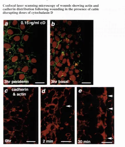
Fig 9 Confocal laser scanning microscopy of wounds showing actin and cadherin distribution following wounding in the presence of cable disrupting doses of cytochalasin D
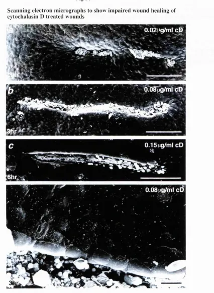
F ig 10 Scanning electron m icrographs to show impaired w ound healing of cytochalasin D treated wounds
Fig 11 Scanning electron micrographs o f BDM treated epithelial wounds
Fig 12 Confocal laser scanning m icroscopy of BDM and control wounds early in the wound healing response
Fig 13 Confocal laser scanning microscopy and cartoon to dem onstrate the m echanism of zipping up at the end of incisional slash wounds using actin cable and anchoring adherens junctions
Fig 14 Confocal laser scanning microscopy of repairing epithelial wounds m ade to the right hind limb of late stage chick embryos (E8-B10)
Fig
15 Resin histology of repairing wound made to the right hind limb flank of an ElO chick embryoFig 16
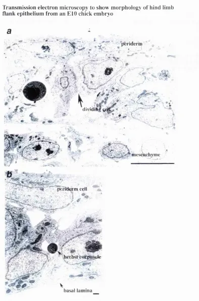
Transmission electron microscopy to show m orphology of hind limb flank epithelium from an ElO chick embryoFig 17
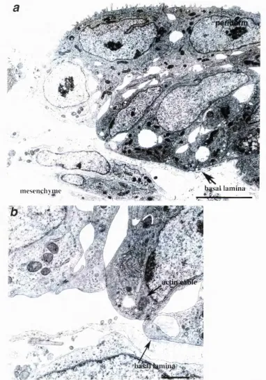
Transm ission electron microscopy to show detailed m orphology o f cabling repairing epithelium from incisional wounds made to the hind limb thigh of ElO chick em bryosFig 18
Transmission electron microscopy to show detailed morphology o f craw ling repairing epithelium from incisional wounds made to the hind limb thigh of ElO chick em bryosC hapter 4
Introduction diagram of the Ras-like GTP binding protein Ras, showing its activators, inhibitors, and some of the effectors of active Rho-GTP
R e s u lt s
Fig 1 Confocal laser scanning microscopy dem onstrating dextran loading o f wound margin cells
Fig 2 Confocal laser scanning microscopy dem onstrating normal repair of dextran loaded w ounds
Fig 3 Confocal laser scanning microscopy of incisional epithelial wounds, C3 transferase treated to block endogenous rho activity
Fig 4 Scanning electron microscopy o f epithelial wounds fixed 3 hr after w ounding in the presence of Rho inactivating C3 transferase or dominant negative N17Rac
Fig 5 Scanning electron microscopy o f epithelial wounds fixed 6 hr after w ounding in the presence of Rho inactivating C3 transferase or dominant negative N lTR ac
Fig 6 Scanning electron microscopy to show epithelial w ound edge m oiphology in control and C3 treated wounds
Fig 7 Resin histology of C3 treated incisional wounds
Fig 8 Fluorescent microscopy of D il labeled excisional wounds made to the dorsum of chick wing buds in the presence of C3 transferase, to assess affect of blocking Rho activity in wounded mesenchyme cells.
C hapter 5
Introduction Fig 1 Cartoon diagram o f the interm ediate filam ent m echanism s of anchorage at the cell membrane
R e s u lt s
F ig 1 Confocal laser scanning microscopy of hind limb amputation w ounds m ade to the E l l . 5 mouse embryo, immunostained to reveal keratin-8 filament distribution
F ig 2 Confocal laser scanning microscopy of hind limb amputation w ounds m ade to the E l 1.5 wild-type and mkS-/- mouse embryos, immunostained to reveal keratin-18 filament distribution and filamentous actin
F ig 3 Scanning electron microscopy o f repairing hind limb amputation wounds made to the E l 1.5 mouse embryo
F ig 4 Scanning electron microscopy o f repairing wounds made to keratin-8 deficient, heterozygote and wild-type mouse embryos
Fig 5 Scanning electron microscopy showing morphology of repairing epithelial wound edges in keratin-8 deficient, heterozygote and wild-type mouse embryos
Fig 6 Fluorescence microscopy showing similar wound mesenchym e contraction in keratin-8 deficient, heterozygote and wild-type mouse embryos 24hr after wounding
C hapter 6 R e s u lts
Fig 1 Confocal laser scanning microscopy of monolayers of Caco-lggE cells both wounded and unwounded
Fig 2 Confocal laser scanning microscopy of repairing small and m edium size wounds made to monolayers of Caco-2gBE cells
Fig 3 Graph to show rate o f Caco wound repair
Fig 4 Scanning electron microscopy of repairing medium size wounds m ade to monolayers of Caco-2gBE cells
Fig 5 Confocal laser scanning microscopy of early repair in monolayers of Caco-2BBE cells wounded in the presence of C3 transferase
F ig 6 Confocal laser scanning microscopy of repair in monolayers of Caco-2BBE cells wounded in the presence of C3 transferase
F ig 7 Confocal laser scanning microscopy of m onolayers wounded in the presence of myc-tagged NlVRac.
Fig 8 High magnification confocal laser scanning microscopy of m onolayers wounded in the presence of myc-tagged NlVRac
C hapter 7
Fig 1 Confocal laser scanning microscopy of transverse and longitudinal sections of neural tube stage chick embryos demonstrating actin filament bundles localising to neural folds
C hapter 1
G en era l In tr o d u c tio n
This thesis investigates the mechanism of re-epithelialisation of em bryonic wounds. Studying embryonic tissue repair has two main purposes. Firstly, it is a sim ple system for studying general mechanisms of tissue repair and secondly, re-epithelialisation utilises an actin purse-string contractile machinery which is likely to be employed during the morphogenetic movements o f development. Therefore it is a controllable model for studying the genetic regulation of morphogenesis. I describe a system in em bryos for studying the rapid changes in the cytoskeleton that occur in wound edge cells immediately following wounding, characterising the contractile machinery and investigating the signals which trigger its assembly. I investigate the importance of the contribution from different cytoskeletal networks to the repair process and introduce the use of a novel cell line which can be used in concert with in vivo studies to dissect more fully, the genetic regulation underlying re-epithelialisation of wounds.
This introductory chapter is divided into 3 sections which review the current understanding of:
1) The mechanisms underlying adult tissue repair
2) The mechanisms underlying embryonic and foetal tissue repair
3) The cytoskeletal actin microfilament network and its regulation by small GTP binding proteins
T h e M ech a n ism s U n d erly in g A d u lt T issu e R ep a ir
~ adult tissue repair requires re-epithelialisation, fibroplasia and
angiogenesis
Repair following injury to skin requires the rapid regeneration of lost and dam aged tissues and their subsequent reorganisation into normal, quiescent dermal and epidermal architecture. Rapidity of repair is essential along with a strong inflammatory response to minimise the chances of infection from the external environment. The processes
underlying adult repair are a well chronicled sequence of events which are similar throughout vertebrate phyla.
The process of tissue restoration following wounding can be divided for ease of study and description into its tributary components. The wound site tissue is described as granulation tissue due to its granular appearance and consists of fibroblasts and
myofibroblasts with their surrounding extra-cellular matrix, together with large numbers o f invading inflammatory cells (macrophages and neutrophils) and a network of new capillaries. The wounded epithelium around the wound site reseals by a process called re- epithelialisation. Fibroblasts in the surrounding dermis proliferate and migrate in to
replace lost fibroblasts and secrete a temporary scaffold of various extra-cellular matrix (ECM) molecules which supports them and allows their movement. For this job, fibroblasts are helped by the recruitment of inflammatory cells to the wounded site. Together, fibroblasts and macrophages can then reorganise dermal architecture. This process of restoring dermal architecture is called fibroplasia. Fibroplasia and re- epithelialisation of the granulation tissue requires huge resources of energy, so the granulation tissue must be supplied by new in-growth of blood vessels from the borders o f the wounded area, which can then be remodelled long after the epidermis has resealed to suit the requirements of the quiescent dermal architecture; the process of blood vessel in-growth is called angiogenesis.
~ wounds rapidly reseal and bleeding is stemmed by a fibrin plug
After cutaneous wounding leading to blood loss, damaged blood vessels and tissue are covered and bleeding stemmed by coagulation of platelets and activation of a blood clotting cascade. A clot rapidly forms covering the entire wound site. Platelets in the coagulated plug degranulate upon exposure to thrombin and to sub-endothelial collagen releasing the large adhesive glycoproteins, fibrinogen, fibronectin, Von W illebrand factor and thrombospondin, which ensure the glue like sealing o f damaged vessels and the initiation of an enzymatic coagulation cascade (reviewed by Yamada & Clark, 1996). This ensures both fibrin production and release of a plethora of cytokines and growth factors into the local tissue. As will be discussed in some detail later, these growth factors serve as the early signals to activate the machinery of repair, both of the epidermis and the wound connective tissue. The platelet plug is quickly strengthened by fibrin deposition, thoroughly sealing off the wounded region from the potentially pernicious environment.
- an inflammatory response is initiated upon wounding adult tissue
Blood borne neutrophils and macrophages are rapidly attracted to the wound site by the chemotactic influence of PDGF, TGF-p and other growth factors released from
proliferation and effect tissue remodelling by their synthesis and release of many growth factors and cytokines (reviewed by Clark, 1996).
M acrophages are rapidly recruited in large numbers in the first of two phases, within 6 hours of wounding (Van Furth et al, 1985; M allucci, 1996). Both this phase and the second phase which occurs 7 days following wounding, are absolutely essential for, and central to, the repair process. If the macrophage influx is blocked by adm inistration of anti-macrophage serum or steroids, wound healing is severely hindered (Leibovich & Ross, 1975). In the presence of this acute inflamm ation, new tissue production and its subsequent remodelling proceeds.
The mechanism underlying adult wound re-epithelialisation
~ epithelial cells migrate into the wound site and proliferate
Following initiation of re-epithelialisation, levels of cell proliferation at the wound margin upregulate. Basal cells of the stratified layer already have a high mitotic index, and this is increased in cells just behind the migrating edge (reviewed by Schaffer and Nanney,
1996). Migrating kératinocytes have a phenotype intermediate between basal cells and differentiated kératinocytes since although they disassemble stratified epidermal keratins, some do possess transglutaminase and involucrin, two proteins associated with a
differentiated state and therefore supra-basal location (M ansbridge & Knapp, 1987; Paladini et al, 1996). Kératinocytes are accompanied in their migration by wound induced proliferation of cells from the outer root sheet of hair follicles and sweat gland ducts (dermally located appendages) adjacent to the wound site. The epithelial cells in dermally located appendages are especially important in rapid re-epithelialisation of large wounds (reviewed by W oodley, 1996)
~ the basal lamina is usually absent from beneath migrating epithelial cells
All basal layer epithelial cells sit on basement membrane which at electron microscope level is comprised of two distinct layers, the basal lam ina and reticular lamina. The basal lamina consists of lamina den sa, lucida and diffusa, secreted by, and immediately adjacent to the basal epithelial cells and containing a variety of adhesive ECM
glycoproteins, including collagen IV, laminin, calinin, fibronectin and proteoglycans (M osher et al, 1992). The deeper reticular lamina of the basement membrane is produced by fibroblasts and contains fibrillar collagens. In all but the most superficial w ounds or blister wounds, the basement membrane has been cut through and is absent from the wound. Hence, rather than their normal attachment via hemidesmosomes to the basal lamina, wound margin epithelial cells form contacts with and migrate across the fibronectin/collagen I rich granulation tissue. During re-epithelialisation, calinin, one
component of the basal lamina is deposited behind the migrating tip of epithelial cells (Clark, Pers. Comm.). Reconstitution of the basal lam ina is patchy during re-
epithelialisation and appears to begin from the outer borders of the wound progressing inwards (reviewed by Uitto et al, 1996). In response to cytokines and grow th factors, epidermal cells synthesise and secrete type IV collagen and laminin (Kahari et al, 1991), but the full strength of the dermal-epidermal junction is restored only after the wound is completely re-epithelialised.
~ kératinocytes extend lamellipodia and crawl across open wound
granulation tissue to heal a wound
Epithelial crawling to repair a wound literally involves a keratinocyte throwing out a finger or toe, called a filopodia, and then reinforcing this by the rest of the foot or lamellipodium. Lamellipodia are specialisations of the membrane plus actin cytoskeleton which tissue culture studies have shown assemble as a response to external cues such as exposure to growth factors like PDGF (Ridley et al, 1992). In order to crawl forward, kératinocytes in vivo must switch their attachment from basal lamina to the major
extracellular matrix (ECM) products o f granulation tissue including fibrin and fibronectin (Clark et al, 1985b). Cell attachment is mediated by transmembrane integrin receptors, which can selectively and specifically recognise and tightly bind cells to small amino-acid sequences on the various ECM products (Hynes, 1992). The lamellipodium attaches tightly to the provisional wound matrix, forming contact adhesions through which the cell generates tension via contraction of the actin microfilament network to draw the rear of the cell forwards (Heath & Holifield, 1993; M itchison & Cramer, 1996).
~ kératinocytes secrete ECM "stepping stones" on which they can crawl
and they change integrin expression to facilitate their own migration across
inhospitable granulation tissue
When human kératinocytes are wounded in culture, they facilitate their own migration by secreting fibronectin (FN) - the major matrix component of granulation tissue in vivo and originally derived from serum coagulation. By secreting their own FN, kératinocytes are laying stepping stones before them to walk across a rough wound bed (Kubo et al, 1984; Clark et al, 1985a). Normal adult skin kératinocytes cannot migrate nor even attach down when placed in vitro on fibronectin, since they lack integrins which can recognise it (Crinnell, 1994). They need to upregulate gene expression from only a basal lamina recognising integrin pattern ( a2p] and agP] for collagen IV and a ^ p4 for
epidermal cells back from the leading edge leaving the migrating tip of kératinocytes free to attach to collagen I in the provisional wound matrix via the a2(3] integrins already expressed in basal kératinocytes - those originally used for attachment to basal lamina (Clark et al, 1996; Pers.Com m.).
The importance of other matrix products in facilitating or inhibiting keratinocyte migration is currently under investigation, and it has recently been demonstrated that cultured kératinocytes secrete laminin 5, within an hour of w ounding (Randell Kramer, Pers. Comm.). Since cells already possess laminin recognising a sp ] integrins, this gives kératinocytes at the wound edge the capacity for an extremely rapid response to the wounding process, permitting their initial migration well ahead of keratinocyte
proliferation. However, earlier in vitro studies have suggested that Laminin V, like the other members of the laminin family, although promoting strong attachm ent to the wound matrix, actually inhibits human keratinocyte migration and may function more as a brake for keratinocyte motility (Marinkovich et al, 1994).
The mechanism underlying adult wound fihroplasia
~ connective tissue cells rapidly proliferate, migrate into the wound space
beneath the fibrin clot and then synthesise and secrete ECM in the wound
site
During the first three days following injury, the resident fibroblasts in and around a wound site are stationary and proliferative. Pre-migratory fibroblasts, like kératinocytes are initially blind to the major matrix components of granulation tissue since they do not express fibronectin (a$P]) receptors, nor vitronectin integrin receptors (avPs).
However, within 3 days of wounding they upregulate expression of appropriate integrins perm itting their migration along FN (a ^ p i), VN (a^pg, ŒvPi and a y p s) and collagen I ( a2pi), and subsequently they can infiltrate the provisional wound matrix (Grinnell,
1992; 1994). Fibroblasts cleave a path for migration by producing enzym es such as matrix metalloproteinases 1, 2 and 3 (M MP-1, M M P-2, M M P-3) and plasm inogen activators which cleave serum derived plasminogen into active plasmin. Plasmin digests the fibrin mesh and MMPs are used by fibroblasts to degrade excess matrix in their path (Grant et al, 1987). Similarly kératinocytes in response to TG F-p stimulation, facilitate their migration by secreting matrix metalloproteinase 2 (gelatinase) which can degrade many ECM products including FN, elastin, gelatin, type IV collagen, laminins and proteoglycans (Overall et al, 1991; Salo et al, 1991).
Between 4-7 days post injury, large amounts of type I procollagen and fibronectin, proteoglycans (GAGs) and hyaluronic acid (HA) are all synthesised and secreted by the
recently migrated fibroblasts (Ignotz & M assague, 1986; Kurkinen et al, 1980). This provides a provisional ECM to replace the initial fibrinogen/vitronectin plug and characteristically it has an open structure, greatly aided by the HA and GAGs which, through their large size and water content, help prevent matting of the collagen and fibronectin fibres greatly aiding fibroblast and macrophage infiltration of the provisional wound matrix (reviewed in Clark, 1996).
~ wound fibroblasts differentiate into myofibroblasts and contract the ECM
Even following a m ajor skin injury, fibroblasts have secreted and accumulated enough ECM to replace lost tissue bulk by about 7 days after wounding. The fibroblasts gathered in the wound site reduce protein production, inhibited partly by the high levels of
collagen in the granulation tissue (Grinnell, 1994; Clark et al, 1995) and transdifferentiate into "myofibroblasts" which characteristically express a-sm o o th muscle actin ju st like smooth muscle cells (Gabbiani et al, 1978). Using the tightly bundled actin fibrils within extended pseudopodia, the myofibroblasts contract and pull closer together and draw the surrounding ECM towards them condensing the granulation tissue and significantly contributing to wound closure throughout the second week following injury
(Desmouliere et al, 1993).
In addition to upregulation of a-sm ooth muscle actin, transdifferentiation involves a switch from a mostly vitronectin based to predominantly fibronectin and collagen I based attachment. Such adhesions promote a strong attachment to the predominantly fibronectin and collagen matrix. Fibroblasts increase their intercellular adherens junction and gap junction connections and align radially across the wound bed resulting in a near
synchronous contraction (Welch et al, 1990; Gabbiani et al, 1978). Following granulation tissue contraction, myofibroblasts undergo apoptosis (W illiams, 1991; Desmouliere et al, 1995), a phenomenon which marks the transition from a fibroblast- rich granulation tissue to a relatively acellular scar.
~ angiogenesis is stimulated concurrent with ECM deposition by fibroblasts
4-7 days post-injury, when new collagen and FN fibrils are being secreted by fibroblasts, endothelial cells responding to angiogenic chemotactic and proliferative signals invade the granulation tissue in a process called neo-vascularization. Both growth factors and the hypoxic wound environment stimulate endothelial cell proliferation and migration of cells into the provisional wound matrix (Knighton et al, 1983). Endothelial cells, initially from venules bordering the damaged site, secrete collagenases and plasminogen activator allowing the cells to migrate through fragmented basement membrane (Kalebic et al,
upwards forming capillary loops and coils in the granulation tissue (Pierce et al, 1992). New sprouts from the capillary loops extend to form a dense plexus of vessels supplying the active cells at the wound site. Invasion of the wound site by sprouting blood vessels involves their migration along fibrin, FN and vitronectin; a process which requires integrins (Brooks et al, 1994; Clark et al, 1995). W hen fibroplasia is com plete, the capillaries regress to a level sufficient to support the new scar tissue and the unnecessary endothelial cells apoptose and are cleared up by macrophages (Ausprunk et al, 1978; Desmouliere et al, 1995).
~ regeneration of dermally located epithelial appendages is rare at a
wound site
Once the epidermis has resealed, the monolayer o f kératinocytes begins to stratify and differentiate into the keratin-rich supra-basal layers; concomitantly the basal lam ina is fully reconstituted. In mammals and birds there is rarely a regeneration o f hair follicles, sense organs or secretory glands in the wounded dermis although w ounded skin is often subsequently hyper-innervated (Reynolds & Fitzgerald, 1995). Only in am phibians and reptiles is regeneration of skin appendages and even whole limbs com mon e.g. Fish can regenerate normal scales (Freitsche & Bailey, 1980), and Gekkonid lizards can
regenerate a tail from a stump of de-differentiated blastema cells resulting in a perfect regeneration of sense organs, pilose pads and alm ost perfect scale growth (M aderson,
1971).
~ dermal tissue is remodelled from provisional matrix to collagenous scar
The provisional wound matrix formed by the fibrin plug is rapidly degraded during fibroplasia and replaced with a fibronectin/collagen rich matrix containing many
additional proteins including, GAGs, H A and other proteoglycans. Once tissue volume is restored and wound contraction mediated by myofibroblasts is complete, the wound matrix is remodelled, invariably leaving scar tissue rather than perfectly organised dermal architecture. Remodelling of the dermis involves death, degradation and cell clearance, balanced against cell proliferation, ECM production and neovascularization, and is occurring right from the earliest stages of wound repair. The first cells to apoptose are endothelial, and then myofibroblasts degenerate leaving a mostly acellular scar (Compton et al, 1989; Desmouliere et al, 1995). Fibronectin and HA are slowly removed. It is essential to constantly strengthen the former wound site tissues and this is achieved by collagen remodelling where collagenase mediated destruction of unwanted fibrils
combines with bundles of collagen fibrils increasing in size and reorienting in response to stresses. Any macrophages remaining in the tissue as well as epidermal cells and
fibroblasts, secrete collagenases including matrix metalloproteinases 1, 2 and 3 as well as tissue inhibitor of metalloproteinases (TIMP) to regulate the process of creating an open
basket weave appearance of ECM fibres (Hasty et al, 1986). Proteoglycans are also deposited which increases wound resilience to deformation and some, including decorin, have been proposed to play an important role inhibiting mature wound fibroblasts from proliferating (Yamaguchi & Ruoslahti, 1988). The process of remodelling can take many months and it is estimated that at best it leaves scar tissue with only 70% o f the strength o f intact skin (Clark, 1996).
Growth factors initiate and co-ordinate re-epithelialisation, fibroplasia and
angiogenesis
Sessile kératinocytes, fibroblasts and macrophages need to be activated to proliferate and migrate. In fact, all the activities of wound repair described above are initiated and co ordinated by growth factors released at the local wound site largely by degranulating platelets and macrophages. In the following section I review the likely growth factors that trigger re-epithelialisation and control fibroplasia.
~ EOF and TGF-a found at the wound site are potent stimulators of
epidermal proliferation
Epidermal Growth Factor (EOF) and the closely related family members Transforming Growth Factor alpha (TG F-a), and Heparin Binding Epiderm al Growth Factor (HB- EGF), are released at the wound site and are almost certainly involved in stim ulating re- epithelialisation. All three act through the EGF tyrosine kinase receptor (EGFR) which dimerises upon ligand binding, initiating autophosphorylation which then triggers a phosphorylation cascade of effector substrates such as phospholipase Gy and Ras
GTPase (Carpenter and Cohen, 1990). EGFR is expressed most abundantly on epithelial cells and it is interesting to note that the small GTPase Rac, which stimulates
lamellipodial extension and cell crawling in both epithelial cells and fibroblasts is activated by EGF (Ridley et al 1992; Ridley, 1995).
EG F is found from the earliest stages in wound repair, released by platelets w here ever there are damaged blood vessels (Oka & Orth, 1983; Pesonen et al, 1989). T G F -a is released from damaged kératinocytes (Coffey et al, 1987; James et al, 1991) and activated macrophages (Rappolee et al, 1988). Both have similar effects directly stim ulating
keratinocyte migration and proliferation (Barrandon & Green, 1987). H B-EG F is expressed at high levels by activated macrophages, stimulating proliferation of
~ topical application of EGF and TGF-a at the wound site speeds up
keratinocyte migration
Several studies have demonstrated a clear role for both EG F and T G F -a in stim ulating migration of kératinocytes. Application of EG F and T G F -a to excisional and bum wounds made to the back of pigs results in a dose dependent (from lOOng-10 pg/m l) increase in the rate of re-epithelialisation, with T G F -a being more potent at accelerating burn wounds (Brown et al, 1986; Shultz et al, 1987). EG F has been used in clinical trials to enhance repair in chronic wounds and in partial thickness dei*matome wounds (Brown et al, 1989; Brown et al, 1991; Falanga et al, 1992). An efficient means of delivering high, sustained levels of EGF via plasmids containing human EG F gene, which are then transfected into kératinocytes at the wound edge has also proven effective in enhancing skin healing (Andree et al, 1994).
~ FGFs regulate re-epithelialisation and angiogenesis
O f the many members of the Fibroblast Growth Factor (FGF) family, two, FG F2 (or basic FGF; bFGF) and FGF7 (or K eratinocyte Growth Factor; KGF) are found
upregulated in repairing tissues. FGFs bind heparin but with differing affinities and have potent affects on kératinocytes, fibroblasts and endothelial cells first identified as early as 1974 (Gospodarowicz, 1974). As with EG F receptors, FG F receptors (FG FRs 1-4) are dimerising transmembrane tyrosine kinases binding FGFs with different affinities (Dionne et al, 1990; Partanen et al, 1991). For effective signalling, an additional low affinity binding between FGF and heparan sulphate proteoglycan (HSPG) on the surface o f the responsive cell is required (Yayon et al, 1991).
K GF (FGF7) is synthesised in vivo in very large quantities (upregulated 160 fold) by wounded dermal fibroblasts within 1 day of injury (W erner et ai, 1992). Its affect is paracrine acting on kératinocytes via a constitutively expressed spliced variant o f FGFR2 inducing both keratinocyte proliferation and migration early in the wound healing
response (Finch et al, 1989). When a dominant negative form of the FGFR2 is expressed in transgenic mice targeted at the basal keratinocyte layer, the cells are made unresponsive to KGF and fail to proliferate and migrate, resulting in hugely delayed re-epithelialisation (W erner et al, 1994). As with T G F -a and EGF, topical application of K GF also
enhances re-epithelialisation of com eal wounds in vivo, by doubling the rate of proliferation at the wound margin (Sotozono et al, 1995). H owever, K G F does not appear to be mitogenic for fibroblasts or endothelial cells. As a further aid in re- epithelialisation, KGF also stimulates secretion of plasminogen activator by migrating kératinocytes, thereby enabling cells to cut through the provisional wound matrix debris speeding up their migration (Tsuboi et al, 1993). The upregulation in K GF is in part
stimulated by components of serum and also by inflammatory cytokines such as IL-1 released from neutrophils and macrophages (Brauchle et al, 1994).
Endothelial cells synthesise but can not secrete FGF2 (18-25KD) since it lacks an appropriate secretory signal peptide. Instead, it is released into the wound site upon cell damage, when it is deposited in the sub-endothelial ECM bound to the basem ent membrane via heparin-like glycosaminoglycans (M uthukrishnan et al, 1991). Active FGF2 is released by heparanase activity from degranulating platelets and activated neutrophils. The most potent effect of FG F2 is angiogenic directing endothelial cell migration and inducing endothelial cell proliferation both in vitro and in vivo (Folkman & Klagsburn, 1987; Bjornsson et al, 1991). A later supply of FGF2 is delivered by the invading macrophages ensuring a sustained stimulus for angiogenesis throughout the repair period (Rappolee et al, 1988).
~ PDGF is a major co-ordinator of fibroplasia
PDGF is found at the wound site from the earliest stages until repair is com plete. It is a glycoprotein composed of two disulphide linked polypeptide chains (A and B) that can be combined as AA (found predominantly in fibroblasts), BB (expressed in m acrophages) and AB (secreted as the major isoform from platelets) (Ross et al, 1986; H am m acher et al, 1988; Shimokado et al, 1985; Antoniades et al, 1991). Platelets provide the initial release of PDGF when they degranulate and there is also transient PDG F secretion from local macrophages activated by signals from the initial degranulation event. The first phase of macrophage influx peaks about 6 hours after wounding. These early sources of PDGF are received by resident fibroblasts within and around the wound. Alone, it can regulate fibroplasia, stimulating fibroblast proliferation (Ross, 1990), facilitating their migration by being a chemoattractant to fibroblasts (Seppa et al, 1982), and finally by stimulating fibroblasts to secrete ECM products including glycosam inoglycans (GAGS), hyaluronic acid (HA), fibronectin and collagenases (Heldin et al, 1989; Bauer et al,
1985; Blatti et al, 1988). PDGF also autoregulates its own gene expression and induces TG F-p transcription and protein secretion by fibroblasts thereby m aintaining expression o f this growth factor at the wound site (Paulsson et al, 1987). During the first 4 days in vivo, PDGF induces pi integrin expression, which once upregulated, allows fibroblasts
to rapidly respond to PDGF as a chemoattractant and infiltrate the granulation tissue (Kirchberg et al, 1995).
isoforms, dimerising upon ligand binding, inducing autophosphorylation of the receptors and triggering a phosphorylation cascade within the cell, activating products including PI3 kinase and Ras (Heldin et al, 1988; 1991; Hu et al, 1992). It is now clear that the two PDGF receptors exert differential effects on cells: Heldin's group have shown that a a receptor dimers inhibit a chemotactic response by a cell to PDGF, but the (3|3 receptor dimers will stimulate a chemotactic response (Nister et al, 1988). Often a cell's response to PDGF is modulated by the ECM a cell is bound to and thus which integrin types are occupied, or synergistically by other growth factors. Studies by Lynch et al. (1987;
1989) adding exogenous growth factors to excisional wounds in the pig dem onstrated that IGF-1 acts synergistically with PDG F to enhance granulation tissue formation.
Following the second wave of macrophage infiltration into the wound at about 7 days after wounding, the new PDGF released is likely to play a role in stimulating
myofibroblast differentiation (Shaw et al, 1990). PD G F is a likely candidate for regulating at least part of this switch, since it induces collagen (a iP i) and fibronectin ( a5pi) integrin receptor expression on fibroblasts and it can stimulate cultured normal
human dermal fibroblasts to contract a collagen gel (Clark et al, 1989).
- VEGF is a potent angiogenic factor at the wound site
V EG F (Vascular Endothelial Growth Factor) is related to PDGF, but it acts through its own tyrosine kinase receptors called flt-1 and flk-1 found selectively on endothelial cells (Peters et al, 1993; Quinn et al, 1993). The receptor levels upregulate at sites o f tissue and blood vessel damage and VEGF itself is secreted by hyper-proliferating kératinocytes and activated macrophages at the wound site (Berse et al, 1992). Curiously, in genetically manipulated diabetic mice which characteristically exhibit poor wound repair,
macrophages and kératinocytes fail to express V EG F despite the presence o f normal potent wound site growth factor stimulators of V EG F secretion (Frank et al, 1995).
~ TGF-p enhances re-epithelialisation
TG F-p has three mammalian isoforms p l , p 2 and p3, secreted in latent forni as
homodimers. Latent TG Fpl is activated by thrombin, plasmin or extreme pH (Lyons et al, 1988; W akefield et al, 1988). Immediate release of latent TG F-p from platelets upon wounding is probably activated by plasmin - a serum derived enzym e itself activated at the wound site by tissue plasminogen activator (tPA) and urokinase plasm inogen
activator (uPA) derived from local kératinocytes, m acrophages and blood (M ayer, 1990). TGF-P acts on cells through transmembrane serine/threonine kinase receptors - type I and type II which phosphorylate each other and signal downstream, and the type III receptor betaglycan which presents ligand to the signalling type II receptor (Ebner et al, 1993a;
1993b; Lopez-Casillas et al, 1994).
M igrating kératinocytes synthesise and secrete T G F -pi (Kane et al, 1991), which
subsequently regulates re-epithelialisation in two ways. Firstly, studies applying TG F-p to explants of pig skin have demonstrated that it enhances the rate of epidermal outgrowth by acting as a motogen, promoting epithelial migration (Hebda, 1988). Secondly, TG F-p induces new FN recognising integrins on epithelial cells facilitating migration (Heino et al, 1989).
~ TGF-p co-ordinates tissue repair
Platelet degranulation provides an initial large release of TGF-p] into the wound site (Assoian & Spom, 1986). Like PDGF, it is a potent chem otactic factor for both fibroblasts (Postlethwaite et al, 1987) and macrophages (Wahl et al, 1987) recruiting inflammatory cells to the wound site. Further TGF-P is released into the wound site both from activated macrophages and PDG F stimulated fibroblasts. TG F-p when tested in culture can stimulate or inhibit proliferation in fibroblasts, epithelial cells and endothelial cells, but in a wound situation it is more likely that its major role is not as a mitogen, but as a chemotactic signal in the first phase and as an inducer of extracellular matrix
production and myofibroblast differentiation in the second phase (Roberts et al, 1985; Coffey et al, 1988; Desmouliere et al, 1993). TGF-P stimulates ECM production
inducing transcription of type I procollagen (Keski-Oja et al, 1988), fibronectin, (Roberts et al, 1988) tenascin (Pearson et al, 1988) and GAGs which are all essential replacements for lost tissue (Bassois & M assague, 1988; Ignotz et al, 1987). It may also play a role inducing vitronectin receptors required for migration into granulation tissue (Ignotz et al,
1989). In the later stages of wound repair, TGF-P triggers both fibroblast contraction and differentiation of a sub-population of fibroblasts to myofibroblasts (M ontesano & Orci,
1988; Desmouliere et al, 1993)
Exogenous addition of TGF-p] to healing incisional rat wounds, greatly enhances granulation tissue formation and increases tensile strength of the wounds (M ustoe et al,
1987; Quaglino et al, 1991). However, the large amounts of TG F-p normally released at adult wound sites are implicated in the induction of over-fibrosis of wounds causing hypertrophic scarring in skin wounds. In foetal wounds, where TGF-p] expression is low and transient, exogenous addition of TGF-p induces fibrosis in the dermis which would never normally be seen (Krummel et al, 1988; Lin et al, 1995). In fact injecting neutralising antibodies to TGF-p isoforms 1 and 2 into the site of a healing adult rat wound subsequently reduces the level o f fibrosis in the dermis (Shah et al, 1992; 1994).
wound healing response this is directed to facilitate cell migration, but subsequently tissue remodelling predominates to regenerate a normal dermal architecture. To regulate this, TGF-P and PDG F act antagonistically to control expression of collagenases at the wound site; TG F-p induces tissue inhibitor of metalloproteinase (TIM P) (Edwards et al,
1987) and plasminogen activator inhibitor (Keski-Oja et al, 1988), which balances PDG F induction of collagenases thereby preventing excess break up of collagens in the
granulation tissue.
A nother m ember o f the TG F-p superfamily, activin, - com posed of pA and PB subunits, have been identified at the wound site. Activin PA heterodimers are upregulated in the dermal granulation tissue below the migrating epithelial tongue of adult w ounds within 15 hours of wounding, and pB heterodimers are upregulated more m odestly in
hyperproliferative migrating kératinocytes (Hubner et al, 1996). The role o f these activin signals at the wound site is as yet is unknown, but since activin receptors are found constitutively in dermal and epidermal cells, they are likely to be key players.
T h e M ech a n ism s U n d e rly in g E m b ry o n ic A n d F o eta l T issu e
R e p a ir
Embryos heal wounds rapidly, efficiently and perfectly. Studying the cell and molecular biology underlying these repair mechanisms may reveal how embryonic epithelial and mesenchymal tissues bend and fold during normal morphogenesis, and may also suggest why adult healing is less perfect resulting in scarring.
- a brief history of wound healing studies in the embryo
In 1959, W eiss and M atoltsy published one of the first studies on wound healing in chick em bryos looking at repair both in vivo and in vitro. Their observations are curious in that they reported failure to heal o f lesions made in the skin and cornea of early stage chick embryos until a critical transition stage of 10 days gestation. Since that time there have been numerous studies reporting that even relatively large wounds made to the chick embryo flank or limb heal rapidly and perfectly (Thevenet, 1981, 1983; M artin & Lewis,
1992a).
Rapid and perfect wound healing in Xenopus em bryos has helped to make X enopus a favourite for cut and paste manipulation experiments by developmental biologists (Smith & Slack, 1983; Sullivan et al, 1984; Jacobson, 1984; Cooke, 1989). The m ost striking observation from early Xenopus wounding experiments was the apparent lack of lamellipodia visibly extending from epithelial cells at the repairing wound margin (Stanisstreet et al, 1980). This is very different from the adult mechanism o f re- epithelialisation, as discussed earlier, whereby kératinocytes at the wound m argin are seen by TEM to be extending lamellipodia and crawling to repair wounds. The apparent lack of cell crawling was not restricted to Xenopus em bryos, but also described in ElO rat embryos wounded at the neural fold stage (Smedley & Stanisstreet, 1984), and in chick embryos wounded at 5 and 7 days of gestation (Thevenet, 1981 ; 1983). Invariably the wound margin was described as relatively smooth as seen by SEM (Stanisstreet et al, 1980), and light microscopy of transverse wound margin epithelial sections revealed a rounded epithelial wound edge (Thevenet, 1981) in contrast to the tapering edge of crawling adult epithelial wound fronts.
Early experiments making small wounds io Xenopus embryos described significant "cell shape changes" which were speculated to be the driving force for epithelial repair
attempted to assess the importance of the cell shape changes in the repair process and suggested that if the cells were stretched tangential to the wound, as occurs im mediately following wounding, then they would be unable to heal. However, subsequently, w ound margin epithelial cells become radially elongated which allowed them to participate in healing. This tangential stretching is a result of the initial gaping of the epithelium when wounded, which reveals that the intact embryonic epithelium is nomially under tension. A similar gaping open of incisional wounds is observed in all em bryonic species at stages so far studied including rat embryos, Xenopus neurulae and chick prim itive streak
em bryo wounds (Stanisstreet & Panayi, 1980; Stanisstreet et al, 1980; Sm edley & Stanisstreet, 1984).
The driving force underlying changes in epithelial cell shape from stretched tangential to radially elongated, were thought to mediate re-epithelialisation and were presum ed to be actin microfilament based (Stanisstreet & Jumah, 1982). The cell shape changes and subsequent wound repair were both blocked when embryos were cultured in saline containing cytochalasin B, a drug which prevents actin polymerisation (Stanisstreet & Panayi, 1980). However, when rat embryos were treated similarly with cytochalasin B, Smedley and Stanisstreet (1984) found that embryos dissociated before any affect on the healing of incisional wounds was noticed. This observation suggested that possibly different mechanisms of wound repair might be operating in mammalian and amphibian em bryos. Similarly, the discovery that repair of wounds in Xenopus em bryos was blocked in the presence of calcium chelators could not be repeated in rat em bryos since the cells dissociated before any impaired healing was observed in calcium free medium (Stanisstreet & Jumah, 1982; Smedley & Stanisstreet, 1984; Stanisstreet et al, 1986).
In the early 1980s, it was believed that cell proliferation might also be an im portant effector of embryonic epithelial repair, perhaps providing the "pushing power" behind the •cell shape changes observed at repairing wound margins (Stanisstreet et al, 1980). Indeed
these authors reported a burst of mitosis visualised as 'mitotic mid-bodies' with SEM , ju st 30 minutes after wounding a neural fold stage chick embryo (Stanisstreet et al,
1980). Similarly, Thevenet (1981, 1983) reported a significant upregulation in the rate of epithelial proliferation at the margin of excisional wounds made to the back of both E5 and E7 chick embryos.
An important step in understanding the mechanism of re-epithelialisation in embryos cam e in 1988, with a study on wound repair in transected Xenopus neurulae. Rajnicek et al (1988) described wounds which were as large as half the embryo, and w ere able to heal completely within 7 hours by means of a rapid "purse-string-like" contraction of the epithelium; this movement began within 5 minutes of wounding. A similar study in chick