OncoTargets and Therapy 2017:10 3453–3465
OncoTargets and Therapy
Dove
press
submit your manuscript | www.dovepress.com 3453
O r i g i n a l r e s e a r c h
open access to scientific and medical research
Open access Full Text article
PD-1 blockade restores impaired function of
ex vivo expanded cD8
+
T cells and enhances
apoptosis in mismatch repair deficient
epcaM
+
PD-l1
+
cancer cells
rajeev Kumar1,2
Fang Yu1
Yuan-huan Zhen3
Bo li2
Jun Wang1
Yuan Yang1,2
hui-Xin ge4
Ping-sheng hu1,2
Jin Xiu1,2
1clinical research centre, The
affiliated hospital of guizhou Medical University, guiyang, People’s republic of china; 2cancer immunology
and immunotherapy centre, The affiliated hospital of guizhou Medical University, guiyang, People’s republic of china; 3Department of colorectal
surgery, The affiliated hospital of guizhou Medical University, guiyang, People’s republic of china;
4Department of surgery, The affiliated
cancer hospital of guizhou Medical University, guiyang, People’s republic of china
Background: Adoptive T cell therapy has been proven to be a promising modality for the treatment of cancer patients in recent years. However, the increased expression of inhibitory receptors could negatively regulate the function and persistence of transferred T cells which mediates T cell anergy, exhaustion, and tumor regression. In this study, we investigated increased cytotoxic activity after the blockade of PD-1 for effective immunotherapy.
Methods: The cytotoxic function of expanded CD8+ CTLs and interactions with tumor cells investigated after blocking of PD-1. Ex vivo expanded CD8+ CTLs were co-cultured with mismatch repair (MMR) stable or deficient (high microsatellite instability [MSI-H]) EpCAM+ tumor cells. The levels of IFN-γ and GrB were detected by enzyme-linked immunosorbent spot assay. Flow cytometry and confocal microscopy were used to assess CD107a mobilization, cytosolic uptake, and cell migration.
Results: A dramatic increase in PD-1 expression on the surface of CD8+ CTLs during ex vivo expansion was observed. PD-1 level was downregulated by approximately 40% after incubation of the CD8+ CTLs with monoclonal antibody which enhanced the secretion of IFN-γ, GrB, and CD107a. Additionally, PD-1 blockade enhanced cell migration and cytosolic exchange between CD8+ CTLs and MMR deficient (MSI-H) EpCAM+PD-L1+ tumor cells.
Conclusion: The blockade of PD-1 enhanced the cytotoxic efficacy of CD8+ CTLs toward MMR deficient tumor cells. In conclusion, we propose that blocking of PD-1 during the expansion of CD8+ CTLs may improve the clinical efficacy of cell-based adoptive immunotherapy.
Keywords: PD-1, CTLs, MSI-H, EpCAM+PD-L1+, cancer immunotherapy
Introduction
Following the success and approval of anti-CTLA-4 with ipilimumab and anti-PD-1
immunotherapy with nivolumab to treat both metastatic melanoma1,2 and pre-treated
metastatic squamous non-small-cell lung cancer (NSCLC),3,4 there has been interest
in shifting the focus to controlling immunosuppressive mechanisms.5–7 One of the
most important mechanisms underlying the significant clinical benefits of immune checkpoint blockade is associated with activating anti-tumor T cell immunity and
attenuating T cell exhaustion.8,9 The expression of inhibitory receptors (iRs) by CD8+
T cells is generally considered a hallmark of impaired T cell function, especially during
chronic viral infection and cancer.10,11 Over the last decade, the family of iRs has been
identified in T cell exhaustion, including PD-1, CTLA-4, mucin domain protein-3,
LAG3, BTLA, and TIGIT.8,12
correspondence: rajeev Kumar Clinical Research Center, The Affiliated hospital of guizhou Medical University, 28 guiyi street, guiyang, guizhou Province 550004, People’s republic of china
Tel/fax +86 851 674 8718 email rajeev.bioraj@gmail.com
Volume: 10
Running head verso: Kumar et al
Running head recto: PD-1 blockade enhances the cytotoxic effect of ex vivo expanded CD8+ T cells
DOI: http://dx.doi.org/10.2147/OTT.S130131
OncoTargets and Therapy downloaded from https://www.dovepress.com/ by 118.70.13.36 on 25-Aug-2020
For personal use only.
Number of times this article has been viewed
This article was published in the following Dove Press journal: OncoTargets and Therapy
Dovepress
Kumar et al
Although the mechanism of underlying T cell exhaustion is complex, an increasing amount of evidence indicates that blocking PD-1 is thus far the most effective means of
restoring CD8+ T cell function. Recently, the results of a study
of 46 patients with advanced melanoma who received a single agent, pembrolizumab, a PD-1 monoclonal antibody (mAb),
showed that the presence of CD8+ T cells at the invasive
tumor margin was associated with the expression of the PD-1/ PD-L1 immune inhibitory axis, and these cells are predictive
biomarkers for PD-1 mAb treatment.13 Moreover, treatment
with PD-1 mAb successfully attenuates T cell exhaustion,
resulting in the proliferation of intra-tumoral CD8+ T cells
and restoring potent CTL-mediated responses in patients who experienced tumor regression. The presence and localization
of CD8+ T cells within the tumor microenvironment has been
associated with prognostic significance in a number of solid
tumors including melanoma, NSCLC,14 colorectal cancer,15,16
renal cell carcinoma, triple-negative breast cancer,17 ovarian
cancer,18 glioma,19 and urothelial carcinoma,20 in addition,
the successful treatment of anti-PD-1 induces robust clinical responses in these cancer types. Furthermore, a genetic biomarker for the effectiveness of anti-PD-1 treatment called mismatch repair (MMR)-deficiency was recently
identified.21 These findings provide additional evidence
indicating that PD-1 blockade counteracts T cell exhaus-tion, resulting in endogenous anti-tumor immune responses in the form of CTL activity toward mutation-associated neoantigen recognition.
In contrast to using anti-PD-1 antibodies which target endogenous PD-1 on T cells within the human body, adoptive immunotherapy involves the infusion of autologous ex vivo activated and expanded T cells into patients with the aim of
combating the tumor.22 Although the T cells used in
adop-tive immunotherapy can be derived from different sources, such as tumor infiltrating lymphocytes (TILs), peripheral blood mononuclear cells (PBMCs), and tumor draining lymph nodes, the general procedure involves T cell isolation, ex vivo activation/genetic manipulation, expansion, and then
transfusion.23 Previous studies have shown that during the
chronic ex vivo activation and expansion of tumor-specific
T cells24,25 or MHC-independent chimeric antigen receptor
(CAR) T cells,25 the expression of iRs is significantly
upregulated on the expanded T cells, leading to exhaustion and poor in vivo persistence. The autologous transfusion of T cells with high levels of iRs into patients might explain the poor clinical efficacy of adoptive immunotherapy against solid tumors. In the present study, we aimed to determine the
effect of PD-1 blockade on ex vivo expanded CD8+ T cells
which were subjected to the standard activation and expan-sion procedure for adoptive immune cell therapy.
Materials and methods
Patient samples and cell lines
The ethics committee of The Affiliated Hospital of Guizhou Medical University, People’s Republic of China approved this research. Human colon cancer cell lines were procured from American Type Cell Collection (ATCC, Manassas, VA, USA). Primary tumor samples–peripheral blood specimens, were obtained from the colorectal surgical department or from healthy volunteers after written informed consent which was obtained from the patients and donors. PBMCs were separated by density gradient centrifugation using Polymorphprep™ (AXIS-SHIELD PoC AS, Oslo, Norway) cell separation media. Microsatellite instability (MSI) was used to investi-gate the standard Bethesda microsatellite markers (Bat-25, Bat-26, D5S346, D17S250, and D2S123) by using fragment analysis of polymerase chain reaction products which were labeled with fluorescent dyes. Differences in the length of two or more markers were indicative of MSI status. Five high MSI (MSI-H) tumor specimens were selected for tumor epithelial cells’ (TECs’) isolation (Table 1).
Primary Tecs and cell lines
The following TECs and colon cancer cell lines were selected for noted expression of cytokeratin 20 (CK20): MMR deficient (MSI-H) TECs, DLD-1, and MMR
profi-cient (MSS) SW480, SW620.26 TECs were isolated from
post-surgically excised colon cancer tissues as previously
Table 1 Disease characteristics of five colorectal patients selected for Tec isolation
Characteristics Total
age 62
sex Male Female
3 2 aJcc stage
ii iii
3 2 Differentiation
T3n0 M0 T3n1 M0
4 1
chemotherapy –
radiotherapy –
Biomarkers MMr (Msi-h) cytokeratin 20 (cK20)
5 5
Abbreviations: Tecs, tumor epithelial cells; aJcc, american Joint committee on cancer; MMr, mismatch repair; Msi-h, high microsatellite instability.
OncoTargets and Therapy downloaded from https://www.dovepress.com/ by 118.70.13.36 on 25-Aug-2020
described.27 Briefly, tumor tissues were collected in RPMI
1640 medium supplemented with 100 U/mL penicillin/
streptomycin, 10% heat-inactivated FBS, and 50 μg/mL
gentamycin. The tumor tissues were further minced into small pieces in a dissociation solution containing collagenase, DNAse, and trypsin (Thermo Fisher Scientific, Waltham,
MA, USA) for 48 hrs at 37°C. The suspensions were washed
using PBS and centrifuged at 1,200 rpm for 3 min. The cell pellets were resuspended in RPMI 1640 medium supple-mented with 10% FBS, gentamycin, hydrocortisone, and
insulin and then seeded into a T25 flask. EpCAM+ TECs were
purified using microbeads (Miltenyi Biotec Inc., Bergisch Gladbach, Germany) as described by the manufacturer, and the viability of purified TECs was determined by trypan
exclusion assay. The isolated EpCAM+ TECs and colon
cancer cell lines were utilized for further experiments. For the DLD-1 cells, RPMI 1640 medium was used for culture and passage, while Leibovitz media (Thermo Fisher Scientific) was used for culture and passage of SW480 and SW620. Both media were supplemented with 10% FBS (Thermo Fisher Scientific), and the cells were maintained in a humidified,
5% CO2 atmosphere at 37°C.
generation of cTls
PBMCs (1.5×10⁶) were cultured overnight in T175 cell
culture flasks in AIM V medium (Thermo Fisher Scientific)
supplemented with 1% FBS and 500 U/mL IFN-γ. After
18 to 24 hrs, 50 ng/mL anti-CD3/28 antibody (OKT3) coated microbeads and 500 U/mL rIL-2 were added to the media.
The cells were incubated at 37°C in a humidified, 5% CO2
atmosphere. Following the standard procedure of large-scale manufacturing, on day 7, the cell culture from each T175 flask was transferred to a disposable and gas-permeable cell expansion bag (CultiLife, Takara Bio, Otsu, Japan) in a final volume of 4–5 L. Flow cytometry was performed to evaluate the PD-1 levels on the T cells before and after 2 weeks of ex vivo expansion.
apoptosis assay
The CD8+ T cells were harvested on day 14 and were
sequen-tially co-cultured for another 48 hrs with cancer cells at an
effector (CD8+ T cells)-to-target (EpCAM+ cancer cells) ratio
of 25:1 in the presence of 1 μg/mL of anti-PD-1 purified
antibody (eBioscience, San Diego, CA, USA) along with an
isotype control.28 The co-culture was incubated with
neutral-izing PD-1 (nPD-1 mAb; BPS Bioscience, San Diego, CA, USA) antibody for 20 minutes before adding blocking PD-1 for neutralization assay. Cells were harvested and washed
in 2× PBS and centrifuged at 1,500 rpm for 10 min. The
cells were pre-stained with EpCAM PE (BD Biosciences,
San Jose, CA, USA) for 30 min and 5 μL of Annexin V per
0.5×10⁶ was used to reconstitute cells in ice-cold binding
buffer for 15 min at 4°C. The cells were acquired using flow
cytometry (FC500, Beckman Coulter, Brea, CA, USA) and analyzed by CXP software.
Phenotyping and the
cD107a-mobilization assay by flow cytometry
The antibodies used for CTL phenotyping (anti-CD3-ECD, CD4-PC5, CD8-PC7, PD-1-PE, CTLA-4PE, PD-L1 FITC; IgG1-FITC, IgG1-PE) were all obtained from BD Biosciences. To investigate competitive binding of staining antibody with blocking PD-1 mAb, flow cytometric analysis was performed using two different clones of staining antibody after 48 hrs: primary PD-1 (MIH4), secondary IgG1 FITC and PD-1 conjugated with PE.Positive staining for PD-1 and PD-L1 was determined by comparing with the respective isotype control. The cell
suspensions were incubated with mAb for 30 min at 4°C and
then washed twice in PBS. PD-1 mAb-treated co-cultures were incubated with an anti-CD107a antibody and
degranu-lation assay was performed.29 The cells were acquired on
an FC 500 (Beckman Coulter) and analyzed by the CXP analysis software.
enzyme-linked immunosorbent spot
(elispot) assay
To measure IFN-γ and GrB secretion during the co-culturing
of CD8+ T and cancer cells, an ELISpot experiment was
conducted using MultiScreen 96-well plates (Mabtech, Nacka Strand, Sweden) in triplicate. Wells that contained only cancer cells served as negative controls, and those that contained anti-CD28 (1:1,000) served as positive controls. The spots were visualized and counted on an Immunospot Imaging Analyzer (Cellular Technology Ltd, Shaker Heights, OH, USA).
chemotaxis assay
The migration of CD8+ T cells was evaluated in vitro using
a transwell insert (Corning Incorporated, Corning, NY,
USA) with an 8.0 μm-pore size, as described previously.30
Briefly, T cells were pre-treated with anti-PD-1 antibody for 48 hrs and then washed once with PBS. The cell count was
then adjusted to 5×10⁵ cells/mL using RPMI 1640 medium,
and a 100 μL aliquot of the suspension was placed in the
top chamber of the transwell insert. The cancer cells were
OncoTargets and Therapy downloaded from https://www.dovepress.com/ by 118.70.13.36 on 25-Aug-2020
Dovepress
Kumar et al
cultured in RPMI 1640 medium, and conditioned medium was added to the bottom chamber of the transwell plate prior to the addition of the CTLs to the top chamber. After
the cells were incubated for 90 min at 37°C in a 5% CO2
atmosphere, the top chamber was removed. The cells were stained with Giemsa, and the number of T cells which had migrated into the bottom chamber was counted using the trypan blue exclusion assay.
statistical analysis
All quantitative data are presented as the means with stan-dard errors of at least three or more independent experiments. Student’s t-tests were performed to analyze statistical signifi-cance. P-values ,0.05 were considered significant.
Results
extracellular level of epcaM and PD-l1
in the colon cancer cell cultures
Primary tumors (5 μm paraffinized sections) were
chromoge-nically immunostained for PD-L1, and found positive at the invasive front (Figure S1A). MHC-I/II expression levels were assessed by flow cytometry. We found a variable range of MHC-I (mean 94.6%, range 96.6%–90.1%) and MHC-II (mean 4%, range 2%–5%) expression level (Figure S1B). The purified TECs and colon cancer cell lines, including DLD-1, SW480, and SW620, were tested to determine cell viability, EpCAM, and intracellular CK20 levels. The viability of puri-fied TECs was found to be 92.9% (range 89.62%–96.47%;
n=5) before further experiments were pursued. The TECs
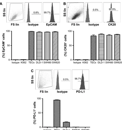
and colon cancer cell lines were observed to be ~100%
positive for EpCAM, and .90% for CK20 (Figure S2A
and B). We further analyzed PD-L1 expression in these cells. We found a significant variation in PD-L1 expression levels on the TECs and colon cancer cell lines: TECs (range
92%–97%, mean =94.33%); DLD-1 (range 13.3%–21.0%,
mean =17.23%); SW480 (,1%); SW620 (,1%); and K562
(,0.5%) (Figure S2C).
PD-1 levels on cD8
+T cells before and
after ex vivo expansion
The CD8+ T cells were expanded using classic methods for 2
weeks using patients’ PBMCs via the timed addition of IFN-γ,
anti-CD3/CD28 coated microbeads, and rIL-2. The median
expansion of the T cells was ~1,000-fold, and the majority
of the cells were CD3+CD8+ T cells (range 64%–87%,
mean =79%) after expansion.31,32 The presence of other
cytotoxic cell types, including CD3-CD56+ NK cells (range
0.1%–5.4%; mean =1.72%) and CD3+CD56+ NKT cells
(range 0.3%–15.1%; mean =6.34%) was also determined
(Figure S3A) and found to be unchanged. CTLA-4 and PD-1 expression was determined and we observed a significant increase in the surface expression of PD-1 in most of the
CD8+ T cells (range 1.6%–31%, mean =9.4% P,0.0001)
(Figure 1A and B), whereas the changes in CTLA-4 expres-sion were unremarkable (Figure S3B). PD-1 expresexpres-sion was
significantly lower (P=0.0037) (Figure 1C) after
incubat-ing with a PD-1 mAb for 48 hrs. Flow cytometric analysis revealed that anti-PD-1 (J105) binds a different epitope than the MIH-4 epitope of purified PD-1 mAb (Figure 1D).
PD-1 blockade enhances the cytotoxic
effect of cD8
+T cells against MMr
deficient EpCAM
+PD-l1
+cancer cells
To test the effect of the PD-1 blockade on the anti-tumoractivity of ex vivo expanded CD8+ T cells, we evaluated
cytotoxicity in colorectal cancer cells in the presence of a PD-1 mAb. We observed an insignificant apoptotic effect in MMR proficient TECs in the presence of PD-1 mAb after 48 hrs (Figure S1C). Therefore, MMR proficient TECs were excluded for further experiments. A dose dependent
treatment was carried out at 0.5, 1.0, 2.5, and 5.0 μg/mL
to select the optimal dose of PD-1 mAb for the subsequent
experiments. The best killing activity of CD8+ T cells
was achieved at doses from 1.0–2.5 μg/mL (Figure 2A).
Therefore, 1.0 μg/mL was chosen as the concentration for
the following experiments. We further selected an effector-to-target (ET) ratio of 25:1 based on the co-incubation of
TECs with CD8+ T cells at different ET ratios (Figure 2B).
Flow cytometric analysis detected a significant increase in apoptosis achieved with or without PD-1 mAb (Figure 2C)
in EpCAM+PD-L1+ cells: MMR deficient TECs (P=0.005)
and DLD-1 cells (P=0.0025) (Figure 2D). Indeed, no
noteworthy apoptotic effect in MMR proficient SW480
(P=0.350) and SW620 (P=0.050) were observed. We
observed dose-dependent (5, 10, 15, and 30 nM) apoptosis to select suitable concentration of nPD-1 mAb, and found no significant effect of blocking PD-1 mAb in the presence of nPD-1 mAb (Figure 2E) in TECs.
PD-1 blockade increases cytotoxic
cytokine secretion and degranulation
of cD8
+T cells against MMR deficient
epcaM
+PD-l1
+cancer cells
We performed quantitative ELISpot assays for both IFN-γ
and GrB after cells were incubated with the PD-1 mAb at
1.0 μg/mL (Figure 3A and B). IFN-γ secretion was higher in
CD8+ T cells which were co-cultured with TECs (P=0.0256)
OncoTargets and Therapy downloaded from https://www.dovepress.com/ by 118.70.13.36 on 25-Aug-2020
or DLD-1 cells (P=0.0361) than in the control (Figure 3C).
Similar results were also observed in GrB+ CD8+ T cells
co-cultured with TECs (P=0.0072) or DLD-1 cells (P=0.0461)
(Figure 3D). To determine the level of degranulation, CD107a mobilization was studied using flow cytometry (Figure 3E). PD-1 blockade enhanced the surface CD107a mobilization on
CD8+ T cells which were co-cultured with TECs (P=0.018)
or DLD-1 cells (P=0.0782), as shown in Figure 3F.
PD-1 blockade enhanced the interaction
and cytosolic uptake between cD8
+T cells and epcaM
+PD-l1
+cancer cells
To analyze the chemotactic interactions between CD8+ T
cells and tumor cells after PD-1 blockade, tumor cells were
stained with the non-invasive dye CMFDA (5 μM), for 20 min
at 37°C and then co-cultured with CD8+ T cells for 48 hrs.33
After culture, the cells were analyzed by flow cytometry. PD-1 blockade significantly increased the proportion of
CD8+CMFDA+ cells among T cells co-cultured with TECs
(P=0.0002) or DLD-1 cells (P=0.05) (Figure 4A). We evaluated
chemotactic attraction using a transwell migration assay.
Pre-treatment with PD-1 mAb at 1.0 μg/mL significantly enhanced
the number of CD8+ T cells which migrated to the lower
cham-ber of the transwell insert in the presence of TECs (P,0.0001)
or DLD-1 cells (P=0.0015), as shown in Figure 4B.
Discussion
In recent years, cancer immunotherapy has emerged as an efficacious treatment modality which produces clinically
valid outcomes in certain types of cancer.34,35 The most
Figure 1 The PD-1 expression on ex vivoexpanded cD8+ T cells obtained from patient’s blood before surgery.
Notes: (A)Mean PD-1 level of cD4 and cD8 cells on day 0 and day 14 (n=11). (B) Representative dot plots of flow cytometry on day 14. (C)Percent normalized PD-1 expression after 48 hrs treatment with PD-1 mab (n=5). (D) Different clones (Mih4 and J105) used for PD-1 staining on cD8+ T cells after treatment by flow cytometry. **P,0.005; ***P,0.0005.
Abbreviations: mAb, monoclonal antibody; ns, not significant.
&'
,VRW\SH ,VRW\SH
3' 3'
&'
&'
&'
&'
&'
&'
%
$
3HUFHQWDJH
&'3' &'3'
,VRW\SH ' QV
' ,VRW\SH ' '
3'P$EJP/
LQKLELWLRQ
±
&
,J* &ORQH0,+±3'P$E
3'P$E
&ORQH-,J* &ORQH0,+
'
OncoTargets and Therapy downloaded from https://www.dovepress.com/ by 118.70.13.36 on 25-Aug-2020
Dovepress
Kumar et al
promising strategies for cancer immunotherapy include a variety of approaches, such as the use of therapeutic antibod-ies which target immune checkpoint molecules, dendritic cell-based vaccines, adoptive transfer of ex vivo activated T cells, and the combinations of these strategies with or
without chemo/radiotherapy.36–38
Although the impressive treatment outcomes have been obtained using TILs and CD19 targeted CAR T cells, demon-strating the promise of T cell-based adoptive immunotherapy, these successes have not been translated beyond early stage
clinical studies.35 It has become apparent that tumors develop
multiple inhibitory networks to effectively evade immune
surveillance.39 The immunosuppressive microenvironment
has been recognized as one of the hallmarks of tumors.40
Strategies aimed at attenuating the endogenous immuno-suppressive environment using non-myeloablative chemo-therapy, irradiation or therapeutic mAbs against immune checkpoint inhibitors have been shown to improve the clinical
efficacy of adoptive T cell immunotherapy for solid tumors.41
However, lower effectiveness of ex vivo expanded T cells which express high levels of iRs is often ignored.
Adoptive T cell therapy generally requires the ex vivo expansion of a large number of T cells to allow an infusion to achieve therapeutic benefits. The CD3/TCR complex
stimulation of tumor reactive T cells mediated by co-stimu-latory signals to retain proliferation and functional properties
of antigen specific T cells.32 This procedure is similar to that
used to expand tumor-specific T cells, non-specific cytotoxic
cells (eg, NK, NKT, CD8+ T), and MHC-independent CAR
T cells. Chronic exposure to cytokines or tumor antigens leads to the expansion of T cells and causes them to develop
artificial exhaustion prior to transfusion.42,43 In our study, we
demonstrate that surface expression of PD-1 on the expanded
CD8+ T cells was dramatically increased (by 10-fold) during
the classic ex vivo expansion procedure with OKT3 and IL-2. This finding supports a previous study which showed that transfused TILs produced a high PD-1 level in a melanoma
animal model.44 We showed that blocking PD-1
interac-tion after ex vivo expansion significantly enhanced the
cytotoxicity of CD8+ T cells toward EpCAM+PD-L1+ cancer
cells, and this increased the release of the cytotoxic IFN-γ
and GrB, as well as CD8+ T cell degranulation. Moreover,
PD-1 blockade improved the interaction and cytosolic
exchange between CD8+ T cells and EpCAM+PD-L1+
cancer cells.
The expression of PD-L1 on cancer cells has been associ-ated with poor prognosis in cancer, demonstrating that PD-1/ PD-L1 pathway-mediated immune tolerance has clinical
$
1RWUHDWPHQW
JP/
$QQH[LQ9
(S&$0
JP/
JP/
JP/
DSRSWRVLV
3'P$EJP/
DSRSWRVLV
±3'P$E
3'P$E
±3'P$E 3'P$E
$QQH[LQ9
(S&$0
%
&
Figure 2 (Continued)
OncoTargets and Therapy downloaded from https://www.dovepress.com/ by 118.70.13.36 on 25-Aug-2020
significance.39,40 The results of a previous study demonstrated
that PD-1 blockade partially enhanced the cytotoxicity of
PD-1+ CD8+ T cells against lymphoma.41
The CK20 identified as a diagnostic and prognostic biomarker that classified in colorectal cancer by MMR status, and the inverse correlations have observed in the higher tumor grade. However, the association between MMR status and
tumor grades has not been specifically investigated.45 In our
study we found typical expression of CK20 in MMR deficient TECs, which were isolated from the primary tumor site.
The clinical trial of PD-1 mAb reached the primary endpoint, showing striking immune related response and significant disease control rate. Perhaps the increased mutation-associated tumor antigens need to be further
dem-onstrated in MMR deficient colon cancer.21,46 In the current
study, we have shown that PD-1 blockade induced a high Figure 2 apoptotic effect of cD8+ T cells on epcaM+ tumor cells after PD-1 blockade for 48 hrs.
Notes: (A)Different concentrations of PD-1 mab used to optimize the dose (n=3). (B) apoptosis measured at different effector-to-target ratios, ranging from 1:1 to 50:1. (C)representative dot plot of apoptosis in epcaM+ Tecs (D)Differential change of apoptosis in tumor cells; Tecs (Msi-h), DlD-1, sW480, and sW620 after PD-1 mab treatment (n=5). (E) cell death induced in dose-dependent manner after PD-l1 epitope blockade of PD-1 protein (left) at 1.0 μg/ml, and PD-1 mab neutralized (right) in presence of nPD-1 antibody (n=3). **P,0.005; ***P,0.0005.
Abbreviations: mab, monoclonal antibody; Tecs, tumor epithelial cells; Msi-h, high microsatellite instability.
100
80
60
40
20
0
– +
***
TECs (MSI-H)
PD-1 mAb (1 µg/mL)
(%) apoptosis
– +
**
DLD-1
PD-1 mAb (1 µg/mL)
100
80
60
40
20
0
SW480
100
80
60
40
20
0
(%) apoptosis
(%) apoptosis
(%) apoptosis
– +
PD-1 mAb (1 µg/mL)
SW620
100
80
60
40
20
0
– +
PD-1 mAb (1 µg/mL)
***
***
nPD-1 mAb (nM)
100
80
60
40
20
0
0 5.0 10.0 15.0 30.0
(%) apoptosis
*** ***
nPD-1 mAb (nM)
–PD-1 mAb +PD-1 mAb
100
80
60
40
20
0
0 30 0 30
(%) apoptosis
D
E
OncoTargets and Therapy downloaded from https://www.dovepress.com/ by 118.70.13.36 on 25-Aug-2020
Dovepress
Kumar et al
3'P$E
±3'P$E
&'7FHOOV7(&V
,)1γ),7& ,)1γ),7&
*U%&\
*U%&\ 3'P$E
±3'P$E
&'7FHOOV'/'
,)1γ),7& ,)1γ),7&
*U%&\ *U%&\
*U%VSRWVî
3'P$E
&'7FHOOV 7(&V &'
7FHOOV &'
7FHOOV
'/'
±
± ±
,)1
γ
VSRWVî
3'P$E
&'7FHOOV 7(&V &'
7FHOOV &'
7FHOOV
'/'
±
± ±
,VRW\SH
&'D
3'P$E
&'7FHOOV
7(&V &'
7FHOOV
'/'
,VRW\SH ±
±
,VRW\SH ±3'P$E
&'D
3'P$E
&'
7FHOOV 7(&V
&'
7FHOOV '/'
$
%
&
'
(
)
Figure 3 effect of PD-1 mab on cytolytic iFn-γ, grB, and cD107a.
Notes: Mobilization after 48 hrs. representative spots of (A) iFn-γ and (B) grB from cD8+ T cells co-cultured with Tecs and DlD-1 respectively. histogram of (C) iFn-γ in cD8+ T cells + Tecs and cD8+ T cells + DlD-1 and (D) grB in cD8+ T cells + Tecs and cD8+ T cells + DlD-1 (n=5). (E) Dot plot of cD107a mobilization and (F) histogram of cD8+ T cells + Tecs and cD8+ T cells + DlD-1 (n=4). *P,0.05; **P,0.005.
Abbreviations: mab, monoclonal antibody; Tecs, tumor epithelial cells.
killing effect in MMR deficient (MSI-H) EpCAM+PD-L1+
cancer cells. Mechanistically, we have shown that
EpCAM+PD-L1+ TECs and DLD-1 cells engage PD-1+ T
cells via the PD-1 ligand. Our results demonstrate that PD-1 blockade enhanced cancer cell apoptosis, the production of
cytolytic molecules, and CD8+ T cell degranulation.
More-over, it was previously shown that blocking PD-1 promoted
the migration of transfused T cells to tumor sites.38 Our
data also indicate that PD-1 blockade promotes the
migra-tion and cytosolic exchange between CD8+ T cells with
EpCAM+PD-L1+ TECs and DLD-1 cancer cells.
Our results offer a viable means of restoring T cell exhaustion during the traditional expansion procedure and
obtaining highly functional CD8+ T cells which can be used
OncoTargets and Therapy downloaded from https://www.dovepress.com/ by 118.70.13.36 on 25-Aug-2020
$
%
&0)'$
&' 7FHOOV
&'7FHOOV
7(&V &'
7FHOOV
'/'
±
3'P$E ±
&'7FHOOV
7(&V &'
7FHOOV
'/'
0LJUDWHGFHOOVî
3'P$E ± ±
Figure 4 cellular interactions, cytosolic uptake, and cD8+ T cells’ migration after 48 hrs.
Notes: (A)cytosolic uptake by cD8+ T cells from TECs and DLD-1 cells validated by flow cytometry (n=3). (B)chemotactic attraction increased after pre-treatment with PD-1 mab; conditioned medium from Tecs and DlD-1 cells was used in the lower chamber of the transwell plate (n=3). *P,0.05; **P,0.005; ***P,0.0005.
Abbreviations: mab, monoclonal antibody; Tecs, tumor epithelial cells.
for adoptive immunotherapy. Furthermore, this function can be restored after blocking PD-1 using mAb, peptides, small molecule inhibitors and advance knock-out technologies
during ex vivo expansion prior to T cell transfusion.47–49 The
transfusion of effective CD8+ CTLs with lower iRs might be
associated with better clinical efficacy, but verification of this hypothesis awaits further human studies.
Conclusion
One of the challenges of adoptive T cell transfusion is the lack of efficient tumor-killing T cells, which results in poor clinical responses. Here, we investigated whether blocking
of PD-1 on CD8+ CTLs during ex vivo expansion could
potentiate the tumor-killing activity and therefore, achieve effective adoptive immunotherapy. We demonstrated that
after ex vivo expansion, the level of PD-1 on CD8+ CTLs
increased dramatically. PD-1 blockade could significantly
potentiate the cytotoxic activity of CD8+ CTLs against
MMR deficient EpCAM+PD-L1+ tumor cells in addition
to increasing the secretion of IFN-γ, GrB, and CD107a.
Moreover, PD-1 blockade enhanced cell migration and
cyto-solic exchange between CD8+ CTLs and EpCAM+PD-L1+
tumor cells. Our findings might provide a new insight that may improve the clinical efficacy of cell-based adoptive immunotherapy.
Acknowledgments
This study was supported by grants from the National Natural Science Foundation of China (project no 81560499 & 81460448), the Guizhou Science & Technology Fund
(project no 20127016), and the Chinese Ministry of Science and Technology (project no 2013ZX09101015). The abstract of this paper was presented at the International Congress of Immunology 2016 (ICI-2016), August 21–26, 2016 in Melbourne, Australia as a poster presentation with interim findings. The poster’s abstract was published in European
Journal of Immunology.
Author contributions
All authors made substantial contributions to conception and design, acquisition of data, or analysis and interpretation of data; took part in drafting the article or revising it critically for important intellectual content; gave final approval of the version to be published; and agree to be accountable for all aspects of the work.
Disclosure
The authors report no conflicts of interest in this work.
References
1. Errico A. Melanoma: CheckMate 067 – frontline nivolumab improves PFS alone or in combination with ipilimumab. Nat Rev Clin Oncol. 2015;12(8):435.
2. Valsecchi ME. Combined nivolumab and ipilimumab or monotherapy in untreated melanoma. N Engl J Med. 2015;373(13):1270.
3. Yaqub F. Nivolumab for squamous-cell non-small-cell lung cancer.
Lancet Oncol. 2015;16(7):e319.
4. Borghaei H, Paz-Ares L, Horn L, et al. Nivolumab versus docetaxel in advanced nonsquamous non-small-cell lung cancer. N Engl J Med. 2015;373(17):1627–1639.
5. Butt AQ, Mills KH. Immunosuppressive networks and checkpoints controlling antitumor immunity and their blockade in the develop-ment of cancer immunotherapeutics and vaccines. Oncogene. 2014; 33(38):4623–4631.
OncoTargets and Therapy downloaded from https://www.dovepress.com/ by 118.70.13.36 on 25-Aug-2020
Dovepress
Kumar et al
6. Byrne WL, Mills KH, Lederer JA, O’Sullivan GC. Targeting regulatory T cells in cancer. Cancer Res. 2011;71(22):6915–6920.
7. Jarnicki AG, Lysaght J, Todryk S, Mills KH. Suppression of antitumor immunity by IL-10 and TGF-beta-producing T cells infiltrating the growing tumor: influence of tumor environment on the induction of CD4+ and CD8+ regulatory T cells. J Immunol. 2006;177(2):896–904. 8. Wherry EJ, Kurachi M. Molecular and cellular insights into T cell
exhaustion. Nat Rev Immunol. 2015;15(8):486–499.
9. Twyman-Saint Victor C, Rech AJ, Maity A, et al. Radiation and dual checkpoint blockade activate non-redundant immune mechanisms in cancer. Nature. 2015;520(7547):373–377.
10. Wherry EJ. T cell exhaustion. Nat Immunol. 2011;12(6):492–499. 11. Legat A, Speiser DE, Pircher H, Zehn D, Fuertes Marraco SA. Inhibitory
receptor expression depends more dominantly on differentiation and activation than “exhaustion” of human CD8 T cells. Front Immunol. 2013;4:455.
12. Jiang Y, Li Y, Zhu B. T-cell exhaustion in the tumor microenvironment.
Cell Death Dis. 2015;6:e1792.
13. Tumeh PC, Harview CL, Yearley JH, et al. PD-1 blockade induces responses by inhibiting adaptive immune resistance. Nature. 2014; 515(7528):568–571.
14. Donnem T, Hald SM, Paulsen EE, et al. Stromal CD8+ T-cell density – a promising supplement to TNM staging in non-small cell lung cancer.
Clin Cancer Res. 2015;21(11):2635–2643.
15. Ling A, Edin S, Wikberg ML, Oberg A, Palmqvist R. The intratu-moural subsite and relation of CD8(+) and FOXP3(+) T lymphocytes in colorectal cancer provide important prognostic clues. Br J Cancer. 2014;110(10):2551–2559.
16. Mei Z, Liu Y, Liu C, et al. Tumour-infiltrating inflammation and prognosis in colorectal cancer: systematic review and meta-analysis.
Br J Cancer. 2014;110(6):1595–1605.
17. Mella M, Kauppila JH, Karihtala P, et al. Tumor infiltrating CD8+ T lymphocyte count is independent of tumor TLR9 status in treat-ment naive triple negative breast cancer and renal cell carcinoma.
Oncoimmunology. 2015;4(6):e1002726.
18. Stumpf M, Hasenburg A, Riener MO, et al. Intraepithelial CD8-positive T lymphocytes predict survival for patients with serous stage III ovarian carcinomas: relevance of clonal selection of T lymphocytes.
Br J Cancer. 2009;101(9):1513–1521.
19. Han S, Zhang C, Li Q, et al. Tumour-infiltrating CD4(+) and CD8(+) lymphocytes as predictors of clinical outcome in glioma. Br J Cancer. 2014;110(10):2560–2568.
20. Sharma P, Shen Y, Wen S, et al. CD8 tumor-infiltrating lymphocytes are predictive of survival in muscle-invasive urothelial carcinoma. Proc
Natl Acad Sci U S A. 2007;104(10):3967–3972.
21. Le DT, Uram JN, Wang H, et al. PD-1 blockade in tumors with mismatch-repair deficiency. N Engl J Med. 2015;372(26):2509–2520.
22. Restifo NP, Dudley ME, Rosenberg SA. Adoptive immunotherapy for cancer: harnessing the T cell response. Nat Rev Immunol. 2012;12(4): 269–281.
23. Wang X, Riviere I. Manufacture of tumor- and virus-specific T lym-phocytes for adoptive cell therapies. Cancer Gene Ther. 2015;22(2): 85–94.
24. Janelle V, Carli C, Taillefer J, Orio J, Delisle JS. Defining novel param-eters for the optimal priming and expansion of minor histocompatibility antigen-specific T cells in culture. J Transl Med. 2015;13:123. 25. Fourcade J, Sun Z, Benallaoua M, et al. Upregulation of Tim-3 and
PD-1 expression is associated with tumor antigen-specific CD8+ T cell dysfunction in melanoma patients. J Exp Med. 2010;207(10): 2175–2186.
26. Yuan Z, Sotsky Kent T, Weber TK. Differential expression of DOC-1 in microsatellite-unstable human colorectal cancer. Oncogene. 2003; 22(40):6304–6310.
27. Oikonomou E, Kothonidis K, Taoufik E, et al. Newly established tumourigenic primary human colon cancer cell lines are sensitive to TRAIL-induced apoptosis in vitro and in vivo. Br J Cancer. 2007;97(1): 73–84.
28. Dons’koi BV, Chernyshov VP, Osypchuk DV. Measurement of NK activity in whole blood by the CD69 up-regulation after co-incubation with K562, comparison with NK cytotoxicity assays and CD107a degranulation assay. J Immunol Methods. 2011;372(1–2):187–195. 29. Hardtke-Wolenski M, Kraus L, Schmetz C, et al. Exchange of
cytoso-lic content between T cells and tumor cells activates CD4 T cells and impedes cancer growth. PloS One. 2013;8(10):e78558.
30. Demmers MW, Baan CC, van Beelen E, Ijzermans JN, Weimar W, Rowshani AT. Differential effects of activated human renal epithelial cells on T-cell migration. PloS One. 2013;8(5):e64916.
31. Trickett A, Kwan YL. T cell stimulation and expansion using anti-CD3/ CD28 beads. J Immunol Methods. 2003;275(1–2):251–255. 32. Teschner D, Wenzel G, Distler E, et al. In vitro stimulation and
expan-sion of human tumour-reactive CD8+ cytotoxic T lymphocytes by anti-CD3/CD28/CD137 magnetic beads. Scand J Immunol. 2011;74(2): 155–164.
33. Liu MF, Weng CT, Weng MY. Variable increased expression of pro-gram death-1 and propro-gram death-1 ligands on peripheral mononuclear cells is not impaired in patients with systemic lupus erythematosus.
J Biomed Biotechnol. 2009;2009:406136.
34. Couzin-Frankel J. Breakthrough of the year 2013. Cancer immuno-therapy. Science. 2013;342(6165):1432–1433.
35. Rosenberg SA, Restifo NP. Adoptive cell transfer as personalized immunotherapy for human cancer. Science. 2015;348(6230):62–68. 36. Melero I, Berman DM, Aznar MA, Korman AJ, Gracia JL, Haanen J.
Evolving synergistic combinations of targeted immunotherapies to combat cancer. Nat Rev Cancer. 2015;15(8):457–472.
37. Maude SL, Frey N, Shaw PA, et al. Chimeric antigen receptor T cells for sustained remissions in leukemia. N Engl J Med. 2014;371(16): 1507–1517.
38. Podrazil M, Horvath R, Becht E, et al. Phase I/II clinical trial of dendritic-cell based immunotherapy (DCVAC/PCa) combined with chemotherapy in patients with metastatic, castration-resistant prostate cancer. Oncotarget. 2015;6(20):18192–18205.
39. Finn OJ. Immuno-oncology: understanding the function and dysfunc-tion of the immune system in cancer. Ann Oncol. 2012;23 (Suppl 8): viii6–viii9.
40. Hanahan D, Weinberg RA. Hallmarks of cancer: the next generation.
Cell. 2011;144(5):646–674.
41. Mahnke K, Skorokhod A, Grabbe S, Enk AH. Opening a niche for therapy: local lymphodepletion helps the immune system to fight melanoma. J Invest Dermatol. 2014;134(7):1794–1796.
42. Fourcade J, Kudela P, Sun Z, et al. PD-1 is a regulator of NY-ESO-1-specific CD8+ T cell expansion in melanoma patients. J Immunol. 2009;182(9):5240–5249.
43. Long AH, Haso WM, Shern JF, et al. 4-1BB costimulation ameliorates T cell exhaustion induced by tonic signaling of chimeric antigen recep-tors. Nat Med. 2015;21(6):581–590.
44. Peng W, Liu C, Xu C, et al. PD-1 blockade enhances T-cell migration to tumors by elevating IFN-gamma inducible chemokines. Cancer Res. 2012;72(20):5209–5218.
45. Lugli A, Tzankov A, Zlobec I, Terracciano LM. Differential diagnostic and functional role of the multi-marker phenotype CDX2/CK20/CK7 in colorectal cancer stratified by mismatch repair status. Mod Pathol. 2008;21(11):1403–1412.
46. Dudley JC, Lin MT, Le DT, Eshleman JR. Microsatellite instability as a biomarker for PD-1 blockade. Clin Cancer Res. 2016;22(4): 813–820.
47. Chang HN, Liu BY, Qi YK, et al. Blocking of the PD-1/PD-L1 interac-tion by a D-peptide antagonist for cancer immunotherapy. Angew Chem
Int Ed Engl. 2015;54(40):11760–11764.
48. Zhan MM, Hu XQ, Liu XX, Ruan BF, Xu J, Liao C. From monoclonal antibodies to small molecules: the development of inhibitors targeting the PD-1/PD-L1 pathway. Drug Discov Today. 2016;21(6):1027–1036. 49. Su S, Hu B, Shao J, et al. CRISPR-Cas9 mediated efficient PD-1
disruption on human primary T cells from cancer patients. Sci Rep. 2016;6:20070.
OncoTargets and Therapy downloaded from https://www.dovepress.com/ by 118.70.13.36 on 25-Aug-2020
$
%
&
,VRW\SH 0+&, 0+&,,
,VRW\SH 0+&, 0+&,,
7XPRUHSLWKHOLDO FHOOV7(&V
±
DSRSWRVLV
7XPRULQYDVLYHIURQW3'/
3W 3W
3'P$EJP/
Figure S1 information on primary tumor.
Notes: (A) PD-l1 staining on 5 μM tumor sections utilized for Tecs’ isolation. (B) MHC-I/II staining of isolated TECs shown by flow cytometry, and (C) apoptotic effect of cD8+ T cells on Tecs (Mss).
Abbreviations: TECs, tumor epithelial cells; Pt, patient; MSS, MMR proficient; MMR, mismatch repair.
Supplementary materials
OncoTargets and Therapy downloaded from https://www.dovepress.com/ by 118.70.13.36 on 25-Aug-2020
Dovepress
Kumar et al
Figure S2 The representative dot plot of flow cytometry staining of tumor antigens, and average level on TECs, DLD-1, SW480, and SW620 along with K562 as a negative control (n=3).
Note: (A) extracellular epcaM, (B) intracellular cK20, and (C) intracellular PD-l1.
Abbreviations: Tecs, tumor epithelial cells; cK20, cytokeratin 20; ss lin, side scatter linear; Fs lin, forward scatter linear.
3'/
FHOOV
,VRW\SH 3'/
)6OLQ
66OLQ
,VRW\SH 7(&V '/' 6: 6:
&
$
)6OLQ
(S&$0
FHOOV
,VRW\SH . 7(&V '/' 6: 6:
66OLQ
,VRW\SH (S&$0
&.
FHOOV
,VRW\SH . 7(&V '/' 6: 6:
66OLQ
)6OLQ ,VRW\SH &.
%
Figure S3 Percentages of (A) killer immune cells, and (B) intracellular CTLA4 before and after ex vivo T cell expansion by flow cytometry (n=11).
$
%
LPPXQHFHOOV
'D\
&'
&' 1. 1.7 &' &' 1. 1.7
'D\
&7/$
FHOOV
,VRW\SH ' ' ,VRW\SH ' '
&'&7/$ &'&7/$
OncoTargets and Therapy downloaded from https://www.dovepress.com/ by 118.70.13.36 on 25-Aug-2020
OncoTargets and Therapy
Publish your work in this journal
Submit your manuscript here: http://www.dovepress.com/oncotargets-and-therapy-journal OncoTargets and Therapy is an international, peer-reviewed, open access journal focusing on the pathological basis of all cancers, potential targets for therapy and treatment protocols employed to improve the management of cancer patients. The journal also focuses on the impact of management programs and new therapeutic agents and protocols on
patient perspectives such as quality of life, adherence and satisfaction. The manuscript management system is completely online and includes a very quick and fair peer-review system, which is all easy to use. Visit http://www.dovepress.com/testimonials.php to read real quotes from published authors.
Dove
press
OncoTargets and Therapy downloaded from https://www.dovepress.com/ by 118.70.13.36 on 25-Aug-2020