TITLE
‘The influence of membrane cholesterol on the rat
GABA
a
receptor’
A thesis submitted for the Degree of Doctor of Philosophy, University of London.
Peter John Bennett, B.Sc. (Hons). September 1996
Department of Pharmacology, School of Pharmacy,
All rights reserved
INFORMATION TO ALL USERS
The quality of this reproduction is dependent upon the quality of the copy submitted.
In the unlikely event that the author did not send a complete manuscript and there are missing pages, these will be noted. Also, if material had to be removed,
a note will indicate the deletion.
uest.
ProQuest 10104219
Published by ProQuest LLC(2016). Copyright of the Dissertation is held by the Author.
All rights reserved.
This work is protected against unauthorized copying under Title 17, United States Code. Microform Edition © ProQuest LLC.
ProQuest LLC
789 East Eisenhower Parkway P.O. Box 1346
Abstract
The G A B Aa receptor is an integral membrane protein surrounded by synaptic
membrane lipids that may exert a biological control upon the receptor. An important component of the lipid milieu is cholesterol that constitutes over 2 0% of the membrane lipids which may interact with the neurosteroid binding sites of the G A B AaR. To
investigate this possibility synaptic membranes were enriched with cholesterol by incubating with liposomes comprising 50 phosphatidylcholine : 50 cholesterol in the presence of 1% BSA. The effects of cholesterol and modulatory drugs on the G A B Aa
channel were assessed via changes induced in ['HJ-FNZ, [^H]-muscimol or [^H]-TBOB binding. Cholesterol enrichment did not affect the affinity of FNZ, muscimol or TBOB for their binding sites; however, the enhancement of [^H]-FNZ binding by pregnanolone, muscimol or propofol; the modulation of [^H]-muscimol by propofol and the modulation of [^H]-TBOB binding by FNZ, pregnanolone, muscimol or propofol were affected.
In enriched cerebral cortex membranes, the potency of pregnanolone to enhance [^H]-FNZ binding was reduced. By contrast, in membranes from spinal cord, the potency of pregnanolone was increased following cholesterol enrichment. In membranes from cerebellum, there was little overall change in pregnanolone potency although the effects of threshold concentrations were increased.
In whole brain membranes modulation of [^H]-TBOB binding by FNZ, pregnanolone, muscimol or propofol was reduced upon cholesterol enrichment
These results provide little evidence for a selective competition between cholesterol and pregnanolone at its binding site. Rather, they suggest an influence of membrane cholesterol on the functional coupling between the modulatory sites on the GABAa channel. The detailed pattern of influence on the enhancement of [^H]-FNZ
Table of Contents
A b s tra c t... 2-3
Table of C o n te n ts...4-9
List of Figures... 10-13
List of T a b le s... 14
C hapter 1 General In tro d u ctio n ...16-34
1.1 Plasma membrane structure... 17 1.2 Cholesterol’s interaction with the nAChR...18-19 1.3 Nature of cholesterol’s binding site at the nAChR...19-20 1.4 Superfamily of ligand-gated ion channels... 20-21 1.5 Molecular biology of the GABAa receptor... 21-26
2.1 Introduction...36-38 2.2 Methods... 38-43 2.2.1 Membrane preparation...38-40 2.2.2 Non specific-Iipid transfer protein extraction...40-41 2.2.3 Liposome preparation...41-42 2.2.4 Cholesterol enrichment of synaptic membranes... 42-43 2.3 Results... 44-46 2.3.1 Cholesterol enrichment... 44 2.3.2 Site of cholesterol enrichment...44-45 2.3.3 Cholesterol depletion of synaptic membranes...45-46 2.3.4 Cholesterol enrichment of discrete areas of the C N S ... 46 2.4 Discussion... 46-51
C hapter 3 The effect of cholesterol enrichm ent of synaptic m em branes upon
pregnanolone potentiation of [ H]-FNZ binding... 59-80
3.2.4 Binding experiments... 62-64 3.3 Results... 64-67 3.3.1 The effects of cholesterol enrichment on FNZ binding... 64 3.3.2 The effect of cholesterol enrichment upon the modulatory action of pregnanolone at the G A B Aareceptor... 64-65
3.3.3 The effect of PC liposome incubation upon the modulatory action of pregnanolone at the GABAa receptor... 65-66 3.3.4 Scatchard analysis of pregnanolone’s modulation of [^H]-FNZ binding...66-67 3.4 Discussion... 67-70
Chapter 4 Influence of cholesterol enrichment on the potentiation of [^H]-FNZ
binding by propofol and muscimol... 81-96
4.4 Discussion...87-89
Chapter 5 Influence of cholesterol enrichment on the modulation of [^H]-muscimoI
binding... 97-109
5.1 Introduction ... 98-99 5.2 Methods... 99-101 5.2.1 Membrane preparation...99 5.2.2 Liposome preparation...99 5.2.3 Cholesterol enrichment of synaptic membranes... 100 5.2.4 Binding experiments... 100-101 5.3 Results... 101-103
5.3.1 Influence of cholesterol enrichment on unlabelled muscimol
displacement of -muscimol...101
5.3.2 Influence of cholesterol enrichment on the modulation of [^H]- muscimol binding by picrotoxinin...1 0 1 -1 0 2
Chapter 6 The influence of cholesterol enrichment upon the modulation of
[^H]-TBOB binding... 110-127
6.1 Introduction... 111-112 6.2 Methods...112-114 6.2.1 Membrane preparation...112 6.2.2 Liposome preparation...112 6.2.3 Cholesterol enrichment of synaptic membranes... 112 6.2.4 Binding experiments... 112-114 6.3 Results...114-117 6.3.1 Influence of cholesterol enrichment upon picrotoxinin displacement of[^H]-TBOB...114 6.3.2 Influence of cholesterol enrichment upon FNZ modulation of
[’H]-TBOB binding... 114-115 6.3.3 Influence of cholesterol enrichment upon muscimol modulation of [^H]-TBOB binding... 115-116 6.3.4 Influence of cholesterol enrichment upon pregnanolone modulation of [’H]-TB0B binding...116 6.3.5 The influence of cholesterol enrichment upon propofol modulation of [^H]-TBOB binding... 116-117 6.4 Discussion...117-121
7.4 Cholesterol has a general effect upon the GABAa receptor... 132-134 7.5 Clinical importance of this w ork...134-136 7.6 In conclusion... 136
Acknowledgments...138
List of figures
Figure 1.1 Generic GABAa receptor protein subunit sequence and topological
structure... 32
Figure 1.2 Helical net plot of the amino acid sequence around M2 of the GABAa
receptor subunit and the Torpedo AChR a subunit... 33
Figure 1.3 Diagram illustrating how the gate of a closed channel might be
constructed... 34
Figure 2.1 The time course of a cholesterol enrichment of rat whole brain membranes from in-house after incubating with 50 PC : 50 cholesterol liposomes or in their absence... 52
Figure 2.2 The time course of a cholesterol depletion of rat whole brain membranes from in-house after incubating with 50 PC : 50 sphingomyelin liposomes or in their absence... 53
Figure 2.3 The cholesterol content of rat whole brain membranes following incubation with or without 50 PC : cholesterol liposomes for 4 hours in the presence of 1% BSA at 2>TC...54
1% BSA at 37°C... 56
Figure 3.1 [’H]-FNZ displacement by unlabelled FNZ in cholesterol enriched and
unenriched whole brain membranes from in-house...71
Figure 3.2 Potentiation of ['H]-FNZ binding by pregnanolone in cholesterol enriched and unenriched membranes from cerebral cortex, cerebellum and spinal cord of in-house rats... 72-73
Figure 3.3 Potentiation of [^H]-FNZ binding by pregnanolone in cholesterol enriched and unenriched whole brain membranes from in-house...74
Figure 3.4 Potentiation of [^H]-FNZ binding by pregnanolone in phosphatidylcholine incubated and control cerebral cortex membranes from in-house... 75
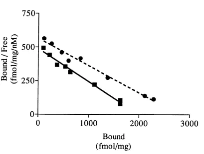
Figure 3.5 Scatchard plot showing the influence of cholesterol enrichment upon
[^HJ-FNZ binding in the presence of 0.45% DM SO to cerebral cortex membranes 76
Figure 3.6 Scatchard plot showing the influence of cholesterol enrichment upon
[^HJ-FNZ binding in the presence of 1 |iM pregnanolone to cerebral cortex
membranes...77
Figure 3.7 Scatchard plot showing the influence of cholesterol enrichment upon [^HJ-FNZ binding in the presence of 0.45% DMSG to spinal cord membranes 78
Figure 3.8 Scatchard plot showing the influence of cholesterol enrichment upon
Figure 4.1 Potentiation of ['HJ-FNZ binding by muscimol in cholesterol enriched and unenriched whole brain membranes from in-house... 90
Figure 4.2 Potentiation of [^HJ-FNZ binding by muscimol in cholesterol enriched and unenriched cerebral cortex membranes from Harlan U.K...91
Figure 4.3 Potentiation of [^HJ-FNZ binding by muscimol in cholesterol enriched and unenriched spinal cord membranes from Harlan U.K...92
Figure 4.4 Potentiation of [^HJ-FNZ binding by propofol in cholesterol enriched and unenriched whole brain membranes from in-house...93
Figure 4.5 Potentiation of ['HJ-FNZ binding by propofol in cholesterol enriched and unenriched cerebral cortex membranes from Harlan U.K...94
Figure 4.6 Potentiation of ['HJ-FNZ binding by propofol in cholesterol enriched and unenriched spinal cord membranes from Harlan U.K...95
Figure 5.1 ['HJ-muscimol displacement by unlabelled muscimol in cholesterol enriched and unenriched whole brain membranes from in-house...106
Figure 5.2 Modulation of [^HJ-muscimol binding by picrotoxinin in cholesterol enriched and unenriched whole brain membranes from Harlan U.K... 107
Figure 5.3 Potentiation of [^HJ-muscimol binding by propofol in cholesterol enriched and unenriched whole brain membranes from Harlan U.K... 108
Figure 5.4 Potentiation of [^HJ-muscimol binding by pregnanolone in cholesterol
enriched and unenriched whole brain membranes from Harlan U.K... 109
Figure 6.1 [^HJ-TBOB displacement by picrotoxinin in cholesterol enriched and
Figure 6.3 Modulation of [^H]-TBOB binding by FNZ in the presence or absence of 100
|iM bicuculline in unconditioned whole brain membranes from Harlan U.K... 124
Figure 6.4 Modulation of ['H]-TBOB binding by muscimol in cholesterol enriched and unenriched whole brain membranes from Harlan U.K... 125
Figure 6.5 Modulation of [^H]-TBOB binding by pregnanolone in cholesterol enriched and unenriched whole brain membranes from Harlan U.K... 126
List of tables
Table 2.1 Changes in cholesterol and in [^H]-cholesterol specific activity after a 4 hour [^H]-cholesterol enrichment and a further 4 hour incubation with or without PC liposomes... 57
Table 2.2 Total membrane cholesterol following a 4 hour incubation at 37°C with 50 PC : 50 cholesterol liposomes and without liposomes...58
Table 3.1 Changes in the Kd and Bmax of [^H]-FNZ binding in the presence of cholesterol enriched or unenriched cortex and spinal cord membranes and incubated with 0.45% DMSO or 1 )iM pregnanolone...80
Table 4.1 Changes in the ECso and Bmax of muscimol potentiation of [^H]-FNZ binding between cholesterol enriched or unenriched cerebral cortex and spinal cord
membranes...96
Table 7.1 Summary of binding experiments showing the effect of cholesterol
BENNETT, P J. & SEMMONDS M.A. (1996). ‘The influence of membrane cholesterol on the GABAa receptor’. Br. J. Pharmacol., 117 (1), 87-92.
BENNETT, P.J. & SIMMONDS M.A. (1995). ‘Cholesterol enrichment of neuronal membranes alters the enhancement of [^H]-flunitrazepam binding by pregnanolone. ’ Br. J. Pharmacol., 114, 294P.
Abbreviations used: ANOVA, analysis of variants; BNZ, benzodiazepine; BSA, bovine serum albumin; Q ', chloride; CNS, central nervous system; DMSO, dimethylsulfoxide; FNZ, flunitrazepam; G ABA, y-aminobutyric acid; GABAaR, y-aminobutyric acid type A receptor; nAChR, nicotinic acetylcholine receptor; nsL-TP, non-specific lipid transfer protein; PC, phosphatidylcholine; pre-nsL-TP, precursor form of the non-specific lipid transfer protein; pregnanolone, 3a-hydroxy-5P-pregnan-20-one; TBOB, t- butylbicycloorthobenzoate; TBPS, t-butylbicyclophosphorothionate.
Chapter 1
Chapter 1
General Introduction
1.1 Plasma membrane structure
j
Chapter I: General Introduction
1.2 Cholesterors interaction with the nicotinic Acetylcholine Receptor (nAChR)
Cholesterol and negatively charged phospholipids are required to maintain ion- gating activity of the nAChR (Fong & McNamee, 1986). A minimum of 45 molecules of hpid are necessary to prevent irreversible loss of both the aUosteric transitions and the ion gating functions (Jones & McNamee, 1988). The agonist dissociation constants are not affected by change of the lipid environment since the agonist binding sites are located at the extracellular domain of the protein above the membrane surface. Meanwhile, the conformational state interconversion of the receptor is dependent upon the nature of the hpid environment, which affects the abihty of the nAChR to undergo low to high affinity state transition. This state transition allows desensitization of the receptor (Lena & Changeux, 1993) causing a difference in the protein structure between the resting and the agonist induced desensitized conformations of the nAChR, both in AChR rich native membranes from Torpedo and in reconstituted hpid vesicles containing purified nAChR as the sole protein component (Femandez-Ballester et a i, 1994). Cholesterol though is not required for the agonist to produce structural changes leading to nAChR desensitization (Femandez-Ballester et a i, 1994). Apart from affecting the transition states, the secondary structure of the protein is also altered by cholesterol. The a helix content of the receptor is increased when reconstituted into membranes containing cholesterol (Butler & McNamee, 1993).
functioning. The interactions between cholesterol and negative phosphoUpids thus allow dynamic changes in the conformation of the receptor which control the opening and closing of the channel.
1.3 Nature of cholesterol’s binding site at the nAChR
Neural membranes comprise of up to 50 % cholesterol on a molar basis (Jones & McNamee, 1988) making the interaction between the cholesterol molecule and nAChR
particularly important. There seems to be two pools of cholesterol within the membranes: a fraction influencing the bulk lipid fluidity and a more tightly bound fraction thought to be associated with the receptor. Fluorescence quenching studies suggests that two lipid binding sites exist at the nAChR; annular sites at the hpid-protein interface and non-annular binding sites which occlude lipids. Negatively charged phospholipids preferentially interact with the receptor at the annular sites while cholesterol appears to interact strongly with the non-annular sites but less well with the annular sites due to its rigid planar structure. The non-annular binding sites may be at the interstices between the subunits which form hydrophobic pockets (Sunshine & McNamee, 1992) with 5-10 per nAChR.
Chapter 1: General Introduction
the M l, M3, and M4 transmembrane domains of the receptor, in the vicinity of a cysteine residue (Narayanaswami & McNamee, 1993; Fong & McNamee, 1987; Femandez- Ballester et al., 1994; Butler & McNamee, 1993). This stabihzation of the nAChR. structure is responsible for supporting the ion-gating activity. Additionally the non- annular sites may be pharmacologically important in respect to local and general anaesthetics. Local anaesthetics, such as procaine and tetracaine, may compete for the cholesterol binding sites (Sunshine & McNamee, 1992).
1.4 Superfamily of ligand-gated ion channels
The nAChR belongs to a superfamily of receptors to which five families of ligand-gated receptors belong. Notably this group includes the inhibitory neuronal y-amino butyric acid Type A (GABAa) channel which conducts Q ' ions. This superfamily of
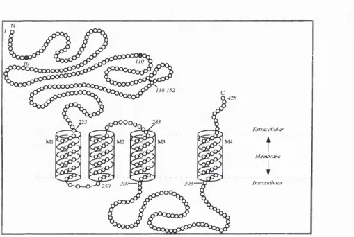
receptors share common features of four putative spanning regions denoted Ml-M4<j
i for each subunit
(Figure 1.1) (Schofield et a/., 1987); strong homology sequence in these hydrophobic transmembrane sequences especially in M2 the hydrophobic stretch of 20 amino acids that lines the ion conducting channel; and a quasi-symmetric pentameric arrangement of similar or identical subunits (Bertazzon et aL, 1992; Nayeem et a/., 1994; Stroud et a/., 1990; Unwin, 1993).
and the nAChR subunits. The a and p subunits of the G A B Aareceptor are homologous
to the nAChR subunits throughout their length (Schofield et al., 1987). In the AChR serines and threonines found in the M2 region of the anion conducting channel are found in analogous positions of the G A B Aa receptor. There is however, a higher number of
serine, threonine or polar residues in the G A B Aareceptor, about 2 per turn compared to the 1.5 per turn in the nAChR. The G A B Aa receptor M2 domains are also richer in
threonine than serine compared to the nAChR. The nAChR vestibule that is above the membrane is negatively charged to gain channel selectivity by repelling anions (Stroud et al., 1990). Meanwhile, the G A B Aareceptors vestibule displays a positive charge to repel
cations (Schofield et al., 1987). Both receptors undergo desensitization if an agonist is present continuously and require two agonist molecules to bind for maximal ion flux (Macdonald & Olsen, 1994; Stroud et al., 1990).
The common features shared by the GABAaR and the nAChR are not surprising since they both evolved from a common ancestor (Orteils & Lunt, 1995). Therefore, it might be likely that binding sites for cholesterol at ‘non-annular’ sites within the GABAa
receptor exist.
1.5 Molecular biology of the GABA^ receptor
Chapter 1: General Introduction
Bicuculline insensitive receptors display neither GABAa or GABAb receptor pharmacology and the p subunit also displays the greatest divergence among the subunit subtypes. For these reasons it has been suggested that the p subunit may constitute a homo-oligomeric receptor that belongs in the GAB Ac category (Rabow et al., 1995). With the advent of molecular biology evidence has accumulated for the existence of six a , four p, three y, two p and one 5 subunit. This means that hundreds to thousands of different combinations are possible but only a small number are presumably used. The exact composition of even one native GABAa receptor is as yet unknown.
The amino acid sequence of the subunits show 20-40% homology to each other but subunits of the same group such as the a subunit show 70% homology (Olsen & Tobin, 1990). Subunits can be combined in a multitude of ways giving rise to different pharmacological profiles. The function of the subunits and the way that they alter the pharmacology of the receptor is outlined below:
i) The p subunit is required for neurosteroid activity, but the type expressed has little impact on steroid potentiation of G ABA evoked currents. This is not true however for BNZ modulation of [^^S]-TBPS binding in the constmct ttipxYz. Here the rank order of modulation is pi>Pz>p3. When an as subunit is expressed this must be coupled with a p3
subunit to gain [^^S]-TBPS binding. The action of the anticonvulsant loreclezole is also dependent on the type of P subunit present. This compound potentiated the action of GAB A with a higher affinity in receptors containing Pz or p3 variants compared to Pi subunits.
ii) The type of a subunit present gives no differential interaction of neurosteroids between cxiPiYz, a3pi72 and aspiYz subunit combinations in electrophysiology experiments. However, the potentiation of G ABA evoked currents by allopregnanolone (5a-pregnan- 3a-ol-20-one) was greater in recombinants with the a% subunit and the enhancement of [^H]-FNZ binding by pregnanolone preferred a3 rather than ai. The a subunit variants are also responsible for differential BNZ pharmacology. The type of a subunit present effects BNZ agonist affinities but not the affinities of the antogonists and inverse agonists.
v) When either p subunit is expressed this produces a receptor that is not modulated by pentobarbital or BNZs and is insensitive to bicuculline and baclofen. Due to this pharmacology the p subunit maybe part of the BNZ insensitive retinal specific GABAaR that resembles a G A B A cR .
(Lambert a/., 1995; Luddens et a i, 1994; Sieghart, 1995)
With expression studies forming homooligomeric ion channels it was seen that some of them can be activated by G ABA and inhibited by bicuculline (Burt & Kamatchi, 1991). Such channels display multiple conductance states and desensitization. Channels containing two subunits are formed more easily and can be activated by a lower G ABA concentration, and the induced ion fluxes were greater (Sigel et al., 1990; Knoflach et al.,
1992). The cooperativity though of the GABAa receptor was absent in most of these dual subunit combinations. Coexpression of a , p, and 72 subunits resulted in large G ABA gated C r flux which could be inhibited by bicuculline. Cooperativity was now present, but as in previous studies the conductance properties were dependent upon the subunit combination used (Sigel et al., 1990; Knoflach etal., 1992).
These studies show that the binding sites for picrotoxinin, barbiturates and possibly steroids seem to be formed by the assembly of most GABAa receptor subunits. Modulation of the channel by BNZs was observed when a , p and 72 subunits were expressed together in a single cell (Burt & Kamatchi, 1991; Sigel et al., 1990; Knoflach et al., 1992). From these studies it was concluded that receptors comprised of a, p, and 7
Chapter 1: General Introduction
Regions of the subunits contain conserved and variable amino acid sequences suggesting the presence of structural and functional domains. All the subunits have four transmembrane domains M1-M4 that are a helices or p-sheet configurations with 22-23 amino acids as shown by hydrophobicity studies, occurring at residues 225-246, 251-272, 284-306 and 394-415 in the carboxyl terminal half of the protein which are conserved (Figure 1.1) (Olsen & Tobin, 1990; Unwin, 1993).
The hydrophilic amino terminal half of the subunit has potential aparagine- glycosylation sites at 10 and 110 amino acids of the tti subunit as well as a conserved cystine bridge between two cysteine residues (Figure 1.1) that may help in hgand binding. In other subunits it is assumed there are other glycosylation sites (two in the a subunit and three in the p subunit), since the peptide sizes determined by SDS-PAGE are larger than the deduced sequences (Mamalaki et a l, 1987; Sweetnam & TaUman, 1986). The C- terminus ends shortly after the M4 hydrophobic transmembrane domain as a hydrophilic chain on the extracellular side (Figure 1.1). Between M3 and M4 a large hydrophilic loop responsible for subunit specificity is prone to phosphorylation in the P subunit at the consensus Arg-Arg-Arg-Ala-Ser and the chain length differs in size between the a and p subunits (87 and 124 amino acids in the a and P subunits respectively) (Feramisco et al.,
1980).
(Bormann et al., 1987; Unwin, 1993). The M2 domain of the GABAa receptor subunits by analogy to that of the M2 domain in the AChR is tliought to line the ion channel pore. Evidence for the M2 transmembrane domain lining the channel pore comes from the rich content of threonine and serine residues which form a hydrophilic environment within the pore conducive to ion conductance (Schofield et a i, 1987; Mamalaki et al., 1987). The M2 transmembrane domain in the tti and pi subunits of the GABAa receptor and the
and Pz subunits of the nAChR have an invariant leucine residue that introduces a bend into the transmembrane helix (Figure 1.2) (Unwin, 1993). The leucine 251 on the a subunit of the AChR and the leucine residues at homologous positions on other subunits project into the centre of the pore and associate by side to side interactions of their branched chains (Figure 1.3). This creates a tight hydrophobic ring preventing the passage of hydrated ions until the GABAa receptor is bound by G ABA (or acetylcholine in the case of the AChR) at its binding sites (Unwin, 1993). When this occurs the stability of this weak association would presumably be affected by nearby hydrophobic and electrostatic interactions causing a conformational change in the helix and opening of the gate (Alber,
1992). The M4 domain interacts with the lipids at the protein-lipid boundary which may also regulate the receptor.
Chapter 1: General Introduction
domains that have ten potential N-glycosylation sites per receptor complex. Evidence for the binding sites locations comes from the fact that deglycosylation of rat GABAa
receptors causes modification in the BNZ and GABA agonist and antagonist binding (Sweetnam & Tallman, 1986).
1.6 Pharmacology of the GABA^ receptor
The various binding sites of the receptor are outlined below. These binding sites are all specific and distinct from one another upon the GABAa receptor. Allosteric
interactions between these binding sites occur thus modulating the gating of the GABAa
channel (Sieghart, 1992).
1.6.1 GABA binding sites
Binding of GABA to the GABAa receptor increases the neuronal conductance of
the Cl ions resulting in membrane hyperpolarization and a reduction in neurone excitability (Study & Barker, 1981; Bormann, 1988). This can be competitively inhibited by bicucuUine (Bormann, 1988).
binding may represent a conformational change of the receptor occurring (Schumacher & McEwen, 1989). It is presumed that the low affinity sites activate the Cl channel (Schumacher & McEwen, 1989) since micromolar concentrations of GABA or its analogues are required for this (Segal & Barker, 1984) and to modulate the other binding sites at the GABAa receptor (Olsen 1982; Squires et al., 1983). The high affinity state of binding could be a desensitized form of the receptor.
1.6.2 Benzodiazepine binding site
These compounds are widely used for their anxiolytic, anticonvulsant, muscle relaxant, sedative and antidepressant properties. BNZs enhance the action of GABA at the GABAa receptor by increasing the frequency of the Q ' ion channel opening (Study & Barker, 1981). Binding experiments have shown that these sites are found on membranes closely associated with the GABAa receptors (Olsen, 1982). Binding of [^H]- flunitrazepam (FNZ) to brain membranes was potentiated by GABA (and the vice versa could be seen) (Skerrit et at., 1982), which could be inhibited by (+)-bicuculline (Olsen,
1982).
There is a high correlation between the clinical potency of the benzodiazepines and their affinity for the [^H]-FNZ binding site which brought the conclusion that these are the binding sites by which the benzodiazepines exert their clinically important actions (Haefely
ABA-Chapter 1: General Introduction
induced Cl' flux but antagonize benzodiazepine agonists or inverse agonists (Haefley et al., 1985).
1.6.3 Pierotoxinin/TBPS binding site
These compounds are convulsants which antagonize the Cl' ion influx induced by GABA (Olsen, 1982; Bormann, 1988). Their binding site seems to be closely associated with the Q ' channel of the G A B Aa receptor since GABA and BNZ binding was not
inhibited by these compounds (Olsen, 1982). Also they directly reduce the Q ' conductance by sterically inhibiting the passage of Cl' ions (Gee, 1988). GABA and compounds that modulate the G A B Aa receptor such as the BNZs, barbiturates and
neurosteroids allosterically inhibit [^^S]-TBPS binding by decreasing its affinity (Gee, 1988). Compounds that reduce the efficacy of GABA at G A B Aa receptors like the
convulsant p-carbolines, enhance [^^S]-TBPS binding affinity through specific interactions with the BNZ binding site. This points to high affinity TBPS binding being associated with a closed conformation of the Cl' ion channel (Gee, 1988).
1.6.4 Barbiturate binding site
order of their potency as anaesthetics and hypnotics. They also inhibit binding of TBPS in a similar rank order (Olsen, 1982; Squires et a i, 1983). The barbiturates seem to interact allosterically with the TBPS binding sites, suggesting that the depressant barbiturates enhance Cl ion flux by interacting with a specific binding site.
1.6.5 Propofol binding site
Propofol exerts its actions through the GABAa receptor. Propofol dose dependently potentiates GABA activated currents, and at higher concentrations directly activates the receptor by increasing the channel conductance to Cl ions in a bicucuUine sensitive manner (Hales & Lambert, 1991). Propofol inhibits aUostericaUy the binding of [^^S]-TBPS and enhances the binding of [^H]-muscimol and [^H]-FNZ (Concas et al.,
1990, 1991; Prince & Simmonds, 1992).
1.6.6 Neurosteroid binding site
Steroids intensify GABA Cl ion conductance in brain membranes by prolonging the open time of the Q ' channels (Peters et a i, 1988). They also increase the binding affinity of GABA and BNZ agonists, but inhibited binding of TBPS (Gee, 1988; Schumacher & McEwen, 1989).
Chapter I: General Introduction
G A B Aa channel though, suggest structure-activity relationships: the requirement of a 3-
hydroxyl group in the a configuration; a keto group in the 2 0 position of the pregnanes and in the 17 position of the androstanes; and the position 17 side chain must be in the P configuration to maintain GABA potentiating activity (Franks & Lieb, 1994; Simmonds & Prince, 1992; Harrison et al., 1987). Furthermore, Oliver et al. (1991), have shown that sensitivity to steroid anaesthetics at the G A B Aareceptor only appeared late in evolution.
Pregnanolone only affects organisms belonging to the phylum Chordata. The insensitivity of invertebrates that do possess G A B Aa receptors excludes a lipid bilayer site for the
actions of neurosteroids. Removal of most of the membrane lipids after solubilization and purification of the G A B Aa receptor, maintains pregnanolone potentiation of [^H]-
muscimol binding, further supporting an interaction with the channel protein (Olsen, 1990). By analogy to the acetylcholine receptor (Jones & McNamee, 1988), cholesterol may bind at the axial clefts between the G A B Aa receptor’s subunits which may be the
locus of certain drug binding sites (Changeux, 1990), such as the neurosteroids. These sites would be accessible to membrane cholesterol and would suggest a model for neurosteroid binding via diffusion through the membrane to a hydrophobic pocket at the
G A B Aareceptor subunit interstices.
1.7 Effect of the lipid environment upon the GABA^ receptor
of a natural brain lipid extract and the cholesterol analogue cholesteryl hemisuccinate (Bristow & Martin, 1987). Cholesterol also seems to be an essential requirement for reconstitution of the protein into liposomes.
1.8 Aims and objectives
Considering that cholesterol binding sites have been shown in the nAChR and there is a requirement for cholesterol if modulation of the GABAa receptor by the allosteric sites is to be retained after solubilization and reconstitution of the receptor into liposomes, the aim of this PhD thesis is to investigate ways of enriching synaptic membranes with exogenous cholesterol and to determine the nature of this enrichment Upon achieving cholesterol enrichment, evidence will be sought to see if cholesterol competes for the neurosteroid binding site or interacts more generally with the GABAa
receptor. Such a competition between cholesterol and neurosteroids or a more general influence of the cholesterol molecule upon the GABAa receptor will be investigated using
Chapter 1: General Introduction
Extnjuellular
Membrane
Iri.'raceuulai
Chapter I: General Introduction
Chapter 2: Cholesterol Enrichment of Synaptic Membranes
Chapter 2
Cholesterol enrichment of svnaptic membranes
2.1 Introduction
The lipid content of the plasma membrane has a considerable influence on the membrane viscosity (Shinitzky & Inbar, 1976) and also the lipid microenvironment surrounding the integral lipid proteins (Benga, 1985). Increasing the cholesterol content increases the membranes microviscosity and the degree of order in the system. With increasing cholesterol content the physical properties of the membrane change abruptly with different superlattices being formed (Virtanen et al., 1995). Changes of this kind may induce or alter various cellular functions which depend on the dynamics of the cell surface membrane such as transport processes (Shinitzy & Inbar, 1976).
In the AChR an optimum fluidity is proposed to account for conformational transition properties between high and low affinity states of the receptor (Fong & McNamee, 1986). Only AChRs in membranes containing both cholesterol and negatively charged phospholipids retain ion gating properties, due to these lipids interacting strongly and stabilizing the amphipathic helical structure of the AChR whilst the ion channel is
The role of cholesterol in stabilization
opened upon agonist binding. i is important since, synaptic
inhibit AChR function by perturbing interactions between AChRs and cholesterol and/or negatively charged phospholipids (Andreasen & McNamee, 1980; Pjura et al., 1982). The dissociation constants of agonists for the AChR will not be altered significantly by different lipid environments since the agonist binding sites project into the extracellular domains above the membranes.
Modulation by cholesterol is also seen in other integral membrane proteins. The catalytic activity of Ca^^-ATPase is inhibited by cholesterol (Warren et al., 1975) and a critical concentration of cholesterol is required for maximal catalytic activity of adenylate cyclase (Whetton et al., 1983). Cholesterol depletion from liver plasma membranes results in an increased 5’-nucleotidase activity (Whetton & Hously, 1983).
Chapter 2: Cholesterol Enrichment o f Synaptic Membranes
non-specific sterol carrier between donor and acceptor membranes eliminating a chemical gradient by mediating both net mass transfer and a random exchange of lipids. This nsL- TP has now been expressed in the E. Coli strain BL21 (DE3) as the pre-nsL-TP and purified by anion exchange chromatography (Ossendorp et aL, 1992). Unesterified cholesterol will also undergo spontaneous exchange between membranes in one kinetic pool, even though distributed between two leaflets (Kan & Bittman, 1991; Fugler et a/., 1985). The rate of spontaneous transfer is dependent upon the degree of saturation of the fatty acid acyl chains, the sphingomyelin content of the membrane (Phillips et aL, 1987; Bittman, 1988), and also the curvature of the surface from which desorption occurs (McLean & Phillips, 1984; Fugler et aL, 1985)
Here I report the use of the nsL-TP, pre-nsL-TP and spontaneous lipid transfer to study the transport of cholesterol from PC : cholesterol liposomes to synaptic rat whole brain membranes to evaluate the best system. Also I have determined the nature of this cholesterol enrichment whether the cholesterol enriches as a single pool and mixes with the endogenous cholesterol or enriches to a separate pool.
2.2 Methods
2.2.1 Membrane preparation
Chapter 2: Cholesterol Enrichment of Synaptic Membranes
EDTA, pH 7.4). The final pellet was resuspended in ice cold wash buffer and stored at - 20°C until required.
2.2.2 Non specific - lipid transfer protein extraction
The nsL-TP was extracted by the method of Crain & Zilversmit (1980) as follows, but with the omission of the heat treatment and octylagarose column chromatography steps. Porcine liver (2 kg) was diced and the excess fat removed prior to rinsing with 0.25 M sucrose to remove the blood. The tissue was then homogenized in a Waring blender yielding a 35% suspension of tissue in SET buffer (0.25 M sucrose, 1 mM EDTA, 50 mM
(v/v)
Tris-base, 0.02% sodium azide, pH 6.9 at 25°C). The homogenate was centrifuged at (w/v)
13000 g for 30 min and the supernatant carefully decanted, this was then brought to pH 5.1 with 3 M HCl and stirred for 30 min at room temperature. The solution was then stirred for 30 min on ice and spun at 13000 g for 30 min after which the pellet was discarded. The supernatant was adjusted to pH 7.4 with 3 M NaOH and stored in 600 ml aliquots at -20°C for a maximum of 6 months.
When required the supernatant was thawed overnight and the pH altered to 5.1. Over a period of an hour ammonium sulphate was added to a 40% saturation and stirred
I (w/v)
for a further hour. The protein precipitated by this step was pelleted by centrifugation at 13000 g for 30 min and then the clear supernatant brought to 90% ammonium sulphate
I (w/v)
The pellet from this step was resuspended in 40 ml resuspension buffer (25 mM sodium sulphate, 10 mM mercaptoethanol, 0.02% sodium azide, pH 7.4 at 4°C) and dialyzed
I (w/v)
extensively against dialysis buffer (5 mM mercaptoethanol, 5 mM sodium sulphate and 0.02% sodium azide). This involved three changes of 2 1 of dialysis buffer at intervals of 4
(w/v) or 12 h.
After dialysis the solution was applied to a PDIO sephadex G-25 column pre-equihbriated with equilibrium buffer (5 mM sodium phosphate, 5 mM mercaptoethanol, 0.02% sodium azide). The column was washed with 160 ml equilibrium buffer and the
(w/v)
bound protein eluted with 160 ml column wash buffer (0.25 mM sodium phosphate, 45 mM sodium chloride, 5 mM mercaptoethanol, 0.02% sodium azide, pH 7.4 at 4°C). The
! (w/v)
elutant was stored at 0-4°C until required. Immediately before use the transferase protein was concentrated 5-10 times using a centrifugal concentrator centrifuged at 5000 g for 45 min. To remove any residual protein the concentrated transferase was then centrifuged at 48000 g for 15 min.
2.2.3 Liposome preparation
Chapter 2: Cholesterol Enrichment o f Synaptic Membranes
mM EDTA, 50 mM Tris-Base, 0.02% sodium azide, pH 6.9 at 25°C) was added and the
i
(w/v)lipids allowed to hydrate for 1 h at room temperature. The lipids were then dispersed with a Branson Sonifier 250 fitted with a half inch tip on 80% duty cycle under a stream of nitrogen, whilst incubating on an ice bath. Three periods of 4 min sonication were carried out at 90 W allowing the same time in between each sonication for cooling. After sonication the translucent solution was centrifuged at 48400 g for 20 min at room temperature and the pellet containing titanium fragments and undispersed lipids discarded. This procedure generally incorporated about 80% of the cholesterol into liposomes. Pure PC and PC : sphingomyelin liposomes were prepared by the same procedures.
2.2.4 Cholesterol enrichment of synaptic membranes
Previously prepared membranes were washed at 4°C with wash buffer and centrifuged at 8000 g for 25 min at 4°C and resuspended in SET buffer. Cholesterol-enrichment was achieved by incubating the membranes (0.4 mg protein ml'^) in SET buffer containing 1% BSA, and approximately 0.5 mg total liposome lipids ml'^ (approximately
(w/v)
0.35 mg total liposome lipids ml'^ in preliminary studies) at 37°C. In some preliminary
studies, 5 |ig ml'^ of the lipid transfer protein pre-nsL-TP (Ossendorp et a i, 1992) was included to determine whether it enhanced the lipid transfer or 1-2 units of nsL-TP. The cholesterol transfer was terminated after 7 h (in initial experiments) or 4 h (in all subsequent studies) by addition of an equal volume of ice cold assay buffer (50 mM Tris-
The pellets were resuspended in wash buffer. Essentially the same procedures were used to expose membranes to pure PC hposomes and PC : sphingomyelin liposomes. For comparative purposes, a portion of the original membrane preparation was subjected to these same procedures but with the omission of the liposomes and these membranes were designated as ‘unenriched’. All membranes were frozen until required.
To monitor the time course of cholesterol enrichment, experiments were performed in which the membranes were sampled at intervals during the incubation, for assay of membrane cholesterol (Sigma diagnostics kit) and protein (BioRad reagent). The stability of enrichment in membranes held at 4°C was assessed by repeated washing of the membranes with the wash buffer.
In experiments to investigate the disposition of the cholesterol enrichment, whole brain membranes were incubated with PC : cholesterol liposomes as detailed above but including [^H]-cholesterol (specific activity 1.86 mCi mmol'^). Following enrichment, the membranes were incubated for a further 4 h with or without PC liposomes (0.25 mg PC ml'^) at 37°C in SET buffer containing 1% BSA to achieve substantial reversal of the
(w/v)
cholesterol enrichment Aliquots were removed for assay of [^H]-cholesterol, total cholesterol and protein after 2 min and 4 h.
Chapter 2: Cholesterol Enrichment of Synaptic Membranes
2.3 Results
2.3.1 Cholesterol enrichment
Preliminary studies using nsL-TP extracted from bovine liver for the cholesterol enrichment of rat whole brain membranes gave a cholesterol enrichment of 140 ±18% and
108 ± 8% above basal levels for the nsL-TP compared to spontaneous enrichment respectively (incubated with liposomes comprising 0.30 mg cholesterol and 0.75 mg PC per ml of SET buffer added. Mean and s.e.m. of 6 experiments). Meanwhile, pre-nsL-TP yielded an enhancement no greater than that of spontaneous cholesterol enrichment (196% and 208% increase above basal, respectively) in the presence of the 0.75 mg PC and 0.75 mg cholesterol per ml of SET buffer liposomes. Therefore in subsequent studies, spontaneous cholesterol transfer was routinely used.
A time course study of the spontaneous enrichment of whole brain membranes with cholesterol from liposomes comprised of 50 PC : 50 cholesterol showed that the enrichment reached equilibrium after 3 h (Figure 2.1). On the basis of these results all subsequent cholesterol enrichment studies involved 4 h incubations. Washing of the membranes to remove liposomes and a further 4 washes at 4°C with wash buffer resulted in a loss of about 2 0% of the cholesterol enrichment.
2.3.2 Site of cholesterol enrichment
possibility that the exogenous cholesterol was associated with the membrane as a separate pool. Whole brain membranes were enriched with [^H]-cholesterol and the specific activity (Sp.Act.) of the [^H]-cholesterol in the membranes was measured at intervals during subsequent washing and depletion of cholesterol from the membranes at 37°C with a buffer containing 1% BSA. It was predicted that depletion of [^H]-cholesterol should
(w/v)
result in no change in Sp.Act. of the [^H]-cholesterol remaining in the membranes if depletion was from a homogenous mixed pool of exogenous and endogenous cholesterol. In contrast, depletion from a separate exogenous pool having the same Sp.Act. as the [^H]-cholesterol in the liposomes, should result in a clear reduction in the whole membrane Sp.Act. of the remaining [^H]-cholesterol. When this was tested experimentally the results obtained were as shown in Table 2.1. In the wash period following enrichment with [^H]-cholesterol, a 28-34% depletion of total cholesterol was associated with no change in Sp.Act. of [^H]-cholesterol remaining in membranes. This contrasts with a predicted reduction in Sp.Act. of 14143 ± 1439 d.p.m. qmol'^ cholesterol (6 8%) or 10757 ± 732 d.p.m. fimol'^ cholesterol (52%), depending on the presence or absence of PC liposomes, if the depleted cholesterol had come from a separate exogenous pool.
2.3.3 Cholesterol depletion of synaptic membranes
Chapter 2: Cholesterol Enrichment o f Synaptic Membranes
membranes incubated with these liposomes should show a decrease in the endogenous cholesterol. The results showed no decrease in the cholesterol content after a 4 h incubation in the presence of 1% BSA at 37°C (Figure 2.2).
(w/v)
2.3.4 Cholesterol enrichment of discrete areas of the C.N.S.
In many of the experiments involving the binding of [^H]-FNZ and [^H]-muscimol, membranes from discrete areas of the CNS were used. The cholesterol enrichments in membranes from cerebral cortex, cerebellum and spinal cord following a 4 h incubation with 50 PC : 50 cholesterol liposomes are shown in Table 2.2.
A comparison was made between the cholesterol enrichment of synaptic membranes from in-house rats and those purchased from Harlan U.K. following a 4 h incubation with 50 PC : 50 cholesterol liposomes. In whole brain, cerebral cortex and spinal cord synaptic membranes a lower cholesterol enrichment was achieved with Harlan U.K. rat membranes (Figures 2.3-2.5).
2.4 Discussion
The primary reason for carrying out these studies was to investigate the possibility that cholesterol enrichment of synaptic membranes may alter the binding properties and modulatory coupling of the benzodiazepines, neurosteroids, propofol, GABA and cage convulsants acting at the GABAa receptor in later studies. The nature of this cholesterol
these regions a study was also made of the cholesterol enrichment in cerebral cortex, cerebellum and spinal cord synaptic membranes. The synaptic membranes derived from Harlan U.K. and in-house rats may differ in their endogenous cholesterol content. The endogenous cholesterol can be affected by diet and the way that the CNS regions and whole brain were prepared. Therefore a comparison was made between the cholesterol enrichment of Harlan U.K. and in-house whole brain, cerebral cortex and spinal cord synaptic membranes.
Cholesterol enrichment of synaptic membranes was achieved by cholesterol partitioning between liposomes and synaptic membranes. We found that the lipid transfer protein pre-nsL-TP did not enhance cholesterol transfer above the spontaneous enrichment obtained in its absence, in agreement with Nichols & Pagano (1983). However, the nsL-TP did show an enhancement over spontaneous cholesterol transfer with the lower cholesterol concentration in the liposomes over a 7 h period. A draw back of the nsL-TP is that the protein aggregates upon storage thus preventing interactions with the membrane and decreasing the overall transfer activity. The rapid spontaneous transfer required the presence of 1% BSA at 37°C, the BSA presumably acting as a carrier for the
(w/v)
cholesterol (Bartholow & Geyer, 1982; Lundberg & Suominen, 1985), since it stimulates the rate of exchange (Clejan & Bittman, 1984). Both enrichment and de-enrichment could be achieved under these conditions. Although the enrichment thus achieved was stable at
Chapter 2: Cholesterol Enrichment of Synaptic Membranes
plasma membranes. With the lability of higher levels of cholesterol, it is not known whether the membranes in vivo contained cholesterol above the basal level, the extra cholesterol having been lost during the isolation procedure. Meanwhile, Phillips et al.
(1987) have reported that cholesterol depletion is possible with small unilamellar vesicles of egg PC being present at a concentration of more than 1 mg ml'^ of medium to give a maximum flux. The data here may reflect the low concentration of PC : sphingomyelin liposomes in the buffer medium used for cholesterol depletion.
In manipulating the lipid composition of membranes, it cannot be assumed that the various components are homogeneously distributed. Membrane proteins have specific affinities for different lipids, thus creating an intimate shell of lipids that has a different composition from the bulk phase. This may lead to the presence of cholesterol rich domains within the plasma membrane (Lund-Katz, 1995). Nevertheless, the composition of the shell is likely to be influenced by the availability of specific lipids in the bulk phase (Benga, 1985). The nicotinic acetylcholine receptor is reported to interact strongly with sterol molecules but they are absent from the lipid shell. The physical chemical evidence suggests that cholesterol may bind in the axial clefts between the protein subunits (Jones & McNamee, 1988). The axial clefts may also be the locus of the binding sites for certain drugs (Changeux, 1990). Since the GABAa receptor is structurally related to the nicotinic acetylcholine receptor, an analogous model may be applied to the neurosteroid sites on the GABAa channel protein. A requirement for the presence of cholesteryl hemisuccinate a
shown and may well be due to an interaction between cholesterol and the GABAa
receptor.
The nsL-TP wiU transfer a variety of phospholipids (Bloj & Zilversmit, 1983; Crain & Zilversmit, 1980) between membranes by way of a carrier mechanism (Wirtz & Gadella, 1990). The model proposed involves nsL-TP being instrumental in juxtaposing two membrane interfaces (Van Amerongen, 1989). NsL-TPs tendency to form dimers (Van Amerongen, 1985), may indicate that dimers are essential for the coUisional complex between membranes, enabling lipids to redistribute subsequently at these collision sites. If a particular phospholipid though, is strongly associated with a membrane protein it may not be removed by this transfer protein (Dawidowicz, 1987). The pre-nsL-TP of rat liver supplied by Prof. Wirtz was expressed in E.Coli (strain BL21) as a 15 kDa precursor to the nsL-TP. It has a presequence of 20 amino acids but still retains the lipid transfer activity identical to the mature nsL-TP (Ossendorp et al., 1992).
Spontaneous cholesterol transfer occurs by aqueous diffusion from a donor particle (Pownall et aL, 1983; Phillips et aL, 1987; Dawidowicz, 1987; Bittman 1988). Cholesterol forms the transitional state when it is attached to the outer surface of the liposome by its hydrophobic tail, this then desorbs from the outer leaflet to the aqueous phase which is the rate limiting step (Phillips et aL, 1987). Only a single pool of cholesterol for desorption has been detected (Kan & Bittman, 1991) indicating cholesterol exchanges between the two leaflets rapidly. Exchange will be first order provided an
Chapter 2: Cholesterol Enrichment of Synaptic Membranes
acyl chains and bending of them (if cis double bonds are present) which decreases possible Van der Waal associations with cholesterol causing looser packing (Fugler et al., 1985). PC also undergoes exchange between the liposomes and synaptic membranes but to a lesser degree (six times lower than cholesterol), since it is less soluble in the aqueous phase (Pownall et a i, 1983; Phillips et al., 1987). Lundberg & Suominen (1985) have reported that inclusion of BSA will further decrease this transfer but wiQ increase cholesterol transfer. The small unilamellar vesicles (liposomes) used produce highly curved membranes leading to looser packing of PC fatty acyl chains due to the assymetry of the phospholipid packing within the bilayer. The polar heads of the PC take up a large area compared to the acyl chains causing the decrease in lateral packing (Fugler et al.,
gradient where it becomes closely associated with the sphingomyelin resulting in cholesterol depletion.
In summary it has been shown that Harlan U.K. and in-house synaptic membranes from specific CNS regions can be enriched with cholesterol by spontaneous transfer mediated by 1% BSA with a routine incubation with PC : cholesterol liposomes for 4 h at
(w/v)
Chapter 2: C holesterol Enrichm ent o f Synaptic M em branes C O o . a
4
-3
-oU
I
1
-r
0
100
200
300
Time (min)
400
500
Figure 2.1 The time course ol a cholesterol enrichment o f rat whole brain membranes from in-house iii'ter incubating with 50 PC : 50 cholesterol liposomes (squines) or in their absence (triiuigles). Cholesterol enrichment was rapid at first but plateaued after 180 minutes. The control incubation showed no change in cholesterol content. The values shown are the means o f cholesterol content measured in triplicate ± s.e. and metm. Similar results were obtained in 3 other experiments.
1.00-1
o
0.75-^
0.25-0.00
-0.0 0.5 1.0 1.5 2.0 C tio le s te r o l c o n c e n tr a tio n ( n iM )
Standard Cholesterol curve number
Slope Mean 95%
Confidence interval
Confidence Interval %
mean
1 0.558 0.013 2 J 3
2 0.605 0.054 8.313
3 0.616 0.141 22.30
4 0.595 0.018 3.08
Overall mean 0.593 ±0.01 0.057 ± 0.06 9.161 ± 4.77
Above is a typical cholesterol standard curve obtained with the Sigma cholesterol assay kit used to determine the cholesterol content o f synaptic membranes. The data points show the mean o f 3 readings ± s.e. and mean with the error btirs lying within the symbols. The cholesterol standards and samples were incubated with the reagent for 5 mins at 37°C which caused a colour change due to the production o f a quinoneim ine dye that was measured at 500mn.
(D c/3
II
O CD 4-» </5 <ue
s 'l.O O i
0
.75
-0
.50
-0
.25
-T
2
X3
X4
X5
X6
17
T im e (H o u r s )
Chapter 2: Cholesterol Enrichment of Synaptic Membranes
7.5-1
3 5.0
-a .
2.5
-0.0
oo
<N
(N fN ON
(N
Dale
c (U c o CJ 0 (U 4—) C/0 0)
1
uI
i n yn
oc (N
On
OO
D a t e
Chapter 2: Cholesterol Enrichment o f Synaptic Membranes
1.5-1
c1
0 O2
(/) (D1
(/) Oîl
01
ON 0 4 r-HD a t e
i n o \
00
i n G\
Table 2.1 Changes in cholesterol and in [^H]-cholesterol specific activity after a 4 h [^H]-cholesterol enrichment and a further 4 h incubation with or without PC liposomes both carried out with SET buffer.
Membrane cholesterol (jimol mg'^ protein)
% change from 2
min wash
Specific activity of membrane [^H]-cholesterol
(d.p.m.pmor^ cholesterol)
% change from 2
min wash
Before incubation 2.10 ±0.10 0
After incubation with [^H]- cholesterol : PC liposomes
2 min wash 3.22 ±0.12 20887 ±1197
4 h wash with PC liposomes 2.17 ±0.07 -34% 20914 ±504 -0.1%
4 h wash without PC liposomes 2.33 ±0.16 -28% 20852 ±476 0.2%
Chapter 2: Cholesterol Enrichment o f Synaptic Membranes
Table 2.2 Total membrane cholesterol following a 4 h incubation at 37°C with 50 PC : 50
cholesterol liposomes (enriched) and without liposomes (unenriched).
Cholesterol content
(|imol cholesterol mg'^ protein)
CNS region Unenriched Enriched
Cortex 2.25 ±0.04 4.54 ± 0 .0 9
Cerebellum 1.96 ±0.10 3.63 ±0.13
Spinal cord 2.64 ±0.13 4.23 ±0.17
The Effect of Cholesterol Enrichment of
Synaptic Membranes upon Pregnanolone
Chapter 3: The Effect of Cholesterol Enrichment of Synaptic Membranes upon Pregnanolone Potentiation o f [^HJ-FNZ Binding
Chapter 3
The effect of cholesterol enrichment of svnaptic membranes
uoon oreananolone ootentiation of f^HI-FNZ binding
3.1 Introduction
potentiate the enhancement of [^H]-Flunitrazepam (FNZ) binding by GAB A and at higher concentrations to enhance [^H]-FNZ binding directly (Harrison et a i, 1987; Majewska et al., 1986) with a dependence on the presence of Q ' ions (Prince & Simmonds, 1992a). Clear structural activity requirements have been seen for neuro steroid potentiation of GABAa receptor mediated responses (Simmonds, 1991). AH this is compatible with the idea that there are specific recognition sites for the neurosteroids on the GABAa channel protein.
An interaction may occur between the high concentration of cholesterol in the lipid membrane and the neurosteroid site due to their similar structures. The alternative scenario is that cholesterol may interact more generally with the GABAa receptor. Such an association has been seen between cholesterol and the nicotinic acetylcholine receptor (Jones & McNamee, 1988). To investigate these possibilities neuronal membranes have been enriched with cholesterol and the effect this has upon FNZ binding to the GABAa
receptor and the potentiation of [^H]-FNZ binding by pregnanolone studied.
3.2 Methods
3.2.1 Membrane preparation
Membranes were prepared either from the whole brain, cerebral cortex, cerebellum, or spinal cord of male Wistar rats (130-200 g) as in chapter 2 (see section
Chapter 3: The Effect of Cholesterol Enrichment o f Synaptic Membranes upon Pregnanolone Potentiation o f fHJ-FNZ Binding
3.2.2 Liposome preparation
PC and cholesterol (0.75 mg of each per ml of buffer added) liposomes were prepared as in chapter 2 (see section 2.2.3).
3.2.3 Cholesterol enrichment of svnaptic membranes
Cholesterol enrichment of synaptic membranes was achieved by the standard incubation set out in Chapter 2 (see section 2.2.4).
To monitor the cholesterol enrichment, membranes were sampled at 5 min and 4 h during the incubation, for assay of membrane cholesterol (Sigma diagnostics kit) and protein (BioRad reagent).
3.2.4 Binding experiments ^
Experiments were performed to determine the effects of FNZ and pregnanolone on [^H]-FNZ binding to cholesterol-enriched, PC-incubated and unenriched membranes. On the day of assay the membranes were thawed and centrifuged at 48400 g for 20 min at 4°C and the pellets were resuspended in assay buffer to give a final protein concentration
of about 1 mg m l'\ Aliquots of membranes (100 |il) were pre-incubated with FNZ 10
pM-1 |iM or pregnanolone 30 nM-10 |iM in assay buffer at 37°C for 10 min. [^H]-FNZ (3a-hydroxy-5p-pregnan-20-one)
Brandell Cell Harvester. The filter mat was washed a further four times with 2 ml wash buffer and the radioactivity determined by standard hquid scintillation techniques (Beckman 5801).
In Scatchard experiments to determine the nature of pregnanolone’s modulation of [^H]-FNZ binding, essentially the same procedures as above were used, except 1 |iM pregnanolone or 0.45% DMSO were pre-incubated with synaptic membranes from cerebral cortex or spinal cord that had been enriched with cholesterol or were unenriched. [^H]-FNZ was added to give a concentration range of 0.2-20 nM in a final volume of 0.5 ml.
Non-specific binding was determined by use of 1 |iM FNZ and was in the region of 5-10%. FNZ was initially dissolved in ethanol and pregnanolone was dissolved in dimethylsulphoxide (DMSO). The final concentration of ethanol was less than 0.4% and DMSO was 0.45%. Since DMSO caused a consistent reduction in [^H]-FNZ binding by about 30%, it was present in all assay tubes for experiments involving pregnanolone. [^H]-FNZ (1 nM) was used as a trace concentration which should have no functional effect on the GABAa receptor. The incubation was carried out on ice, since a decrease in [^H]-FNZ binding affinity occurs with increasing temperature though this does not affect the Bmax (Quast et al., 1982). EDTA (1 mM) was included in the wash buffer to chelate divalent cations.
Chapter 3: The Effect of Cholesterol Enrichment of Synaptic Membranes upon Pregnanolone Potentiation o f fH ]-FNZ Binding
Data were plotted using the Prism package (Graphpad Software). Statistical comparisons were made by Student’s t test for independent samples and ANOVA.
3.3 Results
3.3.1 The effect of cholesterol enrichment on FNZ binding
In initial experiments to determine the effect of cholesterol enrichment on FNZ binding, displacement of [^H]-FNZ by unlabeUed FNZ (10 pM-1 |iM) was measured in rat whole brain membranes. Although [^H]-FNZ binding was slightly lower in enriched membranes, the displacement by unlabelled FNZ was found to be unaffected by cholesterol enrichment (Figure 3.1).
3.3.2 The effect of cholesterol enrichment upon the modulatory action of pregnanolone at the GABA^ receptor
The foregoing result allowed an investigation into the influence of cholesterol enrichment upon the modulatory action of pregnanolone on the GABAa receptor by
pregnanolone increased with concentrations up to 3 |xM. These concentration-effect relationships were compared with analogous data from enriched membranes. Where cholesterol enrichment caused lateral shifts of the concentration-response curves, the log shifts were calculated to determine changes in the potency of pregnanolone. In whole brain and cerebral cortex membranes, enrichment with cholesterol was associated with a 3.1 and a 3.2 fold reduction in pregnanolone potency, respectively (Figures 3.3 and 3.2). In cerebellar membranes, enrichment with cholesterol was associated with no change in pregnanolone potency over most of the concentration range. However, at the threshold of the concentration-effect curve in the enriched cerebellar membranes, concentrations of 0.1
and 0.3 |iM pregnanolone gave a significant enhancement of [^H]-FNZ binding to 108.2 ±
1.4% of control compared with no enhancement in unenriched membranes (mean ± s.e.mean, n=4) (Figure 3.2). In spinal cord membranes, enrichment with cholesterol was, by contrast, associated with an 8.4 fold increase in pregnanolone potency (Figure 3.2).
3.3.3 The effect of PC liposome incubation upon the modulatory action of pregnanolone at the GABA^ receptor
Chapter 3: The Effect of Cholesterol Enrichment o f Synaptic Membranes upon Pregnanolone Potentiation o f ^H]-FNZ Binding
membranes not exposed to PC (Figure 3.4). Likewise, in cerebellar membranes, potentiation of [^H]-FNZ binding by 1 |xM pregnanolone was found to be 116.7 ± 4.28%
of control binding for membranes pre-incubated with PC liposomes and 110.1 ± 4.46% of control binding for membranes not exposed to PC (mean ± s.e.mean, n=4). A similar
experiment in spinal cord membranes showed 1 |iM pregnanolone potentiation of [^H]- FNZ binding to be 140.1 ± 15.92% of control binding and 128.3 ± 12.75% of control binding, respectively, for membranes pre-incubated with PC liposomes and membranes not exposed to PC liposomes (mean ± s.e.mean, n=7). In neither case was the difference statistically significant
3.3.4 Scatchard analysis of pregnanolone’s modulation of l^Hl-FNZ binding
![Figure 1.2 Helical net plot of the amino acid sequence around M2 of the GABAaŒ] subunit (A) and the Torpedo AChR a subunit (B)](https://thumb-us.123doks.com/thumbv2/123dok_us/8626733.1416441/34.595.29.533.73.524/figure-helical-amino-sequence-gabaaoe-subunit-torpedo-subunit.webp)


![Figure 3.1 PH]-FNZ displacement by unlabelled FNZ in cholesterol enriched (squares) and unenriched (circles) whole brain membranes from in-house](https://thumb-us.123doks.com/thumbv2/123dok_us/8626733.1416441/72.596.44.443.97.399/figure-displacement-unlabelled-cholesterol-enriched-squares-unenriched-membranes.webp)

![Figure 3.4 Potentiation of [^H]-FNZ binding by pregnanolone in phosphatidylcholine incubated (squares) and control (circles) cerebral cortex membranes from in-house](https://thumb-us.123doks.com/thumbv2/123dok_us/8626733.1416441/76.595.51.498.107.392/figure-potentiation-binding-pregnanolone-phosphatidylcholine-incubated-cerebral-membranes.webp)