Breast Cancer - Targets and Therapy 2018:10 1–11
Breast Cancer - Targets and Therapy
Dove
press
submit your manuscript | www.dovepress.com 1
O R I G I N A L R E S E A R C H
open access to scientific and medical research
Open Access Full Text Article
The effects of black cohosh on the regulation
of estrogen receptor (ER
α
) and progesterone
receptor (PR) in breast cancer cells
Monica Szmyd1–3
Victoria Lloyd1–3
Kelly Hallman1–3
Katie Aleck1–3
Viktoria Mladenovik1–3
Christina McKee1–3
Mia Morse1–3
Tyler Bedgood1–3
Sumi Dinda1–3
1Biomedical and Therapeutic Sciences, School of Health Sciences, 2Prevention Research Center, 3Center of Biomedical Sciences, Oakland University, Rochester, MI, USA
Abstract: The North American plant Cimicifuga racemosa, also known as black cohosh (BC), is a herb that recently has gained attention for its hormonal effects. As the usage of hormone replacement therapy is declining due to its adverse effects in women with cancer, many are turning to herbal remedies like BC to treat menopausal symptoms. It is crucial to determine whether the effects of BC involve estrogen receptor-alpha (ERα). Previous studies from our laboratory have shown ERα to be a possible molecular target for BC. In this study, we examined the effects of BC (8% triterpene glycosides) alone and in combination with hormones and antihormones on the cellular viability, expression of ERα and progesterone receptor (PR)-A/B, and cytolocalization of ERα in ER (+) and PR-A/B (+) T-47D breast cancer cells. Cells were cultured and proteins were extracted and quantified. Western blot analysis revealed alterations in the expression of ERα
and PR after treatment with BC (5–100 µM). BC induced a concentration-dependent decrease in ERα and PR protein levels when compared to the control. Image cytometric analysis with propidium iodide staining was used to enumerate changes in T-47D cell number and viability. A decrease in T-47D cell viability was observed upon treatment with 5–100 µM BC. The ideal concentration of BC (100 µM) was used in combination with hormones and antihormones in an effort to further understand the possible similarities between this compound and other known effectors of ERα and PR. After a 24-hour concomitant treatment with and/or in combination of BC, estradiol, ICI 182, 780, and Tamoxifen, downregulation of ERα and PR protein levels was observed. Delineating the role of BC in the regulation of ERα, PR, as well as its mecha-nisms of action, may be important in understanding the influence of BC on hormone receptors in breast cancer.
Keywords: black cohosh, breast cancer, ERα, progesterone receptor, hormone replacement therapy
Introduction
A woman’s health is greatly impacted by hormone levels to maintain physiological order so vital biological functions are carried out. The main sex hormones in women are estrogen and progesterone. Estrogen is responsible for secondary sex characteristics
and cell growth and progesterone balances estrogen’s proliferative effect.1 Hormonal
imbalances may lead to women becoming susceptible to health issues, such as breast cancer. Breast cancer is the leading cause of death among women over the age of 50 in USA.2 Estrogen receptor (ER) expression is the main indicator of potential responses to
hormonal therapy, and ~70% of human breast cancers are hormone dependent and
ER-positive.2 Recently, there has been a rise in interest regarding the benefits of hormone
replacement therapy, motivating researchers to find effective, nonhormonal approaches Correspondence: Sumi Dinda
School of Health Sciences, 3164 HHB, 433 Meadow Brook Road, Oakland University, Rochester, MI 48309-4476, USA
Tel +1 248 364 8676 Fax +1 248 364 8657 Email sdinda@oakland.edu
Journal name: Breast Cancer - Targets and Therapy Article Designation: ORIGINAL RESEARCH Year: 2018
Volume: 10
Running head verso: Szmyd et al
Running head recto: Effects of black cohosh on ERα and PR DOI: http://dx.doi.org/10.2147/BCTT.S144865
Breast Cancer: Targets and Therapy downloaded from https://www.dovepress.com/ by 118.70.13.36 on 20-Aug-2020
For personal use only.
Dovepress
Szmyd et al
to treat menopause-related symptoms. Complementary and alternative medicine, such as botanic medicines, has grown increasingly popular in the last decade as an approach to
hormone therapy.3 Sales of herbal dietary supplements for
personal well-being reached $6.4 billion in 2014.4 According
to HerbalGram’s 2014 Herb Market Report, black cohosh (BC) ranked fourth as one of the most popular botanicals,
especially for menopausal symptom relief.4
Cimicifuga racemosa, also known as BC, is a herbaceous
perennial plant native to North America.5 BC has been used
for centuries across numerous cultures for its great range of health benefits. It has been widely used as a pain relieving, fever reducing, and anti-inflammatory agent, as well as for its ability to treat infectious diseases. Recently, BC has acquired significant attention for its hormonal effects, which have the possibility to alleviate female medical conditions, including menopausal symptoms, such as hot flashes, profuse sweating, and sleep disturbances.6,7
As the usage of hormone replacement therapy is declining due to its adverse effects in cancer patients, women are turn-ing to herbal remedies such as BC to treat their menopausal conditions.8,9 Studies indicate that flavonoids, like BC, may
act as a selective estrogen receptor modulator (SERM), thus inducing inhibitory growth effects on hormone-dependent
cancer cells.10 SERMs are compounds that interact with
intracellular ERs in target organs, such as skeletal or repro-ductive organs, where they have agonistic or antagonistic effects, respectively. SERMs are being intensively studied and have proven to be an effective treatment for different conditions related to postmenopausal women’s health, such
as hormone-responsive cancer.11
Steroid hormones, such as estrogen, have well- documented effects on the proliferation of breast tissue. As
estradiol (E2) diffuses across the epithelial membrane of
breast tissue, it binds to its receptor in the nucleus causing further activation of estrogen-responsive genes. Of the two
ER types, ERα has clinical relevance which is coded by
ESR1 gene. A similar molecular pathway is observed with
progesterone on progesterone receptors (PRs). Two isoforms are expressed, PR-A and PR-B, both of which are coded by the same PR gene, but PR-A is a truncated version of PR-B.12 Transcription of both isoforms is indirectly induced
through activation of ER. PR-A serves as a transcriptional inhibitor of steroid hormone receptors and PR-B functions to provide transcriptional activation of progesterone-responsive genes.13 The regulation of cellular proliferation occurs by cell
cycle-specific actions in cells undergoing G1 phase. Estro-gen has early stimulatory effects in the G1 phase allowing
only limited time for antiestrogens to inhibit further growth. As estrogen binds to its ER, it allows for further activation and continued proliferation of breast tissue.14 It is vital to
determine whether the effects of BC encompass ERα and
PR-A/B. Previous studies from our laboratory have shown ERα to be a possible molecular target for breast cancer.15,16
In this study, we have examined the effects of 8% BC alone and in combination with hormones and antihormones on the cellular viability, expression of ERα and PR-A/B, and
cytolocalization of ERα in ER (+) and PR-A/B (+) T-47D
breast cancer cells. Understanding the role of BC during
regulation of ERα and PR-A/B, as well as its mechanisms of
action, may be crucial in understanding the impact that BC has on steroid hormone receptors in breast cancer.
Materials and methods
Cell culture and treatment with ligands
The human breast cancer cell line, T-47D, was obtained from American Type Culture Collection (Manassas, VA, USA). These cells were routinely cultured following thesame protocol as previous studies in our laboratory.15,17–19
Cells were incubated at 37°C in an incubator with 5% CO2
in RPMI-1640 media (Hyclone, Logan, UT, USA) and 10% fetal bovine serum (FBS; Hyclone) that contain growth fac-tors and exogenous steroids which assist in cell growth and proliferation. The medium was replaced every 48 hours. Once the cells acquired proper confluency, the medium was changed to a 5% dextran-coated charcoal (DCC)-stripped FBS. The purpose of the stripped serum is to diminish cells of any endogenous steroids and growth factors. Therefore, this lets the cells remain at their basal metabolic rate dur-ing treatment with the compound, which will certify that the effects perceived on the cells are exclusively due to the treatment and not due to other factors within the media. The cells were maintained in charcoal-stripped serum for 6 days. On the sixth day, the 6-well plates were treated with 2 µL of ligands for 24 hours. Varying concentrations (5–100 µM) of BC were used for the concentration dependency studies. For the hormone studies, hormones and antihormones were combined with 100 µM BC.
Protein extraction and quantification
After the treatment duration of 24 hours, the cells’ proteins were extracted following the same protocol as in previousexperiments done in our laboratory.15,17–19 The 5% stripped
serum was aspirated, washed with Hanks balanced salt solu-tion, and aspirated. The cells were lysed with an extraction buffer composed of radioimmunoprecipitation assay lysis
Breast Cancer: Targets and Therapy downloaded from https://www.dovepress.com/ by 118.70.13.36 on 20-Aug-2020
Dovepress Effects of black cohosh on ERα and PR
buffer, PMSF, and protease inhibitor cocktail (Santa Cruz Biotechnology, Inc, Dallas, TX, USA). After the addition of the buffer, a high-speed supernatant of the extracts was prepared by centrifuging at 15,000 rpm for 15 minutes at 4°C. The supernatant of each sample was separated and used to prepare a protein assay based on the Bradford method (Bio-Rad Laboratories Inc, Hercules, CA, USA). The data generated by the protein assay were used to quantify and normalize the amount of protein within each sample.
Sodium dodecyl sulfate-polyacrylamide
gel electrophoresis (SDS-PAGE) and
Western blot analyses
The extracted proteins were then separated according to molecular weight by running SDS-PAGE, therefore allowing the protein of interest to be isolated using Western blot analy-sis. The same technique performed in previous studies in our laboratory was utilized.15,17–19 Protein in the supernatant was
heated to 85°C for 3 minutes. Each sample was then loaded into 7.5% polyacrylamide gel in equivalent concentrations as determined from the previously mentioned protein assay. A method known as electroblotting allowed the proteins in the gel to be transferred to an Immobilon PVDF membrane (EMD Millipore, Billerica, MA, USA). The membrane was then washed for 30 minutes in Tris-buffered saline (TBS)-Tween (0.1%) and then blocked with 5% nonfat dry milk for 1 hour to block nonspecific proteins on the membrane. In order to
detect ERα and PR-A/B, the primary antibodies – anti-mouse
monoclonal antibody (Santa Cruz Biotechnology, Inc) and anti-rabbit PR-A/B polyclonal (Cell Signaling Technology Inc, Danvers, MA, USA) – were used followed by 30 minutes with three changes of TBS-Tween and re-blocked with 5% nonfat dry milk for 30 minutes. In order to distinguish the primary antibodies, secondary goat-anti-mouse IgG2A antibody and secondary anti-rabbit antibody (Jackson Laboratory, Bar Harbor, ME, USA) were used, respectively. The specific bands
for ERα and PR-A/B could be visualized by the enhanced
chemiluminescence technique according to instructions from Advansta Inc (Menlo Park, CA, USA). The protein bands were then viewed using the Bio-Rad ChemiDoc imaging system (Bio-Rad Laboratories Inc). After immunoblotting, the PVDF membranes were stained with Coomassie Blue to confirm accurate normalization against total protein levels and full transfer of protein. The protein band density from the Western blots was then quantified using the Image Studio Lite program version 3.1 (LI-COR Biosciences, Lincoln, NE, USA).
Reverse transcription quantitative
real-time polymerase chain reaction
(RT-qPCR)
Total RNA was extracted from T-47D cells using “Trizol” reagent (Thermo Fisher Scientific, Waltham, MA, USA) as per the manufacturer’s protocol. gDNA-free total RNA was reverse transcribed using iScript cDNA Synthesis Kit (Bio-Rad Laboratories Inc) according to the manufacturer’s instructions. Prior to RT-qPCR analysis, cDNA was diluted 10-fold in PCR-grade water. qPCR of reverse transcribed cDNA (RT-qPCR) was performed in 96-well format in the Bio-Rad CFX384 Real Time System. The assay included no template, no template during RT, and no RT controls to detect reagent contamination and presence of genomic DNA. The 96-well plates were arranged in randomized treatment maximization blocks. For data analysis, the quantification cycle (Cq) value was determined and specific gene expression normalized to endogenous control using
ΔΔCq method. Expression of ACTB and HPRT-1 genes
was set as endogenous controls (reference genes). The
normalized ΔCq from treated samples was compared with
the stripped control (Cs) to obtain ΔΔCq values and used
to calculate relative fold change compared with control. ESR1 mRNA levels were determined by RT-qPCR. T-47D cells were treated in the presence or absence of 100 µM
BC, E2, and ICI 182, 780 (ICI) for 24 hours. Results are
shown as the mean ± standard error of the mean (SEM) of
at least three independent experiments with three replicates in each experiment.
Cell viability assays
Cell viability assays were used to show the total number of live cells post a 6-day treatment of ligands at varying concentrations. The same protocol was followed as from
previous studies in our laboratory.17–19 Studies were
orga-nized in 12-well culture plates with an initial cell count of
3.0×10⁴ cells per well. The cells were maintained in 1 mL
culture medium containing 10% FBS for 2 days. For the next 6 days, growth factor media were replenished with DCC-FBS media and treated with ligands over 2-day intervals for 6 days. Treatments of 5–100 µM BC were implemented, followed by performance of a cell viability assay. The cells were trypsinized, extracted from their wells, stained with propidium iodide, and underwent image cytometry using the Cellometer Vision CBA software (Nexcelom Bioscience LLC, Lawrence, MA, USA).
Breast Cancer: Targets and Therapy downloaded from https://www.dovepress.com/ by 118.70.13.36 on 20-Aug-2020
Dovepress
Szmyd et al
Immunofluorescence and confocal
microscopy
T-47D cells were plated on coverslips in 12-well plates as described in the “Cell viability assays” section.
Immunolabel-ing was performed for ERα in T-47D cells. The distribution
of three-dimensional fluorescent structures was examined using a Nikon Digital Eclipse C1 plus confocal microscope (Nikon Instruments Inc, Melville, NY, USA). NIS Elements AR software (Nikon Instruments) was used for noise reduc-tion and three-dimensional reconstrucreduc-tion of the images. The same protocol was followed as previous studies performed in our laboratory.15
Statistical analyses
The results are expressed as mean ± SEM. Statistical signifi-cance was determined by Kruskal–Wallis test followed by post hoc analysis using Mann–Whitney U test. Differences are con-sidered significant at p<0.05. In all figures, *p<0.05, **p<0.01, ***p<0.001. Statistical analyses were carried out using SPSS for Windows, version 11.5 (SPSS Inc, Chicago, IL, USA).
Results
Concentration-dependent effects of BC
on ER
α
levels and PR-A/B levels
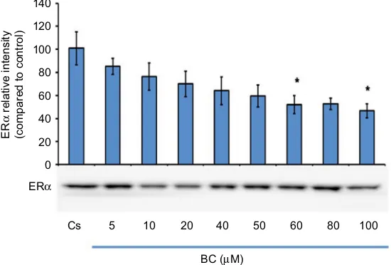
Figure 1 displays the data of the concentration dependency
study on the levels of ERα protein. T-47D cells were
cul-tured in RPMI-1640 medium supplemented with 10%
FBS for 2 days followed by 6 days in media containing 5% DCC-stripped FBS with media changed every 48 hours. On the seventh day, cells were treated with BC for 24 hours at concentrations of 5–100 µM. Concentration-dependent
downregulation of ERα protein expression was detected
as compared to the control (denoted Cs), which was grown in 5% charcoal-stripped serum during the experiment. The results indicate an optimal concentration at 100 µM BC due
to the decrease in ERα protein expression that is detected
with a 57% decrease compared to the control.
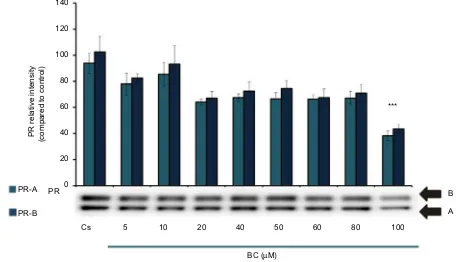
Figure 2 represents the results of PR-A/B levels when T-47D cells were cultured, as previously described, and treated for 24 hours with varying concentrations of BC from 5 to 100 µM. With increasing concentrations of BC, the results indicate that BC exerts a concentration-dependent downregulation of PR-A/B levels. The greatest decrease of protein expression is observed when cells were treated with 100 µM BC. The results from the concentration-dependent
effects of BC on both ERα and PR-A/B protein expression
indicate 100 µM BC to be the optimal concentration to test
ERα and PR-A/B expression in the presence of hormones
and antihormones for this study.
Concentration-dependent effects of BC
on cell viability
Cellular viability assays determine the amount of cells that maintain their viability after treatment with varying BC
Figure 1 Concentration-dependent effects of BC on ERα levels.
Notes: T-47D cells were cultured in RPMI-1640 medium supplemented with 10% FBS for 2 days followed by 6 days in media containing 5% DCC-stripped FBS with media changed every 48 hours. On the seventh day, cells were treated with BC for 24 hours at concentrations of 5–100 µM. Cellular protein extracts were prepared followed by
protein quantification, SDS-PAGE, and Western blot analysis. The control lane, Cs, represents cells grown in the absence of ligands in media containing 5% DCC-stripped FBS.
The relative intensity of ERα protein, as compared to Cs, is displayed as the mean ± SEM. The asterisk indicates significant difference with respect to the control. *p<0.05 (Kruskal–Wallis test followed by post hoc analysis using Mann–Whitney U test). Three independent experiments are displayed in the representative blots.
Abbreviations: BC, black cohosh; Cs, control; ERα, estrogen receptor-alpha; SEM, standard error of the mean; SDS, sodium dodecyl sulfate; PAGE, polyacrylamide gel electrophoresis; DCC, dextran-coated charcoal; FBS, fetal bovine serum.
ERα
Cs 5 10 20 40 50
BC (µM)
60
* *
80 100
ER
α
relative intensit
y
(compared to control)
0 20 40 60 80 100 120 140
Breast Cancer: Targets and Therapy downloaded from https://www.dovepress.com/ by 118.70.13.36 on 20-Aug-2020
Dovepress Effects of black cohosh on ERα and PR
concentrations. Figure 3 demonstrates the results of cellular influence of BC at varying concentrations. To determine this, T-47D cells were cultured in 12-well plates with 30,000
cells per well in FBS media containing growth factors for 2 days. DCC-stripped FBS containing media supplements with ligands of varying concentrations from 5 to 100 µM BC were
Figure 2 Concentration-dependent effects of BC on PR-A/B levels.
Notes: T-47D cells were cultured in RPMI-1640 medium supplemented with 10% FBS for 2 days followed by 6 days in media containing 5% DCC-stripped FBS with media changed every 48 hours. On the seventh day, cells were treated with BC for 24 hours at concentrations of 5–100 µM. Cellular protein extracts were prepared followed by
protein quantification, SDS-PAGE, and Western blot analysis. The control lane, Cs, represents cells grown in the absence of ligands in media containing 5% DCC-stripped
FBS. The relative intensity of PR-A/B protein, as compared to Cs, is displayed as the mean ± SEM. The asterisk indicates significant difference with respect to the control. ***p<0.001 (Kruskal–Wallis test followed by post hoc analysis using Mann–Whitney U test). Three independent experiments are displayed in the representative blots.
Abbreviations: BC, black cohosh; Cs, control; SEM, standard error of the mean; SDS, sodium dodecyl sulfate; PAGE, polyacrylamide gel electrophoresis; DCC, dextran-coated charcoal; FBS, fetal bovine serum; PR, progesterone receptor.
0 20 40 60 80 100 120 140
Cs PR PR-A
PR-B
5 10 20 40 50
BC (µM)
60 80 100
B ***
A
PR relative intensit
y
(compared to control)
Figure 3 Concentration-dependent effects of BC on cell viability.
Notes: T-47D cells were cultured in 12-well plates containing ~30,000 cells per well. For 2 days, cells were maintained in 10% FBS media containing growth factors for growth. For the following 6 days, growth factor media were replenished with DCC-FBS media and treated with ligands at 2-day intervals over 6 days. The treatments consisted of 5–100 µM BC and were followed by a cell viability assay utilizing propidium iodide staining and image cytometry via the Nexcelom Cellometer Vision on the
seventh day. *p<0.05 and ***p<0.001 (Kruskal–Wallis test followed by post hoc analysis using Mann–Whitney U test). Three independent experiments are displayed in the graph.
Abbreviations: BC, black cohosh; Cs, control; DCC, dextran-coated charcoal; FBS, fetal bovine serum.
20
Cell viability (% of control)
0 40 60 80 100 120
Cs 5 10
* * *
* *
***
20 40 50 60 80 100
BC (µM)
Breast Cancer: Targets and Therapy downloaded from https://www.dovepress.com/ by 118.70.13.36 on 20-Aug-2020
Dovepress
Szmyd et al
added and replaced for 6 days in 2-day intervals followed by extraction. Image cytometric analysis with propidium iodide staining was used to quantify alterations in T-47D cell number and viability via the Nexcelom Cellometer. A 23%–61% decrease in T-47D cell viability was observed post-incubation of 5–100 µM BC, respectively. Results indicate a significant decrease in cell viability when treated with 20–100 µM BC, with the greatest significance witnessed at 100 µM BC.
Hormonal and antihormonal effects of
BC on ER
α
levels and PR-A/B levels
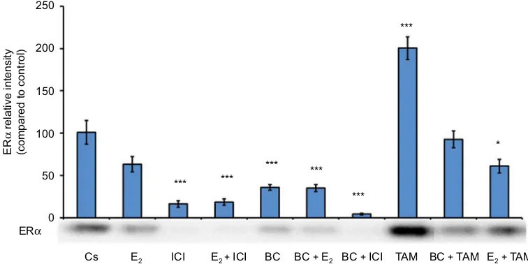
T-47D cells were cultured in RPMI-1640 medium supple-mented with 10% FBS for 2 days, followed by 6 days in media containing 5% DCC-stripped FBS with media changed every 48 hours. On the seventh day, cells were treated for 24 hours with or in combinations of 100 µM BC and 10 nM
E2, as well as antihormones at 1 µM of pure ER antagonist,
ICI, and 1 µM Tamoxifen (TAM). Treatment combinations are as follows: E2, ICI, E2 + ICI, BC, BC + E2, BC + ICI,
TAM, BC + TAM, E2+ TAM. Following protein extraction,
quantification via the Bradford method, SDS-PAGE, and Western blot analysis were performed. Figure 4 illustrates
the results from the Western blot for ERα protein
expres-sion of the previously mentioned treatment combinations.
In comparison to control, treatments of E2 alone and BC +
TAM showed a slight decrease of protein expression, but not
of significance. Treatment of E2+ TAM shows significant
downregulation of ERα protein expression in comparison
to control. However, when compared to control, ICI, E2+
ICI, BC, BC + E2, and BC + ICI showed highly significant
downregulation of ERα protein expression. Treatment with
TAM alone showed exceedingly significant upregulation of
ERα protein expression.
Figure 5 illustrates the results of PR-A/B expression with cell culture and treatment combinations as previously described. When compared to the control, E2 and E2 + TAM show signifi-cant upregulation of PR levels. Signifisignifi-cant downregulation of
PR is observed with treatment combinations BC, BC + ICI,
and BC + TAM. Treatments of ICI, E2+ ICI, and TAM show
a slight, but not significant, decrease in PR levels. There is a
minor increase in PR levels when cells are treated with BC +
E2 that is also not of significance when compared to the control.
Effects of hormones and antihormones
with BC on cell viability
T-47D cellular viability was also assessed under treatment combinations that were previously mentioned with hormones and antihormones. Cells were cultured using the experimental approach as indicated in “Cell viability assays” in “Materi-als and methods” section. Figure 6 illustrates the results of
Figure 4 Effects of hormones and antihormones with BC on ERα levels.
Notes: T-47D cell cultures were prepared as stated in Figures 1 and 2. On the seventh day, cells were treated with or in combinations of 100 µM BC, 10 nM E2, 1 µM of
pure ER antagonist, ICI, and 1 µM TAM and incubated for 24 hours. Treatment combinations are as follows: E2, ICI, E2+ ICI, BC, BC + E2, BC + ICI, TAM, BC + TAM, E2+ TAM. Cellular protein extracts were prepared followed by protein quantification, SDS-PAGE, and Western blot analysis. The control lane, Cs, represents cells grown in the
absence of ligands in media containing 5% DCC-stripped FBS. The relative intensity of ERα protein, as compared to Cs, is displayed as the mean ± SEM. The asterisk indicates
significant difference with respect to the control. *p<0.05 and ***p<0.001 (Kruskal–Wallis test followed by post hoc analysis using Mann–Whitney U test). Three independent experiments are displayed in the representative blots.
Abbreviations: BC, black cohosh; Cs, control; ERα, estrogen receptor-alpha; SEM, standard error of the mean; SDS, sodium dodecyl sulfate; PAGE, polyacrylamide gel electrophoresis; DCC, dextran-coated charcoal; FBS, fetal bovine serum; ICI, ICI 182, 780; TAM, Tamoxifen; E2,estradiol.
0
*** ***
*** ***
***
***
*
Cs E2 ICI E2 + ICI BC BC + E2 BC + ICI TAM BC + TAM E2+ TAM
50 100 150 200 250
ERα
ER
α
relative intensit
y
(compared to control)
Breast Cancer: Targets and Therapy downloaded from https://www.dovepress.com/ by 118.70.13.36 on 20-Aug-2020
Dovepress Effects of black cohosh on ERα and PR
Figure 5 Effects of hormones and antihormones with BC on PR-A/B levels.
Notes: T-47D cell cultures were prepared as stated in Figures 1 and 2. On the seventh day, cells were treated for 24 hours with or in combinations of 100 µM BC, 10 nM E2, 1 µM of pure ER antagonist, ICI, and 1 µM TAM. Treatment combinations are as follows: E2, ICI, E2+ ICI, BC, BC + E2, BC + ICI, TAM, BC + TAM, E2+ TAM. Cellular protein extracts were prepared followed by protein quantification, SDS-PAGE, and Western blot analysis. The control lane, Cs, represents cells grown in the absence
of ligands in media containing 5% DCC-stripped FBS. The relative intensity of PR-A/B protein, as compared to Cs, is displayed as the mean ± SEM. The asterisk indicates
significant difference with respect to the control. *p<0.05 and ***p<0.001 (Kruskal–Wallis test followed by post hoc analysis using Mann–Whitney U test). Three independent experiments are displayed in the representative blots.
Abbreviations: BC, black cohosh; Cs, control; PR, progesterone receptor; ER, estrogen receptor; SEM, standard error of the mean; SDS, sodium dodecyl sulfate; PAGE, polyacrylamide gel electrophoresis; DCC, dextran-coated charcoal; FBS, fetal bovine serum; ICI, ICI 182, 780; TAM, Tamoxifen; E2,estradiol.
PR relative intensit
y
(compared to control)
0 50 100 150 200
250 *
*
***
***
* 300
350 400 450 500
PR
PR-A
PR-B Cs E2 ICI E2 + ICI BC BC + E2 BC + ICI TAM BC + TAM E2+ TAM
B
A
Figure 6 Effects of hormones and antihormones with BC on cell viability.
Notes: T-47D cells were cultured as mentioned in Figure 3. The treatments, alone or in combination, consisted of 100 µM BC, 10 nM E2, 1 µM of pure ER antagonist, ICI,
and 1 µM TAM. Treatment combinations are as follows: E2, ICI, E2+ ICI, BC, BC + E2, BC + ICI, TAM, BC + TAM, E2+ TAM. This was followed by a cell viability assay utilizing propidium iodide staining and image cytometry via the Nexcelom Cellometer Vision on the seventh day. ***p<0.001 (Kruskal–Wallis test followed by post hoc analysis using Mann–Whitney U test). Three independent experiments are displayed in the graph.
Abbreviations: BC, black cohosh; Cs, control; ER, estrogen receptor; ICI, ICI 182, 780; TAM, Tamoxifen; E2,estradiol.
0 20 40 60 80 100 120 140 160
Cs
*** *** ***
ICI BC BC + E2 BC + ICI TAM BC + TAM E2+ TAM
E2 E2 + ICI
Cell viability (% of control)
Breast Cancer: Targets and Therapy downloaded from https://www.dovepress.com/ by 118.70.13.36 on 20-Aug-2020
Dovepress
Szmyd et al
the treatment combinations compared to the control. As
mentioned previously, E2 is an ER agonist; therefore, it is
expected that this compound will induce cell proliferation in hormone-dependent T-47D cells. Results in Figure 6 also signify that treatment of BC alone significantly reverses cell proliferation; it is also seen with treatments of ICI alone and in the combination of BC + ICI. Treatment with E2 + ICI, BC
+ E2, TAM, BC + TAM, and E2 + TAM, when compared to
control, shows no cellular reduction.
Effects of BC on ESR1 levels
RT-qPCR was utilized to study the effects of BC on the levels
of ERα under various conditions. ESR1 gene expression was
normalized to reference genes, HPRT-1 and ACTB, in T-47D cells exposed to treatment conditions for 24 hours. These results depicted in Figure 7 show that ESR1 expression was
reduced with 10 nM E2 but increased with 100 µM BC, and
in combination with E2 and BC. The effects observed by
BC and ICI combination induced a 30% decrease in ESR1
Figure 7 Effects of BC on ESR1 gene expression.
Notes: (A) The effect of BC, E2, and ICI alone and in combination on ESR1 mRNA levels in T-47D breast cancer cells was determined by RT-qPCR. T-47D cells were treated
in the presence or absence of 100 µM BC, 10 nM E2, and/or 1 µM ICI for 24 hours. Results are shown as the mean ± SEM of at least three independent experiments with three replicates in each experiment. *p<0.05 (Kruskal–Wallis test followed by post hoc analysis using Mann–Whitney U test). (B) Calculation of PCR efficiencies. RT-qPCR efficiencies of reference ACTB and HPRT-1 genes and target gene (ESR1) were determined. Cq was plotted against the log amount of cDNA input. Amplification efficiencies
were calculated according to the equation E = 10(-1/slope).
Abbreviations: BC, black cohosh; SEM, standard error of the mean; ICI, ICI 182, 780; E2,estradiol; RT-qPCR, reverse transcription quantitative real-time polymerase chain reaction; Cq, quantification cycle; DMSO, dimethyl sulfoxide; SYSR, SsoAdvanced SYBR Green Supermix.
A
B
Normalized expression of ESR
1
ICI BC BC + E2 BC + ICI
E2
DMSO 0
0.5
**
**
**
** **
1 2 3
1.5 2.5 3.5
26
–3 –2
Log starting quantity Standard
unknown
SYSR E–103.3% R*2–0.957 Slope – –3.133 y–Int–25.279
ESR1
–1 0
28 30
Cq 32
34 36
Standard curve
Breast Cancer: Targets and Therapy downloaded from https://www.dovepress.com/ by 118.70.13.36 on 20-Aug-2020
Dovepress Effects of black cohosh on ERα and PR
expression levels as compared to the dimethyl sulfoxide-treated control.
Effects of BC on the cellular localization
of ER
With the use of confocal microscopy, the cytolocalization of ERα was determined to be located within the nuclei of T-47D cells as shown in Figure 8. To stain the nuclei of the cells,
4′,6-diamidino-2-phenylindole (DAPI; blue)
immunofluores-cent stain was used, and Cy3 (red) immunofluoresimmunofluores-cent stain
was used for ERα protein. Furthermore, the control group
(Cs) is a representation of cells cultured in 5% DCC-FBS. Treatments included 100 µM BC alone or in combination
with 10 nM E2, 1 µM ICI, and 1 µM TAM. Treatment of cells
with 10 nM E2 decreased ERα nuclear intensity compared to
Cs. The addition of 1 µM ICI with E2 reduced the intensity
of immunolocalization of ERα to a greater extent from the
E2-induced treatment. When cells were treated with 100 µM
BC, a reduced level of ERα intensity was observed. E2 and
BC treatment did not increase the ERα level compared to E2
alone. Treatment with ICI, TAM, and BC reduced the ERα
levels compared to Cs, also indicating an ERα-dependent
mechanism at this concentration.
Discussion
Many women are taking botanicals such as BC to relieve menopausal symptoms of hot flashes, profuse sweating, and sleep disturbances.6,7 They have turned to botanicals due to
the increased anxiety over the risks of traditional hormonal
therapy.20 BC is a phytoestrogen, which may possess
estro-genic or antiestroestro-genic effects in the human body. Phytoes-trogens are a class of chemicals found naturally in certain plants that mimic the action of estrogen.10 In this study, the
effects of BC alone and in combination with hormones and
antihormones were examined with cellular viability and
expression of ERα and PR-A/B in ER (+) T-47D breast
cancer cells.
Our results demonstrated that 8% BC induced a
concentration-dependent decrease in both ERα and PR-A/B
protein levels, with optimal reduction occurring at 100 µM. As for effects of hormones and antihormones with BC on ERα protein levels, the results of this study demonstrate that
ERα protein levels were downregulated with the treatment
of 10 nM E2. Likewise, when BC was used alone, ERα was
downregulated when compared to the control, which may
suggest a possible ERα-mediated mechanism of action.
When treatments of ICI were used alone or in combina-tions of ICI + E2 and BC + ICI, its effects on ERα resulted in downregulation when compared to the control, which is expected, due to ICI’s pure antagonist properties. When BC
was used with TAM, ERα protein levels were downregulated
when compared to the control, as they are seen when 10
nM E2 and TAM were used. This, once again, may suggest
a possible ERα-mediated mechanism of action. As for the
effects of hormones and antihormones with BC on PR-A/B levels, E2 treatment alone and in combination (BC + E2 and
E2 + TAM) upregulates PR-A/B protein levels when compared
to the control, demonstrating the effects estrogen has on PR
levels. Treatment of BC alone and BC + TAM downregulates
PR-A/B levels when compared to the control. This may sug-gest that the properties of BC act through an independent pathway, E2 on PR.
Cell viability assays showed a decrease in cell viability pattern upon treatment with 5–100 µM of BC as compared to the control. The maximal effect was observed at 100 µM with only 28% of viable cells remaining, indicating that high concentrations of BC inhibited cell division. These results indicate that the reduction of cell viability, after treatment
Figure 8 Effects of BC on the cellular localization of ERα.
Notes: Treated T-47D cells were grown in 12-well growth plates, each well containing ~30,000 cells on coverslips. The cells were sustained for 2 days in whole media containing 10% FBS. They were then withdrawn from endogenous growth factors by culturing in DCC-FBS for 6 days. E2, ICI, BC, and TAM were added in 2-day intervals alone or in combination for a period of 6 days. Cells were treated with Cy3 (red) and DAPI (blue) immunofluorescent stains and the cytolocalization of ERα protein was determined using confocal microscopy.
Abbreviations: BC, black cohosh; Cs, control; ERα, estrogen receptor-alpha; DCC, dextran-coated charcoal; FBS, fetal bovine serum; ICI, ICI 182, 780; TAM, Tamoxifen; E2,estradiol.
DAPI
ERα
Merge
Cs ICI BC BC + E2BC + ICI
BC + TAM
E2 E2 + ICI
E2 +
TAM
Breast Cancer: Targets and Therapy downloaded from https://www.dovepress.com/ by 118.70.13.36 on 20-Aug-2020
Dovepress
Szmyd et al
with BC, may correlate with the downregulation of ERα and
PR-A/B protein expression, as observed in the Western blot
analyses. The proliferative effect of E2 in T-47D cells was
reversed by treatments with ICI, BC, and the combination
of BC + ICI, demonstrating a significant reduction of cell
viability. These results may suggest that BC has an antipro-liferative effect.
Images acquired through confocal microscopy reveal the
cytolocalization of ERα remains within the nuclei of T-47D
cells. Treatment of cells with 10 nM E2 decreased ERα nuclear intensity compared to the control (Cs). The addition of 1 µM ICI with E2 significantly reduced the extent and intensity of
immunolocalization of ERα from the E2-induced treatment.
When cells were treated with 100 µM BC, a reduced level of ERα intensity was observed and this correlates with the results of the Western blot analyses. BC + E2 treatment decreased the ERα intensity as compared to the control, once again correlat-ing with the results of the Western blot analyses. Treatment
with BC + ICI decreased the ERα nuclear intensity, as is
expected, due to ICI’s pure antagonist properties. TAM and
BC reduced the ERα levels compared to Cs, also indicating
an ERα-dependent mechanism at this concentration.
ESR1 gene expression levels were reduced with the treat-ment of 10 nM E2, 100 µM BC, and BC in combination with E2. Consistent with our findings, BC appears to decrease proteins that are involved in translation. Based on the observations of BC effects on cell viability and ER and PR protein expression, concentration-dependent alterations in the sensitivity of both estrogens and antiestrogens in our results, along with our labo-ratory’s previous study results, there is a possibility that a dual
mechanism for BC is both ER dependent and independent.17,19
Conclusion
BC may be a modulator for a receptor that exhibits concen-tration-dependent functional selectivity as well as cross-talk
with ERα and PR-A/B. It is clear that BC regulates the
steroid receptors on a molecular level; understanding the dose–response relationship of BC may aid in the development of more selective ER agonist and antagonist mechanisms. While further studies are necessary, our results support the potential of BC as a preventative measure against breast cancer initiation and progression.
Acknowledgments
We would like to thank Meghan Quigley for her technical assistance. We would also like to thank Naturex (NJ, USA) for their donation of BC. Funding was provided by the Center
of Biomedical Research and Prevention Research Center at Oakland University (Rochester, MI, USA).
The abstract of this paper was presented at the 97th Annual Meeting of the Endocrine Society at San Diego, CA, USA, on March 5–8, 2015, as a poster presentation with interim findings (presentation number: FRI-298; http://www.
endocrine.org/meetings/endo-annual-meetings/abstract-details?ID=20489). The poster’s abstract was published in
“Poster Abstracts” in the meeting proceedings.
Disclosure
The authors report no conflicts of interest in this work.
References
1. Dietz BM, Hajirahimkhan A, Dunlap TL, Bolton JL. Botanicals and their bioactive phytochemicals for women’s health. Pharmacol Rev.
2016;68(4):1026–1073.
2. Lumachi F, Brunello A, Maruzzo M, Basso U, Basso SM. Treat-ment of estrogen receptor-positive breast cancer. Curr Med Chem.
2013;20(5):596–604.
3. Taylor M. Complementary and alternative approaches to menopause.
Endocrinol Metab Clin North Am. 2015;44(3):619–648.
4. Smith T, Lynch ME, Johnson J, Kawa K, Bauman H, Blumenthal M. Herb supplement sales increase 6.8% in 2014. HerbalGram.
2015;107:52–59.
5. Borrelli F, Izzo AA, Ernst E. Pharmacological effects of Cimicifuga racemosa. Life Sci. 2003;73(10):1215–1229.
6. Blumenthal M. The use of black cohosh to treat symptoms of meno-pause. Sex Reprod Menopause. 2004;2(1):27–34.
7. Einbond LS, Wen-Cai Y, He K, et al. Growth inhibitory activity of extracts and compounds from Cimicifuga species on human breast cancer cells. Phytomedicine. 2008;15(6–7):504–511.
8. Wuttke W, Jarry H, Becker T, et al. Phytoestrogens: endocrine dis-rupters or replacement for hormone replacement therapy? Maturitas.
2003;44(Suppl 1):S9–S20.
9. Manavathi B, Dey O, Gajulapalli VN, Bhatia RS, Bugide S, Kumar R. Derailed estrogen signaling and breast cancer: an authentic couple.
Endocr Rev. 2013;34(1):1–32.
10. Oseni T, Patel R, Pyle J, Jordan VC. Selective estrogen receptor modula-tors and phytoestrogens. Planta Med. 2008;74(13):1656–1665. 11. Maximov PY, Lee TM, Jordan VC. The discovery and development of
selective estrogen receptor modulators (SERMs) for clinical practice.
Curr Clin Pharmacol. 2013;8(2):135–155.
12. Yip CH, Rhodes A. Estrogen and progesterone receptors in breast cancer.
Future Oncol. 2014;10(14):2293–2301.
13. Wen DX, Xu YF, Mais DE, Goldman ME, McDonnell DP. The A and B isoforms of the human progesterone receptor operate through distinct signaling pathways within target cells. Mol Cell Biol.
1994;14(12):8356–8364.
14. Musgrove EA, Hamilton JA, Lee CS, Sweeney KJ, Watts CK, Sutherland RL. Growth factor, steroid, and steroid antagonist regulation of cyclin gene expression associated with changes in T-47D human breast cancer cell cycle progression. Mol Cell Biol. 1993;13(6):3577–3587. 15. Siebert AE, Sanchez AL, Dinda S, Moudgil VK. Effects of
estro-gen metabolite 2-methoxyestradiol on tumor suppressor protein p53 and proliferation of breast cancer cells. Syst Biol Reprod Med.
2011;57(6):279–287.
16. Dinda S, Sanchez A, Moudgil VK. Effects of LY117018 (a SERM analog of raloxifene) on tumor suppressor proteins and proliferation of breast cancer cells. Horm Mol Biol Clin Investig. 2010;2(1):211–217.
Breast Cancer: Targets and Therapy downloaded from https://www.dovepress.com/ by 118.70.13.36 on 20-Aug-2020
Dovepress
Breast Cancer - Targets and Therapy
Publish your work in this journal
Submit your manuscript here: https://www.dovepress.com/breast-cancer---targets-and-therapy-journal
Breast Cancer - Targets and Therapy is an international, peer- reviewed open access journal focusing on breast cancer research, identification of therapeutic targets and the optimal use of preven-tative and integrated treatment interventions to achieve improved outcomes, enhanced survival and quality of life for the cancer patient.
The manuscript management system is completely online and includes a very quick and fair peer-review system, which is all easy to use. Visit http://www.dovepress.com/testimonials.php to read real quotes from published authors.
Dove
press
Effects of black cohosh on ERα and PR
17. Hallman K, Aleck K, Dwyer B, et al. The effects of turmeric (curcumin) on tumor suppressor protein (p53) and estrogen receptor (ERα) in breast cancer cells. Breast Cancer (Dove Med Press). 2017;9:153–161. 18. Hallman K, Aleck K, Quigley M, et al. The regulation of steroid
recep-tors by epigallocatechin-3-gallate in breast cancer cells. Breast Cancer
(Dove Med Press). 2017;9:365–373.
19. Saluzzo J, Hallman KM, Aleck K, et al. The regulation of tumor sup-pressor protein, p53, and estrogen receptor (ERα) by resveratrol in breast cancer cells. Genes Cancer. 2016;7(11–12):414–425.
20. Snelten CS, Dietz B, Bolton JL. Modulation of estrogen chemical carci-nogenesis by botanical supplements used for postmenopausal women’s health. Drug Discov Today Dis Mech. 2012;9(1):47–54.
Breast Cancer: Targets and Therapy downloaded from https://www.dovepress.com/ by 118.70.13.36 on 20-Aug-2020