Copyright1998 by the Genetics Society of America
Genetic Analysis of Organ Fusion in Arabidopsis thaliana
Susan J. Lolle, Wendy Hsu and Robert E. Pruitt
Department of Molecular and Cellular Biology, Harvard University, Cambridge, Massachusetts 02138 Manuscript received December 31, 1997
Accepted for publication March 3, 1998
ABSTRACT
Postgenital organ fusion occurs most commonly during reproductive development and is important in many angiosperms during genesis of the carpel. Although a number of mutants have been described that manifest ectopic organ fusion, little is known about the genes involved in regulating this process. In this article we describe the characterization of a collection of 29 Arabidopsis mutants showing an organ fusion phenotype. Mapping and complementation analyses revealed that the mutant alleles define nine different loci distributed throughout the Arabidopsis genome. Multiple alleles were isolated for the four complemen-tation groups showing the strongest organ fusion phenotype while the remaining five complemencomplemen-tation groups, all of which show only weak floral organ fusion, have a single representative allele. In addition to fusion events between aerial parts of the shoot, some mutants also show abnormal ovule morphology with adjacent ovules joined together at maturity. Many of the fusion mutants isolated have detectable differences in the rate at which chlorophyll can be extracted; however, in one case no difference could be detected between mutant and wild-type plants. In three mutant lines pollen remained unresponsive to contact with the mutant epidermis, demonstrating that organ fusion and pollen growth responses can be genetically separated from one another.
I
N plants the outermost layer of cells covering the dermis to physical contact with other epidermal cells and occurs most commonly during floral ontogenesis shoot and root surfaces offers the first site of contactwith biotic as well as abiotic factors present in the sur- (Cusick1966). Postgenital fusion represents a special case where the developmental potential of the epider-rounding environment. The barrier presented by this
epidermal layer is dynamic and selective, permitting mis is altered so that it no longer remains unresponsive to contact. In Catharanthus roseus, where this develop-some biological interactions while blocking others.
Dur-ing plant growth, for example, epidermal cell interac- mental response has been studied most extensively, it has been demonstrated that organ fusion involves the tions play an important role in the elaboration of the
exchange of small, water-soluble morphogenetic factors shoot by regulating organ fusion, thus contributing to
(SiegelandVerbeke1989). Although many of the epi-variation of the body plan, especially in flowers. During
dermal cells along the fusion suture in C. roseus respond reproductive development epidermal derivatives
pro-to contact by forming a tight cell wall association, a vide a receptive surface for the hydration and
germina-subset of approximately 400 cells dedifferentiate and tion of pollen, an important first step in the fertilization
redifferentiate into parenchyma cells (Walker 1975 process. In some species the specialized epidermal cells
a,b,c;VerbekeandWalker1985, 1986). Both cell wall that interact with pollen (the stigmatic papillar cells)
adhesion and the cellular redifferentiation response are provide the selection point where self-pollen is blocked
known to require recognition events because noncarpel during early development (Ockendon 1972;de
Net-epidermal derivatives will not undergo any of the tancourt 1977;Bell1995), thereby selecting against
changes associated with this contact-mediated response self-fertilization and promoting outcrossing. Clearly,
(Siegel and Verbeke 1989). The details of how this mediating the responsiveness of the shoot epidermis to
developmental pathway is regulated and information external factors is critical to the viability, proper
elabora-about the signals and signalling cascades involved re-tion and growth of the plant, as well as the fertilizare-tion
main elusive. process itself.
To date six mutations have been described which In many plant species primordial structures are known
cause ectopic expression of an epidermal fusion re-to unite following initiation at the shoot apical
meri-sponse: adherent1 (Kempton1920), adherent2 (Neuffer stem. This process, known as postgenital fusion, is
et al. 1997) and crinkly4 (Becraftet al. 1996) in maize achieved by a change in the responsiveness of the
epi-and fiddlehead (Lolleet al. 1992), wax1 and wax2 (Jenks et al. 1996) in Arabidopsis. In all six cases the mutations were identified by virtue of some impairment of normal Corresponding author: Robert E. Pruitt, Department of Molecular and
expansive growth in mutant plants. In maize plants har-Cellular Biology, Harvard University, 16 Divinity Ave., Cambridge,
MA 02138. E-mail: pruitt@billie.harvard.edu boring the crinkly4 (cr4) mutation, for example,
mal cells adhere to one another early in development, occur on all mutants showing the organ fusion pheno-type, supporting the notion that organ fusion and pollen culminating in significantly stunted and misshapen
adult plants (Becraftet al. 1996). Closer examination growth promotion define distinct biological processes. of cr4 mutants reveals that the epidermis itself is atypical
and forms graft-like fusions. The CR4 gene has been
MATERIALS AND METHODS
isolated and encodes a putative receptor protein kinase
(Becraftet al. 1996). In the Arabidopsis fdh-1 mutant, Plant growth conditions:Plants were maintained under a 24-hr light regimen and were illuminated with a mix of
fluo-epidermal morphology is normal (with the exception
rescent and incandescent lights (100–175mmol m22sec21at
of the fusion sutures) although plant growth is also
pot level). Plants were grown in CustomBlen Plus (Griffin
severely affected (Lolleet al. 1992). In addition, analysis
Greenhouse Supplies, Tewksbury, MA) soil mix and watered
of the fdh-1 mutant revealed that the entire shoot surface as needed with distilled water. The environment in the Percival
(I-37LLVL and I-60LLVL) growth chambers (Percival
Manu-was competent to promote pollen hydration,
germina-facturing Company, Boone, IA) was maintained at 258
tion and tube growth (LolleandCheung1993). The
(618) and 50% relative humidity.
pollen response seen on the fdh-1 shoot surface
reiter-Mutagenesis and plant manipulations:All of the new
muta-ates a pollen-stigma type interaction in that the pollen tions isolated in our laboratory as a part of this study were growth response involves recognition events that limit recovered from mutagenesis with ethyl methanesulfonate
(EMS). Seeds were treated with EMS essentially as described
the spectrum of pollen species that can grow on the
in Robinson-Beerset al. (1992). M1 plants were grown on fdh-1 surface to other members of the mustard family
soil and a single silique was harvested from each plant. All of
(LolleandCheung1993). Furthermore, like the
wild-the seeds from a single M1plant were grown as a family and
type stigma, fdh-1 epidermis is unable to bypass the hy- screened for the segregation of organ fusion mutants. In some dration block imposed by some of the cer mutations cases mutations could be recovered directly from selfing of
homozygous mutant plants, while in others they were
recov-(Preusset al. 1993;Hu¨ lskampet al. 1995; Lolleet al.
ered through heterozygous siblings present in the family.
Ap-1997). The wax1 and wax2 Arabidopsis mutants, on the
proximately 15,000 M1 families were screened and 21 new
other hand, show altered epicuticular wax as well as an
organ fusion mutations were recovered (see Table 1).
organ fusion phenotype (Jenkset al. 1996). Whether
Lines polymorphic for molecular markers were generated
or not these wax mutants interact with pollen has not by manually crossing homozygous or heterozygous mutant
plants (in one of three ecotype backgrounds: Landsberg,
Co-been determined.
lumbia or Wassilewskija) to an appropriate wild-type line. F1
Two distinct mechanisms for promoting organ fusion
plants were allowed to self pollinate and set seed. F2plants
are suggested by the studies done on the cr4 and fdh-1
homozygous for the mutation of interest were then collected
mutants. It seems plausible in light of the sequence and tissue stored at2808until processed for DNA-based map-homology shown by the CR4 gene that fusions in these ping analyses.
Complementation analyses were done by crossing
homozy-maize mutants result from disruption of the signalling
gous or heterozygous mutant plants to one another and the
pathway directing epidermal differentiation such that
resulting F1plants scored for the presence or absence of the
cells either do not achieve or do not maintain a terminal
mutant phenotype. In some instances, lines polymorphic for
and unresponsive developmental state typical of epider- molecular markers were used to construct trans-heterozygous mal derivatives. In fdh-1 mutants, on the other hand, a F1plants in order to facilitate identification of both F1and F2
progeny using the molecular markers.
change in the permeability barrier of the outer
epider-Determination of genetic map positions:DNA was prepared
mal cell wall and cuticle is correlated with and may be
from individual F2plant samples according toEdwardset al.
responsible for organ fusion (Lolle et al. 1997). It is
(1991). Briefly, tissue was ground in microcentrifuge tubes
postulated that the factors promoting fusion become using disposable plastic pestles. Following grinding, 400ml of accessible and are exchanged across fdh-1 cell walls be- extraction buffer (200 mmTris-HCl pH 7.5, 250 mmNaCl, 25 mm EDTA, 0.5% SDS) were added and samples vortexed.
tween contacting cells, initiating the cascade of
develop-Samples were centrifuged for 1 min in a microcentrifuge, 300
mental events leading to cell wall adhesion and organ
ml of the supernatant transferred to a fresh tube and an equal
fusion. In the former case a putative receptor in the
volume of isopropanol added. Samples were vortexed and left
signal transduction pathway has been identified while for 2 min at room temperature. Following a 5-min centrifuga-in the latter case early events centrifuga-in signallcentrifuga-ing have become tion, supernatants were discarded and pellets air-dried for
5–10 min at room temperature. DNA preparations were then
constitutive.
resuspended in 100 ml TE (10 mm Tris-HCl pH 8.0, 1 mm
In this article we describe the isolation and
character-EDTA) and stored either at 48or at2208.
ization of 29 independently derived mutations causing
Amplification of simple sequence length polymorphisms
organ fusion in Arabidopsis. The mutations fall into (SSLPs;BellandEcker1994) or codominant amplified
poly-nine complementation groups putatively identifying morphic sequences (CAPS; Konieczny andAusubel1993) was performed using polymerase chain reaction (PCR).
Oligo-nine genes. In the majority of mutants recovered, a
nucleotide primers were obtained from Research Genetics
change in the permeability barrier of the epidermal cell
Inc. (Huntsville, AL) and Taq DNA polymerase and reaction
wall cosegregates with the organ fusion phenotype but
buffer from Perkin-Elmer (Norwalk, CT). Deoxynucleotide
in one case it does not, suggesting that organ fusion triphosphates (dNTPs) were purchased from GIBCO-BRL can be achieved by at least two distinct mechanisms (Gaithersburg, MD). Each 20-ml reaction mixture contained
1ml of DNA, buffer, 200mmol dNTPs, 0.5 units of AmpliTaq
DNA polymerase (Perkin-Elmer, Norwalk, CT) and 5 pmol screen, which would allow the recovery of any fully male each of the forward and reverse primers. and female sterile mutant through heterozygous sibling
Conditions for amplification of SSLPs were as follows: 1
plants. As shown in Table 1, 21 mutants were isolated in
cycle of 2 min at 948, 15 sec at 558, 30 sec at 728, followed by
this screen, which displayed an organ fusion phenotype.
39 cycles of 15 sec at 948, 15 sec at 558, 30 sec at 728. Conditions
for amplification of CAPS were as follows: 1 cycle of 2 min at During the course of our genetic screen we also
ob-948, 15 sec at 558, 2 min at 728, followed by 39 cycles of 30 tained an additional eight mutants from the sources sec at 948, 15 sec at 558, 2 min at 728. Restriction digests of indicated in Table 1. All of the mutant phenotypes de-CAPS products were carried out using the restriction enzymes
scribed segregated in a manner consistent with
mono-specified inKoniecznyandAusubel(1993). PCR products
genic, recessive mutations. Each mutant line was
out-were size-separated by Tris-Borate-buffered agarose gel
elec-trophoresis. Agarose (4%) gels were used to resolve SSLP crossed to an appropriate wild-type line to generate an
products and 1.5–2% agarose gels to resolve CAPS products. F1 line bearing polymorphisms for molecular markers Linkage to specific SSLP or CAPS markers was determined suitable for use in DNA-based mapping procedures. by scoring a sufficient number of F2individuals to give an
Analysis of F2plants for each of these outcrossed mutant
LOD score larger than 3.0 using Mapmaker/EXP version 3.0
lines revealed linkage to nine different SSLP or CAPS
(Landeret al. 1987). Mutations which mapped to similar map
positions were tested for allelism by complementation analysis. DNA markers distributed throughout the genome.
Phenotypic analyses of organ fusion, fertility, porosity and Complementation analysis between mutants with simi-pollen hydration:Plants were visually inspected for evidence of lar map positions confirmed that our collection of fu-organ fusion during seedling, juvenile, adult and reproductive
sion mutants defines nine distinct genes. The genetic
developmental stages. Fusion was scored as positive if two
map positions of these complementation groups are
organs adhered to one another and could not easily be
sepa-rated by gentle physical manipulation. Ovules were analyzed illustrated in Figure 1 and their morphological
pheno-by dissecting open the ovary walls of the gynoecium and view- types are described individually as follows:
ing ovules under a dissecting microscope (Leica/Wild M3C, airhead (ahd) complementation group:Only a single Heerbrugg, Switzerland). Samples analyzed by scanning
elec-allele was isolated in this complementation group. Of all
tron microscopy were fixed in FAA (50% ethanol, 3.7%
formal-the fusion mutants we have characterized, this mutant
dehyde, 10% acetic acid) and dehydrated through a graded
ethanol series to 100% ethanol. Samples were critical point showed one of the weakest organ fusion phenotypes,
dried using a Samdri-PVT-3B unit and sputter coated with which is manifested only in fusion between floral organs gold. Samples were viewed in an AMR 1000 scanning electron (Table 2). Fusions are most frequently observed be-microscope at 10 kV.
tween sepals and petals, resulting in poor emergence
Mutant plants were tested for male fertility by outcrossing
of the latter. Although interorgan fusions within the
to a male sterile/female fertile line (TH154; R. E. Pruitt,
unpublished results) and scoring for silique elongation and flowers are common, no morphological aberrations
seed set. Female fertility was tested by pollinating mutant have been observed in the ovules. Fertility is normal plants with wild-type pollen and scoring for silique elongation (see Table 2).
and seed set. In cases where floral organ fusion was severe
bulkhead (bud) complementation group:Only one
al-mutant flowers had to be dissected open to reveal the
gynoe-lele belonging to this complementation group was
iden-cium.
The rate at which chlorophyll could be extracted as a mea- tified. Like airhead, this allele causes only weak fusion
sure of porosity was determined as described inLolleet al. where fusion events are essentially restricted to floral (1997). Tissue samples were collected following elongation of organs (Figure 2). A striking feature of this mutant, the inflorescence bolt (approximately 4 wk after planting).
however, is seen in the severity of the ovule phenotype.
In all cases where fusion did not prevent physical separation,
As shown in Figure 3, C and D, ovule morphology is
the rosettes were removed from the inflorescence bolt and
the two tissue samples immersed separately in 80% ethanol. highly abnormal. Although the funiculus can be
identi-Samples were agitated gently on a rotating platform and 100- fied for each individual ovule the remaining structure
ml aliquots of the ethanol solution removed at 10 min, 20 is severely compromised and shows little morphological min, 40 min, 60 min, 80 min and 24 hr following immersion.
integrity. Not surprisingly, this mutant is a tight female
Total chlorophyll content in each sample was determined
sterile (Table 2).
using absorption readings taken at 647 and 664 nm using a
UV-1201 spectrophotometer (Shimadzu Corp., Tokyo). Pollen conehead (cod) complementation group:Plants
homo-hydration assays were carried out as described previously zygous for the conehead-1 mutation display either a weak (Lolleet al. 1997).
fusion phenotype similar to bulkhead-1 mutants or a se-vere fusion phenotype like that seen in fdh mutants with approximately equal frequency (Figure 2). We were
RESULTS
unable to separate the weak and strong fusion pheno-types even after 3 backcrosses; when the two phenopheno-types
Mutant isolation and complementation analysis: The
isolation of the original fdh-1 mutant demonstrated the were mapped independently they mapped to the same location. These two distinct phenotypes may represent possibility of identifying genes whose products are
re-quired to suppress interorgan fusion through the isola- modification of the cod mutant phenotype by a second segregating locus, but we have not yet attempted to tion of mutations that promote such fusions. In order
TABLE 1
Summary of organ fusion mutations
Gene Allele Genetic background Mutagen Source
airhead 1 Columbia fast neutrons Alan Sessions(University of California, Berkeley, CA)
bulkhead 1 Landsberg erecta EMS This study
conehead 1 Landsberg erecta EMS This study
deadhead 1 Landsberg erecta EMS This study
2 Landsberg erecta EMS This study
Chris Somerville(Carnegie Institute of Washington,
3 Columbia ? Stanford, CA)
4 Columbia ? Lawrence Hobbie(Adelphi Univeristy, Garden City, NY)
eceriferum10 1 Landsberg erecta X ray Koornneffet al 1989
fiddlehead 1 Landsberg erecta EMS Lolleet al. 1992
2 Landsberg erecta EMS This study
3 Landsberg erecta EMS This study
4 Landsberg erecta EMS This study
5 Landsberg erecta EMS This study
6 Landsberg erecta EMS This study
7 Landsberg erecta EMS David Smyth
hothead 1 Landsberg erecta EMS This study
2 Landsberg erecta EMS This study
3 Landsberg erecta EMS This study
4 Landsberg erecta EMS This study
5 Landsberg erecta EMS This study
6 Landsberg erecta EMS This study
7 Landsberg erecta EMS This study
8 Landsberg erecta EMS This study
9 Wassilewskija T-DNA Stock center (Arabidopsis Biological Resource Center)
10 Landsberg erecta EMS David Smyth(Monosh University, Clayton, Australia)
11 Landsberg erecta EMS This study
pothead 1 Landsberg erecta EMS This study
thunderhead 1 Landsberg erecta EMS This study
2 Landsberg erecta EMS This study
deadhead (ded) complementation group:Four alleles ed a wild-type phenotype while others showed only a waxless phenotype or both a waxless and organ fusion were identified which fell into this complementation
group. All of the mutants in this group show a marked phenotype. Because of the wax phenotype observed in ded mutants, all of these mutations were also comple-surface luster consistent with a waxless or eceriferum
phenotype. As summarized in Table 2, for the ded-1 and mentation tested against cer5-1, the only cer mutant known to map in the same vicinity. All of these crosses ded-2 alleles, fusion can occur upon emergence of the
first true leaves. Only a small fraction of these mutant produced only wild-type F1progeny. Mapping data
con-firmed that cer5-1 mapped to a nearby but distinct map plants grows to produce an inflorescence as fusion often
limits normal vegetative growth. Early fusion events are position relative to the DED gene.
eceriferum10 (cer10) complementation group: The also typical for the ded-4 allele but not the ded-3 allele
where fusion events are commonly limited to the inflo- cer10 locus has been described previously byKoornneef et al. (1989). These mutants have a pleiotropic pheno-rescence and flowers. Ovules show a strongly aberrant
phenotype in ded-1 whereas ovule defects seen in ded-2 type including reduced plant height (Koornneefet al. 1989). However, in these descriptions no mention was mutant plants are less severe. No ovule phenotype was
observed in ded-4 mutant plants (ded-3 ovaries were not made of the organ fusion phenotype. As indicated in Table 2, cer10 mutants manifest a fusion phenotype simi-surveyed). ded-1 and ded-2 are female semi-sterile (Table
2), whereas ded-3 and ded-4 mutants show normal fer- lar to the bulkhead-1 mutant. Although fusion is re-stricted to floral organs, the ovules are not affected tility.
Complementation analyses revealed unusual allelic (Table 2). These mutants are male semisterile, as pre-viously noted byKoornneefet al. (1989) but are female interactions at this locus. As a consequence all possible
pair-wise allele combinations were constructed and F1 fertile (Table 2).
fiddlehead (fdh) complementation group:Five new al-plants scored for their wax and fusion phenotype. As
Figure 1.—Schematic representation of genetic map positions for the nine genetic loci. The distance of each locus from a linked molecular marker is incated, together with the di-rection from that marker, if known. Three of the loci map to chromosome 1 and two each to chromosomes 2, 3 and 5, respectively.
one additional allele was obtained from a colleague Both fdh-5 and fdh-6 manifest a defective ovule pheno-type (Table 2) and all alleles tested (fdh-1, fdh-3, fdh-5 (see Table 1). Like the original fdh-1 mutant, described
previously byLolleet al. (1992), all of these new alleles and fdh-6) are female semisterile (Table 2).
hothead (hth) complementation group:Eleven alleles show a strong organ fusion phenotype (Figure 2).
Al-though organ fusion is variable from one individual define the HTH locus. The fusion phenotype of these mutants is intermediate in strength with organ fusion plant to the next, for every allele it is clear that all organs
are competent to fuse, including the first true leaves. generally being limited to flowers (Table 2). The floral
TABLE 2
Summary of mutant phenotypesa
Juvenile Adult Wild-type
rosette leaf rosette leaf Floral Ovule Spontaneously Female Male pollen cer pollen
Locus fusionb fusionb fusionb abnormalityc fertiled fertilee fertilee hydrationf hydrationf
wildtype 2 2 2 2 11 1 1 2 2
airhead 2 2 1/2 2 11 1 1 1 2
bulkhead 2 2 1 1 2 2 1 2 2
conehead 2 1 1 2 1/2 1 1 1 2
deadhead 1 1 1 1/2 1/2 1/2 1/2 1 2
eceriferum10 2 2 1 2 2 1 2 1 2
fiddlehead 1 1 1 1 2 2 1 1 2
hothead 2 2 1 1/2 1 1 1 1 2
pothead 2 2 1/2 2 11 1 1 2 2
thunderhead 2 1 1 1 2 1/2 1 1/2 2
aThe representative alleles scored for each locus were: ahd-1, bud-1, cod-1, ded-1, ded-2, ded-4, cer10-1, fdh-3, fdh-5, fdh-6, hth-4,
hth-8, hth-10, phd-1, thd-1 and thd-2.
bFor each of the fusion phenotypes:1, fusion events strong enough to seriously distort the shape of the organ;1/2, fusion
events which do not distort the shape of the organ;2, no fusion.
cEach representative allele was scored for the presence or absence of morphologically abnormal ovules (1or2).1/2, some
alleles showed an ovule defect while others did not.
dSpontaneously fertile refers to the ability of the flowers to set seed without human intervention.11, wild-type levels of seed
set;1, seed set less than 50% of wild-type levels;2, no seed set (less than 1 seed/flower);1/2, variation between alleles tested.
eMale and female fertility were scored relative to wild-type in reciprocal crosses.1, at least 50% of wild-type fertility;2, less
than 50% of wild-type fertility;1/2, variation between the alleles tested.
fSamples were scored as positive for hydration if 50% or more of the applied pollen grains became spherical in shape within
Figure2.—Light micrographs showing the fusion phenotypes associated with various mutant alleles. As shown in A, wild-type Landsberg er inflorescences show a typical spiral phyllotaxy where contact between neighboring buds decreases as development progresses. As individual flowers mature petals elongate and emerge unimpeded by the surrounding sepals. In bulkhead (B), hothead (G) and pothead (H) mutant petals are restricted in their growth but do emerge from some flowers (arrow in G). In mutants showing stronger fusion phenotypes, like conehead (C), deadhead (D), thunderhead (E), and fiddlehead (F) mutants, organ fusion alters the configuration normally seen in the inflorescence and blocks petal and anther emergence. The pistil, however, usually protrudes out from the individual floral buds. In thunderhead-1 mutants the first 3–4 rosette leaves (as shown in E) do not adhere and have a normal phyllotactic relationship. Upon emergence of subsequent leaves, however, fusion commences resulting in a central trumpet-like clustering of leaves (arrow in E). The inflorescence is completely sequestered within these leaves but often will emerge as the plant continues to grow. Magnification of panel A, B, D, F and H is the same. Magnification of panels C and G is the same. Bars, 1 cm.
organ fusion phenotype is stronger than that seen in plants was confirmed by demonstrating that they were homozygous for at least three SSLP markers that were either bud-1 or ahd-1 but not strong enough to
com-pletely block petal emergence (see Figure 2) or self- homozygous in the F1 plants and represented both
Landsberg and Columbia alleles. All of these plants were fertilization. Although the majority of gynoecia show no
ovule defects, approximately 10–20% of the gynoecia also progeny tested and shown to segregate 3:1 for the fusion phenotype after selfing. As shown in Table 3 only surveyed in the hth-8 and hth-10 mutants showed
evi-dence of ovule abnormalities while no defects were seen F2progeny derived from hth-8/hth-10 transheterozygotes
segregated wild-type plants. No wild-type progeny were in any of the ovaries sampled from hth-4 mutants.
Mapping data from the HTH locus showed that a found among F2 plants derived from the hth-4/hth-10
transheterozygotes. subset of hth alleles recombined with a flanking SSLP
marker at a higher frequency than the remaining group pothead (phd) complementation group: Only one al-lele for this locus was isolated. Organ fusion in the phd-1 of hth alleles, although crosses between these two groups
of alleles failed to show complementation in the F1gen- mutant plants is limited to the flowers and is even weaker
than that seen in ahd-1 (see Figure 2). As in ahd-1, eration. To determine the frequency of recombination
which took place between these different classes of hot- fusion is most often seen between sepals and petals and interferes with the proper deployment of the petals. head alleles, hth-4, hth-8 and hth-10 mutants polymorphic
for Columbia and Landsberg molecular markers were Ovules are normal in appearance and mutant plants are self-fertile (Table 2).
used to generate F1plants. The identity of these F1plants
as heterozygotes was confirmed by demonstrating that thunderhead (thd) complementation group:Organ fu-sions in plants harboring mutations at this locus initiate they were heterozygous for SSLP markers which were
homozygous in each parental line but polymorphic be- late in vegetative development (see Table 2). Of the two alleles isolated, plants homozygous for the thd-1 tween the parental lines. F2plants derived from
individ-ual F1plants were then scored for segregation of wild- allele show a sharper transition to fusion competence.
Figure3.—Scanning elec-tron micrographs showing normal wild-type ovules and ovule phenotypes of three representative mutants. In all cases, ovary walls were re-moved to reveal the ovules. The boxed areas in A, C, E and G are shown at higher magnification in the adja-cent and corresponding pan-els (B, D, F, and H). In the wild-type ovary (A and B) each locule contains two parallel rows of ovules (o) on either side of the sep-tum. Pollen grains land on the stigmatic papillar cells (p), germinate and grow tubes which penetrate into the ovary and fertilize the ovules. As shown in B, ov-ules are tightly packed within the ovary but manifest no adhesion. Each ovule is an-chored to the ovary only by the funiculus (f ). In bulk-head-1 mutants (C and D) ovules (o) show little mor-phological integrity. The only identifiable structure is the funiculus (f ). In thunder-head-1 mutants (E and F), although the majority of ovules are physically joined to one another at maturity, some ovules develop rela-tively normally. As shown at higher magnification in F, one ovule has developed without attachments to neighboring structures and not only has a funiculus (f ) but also a micropyle (arrow). In thunderhead-2 mutants (G and H), on the other hand, a larger propor-tion of the ovules remain unattached to their neigh-bors and show normal mor-phology. Bars, magnifica-tion in micrometers.
expansion of the first four to five leaves fuse together, mutant ovaries (Figure 3). Although many ovules are affected adversely in the mutants, some ovules in either as do the inflorescence and flowers (see Figure 2). thd-2
mutants, on the other hand, show a more gradual onset case appear morphologically normal and fully differen-tiated. Mutant plants are female semisterile (Table 2). of late vegetative fusion but display similarly severe floral
organ fusions. In plants homozygous for either mutant Pollen hydration:One goal in characterizing a larger collection of fusion mutants was to determine whether allele, ovules are joined to one another at maturity but
the organ fusion phenotype. Table 2 summarizes the results of the pollen hydration assays. Some mutants clearly support a rapid hydration response time similar to that seen on fdh-1 plants (cod-1 and thd-2, data not shown). In other instances hydration takes place but is attenuated (ahd, cer10, ded, hth, data not shown). How-ever, three mutants show no hydration response with wild-type pollen: bud-1, phd-1 and thd-1. Two of these, bud-1 and phd-1, manifest a weak fusion phenotype while the thd-1 mutant has a very strong organ fusion pheno-type. Although the thd-1 mutant does not promote hy-dration, pollen grains hydrate within 10 min when ap-plied to the thd-2 mutant surface. No hydration of the
cer1-147 pollen was observed on any of the mutant plants Figure4.—A summary of the complementation phenotypes
tested, indicating that in all cases the pollen hydration from all pair-wise crosses of the ded alleles. As shown, one
allele combination shows full interallelic complementation
observed requires specificity on the pollen side of the
(ded-2 allele crossed to ded-4) while two combinations show
interaction similar to that seen on the Arabidopsis
partial complementation restoring only the fusion phenotype
stigma (Hu¨ lskampet al. 1995;Preusset al. 1993).
to normal (ded-3 crossed to either ded-2 or ded-4). cer5 comple-Permeability to chlorophylls: Representative alleles ments all ded alleles.
from each of the loci identified in this study were assayed for changes in the rate at which chlorophyll could be
extracted. The results are summarized in Figure 5. As mediated organ fusion is the pollen-stigma interaction. In this case a specific recognition reaction also takes is evident from the graphed data, by this criterion there
exists a wide variation in porosity among the mutants place as well as an exchange of factors between the participating cells. Not only do pollen grains adhere to tested. Clearly, the most permeable samples are found
in the fdh and ded complementation groups (Figure 5, the papillar cell (an epidermal derivative) in a manner akin to an organ fusion-like event, but this interaction B and D). Only one mutant showed a relatively intact
chlorophyll permeability barrier (that is, similar to wild triggers the further development of the male gameto-phyte. Interestingly, lipids have recently been high-type): phd-1 (Figure 5G). The remaining loci show a
range of permeabilities, but all show some enhancement lighted as playing regulatory roles in both processes (Hu¨ lskampet al. 1995;Lolleet al. 1997; Preusset al. of permeability to chlorophyll relative to Landsberg. In
cases where the rosette could be assayed independently 1993).
In this article we describe the isolation of 21 new from the inflorescence, the inflorescence usually
show-ed a greater permeability (see Figure 5, A–D, F and mutations and the further characterization of an addi-tional eight mutants which manifest an organ fusion H). thunderhead plant tissues were further subdivided by
separating the juvenile from adult leaves (or adult leaves phenotype. In addition to the original fdh locus de-scribed previously (Lolleet al. 1992), eight additional plus inflorescence in the case of thd-1). As shown in
Figure 5H, the adult leaves of thd-2 show a permeability loci were identified which are involved in mediating organ fusion. Multiple alleles were found for three loci profile similar to the juvenile leaves of the thd-1 allele.
However, both show approximately equivalent perme- (ded, fdh and hth), two alleles for the thd locus but only single alleles at the remaining five loci (ahd, bud, cer10, ability changes in their inflorescence tissues.
cod, phd). With the exception of the conehead mutant, the four genes with only one representative allele are
DISCUSSION
represented by mutants that all manifest weak organ fusion. Due to the subtle nature of the fusions other Examples of postgenital organ fusion can be found
in many angiosperm species; this fusion is thought im- mutations that fall into these complementation groups may have been overlooked in the course of our genetic portant in both increasing developmental flexibility
dur-ing floral ontogeny and facilitatdur-ing the mechanics of screen. It may be possible in the future, however, to manipulate environmental conditions or to use other the pollination process. The most extensive analysis of
the cell biology of postgenital organ fusion has been sensitized genetic backgrounds such that the frequency or severity of organ fusion is enhanced, simplifying iden-done in C. roseus (Walker 1975a,b,c; Verbeke and
Walker1985, 1986). Based on the results of these stud- tification of other alleles at these loci. The relative map positions as determined using DNA-based mapping pro-ies it is known that organ fusion is an epidermis-specific
interaction, involves reciprocal recognition events and cedures reveal that the genes identified in this article are not clustered but map throughout the Arabidopsis is mediated by small diffusible water-soluble molecules
(Siegel and Verbeke1989). Another type of cell-cell genome.
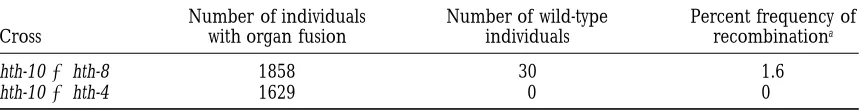
epidermally-TABLE 3
Summary of hothead intragenic recombination frequencies
Number of individuals Number of wild-type Percent frequency of
Cross with organ fusion individuals recombinationa
hth-103hth-8 1858 30 1.6
hth-103hth-4 1629 0 0
aBecause the progeny of these crosses are derived from selfing of the F
1 generation the frequency of
recombination (r) can be estimated from the number of wild-type recombinants: frequency of wild-type recombinants5r/21r/22r2/4. Since r is small this latter expression is approximately equal to r.
notype varies among members in this mutant collection alleles results in both the cer and organ fusion pheno-types only when homozygous or in combination with from a severe phenotype like that of the original
fdh-1-mutant (Lolleet al. 1992), to a very subtle fusion pheno- ded-1. The pattern is further complicated by the fact that plants bearing some combinations of alleles (ded-type such as is the case for the ahd-1 and phd-1 mutants.
Plants with a severe fusion phenotype can suffer serious 3/ded-4; ded-2/ded-3) have an obvious cer phenotype but fail to undergo any organ fusion whatsoever. More growth impairment while plants with a weak phenotype
are relatively normal in appearance. In addition to fu- mutant alleles of this locus are needed to allow a better definition of this complex complementation pattern. sion of organs comprising the external shoot, in some
mutants ovules with abnormal morphology were also Characterization of the mutants presently available as well as molecular analysis of the DED gene may allow found. Although these abnormal structures (in which
adjacent ovules are joined together at maturity) are us to determine the nature of the interallelic comple-mentation seen at this locus.
reminiscent of fused structures seen in other parts of
the plant, determination of the mechanism which leads Of all of the organ fusion mutants we have recovered, only mutations at the DEADHEAD locus show a wax to these structures will require a more detailed
examina-tion of ovule development in these mutants. Surpris- defect. The deadhead eceriferum phenotype suggests a defect in lipid biosynthesis (Aarts et al. 1995; Han-ingly, ovule defects were detected not only in mutants
showing strong fusion between organs but also in mu- noufaet al. 1996; Negruket al. 1996;Xiaet al. 1996), and, as indicated by biochemical analyses of fdh-1 mu-tants like bud-1, which manifest a relatively weak fusion
phenotype. Although the bud-1 mutant is a tight female tants, changes in lipid composition of the outer epider-mal cell wall and cuticle may play some role in promot-sterile, other mutants where ovule abnormalities were
detected (ded-1 and thd-1) are not, indicating that ova- ing organ fusion. Two other Arabidopsis mutants which also show organ fusion and have a wax defect have been ries containing large abnormal structures derived from
many ovules need not block the fertilization process described (Jenkset al. 1996) and may be allelic to our ded mutants, although complementation tests have not in the few remaining normal ovules. The detectable
presence of a funicular structure with relatively normal yet been performed. However, deadhead does not appear to be allelic to cer5 which maps to a similar location in morphology in all mutants that manifest ovule defects
suggests that deviation from normal ovule development the genome. A detailed biochemical analysis of these mutants may reveal the loss or modulation of a lipid occurs after the ovule primordia have elongated and
the integuments have initiated (stage 2-II; Schneitzet component that may be important for maintaining epi-dermal developmental integrity.
al. 1995). Further analysis of ovule development should
help illuminate the ontogeny of these aberrant struc- A second locus which has interesting phenotypic be-havior is CONEHEAD. In this case, plants which are tures and what, if any, cellular consequences these
ab-normalities have for the embryo sac as well as the sporo- homozygous for the mutation fall in equal numbers into two discrete phenotypic classes: those that exhibit strong phytic tissues of the ovule.
The deadhead locus which we have described has an fusion and those that exhibit weak fusion. Both of these phenotypes were present in the original single line fam-interesting pattern of interallelic complementation.
Plants homozygous for any of the individual ded mutant ily in which the mutation was isolated and both pheno-types have persisted through three backcrosses to the alleles have a strong eceriferum phenotype as well as
manifesting relatively severe organ fusion. Heteroallelic parental wild-type line, Landsberg erecta. Although it is possible that this variation in phenotype is due to a combinations have widely varying phenotypes, however,
ranging from completely wild-type plants to plants with genetic modifier which is segregating in this line, it is hard to account for the fact that it has been carried as severe a phenotype as the individual alleles. The
pres-ent complempres-entation analysis fails to define any obvious along through the backcrosses (implying close linkage to the cod mutation) and yet is readily separable from subgroupings of alleles. One allele (ded-1) fails to
Alterna-tively, it is possible that the mutation in COD is itself of organ fusion is still confined to late adult develop-ment. Plants are known to manifest a variety of morpho-responsible for both phenotypes with variable
expressiv-ity of the gene somehow producing two discrete classes logical and biochemical changes during different phases in their development (Lawson and Poethig of plants. The isolation of additional alleles of this locus
and COD gene isolation may help clarify how these 1995) and it may be that at this locus the juvenile to adult phase transition plays some role in regulating THD mixed phenotypes are achieved.
The disproportionately large number of mutations gene expression. Interestingly, the adherent2 locus in maize shows a similar restriction of organ fusion to the recovered in the HOTHEAD complementation group
suggest that this locus may represent an unusually large adult phase of the plant life cycle (Neufferet al. 1997). Although the mutants described in this article were gene (or at least a very large target for the mutagen
EMS). One corroborating piece of evidence comes from all selected on the basis of having an organ fusion phe-notype, epidermal cells in many of them also interact our analysis of recombination frequencies between
dif-ferent hth alleles. Based on estimates derived from these with pollen, as was the case for fdh-1 (LolleandCheung 1993). This fact clearly indicates that these two seem-recombination data the HTH locus may span as much
as 1.6 cM. This is dramatically larger than similar recom- ingly distinct processes are in some way genetically re-lated, since mutations in several different genes bring bination estimates for other Arabidopsis genes (0.07
cM for GA1;Koornneefet al. 1983; 0.01 cM for CSR1; about similar alterations in both processes. On the other hand, three of the new organ fusion mutants which we Mourad et al. 1994). A number of possibilities may
explain this discrepancy. First, it is possible that the have identified, representing three different comple-mentation groups, fail to support pollen hydration on apparent recombinants actually represent reversion of
one of the two mutations (presumably hth-8, since hth- their nonreproductive epidermal surfaces. Taken to-gether, these results indicate that while these two pro-10 was present in both crosses). While we have not
attempted to measure this directly, we consider this an cesses may be related, they are also clearly separable genetically, and it will be very interesting to learn the unlikely explanation since it would indicate a
spontane-ous reversion rate of 1.63 1022. Second, it is possible molecular identities of the gene products which are in
common as well as those which are unique to each of that the HTH locus contains a particularly
recombino-genic region within its borders. In this case the HTH the two pathways.
In the original fdh-1 mutant a striking increase in the locus may be a gene of quite ordinary size despite the
measured recombination frequencies between alleles. permeability of the epidermal cell wall and cuticle was the only alteration detected that distinguished mutant Third, the HTH locus may be split by a large intervening
sequence as is the case in a number of other genes, plants from wild type (Lolleet al. 1997). Although it seemed plausible that this permeability change might such as some of the homeotic genes of Drosophila
(GehringandHiromi1986). Fourth, it remains a for- play a role in the acquisition of fusion competence by the epidermal cells, no corroborating evidence sup-mal possibility that this locus is made up of two distinct
genes that interact in some manner such that they fail ported this hypothesis. The fact that many of the new organ fusion mutations isolated, which fall into a num-to complement one another in a simple cis-trans test.
Although further analysis of recombination frequencies ber of different complementation groups, also show changes in permeability to chlorophyll, however, is con-may reveal how the alleles group on the genetic map,
not until the gene is isolated and characterized will its sistent with this hypothesis. Furthermore, it is clear that thunderhead mutant plants switch to organ fusion compe-structure be revealed.
At the THUNDERHEAD locus, one of the two mutant tence at a discrete time in development and that this switch coincides precisely with a change in permeability. alleles (thd-1) shows a sharp transition from normal
growth to fusion competence, which appears to coincide As such, these data strongly support the notion that changes in the permeability of the epidermal cell wall well with the juvenile to adult phase transition. In these
mutants fusion cannot be detected until emergence of and cuticle to small molecules represent one mecha-nism which can lead to ectopic organ fusion during the fourth or fifth leaf. The mutant plants when mature
show a central trumpet-like clustering of leaves sur- Arabidopsis development.
One of the mutants recovered in this study fails to rounded by a relatively normal rosette. The thd-2 allele
does not show a similarly sharp transition, but the onset show any change in permeability that can be detected
assistance with various aspects of this project. We thank Edward
with our present assay. This mutant may, of course, have
Selingof OEB and MCZ at Harvard for assistance with sample
prepa-permeability changes below the limit of our detection
ration and use of the scanning electron microscope. We also thank or it may have changes in permeability to the relevant Allen Sessions, David Smyth, Lawrence Hobbie, Chris
Somer-signalling molecules that do not affect the permeabil- villeand the Arabidopsis Stock Center for making seeds available
from mutant lines showing a fusion phenotype. We are grateful to ity of our test molecule, chlorophyll. Alternatively, this
Ueli Grossniklausand members of his laboratory as well asGraeme
mutant may represent a class of mutations which
Berlyn for their continued interest in this project and for many
allow organ fusion to take place by some other
mecha-helpful discussions. This work was supported in part by a grant from nism, perhaps one related to the cr4 mutant of maize the Clark Fund at Harvard University, Harvard College Research Foun-(Becraftet al. 1996). Interestingly, an Arabidopsis mu- dation Fellowships and by National Science Foundation (NSF) Grant IBN-9405391 awarded to R.E.P. The main body of the work described tant (blasig) having an epidermal phenotype similar to
in this article was supported by NSF Grant IBN-9596044 awarded to cr4 but manifested only in the ovules has previously
S.J.L. and NSF Grant IBN-9723563 awarded to R.E.P. and S.J.L. been characterized in this lab (Schneitz et al. 1997).
A more detailed examination of the epidermal mor-phology of the mutant showing organ fusion without
LITERATURE CITED
an accompanying change in permeability as well as the
blasig mutant will be required to see if any of the same Aarts, M. B.M., C. J. Keijzer, W. J. Stiekema and A. Pereira,
features characterizing the cr4 epidermis are also mani- 1995 Molecular characterization of the CER1 gene of Arabidopsis involved in epicuticular wax biosynthesis and pollen fertility. Plant fested in these mutants.
Cell 7: 2115–2127.
We undertook a genetic approach to studying organ Becraft, P. W., P. S. StinardandD. R. McCarty,1996 CRINKLY4:
fusion with the goal of identifying as many genes as a TNFR-like receptor kinase involved in maize epidermal differen-tiation. Science 273: 1406–1409.
possible which are involved in this developmental
pro-Bell, C. J.,andJ. R. Ecker,1994 Assignment of 30 microsatellite
cess. Ultimately, we hope to identify a variety of molecu- loci to the linkage map of Arabidopsis. Genomics 19: 137–144.
Bell, P. R.,1995 Incompatibility in flowering plants: adaptation of
lar players in this process either directly by screening
an ancient response. Plant Cell 7: 5–16. for plants showing organ fusion or secondarily by
look-Cusick, F.,1966 On phylogenetic and ontogenetic fusions, pp. 170–
ing for enhancers and suppressors of the primary muta- 183 in Trends in Plant Morphogenesis, edited by E. G. Cutter.
Longmans Green & Co., New York. tions. At the outset we expected to find mutations
alter-de Nettancourt, D.,1977 Incompatibility in angiosperms.
Springer-ing the barrier to diffusion of signallSpringer-ing factors (such
Verlag, Berlin, New York. as described for fdh-1;Lolleet al. 1997), possibly
muta-Edwards, K., C. JohnstoneandC. Thompson,1991 A simple and
rapid method for the preparation of plant genomic DNA for tions identifying the factors involved in the responses
PCR analysis. Nucleic Acids Res. 19: 1349. (the signalling molecules themselves) or mutations
af-Gehring, W. J.,andY. Hiromi,1986 Homeotic genes and the
ho-fecting members of the downstream signal transduction meobox. Annu. Rev. Genet. 20: 147–173.
Hannoufa, A., V. Negruk, G. EisnerandB. Lemieux,1996 The
pathway. The collection of mutants that we present here
CER3 gene of Arabidopsis thaliana is expressed in leaves, stems, clearly contains mutations in a number of different
roots, flowers and apical meristems. Plant J. 10: 459–467. genes which alter the permeability barrier as well as one Hu¨ lskamp, M., S. D. Kopczak, T. F. Horejsi, B. K. KihlandR. E.
Pruitt,1995 Identification of genes required for pollen-stigma
example of a mutation which probably leads to organ
recognition in Arabidopsis thaliana. Plant J. 8: 703–714. fusion by a different type of primary defect. It may be Jenks, M. A., A. M. Rashotte, H. A. TuttleandK. A. Feldmann, that this class of mutations will include the Arabidopsis 1996 Mutants in Arabidopsis thaliana altered in epicuticular wax
and leaf morphology. Plant Physiol. 110: 377–385. homolog of the putative CR4 receptor protein kinase
Kempton, J. H.,1920 Heritable characters of maize V: adherence.
(Becraft et al. 1996). Although the evidence is only
J. Hered. 11: 317–322.
circumstantial, it may be that both processes are part Konieczny, A.,andF. M. Ausubel,1993 A procedure for mapping
Arabidopsis mutations using co-dominant ecotype-specific PCR-of the same developmental pathway where changes in
based markers. Plant J. 4: 403–410.
wall permeability permit exchange of factors that modu- Koornneef, M., C. J. HanhartandF. Thiel,1989 A genetic and
late CR4 receptor kinase activity. In cr4 mutant plants phenotypic description of eceriferum (cer) mutants in Arabidopsis thaliana. J. Hered. 80: 118–122.
epidermal cells may not fully differentiate or only
be-Koornneef, M., J. van Eden, C. J. HanhartandA. M. M. de Jongh,
come partially dedifferentiated and hence be compe- 1983 Genetic fine-structure of the GA-1 locus in the higher tent to form graft-like fusions, while in fdh-1 mutant plant Arabidopsis thaliana (L.) Heynh. Genet. Res. 41: 57–68.
Lander, E., P. Green, J. Abrahamson, A. Barlow, M. Daleyet al.,
plants the epidermis may become primed to exchange
1987 MAPMAKER: an interactive computer package for con-factors that will initiate the cascade of biological events structing primary genetic linkage maps of experimental and natu-that promote cell wall adhesion. Molecular characteriza- ral populations. Genomics 1: 174–181.
Lawson, E. J. R.,andR. S. Poethig, 1995 Shoot development in
tion of the genes identified in this article may help to
plants: time for change. Trends Genet. 11: 263–268.
resolve questions about how these two types of organ Lolle, S. J., G. P. Berlyn, E. M. Engstrom, K. A. Krolikowski,
fusion mutations are related mechanistically. W.-D. Reiteret al., 1997 Developmental regulation of cell
inter-actions in the Arabidopsis fiddlehead-1 mutant: a role for the epider-We are indebted toCeri Batchelder, Martin Hu¨ lskamp, Steve
mal cell wall and cuticle. Dev. Biol. 189: 311–321.
KopczakandKay Schneitzfor their willingness to screen for organ
Lolle, S. J.,andA. Y. Cheung,1993 Promiscuous germination and
fusion mutations while pursuing other mutants of their own. We also growth of wild-type pollen from Arabidopsis and related species thankAngeline Chong, Phyllis Itoka, Katherine Krolikowski, on the shoot of the Arabidopsis mutant, fiddlehead. Dev. Biol. 155:
250–258.
Lolle, S. J., A. Y. CheungandI. M. Sussex,1992 Fiddlehead: An Schneitz, K., M. Hu¨ lskampandR. E. Pruitt,1995 Wild-type ovule
development in Arabidopsis thaliana : a light microscope study of Arabidopsis mutant constitutively expressing an organ fusion
pro-cleared whole-mount tissue. Plant J. 7: 731–749. gram that involves interactions between epidermal cells. Dev.
Siegel, B. A.,andJ. A. Verbeke,1989 Diffusible factors essential
Biol. 152: 383–392.
for epidermal cell redifferentiation in Catharanthus roseus. Science
Mourad, G., G. HaughnandJ. King,1994 Intragenic
recombina-244:580–582. tion in the CSR1 locus of Arabidopsis. Mol. Gen. Genet. 243:
Verbeke, J. A.,andD. B. Walker,1985 Rate of induced cellular
178–184.
dedifferentiation in Catharanthus roseus. Am. J. Bot. 72: 1314–
Negruk, V., P. Yang, M. Subramanian, J. P. McNevinandB.
Lem-1317.
ieux,1996 Molecular cloning and characterization of the CER2
Verbeke, J. A.,andD. B. Walker,1986 Morphogenetic factors
con-gene of Arabidopsis thaliana. Plant J. 9: 137–145.
trolling differentiation and dedifferentiation of epidermal cells
Neuffer, M. G., E. H. CoeandS. R. Wessler,1997 Mutants of maize.
in the gynoecium of Catharanthus roseus. Planta 168: 43–49. Cold Spring Harbor Laboratory Press, Cold Spring Harbor, NY.
Walker, D. B.,1975a Postgenital carpel fusion in Catharanthus roseus Ockendon, D. J.,1972 Pollen tube growth and the site of the
incom-(Apocynaceae). I. Light and scanning electron microscopic study patibility reaction in Brassica oleracea. New Phytol. 71: 519–522.
of gynoecial ontogeny. Am. J. Bot. 62: 457–467.
Preuss, D., B. Lemieux, G. YenandR. W. Davis,1993 A conditional
Walker, D. B.,1975b Postgenital carpel fusion in Catharanthus
ro-sterile mutation eliminates surface components from Arabidopsis
seus. II. Fine structure of the epidermis before fusion. Proto-pollen and disrupts cell signaling during fertilization. Genes Dev.
plasma 86: 29–41. 7:974–985.
Walker, D. B.,1975c Postgenital carpel fusion in Catharanthus ro-Robinson-Beers, K., R. E. Pruitt andC. S. Gasser,1992 Ovule
seus. III. Fine structure of the epidermis during and after fusion. development in wild-type Arabidopsis and two female-sterile mu- Protoplasma 86: 43–63.
tants. Plant Cell 4: 1237–1249. Xia, Y., B. J. NikolauandP. S. Schnable,1996 Cloning and charac-Schneitz, K., M. Hu¨ lskamp, S. D. KopczakandR. E. Pruitt,1997
terization of CER2, an Arabidopsis gene that affects cuticular wax Dissection of sexual organ ontogenesis: a genetic analysis of ovule accumulation. Plant Cell 8: 1291–1304.
development in Arabidopsis thaliana. Development 124: 1367–