Copyright 0 1990 by the Genetics Society of America
Break-Join Recombination in Phage
X
Franklin
W.
Stahl, Maurice
S.
Fox,’ Daryl Faulds‘ and Mary
M.
Stahl
Institute of Molecular Biology, University of Oregon, Eugene, Oregon 97403-1229
Manuscript received December 24, 1989 Accepted for publication April 6, 1990
ABSTRACT
In phage X, when DNA replication is blocked, recombination mediated by the Red pathway occurs only near the double-chain break site, cos, that defines the termini of the virion chromosome. The recombinants initiated by cos contain newly synthesized DNA near cos, in amount corresponding to a few percent of the length of X. A restriction enzyme cut delivered to one parent far from cos results in elevated recombination near the restriction site. Recombinants induced by this cut have a similarly small amount of DNA synthesis in these replication-blocked crosses. When restriction cuts are introduced in the presence of normal amounts of all of the DNA replication enzymes, many of the resulting recombinants still enjoy, at most, a small amount of DNA synthesis associated with the exchange event. Thus, these experiments fail to support the previously considered possibility that Red-mediated recombination in X proceeds largely through a break-copy pathway.
P
revious work (STAHL et al. 1972b) has shown that recombination in phage X is partially dependent on DNA replication. The degree of dependence is related to distance from the ends of the standard linkage map, so that, in crosses blocked for DNA replication, there is a relative paucity of recombinants in the middle of the map. When replication is allowed, the exchange distribution is more nearly uniform.The significance of the relationship between high recombination (when DNA replication is blocked) and the end of the linkage map was established in experi- ments that turn the linkage map inside out (STAHL, KOBAYASHI and STAHL 1982). X strains were con- structed in which cos, the region of the X chromosome that specifies the termini of the packaged chromo- some, was translocated from its normal position to the middle of the normal chromosome. Such “inside out” chromosomes are packaged from the translocated cos,
and have their ends in the region corresponding to the middle of ordinary X. In these phage, the region of high recombination rate was seen to have been translocated along with cos.
Two explanations for the apparent recombinagen- icity of cos have been considered. In one theory,
recombination-initiating events are distributed along X without regard to cos, even among unreplicated chromosomes. Because of a postulated break-copy recombination mechanism, only events initiated near the termini are completed and packaged when repli- cation forks are blocked (but polymerases remain in- tact) (STAHL et al. 1973). In the second theory, cos is
’ Present address: Biology Department, Massachusetts Institute of Tech-
’
Present address: Codon, 213 East Grand Avenue, South San Francisco, nology, Cambridge, Massachusetts 02 139.California 94080.
Genetics 1 2 5 463-474 (July, 1990)
itself an initiator of recombination (RUSSO 1973). Among unreplicated phage, cos is the principal, and perhaps the only, initiator. The act of replication generates other initiators, so that recombination among replicated phages (which may be strictly break- join) is no longer focused at cos.
Subsequent experiments supported the latter inter- pretation without ruling out appropriately adjusted versions of the former (STAHL, KOBAYASHI and STAHL, 1985). It was shown that a functional cos site can stimulate break-join recombination even when the parent with which it is paired carries an uncuttable mutant cos at that locus. Among cos stimulated recom- binants, the cuttable cos became converted to its un- cuttable allele (and the recombinants were packaged via cos sites cloned elsewhere in the two parents). Thus, cos appeared to be initiating recombination by being a double chain break (DCB) site, more-or-less in the sense of the “double-strand-break repair” model of SZOSTAK et al. (1983). This concept led to the proposal that cos is the only DCB site among non- replicated phage and that replication introduces other such sites, or their functional equivalents. Most attrac- tive was the possibility that the end of the tail of a rolling circle replicative form could initiate exchange (WILKINS and MISTRY 1974; SKALKA 1977; STAHL, KOBAYASHI and STAHL 1985; THALER, STAHL and STAHL 1987b).
464 F. W. Stahl et al.
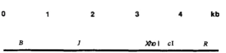
0 1 2 3 4 kb
B I Mnl CI R
FIGURE 1 .-Locations on the X chromosome of genetic elements employed.
TABLE 1
Bacterial strains and plasmids employed
K12SH-28; Su+ indicator to score cI us. cI+ among Ts+ recombi- nants; host for heavy stocks (FANGMAN and NOVICK 1966) FS2769: K12SH-28[pPAORM3.8]; host for heavy, "Xhol-modi-
fied" stocks
FA77: Su- derivative of FA22 (FANGMAN and NOVICK, 1968), a
dnaBts derivative of K12SH-28; host for replication-blocked crosses (MCMILIN and Russo 1972)
blocked crosses with XhoI cutting
blocked crosses with XhoI cutting (FZ14 is Su- dnaBts22 recA56) (STAHL et al. 1972a)
FS1441 = FA77[pPAORM3.8]; RecA+ host for replication FS2594 = FZ14[pPAORM3.8]; RecA- host for replication
FS85 = QR47(XJtsZ5 Rsus5); RecA+ host for repressor crosses FS86 = QK48(XJts15 Rsus5); RecA- host for repressor crosses FS88 = 594(XJtsZ5 imm434 Rsus5); Su- indicator for repressor pPAORM3.8 encodes the PaeR7 restriction system, an isoschizo-
crosse5
mer of XhoI (GINGERAS and BROOKS 1983)
chain ends there. In so doing, it further supports the view that cos is a recombinator by virtue of being a
DCB site.
Our experiments reveal and quantitate a modest a m o u n t of DNA synthesis associated with a cos-initi- ated event and suggest a similar amount of synthesis associated with a centrally located recombination event initiated by an artificially introduced DCB.
Most of our experiments involve X that is wild type
for genes influencing homologous recombination
(red+gam+) recombining in wild type Escherichia coli (ret+). T h u s , they describe the activity of the RecA- assisted Red pathway of X operating in the presence
of Gam-inhibited RecBCD enzyme (for reviews, see SMITH 1983, THALER a n d STAHL 1988). In some experiments, which will be identified, the host is a
recA mutant, so that the Red pathway is mediating exchange without help from the RecA protein.
MATERIALS AND METHODS
Phage and bacteria: A map of X showing features relevant to this work is in Figure 1. Strains of bacteria employed are described in Table 1.
Replication-blocked crosses: Except where noted, crosses were executed in the Su-dnaBts host FA77, or a plasmid-carrying derivative thereof, at 40" (MCMILIN and
Russo 1972). All phages were conditionally defective in the X DNA replication gene P by virtue of the suppressible mutation Psus80. Except where noted, phages were int4,
inactivating X's site specific recombination system.
Crosses with in vivo restriction: In experiments involving cutting of X's XhoI site, the host cell carries the plasmid
pPAORM3.8 (GINGERAS and BROOKS 1983), which encodes the PaeR7 restriction/modification system, an isoschizomer of XhoI. In this paper, both the restriction system and its cut site in X will be referred to as XhoI. Host cell cultures that carry plasmid were grown to 1.5 X 10" cells/ml in ampicillin to hold the plasmid. The cells were removed from the ampicillin and heated at 40" for 10 min. One milliliter of culture was then added to 1 ml of preheated phage mix in which each parent was typically present at 1 .O X 1 O'//ml to give a multiplicity of infection (moi) of 7 of each parent. As in THALER, STAHL and STAHL (1987a), when one parent was cuttable, the uncuttable parent was added 10-15 min before the cuttable one, in order to establish the Red and Gam functions before any DNA cutting occurs. Ninety minutes after infection, the 2-ml infected cultures were lysed with CHCIS and lysozyme, sometimes after removing unad- sorbed phages by centrifugation. The entire volume of each lysate was adjusted to a refractive index of 1.378 with cesium formate and centrifuged in a SW50.1 head at about 30,000 rpm for 16 hr or more. Fractions were collected through a needle hole in the bottom of the tube, usually at two drops per fraction yielding about 75 fractions. Fractions were plaque-assayed on appropriate bacteria as indicated.
Preparation of heavy phage: Density-labeled phage were usually prepared on minimal agar plates in which the glucose was substituted with "C (99%, in most cases) and the am- monium chloride with I5N (99%). Uncuttable parents were modified by preparation of the density labeled stocks in E . coli strain FS2769, a Su+ prototroph carrying pPAORM3.8.
Preparation of radioactive, heavy phage: Heavy phage labeled with "P or "P were prepared by thermal induction of the lysogen K12SH-28 (X int+ cI857 Psus80 Ssus7)lX growing in low phosphate (1 5 mg/liter P) minimal medium. T h e carbon source was [lRC]glucose (-60% "C) at 2 m
/
ml; nitrogen was "NH,CI (98% "N) at 1 mg/ml; "P or 'l"
was present as phosphate at a specific activity of 1-2 mCi/ mg P. Radioactively labeled phage were purified by equilib- rium centrifugation in CsCI. T h e replication-blocked den- sity-transfer experiment was conducted 10 days after label- ing the phage. DNA was released from phage by addition of EDTA (0.01 M) and Sarkosyl (0.1 %) and heating for 6 min at 63
'.
Equilibrium centrifugation of DNA in CsCl was for 40 hr at 38,000 rpm in a SW 50.1 Beckman rotor. Activities of "P, jJP, and 'H were determined by scintilla- tion counting (Packard T r i Carb).DNA extraction for restriction endonuclease digestion:
Cells from 100 ml of infected culture were washed several times with chilled nonradioactive medium containing excess phosphate. The final bacterial pellet was resuspended in citrate buffer to which 3 X 10" nonradioactive phage had been added. Lysis was promoted with CHCI:+ and Brij (0.2%). Phage banded in CsCl were dialyzed against 10 mM Tris, 10 mM EDTA, pH 8.0 (TE). Phage DNA was extracted with cold phenol and dialyzed against TE. To separate X's terminal fragments from each other, endonuclease digests were heated at 65" for 3 min and then quickly chilled before electrophoresis.
RESULTS
XhoI can initiate break-join recombination: h con- tains a solitary XhoI site, at base pair 33,498, which is
0.69 of the distance from the left to the rig ht en d (Figure 1). When one of the two infecting parents
carries the XhoI site in an unmodified (cuttable) state,
Breakjoin Recombination 465
-
20 30Fraction No.(
-
Density )FIGURE 2.-Stimulation of recombination by XhoI in replication- blocked crosses. Crosses of J t s l 5 c1857 R+ X J’ cI+ Rts2 were at restrictive temperature in the E . coli K12 strain FS1441, a Su- dnaEts22 strain carrying the PaeR7 (“XhoI”) restriction system on a plasmid (pPAORM3.8). Both parents were Psus80, so that DNA
replication was “double-blocked.” In cross 1, both parents were uncuttable by virtue of being XhoI-modified. In cross 2, the Rts
phage was cuttable. In cross 3 , theJts CI phage was cuttab1e.J’ R+
recombinants were selected a t 40” on the Su+ indicator K12SH-28,
and plaques were scored as clear (cI), turbid (c+) or mottlers (heteroduplexes cl/c+). T h e data points for each cross, summed according to genotype, are in Table 2. Key: (0) X J’ cI+ R+
recombinants: (0) XJ’ cl R+ recombinants: and (9) XJ’ cl/cI+ R+
recombinants (heteroduplex at cI).
1987a). T h e stimulation can be measured as an in- crease in the recombinant frequency for a marked interval containing the XhoI site relative to the fre- quency of recombinants in a control interval. In the studies of THALER, STAHL and STAHL (1987a) the absolute frequency of recombinants in the test interval increased. Some of their crosses were conducted un- der conditions presumed to block DNA replication, with similar results.
We have performed crosses like those of THALER, STAHL and STAHL (1987a) except that both parents were labeled with heavy isotopes of C and N, allowing us to monitor the block on DNA replication. T h e parents carry conditional lethal mutations in genes J and R, respectively (Figure l ) , so that recombinants can be selected. T h e J-R interval is bisected by a marker at the cI locus, so that recombinants arising in the]-cI interval make clear plaques while those arising in the cI-R interval make turbid plaques. T h e XhoI site is in the J-cI interval. Replication was blocked by the dnaBts mutation in the host and Psus80 in the phages. Three crosses were performed. T h e parental phages were genotypically the same for each of the three crosses, but they differed as to modification at the XhoI site. In cross 1, both parents were modified; in crosses 2 and 3, one or the other parent, respec- tively, was modified. Since DNA cannot be cut by
~3
rt
2
I 1
30
D l
20 30
Fraction No. (-Density )
FIGURE 3.-Stimulation of recombination by XhoI in RecA- rep- lication-blocked crosses. Crosses and markers are as in Figure 2.
T h e host strain, FS2594, was recA56 in addition to being dnaEts22 and carrying the XhoI restriction plasmid. Key as in Figure 2.
XhoI when it is XhoI-modified (and see THALER, STAHL and STAHL 1987a), cross 1 is the control for crosses 2 and 3.
T h e cross lysates were centrifuged in cesium for- mate. Each cross yielded a single peak of recombinant phage, demonstrating that replication was well blocked (Figures
2
and 3).When the host was RecA+ (FS144 l ) , XhoI-stimula- tion of recombination is manifested by a change in the ratio of clear to turbid Ts+ recombinants (Figure 2). Upon cutting either parent, the relative frequency of clear to turbid recombinants increases, implying a relative increase in recombination in theJ-cI interval, which contains the XhoI site. In both crosses involving cutting, the relative frequencies of mottled (cI/cI+ heteroduplex) to turbid (cI+) plaques are increased by cutting, as well. T h e former observation needs no interpretation; we will interpret the latter observation in DISCUSSION.
466 F. W. Stahl et al.
TABLE 2
XhoI stimulated recombination in replication-blocked crosses
Fraction of A recombinant in interval
Crosses J-c 1 cl-R (hets) hetslcl-R
C k l +
RecAf
Cross 1
Expt. 1 Expt. 2
Expt. 1 Expt. 2
Expt. 1 Expt. 2
Cross 2
Cross 3
RecA-
Cross 1 Expt. 1 Expt. 2
Expt. 1
Expt. 2
Expt. 1 Expt. 2 Cross 2
Cross 3
0.15 0.83 0.02 0.16 0.83 0.02
0.76 0.17 0.07 0.75 0.17 0.08
0.63 0.33 0.04 0.38 0.55 0.08
0.12 0.55 0.34 0.14 0.77 0.09
0.76 0.05 0.19 0.76 0.06 0.18
0.22 0.19 0.59 0.24 0.16 0.61
0.02 0.02
0.4 0.5
0.1 0.2
0.6 0.1
3.8 3.0
3.1 3.8 Experiment 1 i s from Figures 2 and 3. In cross 1 , neither parent is cut. In cross 2, the Rts parent i s cut. In cross 3, theJts parent is
cut.
trast to the corresponding RecA+ crosses (Figure 2, cross
2).
T h e set of six crosses was repeated with similar results. For the two sets of crosses the fractions of
J + R + that are
cI,
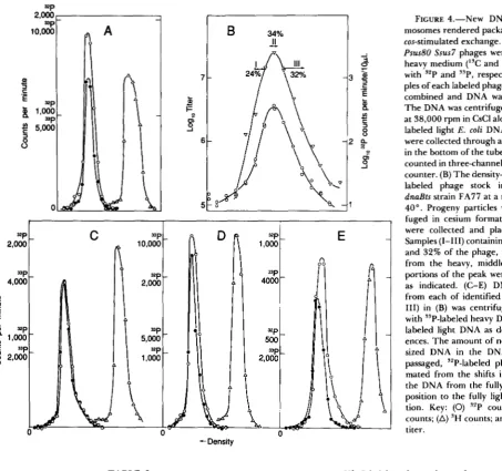
cI+, or cI/cI+ heteroduplex, respec- tively, are reported in Table 2.The progeny X particles from RecA' double- blocked infections contain various small amounts of newly synthesized DNA: Two populations of den- sity-labeled X Psus80 were prepared, one in 32P-con- taining medium and one in 33P-containing medium. T h e populations were calibrated for their relative levels of DNA density label by centrifugation of DNA extracted from the particles (Figure 4A). T h e 32P-
labeled phage was purified and then passed through a double-block cycle at a moi of 10 in strain FA77, and the progeny was centrifuged in cesium chloride. Plaque-assay of the fractions showed that the infection gave a unimodal density-distribution of progeny phages, characteristic of double-block crosses (Figure 4B). Unadsorbed phage (not shown) are well separated from the progeny peak by virtue of their heavy- labeled protein coats. Phage particles from the heavy flank of the progeny peak, from the modal fraction of the peak, and from fractions on the light flank, respectively, were combined with the heavy 33P X and with light 3H-labeled E . coli DNA, and DNA was released from the phage particles. T h e three DNA mixtures were centrifuged in CsCl (Figure 4, C-E).
DNA from the phage particles in the successively lighter fractions is seen to be progressively lighter than the 33P reference. T h e density shifts imply new DNA synthesis to the extent of 1.6% and 4.5% of the
X chromosome in the modal and lighter fractions, respectively.
Under replication-blocked conditions, recombina- tion is prerequisite to chromosome packaging, pre- sumably because it provides the sole route to conca- temer formation (STAHL et al. 1972a). Thus, all of the chromosomes in the lysate of Figure 4B have enjoyed an exchange during their sojourn in the replication- blocked host cell. Since the recombination under these Red+ Gam+ RecA+ conditions is primarily (or exclu- sively) cos-initiated, most, at least, of the chromosomes in Figure 4B have enjoyed an exchange near cos. For the Red system operating in double-blocked RecA- cells, the amount of new DNA in individual progeny
X particles is correlated inversely with the probability of heterozygosity for a marker near the right end of
the chromosome (STAHL and STAHL 1986). That cor- relation was taken to imply that cos-initiated, Red- mediated recombination was accompanied by variable amounts of DNA synthesis at the site of the exchange. In the following section we show that the incorporated label in the Red+ RecA+ double block progeny is concentrated near the termini, where most of the cos-
initiated exchange occurs.
The
"P
incorporated in the X progeny of the Red+ RecA+ double-blocked infection is concentrated in the terminal restriction fragments: FA77 was grown at 26" to 1.5 X 10⁸/ml in Tris-maltose (HERSHEY1955) plus 0.2% Bacto-Peptone (as sole source of nonradioactive phosphorus). T h e 100-ml culture was oxygenated at 39" for 10 min and then infected with nonradioactive "C 14N X int+ cI857 Psus80 Ssus7 at a moi of 20. At this time, glucose was added to a final concentration of 0.1 %, and 5 mCi 32P were added as carrier-free phosphate. Progeny phage were collected at 90 min, at which time the yield is about one phage particle per cell, and purified as described in MATE- RIALS AND METHODS. DNA was extracted with cold phenol and digested with EcoRI or Hind111 as de- scribed in MATERIALS AND METHODS. T h e resulting restriction fragments were sorted by gel electropho- resis. T h e tube gel was stained to identify bands of restriction fragments, and slices 2 mm thick were
scintillation counted. Results are in Table 3. They show that the specific activity of the terminal restric- tion fragments exceeds that of each of the other fragments. These results demonstrate that the new DNA found in the progeny of a double-blocked Red+ RecAf infection is located primarily in the regions of the chromosome where recombination is maximal as a result of cos-initiated exchange.
Break-Join Recombination 467
c a,
c
'E
"pg
1,000-8
5,000 =P 3
A
-Density
TABLE 3
Locations of 'P' incorporated in Red* RecA+ doubleblock infection
~
Position Percent Fragment (kb) length cpm cpm percent length
Percent Percent cpm/
EcoRI A
B
C D E F
Hind111
A B C D E F G
0-21.2 2 1.2-26.1 26.1-31.7 31.7-39.2 39.2-45.0 45.0-48.5
0-23.1 23.1-25.2 25.2-27.5 27.5-36.9 36.9-37.5 37.5-44.1 44.1-48.5
44.5 1444 70.3 1.58
9.8 49 2.4 0.24
11.3 52 2.5 0.22
15.4 110 5.4 0.35
12.1 153 7.4 0.61
6.9 247 12.0 1.74
47.6 850 59.6 1.25 4.3 20 1.4 0.33
4.7 27 1.9 0.40
19.4 200 14.0 0.7
1.2 13.6 170 11.9 0.88
9.1 160 11.2 1.23
shown in a later section that most of the recombinants in a ABts X XRts replication-blocked, XhoI-stimulated
FIGURE 4.-New DNA in chro- mosomes rendered packageable by a cos-stimulated exchange. (A) X cI857 Bus80 Ssus7 phages were grown in heavy medium ("C and "N) labeled with ?'P and "P, respectively. Sam- ples of each labeled phage stock were combined and DNA was extracted. The DNA was centrifuged for 40 hr at 38,000 rpm in CsCl along with 'H-
labeled light E. coli DNA. Fractions were collected through a needle hole in the bottom of the tube, dried, and counted in three-channel scintillation counter. (B) The density-labeled, IsP-
labeled phage stock infected the
dnaBts strain FA77 at a moi of 20 at
40". Progeny particles were centri- fuged in cesium formate. Fractions were collected and plaque-assayed. Samples (1-111) containing 24%, 34%
and 32% of the phage, respectively, from the heavy, middle, and light portions of the peak were identified as indicated. (C-E) DNA isolated from each of identified samples (I- 111) in (B) was centrifuged in CsCl
with "P-labeled heavy DNA and 'H-
labeled light DNA as density refer- ences. The amount of newly synthe- sized DNA in the DNA from the passaged, "P-labeled phage is esti- mated from the shifts in density of the DNA from the fully heavy ("P) position to the fully light ('H) posi- tion. Key: (0) '2P counts; (0) "P counts; (A) 'H counts; and (V) phage titer.
cross are XhoI-initiated, rather than cos-initiated. Thus, DNA synthesis associated with a XhoI-initiated exchange can be estimated by comparing the density of a population of XhoI-initiated recombinants with a control, cos-initiated, population (Figure 5A). The cos-
468 F. W. Sta
tI
-1
I I I I I I
20 30 40 50 20 30 40 Fraction No. (
-
Density )FIGURE 5.-New DNA in chromosomes that have enjoyed a XhoI-stimulated exchange. (A) Density-shift associated with a XhoI- initiated exchange. Heavy modified X Btsl cI+ R+ was crossed with heavy unmodified X B + cI+ Rtsl29 in FS1441. Part of the resulting lysate, i n which most of the B + R+ particles had enjoyed a XhoI- initiated exchange, was spun with the lysate from a cross on the same host of heavy modified X B t s l cI R+ X heavy modified X B + cI Rts129. T h e recombinants in the latter cross are all cos-initiated. T h e gradient was plated a t high temperature on a SuII+ host to score clear and turbid B + R+ recombinants. Light density reference phage (X c1857 Ssus7) were added to the gradient and scored on a SuIll host at temperature restrictive for the Bts and Rts parental genotype phages. (B) Similarity of label of parental phage stocks. T h e pairs of phage stocks used in (A) were tested for equality of
DNA density label by crossing them in the same pairwise combina- tions used i n (A). T h e host used for these crosses was FA77, the same as that used in (A) except lacking the XhoI plasmid. Thus, exchanges generating the B + R+ recombinants are cos-initiated for both crosses. Key: (A) Light density reference X cI857 Ssus7; (0) B +
cl R+; and (0) B + cI+ R+.
binants packaged following a XhoI-initiated exchange have a density distribution like those that have been packaged following a cos-initiated exchange, implying that XhoI-initiated exchange, also, involves only a small amount of DNA synthesis in double block crosses.
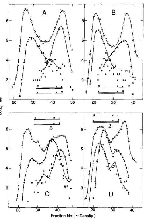
A XhoI cut can overcome the lack of recombinants of intermediate density in a heavy X light cross: T h e extent of DNA synthesis accompanying a XhoI-initi- ated event, in these replication-blocked conditions, is small compared to the length of X. Therefore, if the heteroduplex segments associated with XhoI events are not too long and if degradation from the double chain cut is not too extensive and variable, a XhoI- initiated exchange will produce a discrete density peak in a replication-blocked “profile” cross between a heavy and a light parent.
Our replication-blocked crosses were of the follow- ing form. Parents carried ts markers in the subter- minal genes B and R , respectively (Figure 1). T h e Bts
parent carries the marker cI857. One parent is heavy and the other is light. Btsl substitutes for the Atsl4
marker used in previous papers. As compared to Btsl
and to the several Asus markers examined, Atsl4 used
.hl et al.
I I I I ’ 1 I
L 1 . !
-
r RFz
:o 30 I 40 I 20 50 I I 30 I 40 IP, u
i
I 1 I I I I b 1 I
20 30 40 20 30 40
Fraction No. ( - Density )
FIGURE 6.-Distribution of exchanges in XhoI-cut chromosomes. Replication was blocked by dnaBts22 in the Su- host FS1441 and by PsusBO in all parental phages. Lysates were spun in cesium formate, and density fractions were assayed for total phage on K12SH-28 at 32” and for B + R+ recombinants on the same strain at 4 0 ” . (A) Heavy X Btsl cI26 R+ X light X B+ cI+ Rtsl29 with both parents modified (no XhoI cutting). (B) Light X BtslcI 26 R+ X
heavy X B + cI+ Rtsl29 (both modified). (C) Heavy, modified X Btsl cI26 R f X light, unmodified (XhoI-cuttable) X B + c l + Rtsl2Y. (D)
Light, modified X Btsl cI26 R+ X heavy, unmodified X E + cI+
Rts12Y. Key: (A) Total X; (0) X B + cI+ R+ recombinants; (0) X B + CI
R+ recombinants; and (0) X B + cI/cI+ R+ recombinants (heterodu- plex a t cI).
as a marker reduces recombination near X’s left end in replication-blocked Red+ RecA+ crosses
(J.
M. CRA-SEMANN, personal communication).
In the control crosses (Figure 6, A and B), both parents were modified, so that there was no cutting by XhoI. In Figure 6A, the Bts parent was heavy, while in Figure 6B, the Rts parent was heavy. The nonuni- formity of exchange in replication-blocked crosses
Break-Join Recombination 469
1972a), and half the recombination acts will be be- tween phage of opposite parental genotype (VISCONTI and DELBRUCK 1953), a uniform exchange distribu- tion should result in about one-half of the particles having densities that are distributed uniformly be- tween fully heavy and fully light. Since particles of intermediate density are rare, we can again deduce that most of the exchanges occur near the chromo- some tips, many of them outside the marked interval
In the experimental crosses, Figure 6C corresponds to Figure 6A, except that the Rts parent is unmodified, while Figure 6D corresponds to Figure 6B, with the
Rts parent unmodified. Thus, in Figure 6C the light parent is cut, while in Figure 6D the heavy parent is cut.
Comparing Figure 6C with its control, Figure 6A, we see distributions of Ts+ recombinants that are essentially superimposable except for the peak com- posed predominantly of clear Ts+ recombinants whose density indicates that they are the result of exchange near the XhoI site. T h e same relationship holds be- tween Figure 6, D and B, except for the inversion of these distributions relative to Figure 6, C and A (due to inversion of the density label between the two parental phages). Thus, to a first approximation the results are independent of whether the parent that is cut is heavy or light, as expected from our observation that there is not much DNA synthesis associated with a XhoI-initiated event.
T h e curves involving XhoI cutting add a peak of B+ R+ recombinants without seriously altering the rela- tive amounts of cos-initiated exchanges at the left and right ends leading to B+ R+ recombinants. This feature of the distributions suggests that B+ R+ recombinants manifesting a terminal exchange are primarily “back- ground,” arising by exchanges between the uncuttable parent and a cuttable parent that happened to escape restriction.
A XhoI cut stimulates break-join recombination even when DNA replication forks are unimpeded:
T h e experiments described above demonstrate that a XhoI restriction cut delivered to one parent in a X X
X cross can stimulate break-join recombination. Those crosses were conducted under conditions that strongly block DNA replication by depriving the infected cells of the E. coli dnaB and the
AP
gene products, so they fail to address the possibility that recombination stim- ulated by a double chain break would proceed by break-copy, if that route were available. T o assess this possibility, we conducted XhoI-stimulated crosses in which replication enzymes are fully functional.X repressor impedes DNA replication even in the presence of all the replication enzymes. This imped- ance is due to a requirement for transcription across
X’S origin of replication for maximum “activation” of
(B-R).
1 I l 1
1
,‘i
..(
.
I1
I..
.
20 30 20 30 40 20 30
1
..
IFraction No. (- Density j
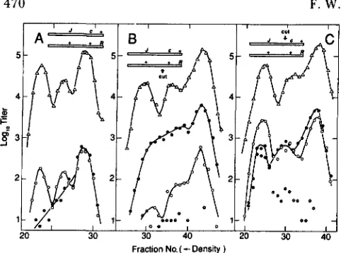
FIGURE 7.-RecA+ repressor crosses. Heavy X Jtsl5 c1857 R + were crossed with heavy XJ’ cI’ Rsus5 in the homoimmune lysogen QR47 (XJtsl5 Rsus5) in the presence of preinfecting heteroimmune helper phageJtsl5 imm434 Rsus5.J+ R’ recombinants were plated selectively on the Su- lysogen 594 (Jtsl5 imm434 Rsus5) at 4 2 ” and scored as ~1857, cI+, or cI/cI+ heteroduplexes by inspection of the plaques. (A) Both parents uncuttable (XhoI-modified). (B) J’ cI’ Rsus5 parent cuttable. (C) Its15 c1857 R + parent cuttable. Key:
Mean fraction number
A B C
0 XJ’ cI’ R + recombinants 24.4 35.7 28.4 0 X J + cI R + recombinants 27.8 35.1 29.4
9 h J + cl/cl+ R + recombinants A Total X immA
ori [see FURTH and WICKNER (1983) for review]. Pre- vious work (e.g., STAHL and STAHL 197 1 ; STAHL et al.
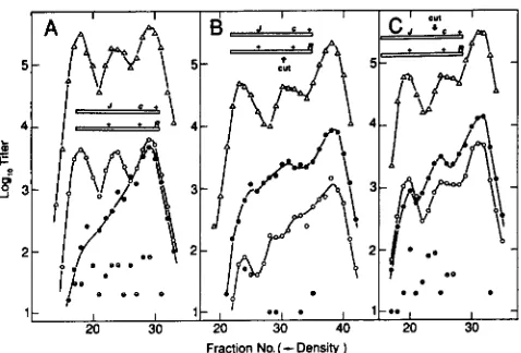
1972a) has exploited this cis-acting effect of X repres- sor to limit the amount of DNA replication of the two infecting X phage in a X X X lytic cycle cross. Conven- tional wisdom (DOVE, INOKUCHI and STEVENS 197 1) says that any replication that might be initiated in an ori-independent fashion will proceed unimpeded in such a cross. Thus, by inhibiting ori-dependent repli- cation we may reveal ori-independent replication ini- tiated by a XhoI-stimulated recombination event.
T h e crosses were J t s l 5 cI857 R+ X
J‘
cI+ Rsus5conducted in bacterial strains that carried X prophage
Jtsl5 Rsus5. Both of the parental phage stocks were heavy-labeled. Repressor made by the resident pro- phage provides a partial block to ori-dependent repli- cation. Since repressor blocks transcription of X genes, a heteroimmune “helper” phage is added to provide Red and Gam recombination functions, 0 and P replication functions, and DNA packaging proteins. T h e imm4j4 helper phage has the genotype J t s l 5 Rsus5 so that it cannot contribute to the formation of J’ R +
470 F. W. Stahl et al.
5
4
5
$ 3 -I
2
1
20 30 30 40 Fraction No. (
-
Density )FIGURE 8.-RecA- repressor crosses. As in Figure 7 except that the host cell was QR48 (X Its15 Rsus5). Key:
Mean fraction number
A B C
0 XJ' c l + R + recombinants 24.9 41.6 31.1 0 X J' cI R' recombinants 26.9 40.3 34.7
9 XJ' cI/cI+ R' recombinants A Total X immA
crosses were performed at 37", which is permissive for Jtsl5.
Figures 7A and 8A present control data, in which neither of the infecting imm' phages is XhoI-cuttable. As seen in previous work (STAHL and STAHL 197 l), this protocol limits DNA replication so that unrepli- cated chromosomes make up about one quarter of the yield of imm' phage. Recombinants in the cI-R inter- val (turbid plaques), at the right end of X, have enjoyed about as many rounds of replication as have the total
imm' phage. Recombinants in the J-cI interval (clear plaques), on the other hand, are rare in the fully heavy peak. Only in the fully light peak are the two recom- binant types found in relative amounts that approxi- mate the relative physical lengths of the intervals involved. This differential association between DNA synthesis and recombination in the two marked inter- vals is quantitated by comparison of the average po- sitions in the density gradient of the two classes of recombinant particles. Recombinants in the J-CI in- terval are lighter on the average than recombinants in the cI-R interval (Figures 7A and 8A).
Experiments in the sections above, in which repli- cation enzymes are missing, argue that failure to find fully heavy J-CI recombinants in double block crosses is due to the lack of double chain ends there. In the experiments of this section, we can anticipate a similar lack of spontaneous initiations among unreplicated chromosomes. We can provide those initiating events with XhoI, however, and note whether the resulting recombinants are present in the heavy peak. If an exchange occurring in the presence of the replication enzymes invariably initiates a replication fork, XhoI- initiated recombinants in the J-CI interval may be
gone from the fully heavy peak. Crosses in which one or the other of the infecting imm' phages is cuttable are in Figures
7,
B and C, and 8, B and C.In Figures 7B and 8B, the Rsus5 parent was cutta- ble. In all density peaks, including the fully heavy one, the J' R+ recombinants now arise primarily in the interva1,J-cI, that contains the XhoI cut site. Because restriction involves a competition between modifica- tion and cutting, it is likely that those heavy, cuttable phages that are cut are cut promptly upon infection of the restricting host cell. Since X replication is de- pendent on supercoiling (for review, see FURTH and WICKNER 1983), the chromosomes linearized by re- striction are probably not able to replicate. We surmise, therefore, that the XhoI stimulated recombi- nants in the light peak replicated after the XhoI- initiated exchange event. T h e J-CI interval recombi- nants, instead of having an average density less than that of the cI-R interval recombinants, as they did in the control, now have an average density that is slightly greater than that of the cI-R recombinants. This argues that XhoI cutting has provided an initia- tion structure for break-join recombination that was previously provided by replication. In the cI-R, con- trol interval, where most initiation is by cos cutting, some initiation structures are probably provided by replication. Thus, these density distributions fail to support the possibility that recombinants arising far from cos, in the J-CI interval, can initiate DNA syn- thesis more extensive than that reported for the dou- ble-block crosses described above. The smeared na- ture of the density distributions, especially of the recombinants arising in the J-CI interval, which con- tains the XhoI site, could imply extensive DNA syn- thesis occurring in some acts of exchange. On the other hand, the smeared recombinant density distri- bution might merely reflect break-join exchanges, near the middle of the chromosome, between heavy restricted chromosomes and chromosomes of the non- restricted parent that had replicated prior to ex- change. T h e latter view seems the more consistent with our other observations.
Break-Join Recombination 47 1
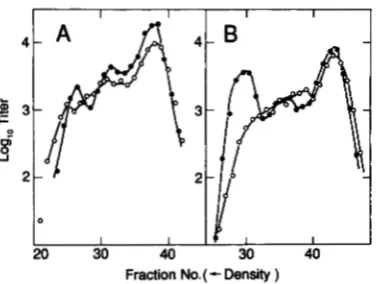
I I I I I I I
Fraction No. (
-
Density )FIGURE 9.-TotalJ+ R + recombinant density distributions from repressor crosses with one XhoI-cuttable parent. (A) RecA+ data of Figure 7, B and C. (B) RecA- data of Figure 8, B and C. Key: (0)
J + cI+ Rsus5 parent cuttable and (O)Jtsl5 cI857 R+ parent cuttable.
in a break-join manner even when all the enzymes required for DNA replication are present in the in- fected cell.
The shapes of the density distributions of total J' R' recombinants (Figure 9, A and B) depend on which of the two parents was cut. Cutting the J cI R' parent yields J' R' recombinants with modal densities cor- responding roughly to heavy, half heavy, and light. Cutting the R parent, however, gives J' R + recombi- nants in which the two heavier modes are not resolved. This difference is compatible with the view that some of the recombinants arise between restricted, heavy chromosomes and chromosomes of the nonrestricted parent that have replicated. When the J cI R' parent is cut, cut-stimulated J' R' recombinants can arise by exchange to the right of the cut, near the right end of the chromosome. The resulting recombinant will have a density not very different from that of the nonrestricted parental chromosome with which the cut fragment bearing the R' gene interacts. On the other hand, when the R parent is cut, J' R + recom- binants will tend to arise by exchange to the left of the cut, near the middle of the h chromosome. Ex- changes between a restricted heavy R chromosome and a half-heavy, nonrestricted J cI chromosome will create a recombinant that is -% heavy. These recom- binants will tend to obscure the heavy and half heavy modes. Thus, the dependence of the density distri- butions on the genotype of the cut parent is under- standable in terms of break-join recombination.
DISCUSSION
Our experiments provide a graphic demonstration that a double chain break in one parent can initiate a break-join exchange with a homologue. They support the view (STAHL, KOBAYASHI and STAHL 1985) that the relative shortage of exchanges in the middle of X when replication is blocked is due to the lack of initiation events there. They give no reason to doubt that all recombination in the Red pathway is normally
A B
+
+ RJ c + I
\I
-
+
Q-
+
R,\r
/ J
-
c +r
+
+
II
+
+
R,,
; J
+
rl
+
"
J c +
_I
'
+ A
n
+
,
A
J c
+>
-=
+
+ 1 , L + * " m dR7
J t C + dI
t s
Q
e
4
--
c m
+
+
+
c +\ 10-
-=-+
FIGURE 10.-Models for DCB-induced recombination. Broken lines indicate DNA synthesis primed by a 3' OH end in a recombi- national intermediate. Arrows indicate the points of cutting that resolve the postulated intermediates to give the indicated products. (A) One-ended, nonreciprocal "crossing over" (STAHL, KOBAYASHIand STAHL 1985). Compare with cross 2 of Figures 2 and 3. (B)
Two-ended, reciprocal crossing over (SZOSTAK et al. 1983). In the crosses reported here, the participating right and left fragments need not come from the same cut phage chromosome. Compare with cross 3 of Figures 2 and 3.
initiated at double chain ends; these ends may be provided as the tails of rolling circles when replication is allowed. The documented similarity between XhoI- initiated events and unpaired cos-initiated events in Red' RecA- crosses supports the view that cos pro- motes exchange by virtue of being a DCB site. (Com- parable experiments for cos-initiated recombination in Red' RecA' crosses have not yielded critical data.)
In the double block crosses of Figures
2
and 3, cI hets are increased in frequency relative to recombi- nants in the cI-R interval in response to cutting. This increase has different implications for the two crosses. For cross 3, in which the cut parent is J-cI (Figures 2472 F. W. Stahl et al.
A B
J
\
Q
n
/
+
c +-=-n
J c
+.
" dc
+"
\
Q
I ./
+
.
+ Ra
\
"
+
J t c +
+
c+"
"
FIGURE 1 1.-A succession of one-ended events (RESNICK, 1976)
can allow both fragments produced by a DCB to contribute to the same crossover chromosome. I n the crosses reported here, the participating right and left fragments need not come from the same cut phage chromosome. Symbols as in Figure 10. Compare with cross 3 of Figures 2 and 3.
recombinant formation by Red, at least in RecA- cells (STAHL and STAHL 1985). In cross
2
(Figures 2 and 3), however, the increase in heteroduplexes is more conveniently understood in terms of the "double- strand-break repair" model of SZOSTAK et al. ( 1 983). T h e left fragment must contribute the J' marker, but cannot contribute the cI+ marker found in the heter- oduplex. Only the right fragment can contribute that. Therefore, the XhoI-induced formation of cI/cI+ het- eroduplexes suggests some invasions of the same un- cut homologue by the two fragments followed by resolution of the resulting intermediate so as to seg- regate the heteroduplex region with the J' marker (Figure 10B). Although the model of SZOSTAK et al.(1983) deals efficiently with these data, other DCB models might do as well. For instance, RESNICK (1976) proposed that DCBR can involve independent, suc- cessive invasions of the same homologue by the DCB ends. A sequence of invasions that could generate the observed heteroduplexes is diagrammed in Figure 1 1. In RESULTS, we proposed that the light XhoI-initi- ated recombinants are a result of postrecombinational replication (Figures
7
and 8). Replication probably requires supercoiled DNA, which seems to imply that the cut chromosome interacted with an uncut chro- mosome to generate a covalently closed, dimeric cir- cle. By this argument, those recombinants that repli- cated arose by "double-strand-break repair"-both ends invaded the same intact homolog, allowing recip- rocal exchange to accompany the repair. T h e ability of Red to promote "double-strand-break repair" has been argued from studies on recombination between inverted repeats in a plasmid growing in the presenceof Red functions (TAKAHASHI and KOBAYASHI 1990). Quantitative features of our XhoI-stimulated RecA- crosses depend strongly on which of the two parents is cut
(4
crosses 2 and 3 in Figures 5 and 6). T h e cI/ cI+ heteroduplex frequencies in those two crossesJ c +
+
+ R+
+ R " J c +-=
rl rl
J
Q
+
is
J
Q
Q
7
-7 C A
7 '
+ A
-, +
+ R J c +rl rl
8 2
+
+
rlQ
J c
+'
-3
;
c.=
c mQ
J
#"=A
r + m m m m d
1 R
+ t rl
a
J t C + /
+
c +-...=
".
.
.
FIGURE 12.-Formation of a symmetric (SZOSTAK et al. 1983)
intermediate predicts that the frequency of cI/cI+ heteroduplexes among XhoI-initiated J' R+ recombinants is independent of which parent is cut by Xhol. (A) Cutting the] cI R+ parent, followed by single-chain degradation rightwards past the cI marker, results in the symmetric intermediate containing two Holliday junctions. There are two equally probable ways of resolving this structure to giveJ+ R + recombinants. Cutting the intermediate horizontally at the left junction and vertically at the right one generates a J + R+
recombinant that is heteroduplex at cI. (B) Cutting the J' c l + R
parent followed by degradation leads to a symmetric structure like that in (A). Resolution of this structure by cutting vertically at the left junction and horizontally at the right one gives a J + R+ recom- binant that is heteroduplex at cI.
C
+
differ. Using a different set of markers, THALER, STAHL and STAHL ( 1 987a) noted a similar change in heteroduplex frequency in Red+ RecA+ crosses. Those authors argued that such differences mean that many recombinants arise by a route that does not involve a symmetric intermediate (see Figure 12 for the argument). Thus, the "double-strand-break re- pair" model of SZOSTAK et al. (1983) does not fully account for the properties of Red-mediated, double chain break-induced recombination in either RecA+ or RecA- cells. Is the synaptonemal complex of re- combining (meiotic) eukaryotic cells a device to ensure that the two ends created by a double chain break will always invade the same homolog?
Break-Join Recombination 473
from a DCB could explain the ability of a DCB to stimulate exchange on the far side of a gross heterol- ogy in RecA+ but not in RecA- crosses (THALER,
Our observation of synthesis associated with cos-
initiated Red-mediated exchange in RecA+ cells sug- gests that the invading 3’ OH end generated at a DCB primes synthesis. In T4 this invasion and priming is a major mechanism for the initiation of chromosome replication-a recombination junction becomes a rep- lication fork (for review, see MOSIG 1987). STAHL and STAHL (1986) supported the view that the small amount of DNA synthesis associated with a Red me- diated exchange in a replication-blocked X cross in RecA- cells also occurs at the site of the exchange. T h e possibiltiy that full-fledged replication forks can arise at Red-mediated recombination junctions is sug- gested by the observation (ENQUIST and SKALKA 1973) that the rate of X DNA replication is higher in Red+ than in Red- infections. Although our work fails to support that possibility, the experiments are not sen- sitive enough to set a low limit on its occurrence. Furthermore, since our trick for initiating exchange is somewhat contrived, our experiments do not rule out the break-copy possibility as a frequent occurrence in standard crosses nor do they bear on the possibility of such “recombination” confined to sister chromo- somes (SKALKA and ENQUIST 1974).
STAHL and STAHL 1987a).
We are grateful to BETH SAMPSON for reminding us that we had an obligation to continue previous work that was aimed at testing the possibility that X, like T4, makes major use of double chain break-promoted recombination to initiate ori-independent DNA replication. Her continued interest in our efforts facilitated this project. IMRAN SIDDIQI and DAVID THALER helped us get our thoughts straight. This work, parts of which were executed in the
1970s, was supported by grants from the National Science Foun- dation and National Institutes of Health. F.W.S. is American Cancer Society Research Professor of Molecular Genetics.
LITERATURE CITED
DOVE, W. F., H. INOKUCHI and W. F. STEVENS, 1971 Replication control in phage lambda, pp. 747-771 in The Bacteriophage Lambda, edited by A. D. HERSHEY. Cold Spring Harbor Lab- oratory, Cold Spring Harbor, N.Y.
ENQUIST, L. W., and A. SKALKA, 1973 Replication of bacterio- phage X DNA dependent on the function of host and viral genes. I. Interaction of red, gam, and rec. J. Mol. Biol. 75: 185- 212.
FANGMAN, W., and A. NOVICK, 1966 Mutant bacteria showing efficient utilization of thymidine. J. Bacteriol. 91: 2390-2391.
FANGMAN, W., and A. NOVICK, 1968 Characterization of two bacterial mutants with temperature-sensitive synthesis of DNA. Genetics 6 0 1-17.
FURTH, M. E., and S. H. WICKNER, 1983 Lambda DNA replica- tion, pp. 145-173 in Lambda 11, edited by R. W. HENDRIX, J.
W. ROBERTS, F. W. STAHL and R. A. WEISBERG. Cold Spring Harbor Laboratory, Cold Spring Harbor, N.Y.
GINGERAS, T . R., and J. E. BROOKS, 1983 Cloned restriction/ modification system from Pseudomonas aeruginosa. Proc. Natl. Acad. Sci. USA 8 0 402-406.
HERSHEY, A. D., 1955 An upper limit to the protein content of the germinal substance of bacteriophage T 2 . Virology 1: 108- 127.
MCMILIN, K. D., and V. E. A. Russo, 1972 Maturation and recombination of bacteriophage lambda DNA molecules in the absence of DNA duplication. J. Mol. Biol. 6 8 49-55.
MOSIG, G., 1987 The essential role of recombination in phage T4
growth. Annu. Rev. Genet. 21: 347-371.
RESNICK, M. A,, 1976 The repair of double strand breaks in DNA: a model involving recombination. J. Theor. Biol. 58:
Russo, V. E. A., 1973 On the physical structure of lambda recom-
SKALKA, A. M., 1977 DNA replication-bacteriophage lambda. Curr. Top. Microbiol. Immunol. 78: 201-237.
SKALKA, A. M., and L. W. ENQUIST, 1974 Overlapping pathways for replication, recombination and repair in bacteriophage lambda, pp. 181-200 in Mechanism and Regulation of DNA Replication, edited by A. R. KOLBER and M. KOHIYAMA. Plenum, New York.
SMITH, G. R., 1983 General recombination, pp. 175-209 in
Lambda 11, edited by R. W. HENDRIX, J. W. ROBERTS, F. W. STAHL and R. A. WEISBERG. Cold Spring Harbor Laboratory, Cold Spring Harbor, N.Y.
STAHL, F. W., I. KOBAYASHI and M. M. STAHL, 1982 Distance from cohesive end site cos determines the replication require- ment for recombination in phage X. Proc. Natl. Acad. Sci. USA
7 9 6 3 18-632 1 .
STAHL, F. W., I. KOBAYASHI and M. M. STAHL, 1985 In phage X, cos is a recombinator in the Red pathway. J. Mol. Biol. 181:
199-209.
STAHL, F. W., and M. M. STAHL, 1971 DNA synthesis associated with recombination. 11. Recombination between repressed chromosomes, pp. 443-454 in The Bacteriophage Lambda, ed- ited by A. D. HERSHEY. Cold Spring Harbor Laboratory, Cold Spring Harbor, N.Y.
STAHL, F. W., and M. M. STAHL, 1985 Non-reciprocal crossing over in phage X. J. Genet. 6 4 3 1-39.
STAHL, F. W., and M. M. STAHL, 1986 DNA synthesis at the site of a Red-mediated exchange in phage X. Genetics 113: 1-12.
STAHL, F. W., K. D. MCMILIN, M. M. STAHL, R. E. MALONE, Y. NOZU and V. E. A. Russo, 1972 A role for recombination in the production of “free-loader’’ lambda bacteriophage particles.
J. Mol. Biol. 68: 57-67.
STAHL, F. W., K. D. MCMILIN, M. M. STAHL and Y. Nozu,
1972b An enhancing role for DNA synthesis in formation of bacteriophage lambda recombinants. Proc. Natl. Acad. Sci.
STAHL, F. W., S. CHUNG, J. CRASEMANN, D. FAULDS, J. HAEMER, S.
LAM, R. E. MALONE, K. D. MCMILIN, Y. Nozu, J. SIEGEL, J.
STRATHERN and M. STAHL, 1973 Recombination, replication, and maturation in phage lambda, pp. 487-503 in Vzrus Re- search, edited by C. F. FOX and W. S. ROBINSON. Academic Press, New York.
STAHL, F. W., K. D. MCMILIN, M. M. STAHL, J. M. CRASEMANN and S. LAM, 1974 The distribution of crossovers along un- replicated lambda bacteriophage chromosomes. Genetics 77:
SZOSTAK, J. W., T. L. ORR-WEAVER, R. J. ROTHSTEIN and F. W. STAHL, 1983 The double-strand-break repair model for re- combination. Cell 33: 25-35.
TAKAHASHI, N., and I. KOBAYASHI, 1990 Evidence for the double- strand break repair model of X recombination. Proc. Natl. Acad. Sci. USA 87: 2790-2794.
THALER, D. S., and F. W. STAHL, 1988 DNA double-chain breaks in recombination of phage X and of yeast. Annu. Rev. Genet.
THALER, D. S., M. M. STAHL and F. W. STAHL, 1987a Double-
97-106.
binant DNA. Mol. Gen. Genet. 122: 353-366.
USA 6 9 3598-3601.
395-408.
474 F. W. Stahl et al.
chain-cut sites are recombination hotspots in the Red pathway VISCONTI, N., and M. DELBRUCK, 1953 The mechanism of genetic of phage A. J. Mol. Biol. 195: 75-87. recombination in phage. Genetics 38: 5-33.
THALER, D. S., M. M. STAHL and F. W. STAHL, 198% Evidence WILKINS, A. S., and J. MISTRY, 1974 Phage lambda’s generalized that the normal route of replication-allowed Red-mediated recombination system, study of the intracellular DNA pool recombination involves double chain ends. EMBO J. 6: 31 7 1- during lytic infection. Mol. Gen. Genet. 129: 275-293.