Toxoplasma gondii
-Specific IgG Antibodies
O. Villard,aL. Breit,aB. Cimon,bJ. Franck,cH. Fricker-Hidalgo,dN. Godineau,eS. Houze,fL. Paris,gH. Pelloux,dI. Villena,hE. Candolfi,a the French National Reference Center for Toxoplasmosis Network
Institut de Parasitologie et de Pathologie Tropicale de Strasbourg, Laboratoire Associé Centre National de Référence de la Toxoplasmose, Université de Strasbourg, Hôpitaux Universitaires de Strasbourg, Strasbourg, Francea
; Laboratoire de Parasitologie-Mycologie, Institut de Biologie en Santé, Centre Hospitalier Universitaire, Angers, Franceb
; Laboratoire de Parasitologie-Mycologie, Hôpital de la Timone, Marseille, Francec
; Laboratoire de Parasitologie-Mycologie, Université Joseph Fourier, Grenoble 1 et Centre Hospitalier Universitaire A. Michallon, Grenoble, Franced
; Laboratoire de Parasitologie-Mycologie, Centre Hospitalier de Saint-Denis, Saint-Denis, Francee ; Laboratoire de Parasitologie-Mycologie, Hôpital Bichat-Claude Bernard, Paris, Francef
; AP-HP, Groupe Hospitalier Pitié-Salpêtrière, Laboratoire de Parasitologie-Mycologie, Paris, Franceg
; Laboratoire de Parasitologie-Mycologie, Centre National de Référence de la Toxoplasmose, Hôpital Maison Blanche, Centre Hospitalier Universitaire, Reims, Franceh
Toxoplasma
infection in pregnant women may cause congenital toxoplasmosis. Diagnosis of infection is based on serological
tests aimed at detecting IgM and IgG antibodies against
Toxoplasma gondii.
However, IgM antibodies are not an accurate
marker for discriminating between acute and latent infection. Detection of residual or persistent IgM may occur months or even
years after primary infection, while the IgG avidity test is a rapid means of identifying latent infections in pregnant women who
exhibit both IgG and IgM anti-
Toxoplasma
antibodies on initial testing during pregnancy. In this study, we assessed and
com-pared the performances of four commercially available
Toxoplasma
IgG avidity tests in immunocompetent and
immunocom-promised patients with acute and latent toxoplasmosis. The positive predictive value of high avidity to confirm latent
toxoplas-mosis was 100% for all the assays, indicating that high avidity is a hallmark of latent infection. However, the negative predictive
value of high avidity ranged from 99.2% (bioMérieux) to 95.3% (Abbott), indicating that acute toxoplasmosis could not be
reli-ably diagnosed based on low IgG avidity alone. Thus, the avidity test provides a rapid means for identifying latent
Toxoplasma
infection in immunocompetent pregnant women presenting both IgG and IgM anti-
Toxoplasma
antibodies on initial testing. In
terms of cost-effectiveness, avidity testing is a powerful tool that optimizes screening and follow-up of pregnant women while
minimizing the costs of screening by avoiding subsequent costly maternal and fetal investigation and unnecessary treatment.
The cheapest assay, Vidas Toxo IgG Avidity, also had the best performance for the diagnosis of latent toxoplasmosis.
T
oxoplasmosis is a widespread parasitic disease that usually
causes no symptoms. However, infection in pregnant women
may result in congenital toxoplasmosis (
1
). In France, a national
program for detection and treatment of toxoplasmosis has
re-duced the rate and severity of congenital infections (
2
,
3
).
Diag-nosis of
Toxoplasma
infection is based on serological tests aimed at
detecting IgM and IgG antibodies against
Toxoplasma gondii
(
1
,
4
). However, these assays have been proven to be poorly reliable
for discriminating between recent and latent infections. Indeed,
detection of specific IgM antibodies, considered to be acute-phase
markers, can lead to false-positive results or the detection of
re-sidual or persistent IgM months or even years after primary
infec-tion, suggesting that IgM is not an accurate acute-phase marker.
In the obstetrical setting, determination of the date of infection is
crucial to judge the necessity for antenatal diagnosis of
toxoplas-mosis (
5
).
For many years, IgG avidity assays have been used in the
sero-logical-screening strategy for pregnant women (
6
,
7
). As these
assays have been shown to be an essential tool for discriminating
between acute and latent stages of infectious diseases, they are
widely used in expert laboratories. Because in-house tests often
lack automation and standardization, the use of commercial IgG
avidity tests is highly recommended. For this purpose, most major
in vitro
diagnostic companies have produced kits based on various
approaches, including recombinant antigen-based technology (
8
–
12
). Since 2006, the objective of the French National Reference
Center for Toxoplasmosis (NRCT) has been to investigate the
methods used for the serological diagnosis of toxoplasmosis, with
the aim of reducing the cost of the French screening program (
13
).
In this study, we assessed the performances of four commercially
available
Toxoplasma
IgG avidity tests in defined populations of
immunocompetent and immunocompromised patients with
acute and latent toxoplasmosis.
MATERIALS AND METHODS
Serum specimens.A total of 206 sera were classified into three groups according to clinical and serological criteria, as follows (14).
(i) Group 1, acute toxoplasmosis.Sixty-seven samples from 56 preg-nant women (one or two sera) corresponded to acute toxoplasmosis in pregnant women with confirmed seroconversion (appearance of IgG and IgM anti-Toxoplasmaspecific antibodies after an initial negative sample) and are therefore precisely dated. No immunocompromised patients were included in this group. The first sera were from untreated pregnant women, with all subsequent sera taken from patients treated with spira-mycin or pyrimethamine-sulfadiazine.
Received29 May 2012Returned for modification9 July 2012
Accepted2 December 2012
Published ahead of print12 December 2012
Address correspondence to O. Villard, ovillard@unistra.fr, or E. Candolfi, candolfi@unistra.fr.
Copyright © 2013, American Society for Microbiology. All Rights Reserved. doi:10.1128/CVI.00356-12
on August 17, 2020 by guest
http://cvi.asm.org/
(ii) Group 2, latent toxoplasmosis with low IgG and negative IgM.
Group 2 comprises 50 sera from 50 subjects with IgG at⬍50 IU/ml and negative for IgM, with a follow-up sample indicating no increase in IgG or presence of IgM. Nine of the patients were immunocompromised. In addition, there were 34 sera from subjects with a positive IgG history for ⬎1 year and no IgM detected, including 11 immunocompromised pa-tients.
(iii) Group 3, latent toxoplasmosis with positive IgG history for>1 year and positive IgM.Group 3 comprises 55 subjects, including 2 im-munocompromised patients and 9 pregnant women more than 6 months pregnant, being treated during pregnancy after toxoplasma seroconver-sion.
All samples were selected using routine tests, including dye tests in reference laboratories from the NRCT network.
Serological diagnosis. (i) Avidity determination.Four kits that are commercially available in France were tested, according to the manufac-turers’ recommendations.
(a)Architect Toxo IgG Avidity (Abbott).The Architect Toxo IgG Avidity assay, European Community approved, is an automated test using a chemiluminescent microparticle immunoassay (CMIA) comprising two single tests that are both two-step immunoassays. One of the aliquots is treated with a blocking agent. The avidity of anti-ToxoplasmaIgG in the sample is calculated using the relative light units (RLUs) of both tests. The percent avidity is obtained from the ratio of RLUs from the sample pre-treated with a blocking agent and those obtained from the unblocked sample. The avidity can be determined for samples tested with Architect Toxo IgG asⱖ1.6 IU/ml. The avidities of specimens are classified as low (⬍50%), gray zone (50 to 59.9%), or high (ⱖ60%). According to the manufacturer, an avidity ofⱖ60% allows the exclusion of an infection of less than 4 months.
(b)Vidas Toxo IgG Avidity (bioMérieux).The Vidas Toxo IgG avid-ity, CE approved, is a semiautomated test combining a two-step enzyme immunoassay sandwich method with a final fluorescence detection (en-zyme-linked fluorescence assay). It uses a dissociation agent, such as urea. The avidity can be determined only if the Vidas Toxo IgG II IgG isⱖ8 IU/ml. Moreover, the Toxo IgG II IgG must be reduced to 15 IU/ml by sample dilution. For IgG titers between 8 and 15 IU/ml, samples can be used undiluted.
The avidity is determined by the ratio of the sample treated with dis-sociated agent to the nontreated sample. This allows the avidity of speci-mens to be classified as low (⬍0.2), gray zone (0.2 to 0.3), or high (ⱖ0.3). According to the manufacturer, low avidity is not a proof of recent infec-tion, whereas high avidity strongly suggests an infection of more than 4 months.
(c)Liaison Toxo IgG Avidity II (DiaSorin).The Liaison Toxo IgG Avidity II, CE approved, is an automated indirect immunoluminometric (competitive luminex immunoassay) test using urea as the dissociation agent. Responses are measured as RLUs, while the avidity is determined by the ratio of RLUs of urea-treated to those of nontreated samples. Avidity can be determined only if the Liaison IgG II IgG isⱖ8.8 IU/ml and must be interpreted with caution if the IgG is⬍15 IU/ml. Avidity is classified as low (⬍0.3), gray zone (0.3 to 0.4), or high (ⱖ0.4). According to the man-ufacturer, low avidity suggests a primary infection acquired within the last 4 months, although latent infection cannot be excluded. In contrast, high avidity excludes primary infection within the last 4 months.
(d)Platelia Toxo IgG Avidity (Bio-Rad).The Platelia Toxo IgG Avid-ity, CE approved, is an indirect enzyme immunoassay in solid phase, which may be automated on a fully automated microplate processor (EVOLIS). It has to be used in association with the Platelia Toxo IgG test. IgG must beⱖ9 IU/ml in order to determine the avidity. The test is based on a standard measurement of IgG followed by the same test after the addition of urea. The avidity is obtained from the ratio of the optical densities (OD) in the samples with and without urea. It allows the avidity to be classified as low (⬍0.4), gray zone (0.4 to 0.5), or high (ⱖ0.5). According to the manufacturer, low avidity suggests recent
infection of less than 20 weeks, although the result does not confirm this diagnosis with certitude, whereas high avidity suggests a past in-fection of over 20 weeks but does not exclude with certitude a more recent infection.
(ii) Retrospective study on the use of avidity assays in 16 French university and general hospitals.We performed a retrospective study for 1 year to evaluate the use of avidity assays in patients presenting with positiveToxoplasmaIgG and IgM results among 16 laboratories in our network. We have evaluated the number of avidity assays performed and their results.
Statistical analysis.For the acute toxoplasmosis population, in an off-label use of the reagents, we estimated (i) the proportion of low-avid-ity results (sensitivlow-avid-ity); (ii) the positive predictive value (PPV) and the negative predictive value (NPV) of a low-avidity result; (iii) the Youden index, measuring the accuracy of the test in detecting acute toxoplasmosis (negative index, ineffective test; index close to 1, effective test); and (iv) Yule’s Q coefficient, measuring the correlation of the IgG avidity index with acute toxoplasmosis (the closer the coefficient to 1, the stronger the correlation). For the latent-toxoplasmosis population, in an approved use, similar biostatistical results were calculated using the proportion of high-avidity results. For all patients, equivocal or intermediate values were considered false negatives in subjects with acute toxoplasmosis and false positives in subjects with latent toxoplasmosis.
RESULTS
Comparison of the sensitivities of the four IgG avidity
immuno-assays for acute toxoplasmosis.
The IgG kinetics, performed in
parallel for the determination of the IgG avidity of each kit,
fol-lowed similar courses with all kits (
Fig. 1A
). It should be borne in
mind that the kinetics of IgG avidity varied considerably between
patients. The Abbott test showed an increase in IgG avidity of
82.8% (23.8 at 2 to 4 weeks to 43.5% at 17 to 36 weeks
postinfec-tion). The bioMérieux test showed an increase of 56.3% (0.064 to
0.1), the Bio-Rad test 42.1% (0.19 to 0.27), and the DiaSorin test
30.1% (0.165 to 0.216). None of the sera exhibited high avidity
even at 36 weeks postinfection (a late stage of infection). However,
we have to bear in mind that all the sera taken from 6 to 8 weeks to
17 to 36 weeks postinfection were from pregnant women treated
for acute toxoplasmosis (
Fig. 1B
)
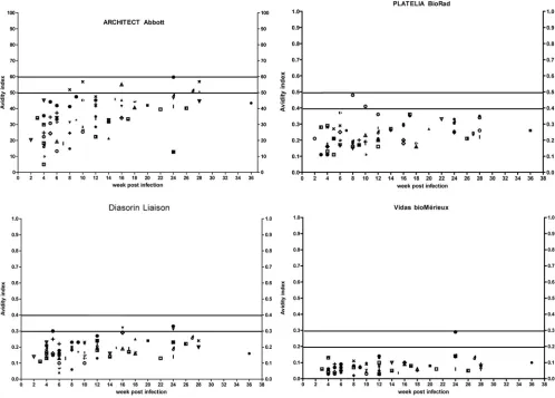
The slow growth avidity maturation shortly postinfection was
evident when the avidities were depicted for individual patients
(
Fig. 2
). However, the existence of equivocal data showed
differ-ences between the four immunoassays (
Table 1
and
Fig. 2
): 6
equivocal results for Abbott, 1 for bioMérieux, 2 for Bio-Rad, and
3 for Diasorin. If these equivocal data were considered false
nega-tives for detecting acute toxoplasmosis, then bioMérieux had the
most appropriate capacity to recognize acute toxoplasmosis, with
98.2% sensitivity, followed by Bio-Rad (96.4%), Diasorin
(94.6%), and finally Abbott (89.3%). Notably, no high-avidity
results were found in this group, confirming the good sensitivity
of these assays for the diagnosis of acute toxoplasmosis. It must be
remembered that none of the kits is recommended for use in
di-agnosing acute toxoplasmosis.
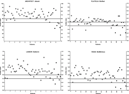
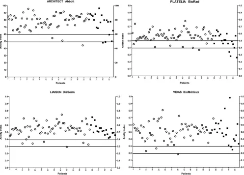
Comparison of sensitivities for latent toxoplasmosis.
Con-sidering all patients with latent toxoplasmosis, the proportions
of high-avidity results were 87.7% with bioMérieux, 87.1%
with Abbott and Diasorin, and 74.8% with Bio-Rad (
Table 2
and
Fig. 3
and
4
). However, better results were obtained in the
group of patients without specific IgM in the serum (group 2),
with the proportion of high-avidity results reaching 94% for
bioMérieux, 91.7% for Abbott and Diasorin, and 83.3% for
Bio-Rad. If the equivocal results were interpreted as
low-avid-Villard et al.on August 17, 2020 by guest
http://cvi.asm.org/
ity results, the sensitivity was greatly diminished for each test,
especially in the case of Bio-Rad, where a high number of
equivocal results were observed. These false-negative results
were mainly accounted for by immunocompromised patients.
Indeed, if only immunocompetent patients were considered,
the sensitivity increased substantially, from 87.1% to 92.5% for
Abbott, 87.7% to 91.6% for bioMérieux, 87.1% to 88.8% for
DiaSorin, and 74.8% to 83.2% for Bio-Rad (
Table 2
).
Comparison of the diagnostic efficacies of avidity assays.
When comparing the PPVs for acute toxoplasmosis, there was a
large variation between kits, ranging from 61.1% for Bio-Rad to
77.5% for bioMérieux (
Table 3
). In contrast, the NPV was more
than 99% for bioMérieux and Bio-Rad but lower for Abbott and
Diasorin (95.3% and 96.8%, respectively). These discrepancies
resulted in a large variation in the Youden index, which measures
the accuracy of the assay, ranging from 0.73 to 0.87. However, the
Yule index, measuring the relationship of IgG avidity with acute
toxoplasmosis, was acceptable, exceeding 0.9 for all of the assays
and ranging from 0.96 to 1 for bioMérieux.
On the other hand, the PPV for latent toxoplasmosis attained
the maximum of 100% with all kits, while Yule’s Q coefficient was
1. However, the NPV was lower, ranging from 61.5% for Bio-Rad
to 77.8% for bioMérieux. In addition, the Youden index ranged
from 0.75 to 0.87 (
Table 3
).
Distribution of avidity results in the 16 French university
and general hospitals.
Among the network hospitals, 11
labora-tories used bioMérieux assays, 3 Bio-Rad, 2 Abbott, and none
DiaSorin. Of 3,885 sera positive for both IgG and IgM, 55.7%
FIG 1Mean kinetics of IgG levels (A) and IgG avidity values (B) with four immunoassays in 67 sera of 56 pregnant women with acute toxoplasmosis.
on August 17, 2020 by guest
http://cvi.asm.org/
exhibited high avidity, 14.8% intermediate affinity, and 29.5% low
avidity (
Table 4
).
DISCUSSION
A number of different methods have been used to determine the
avidity of specific IgG antibodies for dating
Toxoplasma
infection.
The majority of the methods in use for more than 10 years have
involved in-house assays based on the use of protein-denaturing
agents in washing steps or serum diluents. More recently, assays
based on recombinant proteins as blocking agents were
intro-duced (
15
). The IgG avidity assays have become generally
ac-cepted diagnostic tools, with the final goal to exclude recent
infec-tion and risk for the fetus, which is crucial in the case of pregnant
women with suspected acute infection (e.g., IgM-positive sera).
These assays, evaluated using sera from the biobank developed
from the French national screening program, have now been
FIG 2Kinetics of IgG avidity for the four immunoassays in 67 samples taken from 56 pregnant women with acute toxoplasmosis and confirmed seroconversion. All the women were treated for acute toxoplasmosis after the first serum was tested. The horizontal lines represent the upper and lower cutoffs of the gray zone for each assay. Each symbol represents a single patient.
TABLE 1Comparison of the sensitivities of four IgG avidity immunoassays in 56 patients with acute toxoplasmosis (group 1) according to the classification of equivocal results
Assay
Sensitivity when equivocal data considered [% (no./total)]:
Low avidity High avidity
Abbott 100 (56/56) 89.3 (50/56)
bioMérieux 100 (56/56) 98.2 (55/56)
Bio-Rad 100 (56/56) 96.4 (54/56)
DiaSorin 100 (56/56) 94.6 (53/56)
TABLE 2Comparison of the sensitivities of four IgG avidity immunoassays in patients with latent toxoplasmosisa
Assay
Sensitivity [% (no./total)]b
Group 2 Group 3 Total
All patients
Abbott 91.7 (77/84) 80 (44/55) 87.1 (121/139)
bioMérieux 94 (79/84) 78.2 (43/55) 87.7 (122/139)
Bio-Rad 83.3 (70/84) 61.8 (34/55) 74.8 (104/139)
DiaSorin 91.7 (77/84) 80 (44/55) 87.1 (121/139)
Immunocompromised patients and treated pregnant women excluded
Abbott 95.3 (61/64) 88.4 (38/43) 92.5 (99/107)
bioMérieux 95.3 (61/64) 86 (37/43) 91.6 (98/107)
Bio-Rad 92.2 (58/64) 72.1 (31/43) 83.2 (89/107)
DiaSorin 89.1 (57/64) 88.4 (38/43) 88.8 (95/107)
a
Equivocal avidity was considered low avidity.
bGroup 2, IgG positive and IgM negative; group 3, IgG and IgM positive. Villard et al.
on August 17, 2020 by guest
http://cvi.asm.org/
incorporated in decision algorithms used in national
recom-mendations (
4
). Today, several commercial IgG avidity assays
are available, although few cross-evaluations of their
diagnos-tic performance have been published (
11
,
16
–
18
).
One of the primary goals of the French National Center for
Toxoplasmosis was to evaluate the performance of the
commer-cialized assays used in the French national screening program for
congenital toxoplasmosis (
19
). Therefore, we evaluated four
as-says, from Abbott, bioMérieux, Bio-Rad, and DiaSorin, which are
the most widely used in French biology laboratories and in
refer-ence laboratories abroad. These fully automated assays are based
on the exclusion of acute infection, with previous expert advice
reporting good performance of the assays.
Considering these assays within the recommended use (to
ex-clude acute infection when high-avidity antibodies are present),
the PPV for confirming latent toxoplasmosis was 100%. Yule’s Q
coefficient was 1, confirming the strong relationship between high
avidity and latent toxoplasmosis. In our large retrospective study,
in the group of pregnant women with both IgG and IgM
anti-Toxoplasma
antibodies, 55.7% were considered to have a latent
infection with a single test. Therefore, by measuring IgG avidity in
a single first sample in nontreated and immunocompetent
pa-tients, we were able to confirm a latent infection and to exclude a
recent infection (
20
,
21
).
When we used the above-mentioned biobank to compare the
performances of the four assays in detecting acute toxoplasmosis,
noting that they were not designed for this purpose, the kinetics of
IgG maturation had variable performance. Abbott was the most
dynamic assay, probably because it is based on recombinant
pro-teins (SAG1 and GRA8). In contrast to the conventional Bio-Rad,
DiaSorin, and bioMérieux (denaturing) avidity tests, the avidity
competition used in Abbott’s Architect test detects low-avidity
IgG by blocking high-avidity IgG in the sample with a soluble
recombinant antigen (
8
).
In general, the kinetics of avidity maturation were similar for
all of the assays. The observed decrease in the IgG titers after 12
weeks could be due to the effect of treatment, as all the pregnant
women included in this study were treated for acute
toxoplasmo-sis. This could also account for the less dynamic performance of
the three “classical” assays based on a native antigen and the
con-ventional denaturing method for determining avidity (
22
–
24
).
In the context of off-label use, the performances in detection of
acute cases were highly variable. When equivocal results were
con-sidered low avidity, all assays reached 100% sensitivity. The results
FIG 3IgG avidity results with the four immunoassays in IgG- and IgM-positive sera in latent toxoplasmosis (n⫽55). The black squares represent treated or immunocompromised patients; the circles represent the immunocompetent patiens. The horizontal lines represent the upper and lower cutoffs of the gray zone for each assay.
on August 17, 2020 by guest
http://cvi.asm.org/
of this study on pregnant women demonstrated that acute
toxo-plasmosis could not be reliably diagnosed based on low IgG
avid-ity alone. Therefore, only the first sample should be considered a
reference sample when interpreting avidity results, as after the first
determination, all pregnant women were treated with spiramycin
according to French recommendations (
20
,
23
). When we
consid-ered only the first sample in our group of 56 nontreated pregnant
women with acute toxoplasmosis, sensitivity was 100%. Our
re-sults also demonstrate that disequilibrium of host-parasite
dy-namics due to impaired immunity or an anti-
Toxoplasma
treat-ment probably results in a delay of IgG maturation. Therefore,
results from immunocompromised patients or those treated for
Toxoplasma
should be interpreted with caution. This is clearly
indicated only in the Vidas booklet.
In terms of cost-effectiveness, avidity testing is a powerful tool,
allowing optimization of screening and follow-up of pregnant
women (
25
–
27
). This point is confirmed by our retrospective
study among the French network hospitals, which shows that half
of the cases with detection of both IgM and IgG antibodies were
identified as latent toxoplasmosis, thus avoiding unnecessary
sub-sequent maternal and fetal investigation and treatment. For this
reason, the assay price should also be considered. In our study, the
TABLE 3Comparison of PPVs, NPVs, Youden indexes, and Yule’s Q coefficients of four IgG avidity immunoassays for patients with acute or latent toxoplasmosis
Assay PPV (%) NPV (%)
Youden indexc
Yule’s Q coefficientd
Acute toxoplasmosisa
Abbott 73.5 95.3 0.76 0.96
bioMérieux 77.5 99.2 0.87 1
Bio-Rad 61.1 99.1 0.73 0.99
DiaSorin 74.3 96.8 0.8 0.98
Latent toxoplasmosisb
Abbott 100 75.7 0.87 1
bioMérieux 100 76.7 0.88 1
Bio-Rad 100 61.5 0.75 1
DiaSorin 100 75.7 0.87 1
a
For the acute-toxoplasmosis population, in an off-label use of the reagents, we estimated the PPV and NPV of a low-avidity result.
b
For the latent-toxoplasmosis population, in an approved use, similar biostatistical results were calculated using the proportion of high-avidity results. For all patients, equivocal or intermediate values were considered false negatives in subjects with acute toxoplasmosis and false positives in subjects with latent toxoplasmosis.
c
The Youden index measures the effectiveness of the test (negative index, ineffective test; index close to 1, effective test).
d
Yule’s Q coefficient measures the relationship to the IgG avidity index (the closer the coefficient is to 1, the stronger the relationship).
FIG 4IgG avidity results with the four immunoassays in IgG-positive and IgM-negative sera in the latent toxoplasmosis population (n⫽84). The black squares represent treated or immunocompromised patients; circles represent immunocompetent patients. The horizontal lines represent the upper and lower cutoffs of the gray zone for each assay.
Villard et al.
on August 17, 2020 by guest
http://cvi.asm.org/
prices for one run of each reagent (not considering external and
internal quality control sera) for the determination of IgG avidity
were
€
11.63 for Architect (Abbott),
€
5.50 for Vidas (bioMérieux),
€
8.16 for Platelia Toxo IgG Avidity (Bio-Rad), and
€
6.44 for
Liai-son (DiaSorin). The cheapest assay, Vidas (bioMérieux), also had
the best performance for the diagnosis of latent toxoplasmosis. In
any case, avidity testing remains expensive, according to the
eco-nomic evaluations of Stillwaggon et al. (
27
). Thus, we recommend
sending inconclusive sera to an expert laboratory that uses these
complementary methods (
4
). Of interest, the Architect assay,
which employs recombinant antigens, provided the best
perfor-mance for detecting latent infection in the presence of persistent
IgM. This means that the use of recombinant antigens for
toxo-plasmosis assays could be extended in the future, considering that
the type of antigen used in antibody recognition is crucial. For
example, IgGs against antigens recognized early (i.e., GRA7,
GRA8, and ROP1) mature significantly earlier than those directed
against later antigens (i.e., SAG1 and MAG1) (
28
).
In conclusion, the avidity test provides a rapid means for
iden-tifying latent
Toxoplasma
infection in pregnant women who show
both IgG and IgM anti-
Toxoplasma
antibodies on initial testing
during pregnancy. However, there are some limitations in the use
of this method. Avidity assays are not conclusive in some
immu-nocompromised patients and those treated for toxoplasmosis.
Overall, optimal diagnostic performance is achieved by using
ap-propriate combinations of serological, culture-based, and PCR
techniques.
ACKNOWLEDGMENTS
We acknowledge Philippe Thulliez, who performed the dye test, and Elo-die Pernot-Marino and Alexander Pfaff for their critical reviews.
The study was supported by a grant from the Institut National de Veille Sanitaire.
We declare that we have no competing financial interests in this study. Members of the National Reference Centre for Toxoplasmosis and ToxoSurv network, in alphabetical order by location: A. Totet (Hospital and University Centre Amiens); B. Cimon (Hospital and University
Cen-tre Angers); E. Scherer (Hospital and University CenCen-tre Besançon); I. Accoceberry (Hospital and University Centre Bordeaux); G. Nevez and D. Quinio (Hospital and University Centre Brest); J. Bonhomme (Hospital and University Centre Caen); B. Carme and M. Demar (Hospital and University Centre Cayenne); A. Bonnin, B. Cuisenier, and F. Dalle (Hos-pital and University Centre Dijon); N. Desbois (Hos(Hos-pital and University Centre Fort de France); M. P. Brenier-Pinchart, H. Fricker-Hidalgo, and H. Pelloux (Hospital and University Centre Grenoble); L. Delhaes (Hos-pital and University Centre Lille); D. Ajzenberg and M. L. Dardé (Hos(Hos-pital and University Centre Limoges); J. Franck and R. Piarroux (Hospital and University Centre Marseille); P. Bastien, Y. Sterkers, and F. Pratlong (Hospital and University Centre Montpellier); M. Machouart (Hospital and University Centre Nancy); M. Leterrier and F. Morio (Hospital and University Centre Nantes); N. Ferret, C. Pomares, and P. Marty (Hospital and University Centre Nice); A. Angoulvant (Hospital and University Centre Paris Bicêtre); S. Houze (Hospital and University Centre Paris Bichat); T. Ancelle and H. Yera (Hospital and University Centre Paris Cochin); F. Derouin and J. Menotti (Hospital and University Centre Paris St. Louis); M. E. Bougnoux and N. Hassouni (Hospital and University Centre Paris Necker Enfants Malades); F. Touafek and L. Paris (Hospital and University Centre Paris Salpétrière); N. Godineau (Hospital and Uni-versity Centre Paris St Denis); P. Roux and C. Hennequin (Hospital and University Centre Paris St Antoine); S. Azi (Hospital and University Cen-tre Pointe A. PiCen-tre); J. Berthonneau (Hospital and University CenCen-tre Poit-iers); D. Aubert, C. Chemla, and I. Villena (Hospital and University Cen-tre Reims); F. Robert-Gangneux (Hospital and University CenCen-tre Rennes); L. Favennec and G. Gargala (Hospital and University Centre Rouen); P. Flori (Hospital and University Centre St Etienne); E. Candolfi, D. Filisetti, and O. Villard (Hospital and University Centre Strasbourg); J. Filiaux and S. Cassaing (Hospital and University Centre Toulouse); N. Vanlangendonck (Hospital and University Centre Tours).
REFERENCES
1.Montoya JG, Remington JS.2008. Management of Toxoplasma gondii infection during pregnancy. Clin. Infect. Dis.47:554 –566.
2.Couvreur J.1962. Congenital toxoplasmosis. Vie Med.43:1839 –1842. 3.Foulon W, Pinon JM, Stray-Pedersen B, Pollak A, Lappalainen M,
Decoster A, Villena I, Jenum PA, Hayde M, Naessens A.1999. Prenatal diagnosis of congenital toxoplasmosis: a multicenter evaluation of differ-ent diagnostic parameters. Am. J. Obstet. Gynecol.181:843– 847. 4.Villard O, Jung-Etienne J, Cimon B, Franck J, Fricker-Hidalgo H,
Godineau N, Houze S, Paris L, Pelloux H, Villena I, Candolfi E, Le Reseau du Centre National de Référence de la Toxoplasmose. 2011. Sérodiagnostic de la toxoplasmose en 2010: conduite à tenir et interpreta-tion en foncinterpreta-tion des profils sérologiques obtenus par les méthodes de dépisage. Feuillets Biol.52:1–7.
5.Pratlong F.2002. Toxoplasmosis and pregnancy: current trends in sero-logical follow-up. Gynecol. Obstet. Fertil.30:237–243.
6.Hedman K, Lappalainen M, Seppaia I, Makela O.1989. Recent primary toxoplasma infection indicated by a low avidity of specific IgG. J. Infect. Dis.159:736 –740.
7.Roberts A, Hedman K, Luyasu V, Zufferey J, Bessieres MH, Blatz RM, Candolfi E, Decoster A, Enders G, Gross U, Guy E, Hayde M, Ho-Yen D, Johnson J, Lecolier B, Naessens A, Pelloux H, Thulliez P, Petersen E.
2001. Multicenter evaluation of strategies for serodiagnosis of primary infection with Toxoplasma gondii. Eur. J. Clin. Microbiol. Infect. Dis.
20:467– 474.
8.Curdt I, Praast G, Sickinger E, Schultess J, Herold I, Braun HB, Bernhardt S, Maine GT, Smith DD, Hsu S, Christ HM, Pucci D, Hausmann M, Herzogenrath J.2009. Development of fully automated determination of marker-specific immunoglobulin G (IgG) avidity based on the avidity competition assay format: application for Abbott Architect cytomegalovirus and Toxo IgG Avidity assays. J. Clin. Microbiol.47:603– 613.
9.Flori P, Bellete B, Crampe C, Maudry A, Patural H, Chauleur C, Hafid J, Raberin H, Tran Manh Sung R.2008. A technique for dating toxo-plasmosis in pregnancy and comparison with the Vidas anti-toxoplasma IgG avidity test. Clin. Microbiol. Infect.14:242–249.
10. Fricker-Hidalgo H, Saddoux C, Suchel-Jambon AS, Romand S, Fous-TABLE 4Distribution of avidity results in the 16 University and
General Hospitals in patients positive for both IgG and IgM
anti-Toxoplasmaantibodies University hospital No. of sera % High avidity % Equivocal avidity % Low
avidity Assay used
A 602 60.5 16.1 23.4 bioMérieux
B 95 69.5 9.5 21 bioMérieux
C 78 38.5 26.9 34.6 bioMérieux
D 77 58.4 20.7 20.7 bioMérieux
E 245 59.6 10.2 30.2 bioMérieux
F 205 42.9 23.9 33.2 Bio-Rad
G 274 44.5 12.4 43.1 Abbott
H 132 57.6 22.7 19.7 Bio-Rad
I 52 69.2 3.8 26.9 bioMérieux
J 273 60.8 13.6 25.9 bioMérieux
K 1,462 48.5 24.5 27 Bio-Rad
L 68 75 2.9 22.1 bioMérieux
M 44 52.3 11.3 36.3 bioMérieux
N 67 69 15 16 bioMérieux
O 116 42.2 18.1 39.7 Abbott
P 95 42.1 5.3 52.6 bioMérieux
Total 3,885 55.7 14.8 30.2
on August 17, 2020 by guest
http://cvi.asm.org/
sadier A, Pelloux H, Thulliez P.2006. New Vidas assay for Toxoplasma-specific IgG avidity: evaluation on 603 sera. Diagn. Microbiol. Infect. Dis.
56:167–172.
11. Martin C, Morin O.2006. Comparison of liaison automatised diagnostic system (DiaSorin) and Platelia microplate (BioRad) for the serological diagnosis of toxoplasmosis evaluation of the toxoplasma specific immu-noglobulin G avidity. Rev. Francophone Lab.386:71–76.
12. Petersen E, Borobio MV, Guy E, Liesenfeld O, Meroni V, Naessens A, Spranzi E, Thulliez P.2005. European multicenter study of the LIAISON automated diagnostic system for determination of Toxoplasma gondii-specific immunoglobulin G (IgG) and IgM and the IgG avidity index. J. Clin. Microbiol.43:1570 –1574.
13. Villard O, Cimon B, Franck J, Fricker-Hidalgo H, Godineau N, Houze S, Paris L, Pelloux H, Villena I, Candolfi E.2012. Evaluation of the usefulness of six commercial agglutination assays for serologic diagnosis of toxoplasmosis. Diagn. Microbiol. Infect. Dis.73:231–235.
14. Lebech M, Joynson DH, Seitz HM, Thulliez P, Gilbert RE, Dutton GN, Ovlisen B, Petersen E.1996. Classification system and case definitions of Toxoplasma gondii infection in immunocompetent pregnant women and their congenitally infected offspring. European Research Network on Congenital Toxoplasmosis. Eur. J. Clin. Microbiol. Infect. Dis.15:799 – 805.
15. Gay-Andrieu F, Fricker-Hidalgo H, Sickinger E, Espern A, Brenier-Pinchart MP, Braun HB, Pelloux H.2009. Comparative evaluation of the ARCHITECT Toxo IgG, IgM, and IgG Avidity assays for anti-Toxoplasma antibodies detection in pregnant women sera. Diagn. Microbiol. Infect. Dis.65:279 –287.
16. Alvarado-Esquivel C, Sethi S, Janitschke K, Hahn H, Liesenfeld O.2002. Comparison of two commercially available avidity tests for toxoplasma-specific IgG antibodies. Arch. Med. Res.33:520 –523.
17. Bobic B, Klun I, Vujanic M, Nikolic A, Ivovic V, Zivkovic T, Djurkovic-Djakovic O.2009. Comparative evaluation of three commercial Toxo-plasma-specific IgG antibody avidity tests and significance in different clinical settings. J. Med. Microbiol.58:358 –364.
18. Lachaud L, Calas O, Picot MC, Albaba S, Bourgeois N, Pratlong F.
2009. Value of 2 IgG avidity commercial tests used alone or in association to date toxoplasmosis contamination. Diagn. Microbiol. Infect. Dis.64: 267–274.
19. Villena I, Ancelle T, Delmas C, Garcia P, Brezin AP, Thulliez P, Wallon M, King L, Goulet V. 2010. Congenital toxoplasmosis in France in 2007: first results from a national surveillance system. Euro Surveill.15:19600.
20. Candolfi E, Pastor R, Huber R, Filisetti D, Villard O.2007. IgG avidity assay firms up the diagnosis of acute toxoplasmosis on the first serum sample in immunocompetent pregnant women. Diagn. Microbiol. Infect. Dis.58:83– 88.
21. Press C, Montoya JG, Remington JS.2005. Use of a single serum sample for diagnosis of acute toxoplasmosis in pregnant women and other adults. J. Clin. Microbiol.43:3481–3483.
22. Buffolano W, Lappalainen M, Hedman L, Ciccimarra F, Del Pezzo M, Rescaldani R, Gargano N, Hedman K.2004. Delayed maturation of IgG avidity in congenital toxoplasmosis. Eur. J. Clin. Microbiol. Infect. Dis.
23:825– 830.
23. Lefevre-Pettazzoni M, Bissery A, Wallon M, Cozon G, Peyron F, Ra-billoud M.2007. Impact of spiramycin treatment and gestational age on maturation of Toxoplasma gondii immunoglobulin G avidity in pregnant women. Clin. Vaccine Immunol.14:239 –243.
24. Sensini A, Pascoli S, Marchetti D, Castronari R, Marangi M, Sbaraglia G, Cimmino C, Favero A, Castelletto M, Mottola A.1996. IgG avidity in the serodiagnosis of acute Toxoplasma gondii infection: a multicenter study. Clin. Microbiol. Infect.2:25–29.
25. Ancelle T, Yera H, Talabani H, Lebuisson A, Thuillez P, Dupouy-Camet J.2009. How can the cost of screening for toxoplasmosis during pregnancy be reduced? Rev. Epidemiol. Sante Publique57:411– 417. 26. Cozon GJ, Ferrandiz J, Nebhi H, Wallon M, Peyron F.1998. Estimation
of the avidity of immunoglobulin G for routine diagnosis of latent Toxo-plasma gondii infection in pregnant women. Eur. J. Clin. Microbiol. In-fect. Dis.17:32–36.
27. Stillwaggon E, Carrier CS, Sautter M, McLeod R.2011. Maternal sero-logic screening to prevent congenital toxoplasmosis: a decision-analytic economic model. PLoS Negl. Trop. Dis.5:e1333. doi:10.1371/journal .pntd.0001333.
28. Pfrepper KI, Enders G, Gohl M, Krczal D, Hlobil H, Wassenberg D, Soutschek E.2005. Seroreactivity to and avidity for recombinant antigens in toxoplasmosis. Clin. Diagn. Lab. Immunol.12:977–982.
Villard et al.