Electronic Thesis and Dissertation Repository
10-20-2017 10:30 AM
Evaluation of a Microalgal Bioassay Based on Community Level
Evaluation of a Microalgal Bioassay Based on Community Level
Physiological Profiling (CLPP)
Physiological Profiling (CLPP)
Jun-Woo Kim
The University of Western Ontario
Supervisor
Dr. Lars Rehmann
The University of Western Ontario Co-Supervisor Dr. Madhumita B. Ray
The University of Western Ontario
Graduate Program in Chemical and Biochemical Engineering
A thesis submitted in partial fulfillment of the requirements for the degree in Doctor of Philosophy
© Jun-Woo Kim 2017
Follow this and additional works at: https://ir.lib.uwo.ca/etd
Part of the Biotechnology Commons
Recommended Citation Recommended Citation
Kim, Jun-Woo, "Evaluation of a Microalgal Bioassay Based on Community Level Physiological Profiling (CLPP)" (2017). Electronic Thesis and Dissertation Repository. 5042.
https://ir.lib.uwo.ca/etd/5042
This Dissertation/Thesis is brought to you for free and open access by Scholarship@Western. It has been accepted for inclusion in Electronic Thesis and Dissertation Repository by an authorized administrator of
i
Abstract
This thesis examined the use of commercially available Biolog 96-well plates
containing different carbon sources for a microalgal bioassay. Quaternary ammonium
compounds (QACs) were used as potentially toxic model compounds to demonstrate the
applicability of the assay. Toxicity of dodecyl trimethyl ammonium chloride (DTAC) and didecyl
dimethyl ammonium bromide (DDAB) on the growth of Scenedesmus obliquus varied
significantly under autotrophic, heterotrophic and mixotrophic growth conditions. The
concentration for 50% growth inhibition effect (EC50) of DTAC followed the order: autotrophic
(0.48 ± 0.03 mg of DTAC/L) > heterotrophic (1.46 ± 0.04 mg of DTAC/L) > mixotrophic (2.11 ±
0.06 mg of DTAC/L), whereas for DDAB, the order of inhibition was different as: heterotrophic
(0.52 ± 0.02 mg of DDAB/L) > autotrophic (1.18 ± 0.08 mg of DDAB/L) > mixotrophic (1.35 ±
0.02 mg of DDAB/L). Moreover, EC50 values were a function of carbon source and growth
regime, showing that only EC50 values do not fully capture the toxic effects of a potentially
toxic compound might have on algal communities. Therefore, a new assay was developed
based on community level physiological profiling (CLPP). Five different mixtures of artificially
defined microalgal communities were employed and the changes in substrate utilization
patterns by the treatment of hexadecyl trimethyl ammonium chloride (CTAC) were quantified
using principal component analysis (PCA). The toxic effect of CTAC was significant (P < 0.05),
showing 58% inhibition compared to the control and the effect was more pronounced for the
treatment than that obtained by varying the initial composition of the defined algal
communities. The newly developed assay was further applied on wetland water samples,
wastewaters (i.e. primary and secondary), river water, and activated carbon treated and
untreated oil sand process waters (OSPWs). The assay was able to generate a distinguishable
response among these samples, measuring small differences within the respective water
groups and larger differences between them.
Keywords: Mixotrophic and heterotrophic microalgal bioassay, quaternary ammonium
compounds (QACs), freshwater algae, Biolog plates, community level physiological profiling
ii
Contributions of Authors
All experimental designs, data collections, analyses and interpretation of the results
were performed by Jun-Woo Kim. The written manuscripts were carried out in collaboration
iii
Dedicated to my family:
Father: Dong-Suk Kim
Mother: Eun-Ok Kim
Sister: Katie Mi-Kyung Hong
Brother: James Tae-Yeul Kim
Spouse: Chloe Nan-Joo Choi
iv
Acknowledgements
I treasure all the memories I have from working in Drs. Lars Rehmann’s and
Madhumita B. Ray’s lab. I thank all the colleagues who worked alongside me in the lab for all
their support and the knowledge they shared to make me a better researcher. I especially
thank Shreyas Yedahalli and my office mates Adnan Hossain Khan, Tahereh Sarchami and
Dongmin Yun for being good friends, on whom I relied a lot in bad times and shared joys in
good times. I also very much appreciate Siddharth Gupta for the help with TOC analysis and
Tulip Chakraborty for nitrate and phosphate analyses.
Most of all, I would like to thank my supervisors: Drs. Lars Rehmann and Madhumita
B. Ray. Dr. Lars Rehmann guided me as an aspiring researcher. As a supervisor, he provided
me with numerous ideas and taught me that in research, patience and a positive attitude
were important attributes. Dr. Madhumita B. Ray was always there for precious advice and
made me a hard worker to push myself to achieve goals in timely manner. The research would
be incomplete without the guidance of both of my supervisors.
I also appreciate my supervisory committee members, Dr. Amarjeet Bassi and Dr.
Wankei Wan, for their comments. In addition, this scholarly fulfillment would be impossible
without the financial support from the Natural Science and Engineering Council of Canada
(NSERC) CREATE program and a Western Graduate Research Scholarship.
Finally, I would like to acknowledge my family for their endless love and support. I
v
Table of Contents
Abstract ... i
Contributions of Authors ... ii
Acknowledgements ... iv
List of Tables ... viii
List of Figures ... ix
List of Abbreviations ... xi
Nomenclature ... xiv
Chapter 1. Background... 1
1.1 Introduction ... 1
1.2 Literature Review and Research Gaps ... 2
1.2.1 Approach to Contaminated Sites ... 2
1.2.2 Types of Bioassays ... 3
1.2.3 Microalgal Bioassay ... 5
1.2.4 Community Level Physiological Profiling (CLPP) ... 6
1.2.5 Quaternary Ammonium Compounds (QACs) ... 7
1.3 Research Objectives ... 8
1.4 Literature Cited ... 16
Chapter 2. Assessment of the effects of quaternary ammonium compounds (QACs) on Scenedesmus obliquus using Biolog plates under light and dark conditions ... 25
2.1 Introduction ... 26
2.2 Materials and Methods ... 27
2.2.1 Chemicals ... 27
vi
2.2.3 Toxicity tests on microalgae ... 29
2.2.4 Analysis ... 32
2.3 Results and Discussion ... 32
2.3.1 Autotrophic toxicity experiments ... 32
2.3.2 Mixotrophic and heterotrophic toxicity experiments ... 34
2.4 Conclusions ... 46
2.5 Literature Cited ... 47
Chapter 3. Development of microalgal bioassay based on the community level physiological profiling (CLPP) ... 52
3.1 Introduction ... 53
3.2 Materials and Methods ... 55
3.2.1 Microalgae strains ... 55
3.2.2 Culture maintenance... 55
3.2.3 Community Level Physiological Profiling (CLPP) ... 56
3.2.4 Minimum cell density requirement ... 59
3.2.5 Analysis ... 59
3.3 Results and Discussion ... 60
3.3.1 CLPP for individual algal species ... 60
3.3.2 CLPP for synthetic mixed algal communities ... 64
3.3.3 CLPP for toxicity test ... 67
3.4 Conclusions ... 70
3.5 Literature Cited ... 71
vii
4.1 Introduction ... 78
4.2 Materials and Methods ... 79
4.2.1 Water samples ... 79
4.2.2 Microalgal strains and mixed algal community... 80
4.2.3 Sample assessment using Community Level Physiological Profiling (CLPP) ... 81
4.2.4 Analysis ... 81
4.3 Results and Discussion ... 82
4.3.1 Changes in the average well fluorescence development (AWFD) ... 82
4.3.2 Community level physiological profiles (CLPP) for various water samples .... 86
4.3.3 Characterizing CLPP using principal component analysis (PCA) ... 89
4.4 Conclusions ... 91
4.5 Literature Cited ... 92
Chapter 5. General Discussion and Recommendations ... 95
5.1 General Discussion ... 95
5.2 Recommendations for Future Work ... 96
Appendices ... 97
viii
List of Tables
Table 1.1 QACs’ concentrations in wastewater, hospital discharge, surface, river, sea water10
Table 1.2 Effective concentrations (EC50 and EC10) for microalgae under QACs ... 12
Table 2.1 QACs, synonyms, molecular structures and weights ... 28
Table 2.2 Organic carbons and sources in YT Biolog plates in 57 bottom wells ... 31
Table 3.1 Growth rates of five microalgae strains ... 56
Table 3.2 Five mixed algal communities in different ratios. ... 58
Table 3.3 Substrates utilized (metabolic richness) by five individual algal strains... 63
Table 3.4 Euclidean distances for five mixed algal communities through 10th day ... 66
Table 4.1 TOC, nitrate, and phosphate concentrations for various waters... 80
Table 4.2 Euclidean distances for different water samples through 5th day. ... 85
ix
List of Figures
Figure 1.1 Steps for addressing a contaminated site ... 3
Figure 2.1 The growth inhibitions (%) for Scenedesmus obliquus in an autotrophic condition
under the various concentrations of DTAC and DDAB ... 33
Figure 2.2 Toxicity tests on Scenedesmus obliquus for mixotrophic and heterotrophic
conditions under the various concentrations of DTAC and DDAB. ... 36
Figure 2.3 The growth inhibitions (%) for Scenedesmus obliquus in mixotrophic and
heterotrophic conditions under the various concentrations of DTAC and DDAB ... 37
Figure 2.4 The growth inhibitions (%) for Scenedesmus obliquus for different individual organic
carbons in mixotrophic and heterotrophic conditions under the various concentrations of
DTAC and DDAB ... 41
Figure 2.5 Effective concentrations (EC50) for different individual organic carbons in
mixotrophic and heterotrophic conditions under DTAC and DDAB ... 43
Figure 2.6 Effective concentrations (EC50) in range for mixotrophic and heterotrophic
conditions under DTAC and DDAB ... 44
Figure 2.7 Differences in effective concentrations (EC50) between mixotrophic and
heterotrophic conditions for DTAC and DDAB. ... 45
Figure 3.1 Color development over 8 days for 31 wells with different organic carbons ... 61
Figure 3.2 Average well color developments (AWCD) on 31 wells for five algal strains through
11th day... 62
Figure 3.3 Five mixed algal communities in different ratios in PC analysis ... 65
x
Figure 4.1 Ultra-violet (UV) spectrum of various waters. ... 80
Figure 4.2 Average well fluorescence developments (AWFD) for various water samples under
heterotrophic and mixotrophic growth conditions ... 82
Figure 4.3 Fluorescence developments after 4 days for heterotrophic growth condition in the
presence of different individual organic carbons ... 87
Figure 4.4 Fluorescence developments after 3 days for mixotrophic growth condition in the
presence of different individual organic carbons ... 88
Figure 4.5 A mixed algal community under various water samples in PC analysis for
xi
List of Abbreviations
ANOVA Analysis of variance
AWCD Average well color development
AWFD Average well florescence development
BAC Benzalkonium chlorides
BDHAC Benzyl dimethyl hexadecyl ammonium chloride
BDTAC Benzyl dimethyl tetradecyl ammonium chloride
BDDAC Benzyl dimethyl dodecyl ammonium chloride
CLPP Community level physiological profiling
CPCC Canadian phycological culture centre
CC Chlamydomonas center
CCAP Culture collection of algae and protozoa
CPRG Chlorophenol red-β-D-galactopyranoside
CSUP Carbon source utilization pattern
CTAB Hexadecyl trimethyl ammonium bromide
CTAC Hexadecyl trimethyl ammonium chloride
CPB Hexadecyl pyridinium bromide
DTDMAC Ditallow dimethyl ammonium chloride
xii
DTAC Dodecyl trimethyl ammonium chloride
DDAB Didecyl dimethyl ammonium bromide
DDAC Didecyl dimethyl ammonium chloride
DNA Deoxyribonucleic acid
EDDAB Ethyl dodecyl dimethyl ammonium bromide
EHDAB Ethyl hexadecyl dimethyl ammonium bromide
ED Euclidean distance
EDCs Endocrine disrupting compounds
HS High salt
MS Molecular structure
MW Molecular weight
OD Optical density
OECD Organization for economic cooperation and development
OC Organic carbons
OSPW Oil sand processed water
ONPG O-nitrophenyl- β-D-galactopyranoside
PICT Pollution-induced community tolerance
PCA Principal component analysis
xiii
QACs Quaternary ammonium compounds
RNA Ribonucleic acid
SD Standard deviation
STAB Stearyl trimethyl ammonium bromide
STAC Stearyl trimethyl ammonium chloride
TTAB Tetradecyl trimethyl ammonium bromide
TOC Total organic carbon
UTEX University of Texas at Austin
UV Ultra-violet
YES Yeast estrogen screen
xiv
Nomenclature
EC50 Concentration of a toxicant that induces 50% of maximal response (mg/L)
Chapter 1. Background
1.1 Introduction
Contaminated sites are managed by the Canadian federal government by a risk-based
approach. Several components to the risk-based approach include site identification and
characterization, detailed site investigations and risk assessment, evaluation of different risk
management strategies, implementation of a selected management strategy, assessment and
monitoring [1]. Moreover, the Canadian environmental quality guidelines have been
established by the Canadian Council of Ministers of the Environment (CCME) and are available
for the effective management of federal contaminated sites. The guidelines are important as
they are used for evaluating the degree of contamination at the site to decide if further site
investigations and management actions are required.
The guidelines are set based on the scientific information on toxicological studies.
Biological assays are commonly employed in toxicity assessment of various contaminants.
Bioassays are defined as lab cultures of organisms where growth conditions are manipulated
by input of specific chemicals. They complement a chemical analysis in defining the
concentration of contaminant by providing a direct measurement of toxic effects of a specific
compound and detect impacts from many sources for which chemical criteria are poorly
suited to determine synergistic, intermittent and chronic pollutant effects [2]. Moreover, this
is an efficient way of confirming the cause and effect relations between parameters for which
only observational correlations can be achieved from field surveys [3].
Bioassays can work in two ways, in vitro and in vivo. In vitro methods are based on
specific modes or cellular mechanism towards the detection of certain chemicals, while in vivo
methods assess the integrative effects of a toxicant on whole organisms, providing direct
information about the ecological effect. Commonly applied in vitro bioassays include the
umuC and comet for genotoxicity, Ames for mutagenicity and YES and YAS for estrogenicity
and androgenicity detections [4], whilein vivo assays use test species from different trophic
potential toxic compound on growth, reproduction, feeding activity and mortality [5].
Commonly used in vivo standardized tests include the bacteria luminescence inhibition assay
[6], green microalgae growth assay [7], duckweed growth assay [8], Daphnia reproduction
assay [9], and fish egg assay [10].
Among several existing in vivo standardized test systems, microalgae as model
organisms are being used increasingly for bioassay [11,12,13]. They are photosynthetic
organisms with an ability to convert light energy into organic carbon and serve as food source
for organisms of higher trophic levels, such as crustaceans, mussels, other filtering and
detritus feeding invertebrates or vertebrates and small fishes. In addition to their value as
food for higher trophic organisms, algal photosynthesis and respiration can strongly influence
water column oxygen dynamics [14]. Furthermore, they even play a role in nutrient cycling
and serve as habitat for other organisms [15]. The well-being of microalgae is crucial for the
maintenance of a healthy aquatic ecosystem. More importantly, their relatively higher
sensitivity to micropollutants over fish and crustaceans make them popular in environmental
toxicity studies [16]. Other advantages of algal assay’s include simplicity, speed and cost
effectiveness [2].
Despite of the advantages and recent popularity of application of microalgal bioassay
on toxicity assessment, limitations still exist. Therefore, in this Ph.D. research, the gaps in the
current algal toxicity experiment were identified and a new assay based on "community level
physiological profiling (CLPP)" technique was developed. Quaternary ammonium compounds
(QACs) were selected as a model micropollutant stress inducer. The potential of developed
assay was further tested using water samples with complex compositions.
1.2 Literature Review and Research Gaps
1.2.1 Approach to Contaminated Sites
The risk-based approach is acknowledged in more detail through a 10-step process
known as the steps for addressing a contaminated site, which is briefly explained in Figure 1.1
[1]. In addition, the Canadian environmental quality guidelines make important aspects in the
The environmental quality guidelines exist for soil, water and sediments. Generic soil
remediation criteria have been developed for four land uses: agricultural, residential/parkland,
commercial and industrial. Similar, groundwater and surface water criteria have been
established for four water uses: freshwater supporting aquatic life, water used for irrigation,
livestock watering and human drinking water. Freshwater and marine/estuarine sediments’
guideline present for a variety of contaminants. Toxicological studies based on bioassays using
different organisms are playing a crucial role setting guideline baselines for different
contaminants which are initially accessed by the environmental engineers for step 3 in Figure
1.1.
Figure 1.1 Steps for addressing a contaminated site (redrawn from, [1]).
1.2.2 Types of Bioassays
Commonly applied in vitro bioassays include the umuC and comet for genotoxicity,
Ames for mutagenicity and YES (yeast estrogen screen) and YAS (yeast androgen screen) for
estrogenicity and androgenicity detections [4]; these are briefly described here. The umuC
was developed and published in 1985 [17]. A plasmid (pSK 1102) containing the umuC gene
in association with the lacZ receptor gene was constructed and introduced into a
expression of umuC operon and therefore, the lacZ gene. The gene product oflacZis
β-galactosidase and the expression level reflecting genotoxicity was determined using a
colorimetric dye by converting a colourless substrate called ONPG (o-nitrophenyl- β
-D-galactopyranoside) into a yellow solid called o-nitrophenyl. The comet assay, also known as
the single cell gel-electrophoresis assay, is commonly applied for identifying genotoxicity. It
uses eukaryotic cells and involves the encapsulation of cells in a low-melting point agarose
cell suspension, the lysis of the cell membrane in a neutral or alkaline (pH > 13) buffer and the
electrophoresis of lysed cell samples [18]. By the exposure to a genotoxic agent, the damaged
cells take an elongated form resembling the comet, unlike an intact round shape. TheAmes
test is based on the cell stains from Salmonella typhimurium auxotrophic mutants, which are
characterized by a gene mutation that prevents the synthesis of L-histidine, an amino acid
that is necessary for bacterial growth. By exposure of mutants to mutagens, a reversal of the
mutation occurs and synthesize L-histidine for normal growth. The level of mutagenicity is
indicated by the colonies counted following incubation [19]. Yeast estrogen and androgen
screenings (YES and YAS) are based on a genetically modified Saccharomyces cerevisiae
containing human estrogen and androgen receptors coupled to a reporter gene, lacZ.
Endocrine disrupting compounds (EDCs) bind to a receptor and induce the synthesis of β
-galactosidase, which can be detected using a colorimetric dye (chlorophenol red-β
-D-galactopyranoside; CPRG) changing a color from yellow to purple [20].
Commonly used in vivo standardized tests are mainly bacteria luminescence
inhibition assay [6], green microalgae growth assay [7], duckweed growth assay [8], Daphnia
reproduction assay [9], and fish egg assay [10]. Most bacterial tests are based on the
gram-negative bacillus Vibrio fischeri. It bioluminescences when it is metabolically active. The
bacteria are obtained in a freeze-dried form and a solution of NaCl is added to create an
osmotic pressure. The acute toxicity of a substance can be determined after incubation,
bioluminescence level measurement and the calculation of the EC50 parameter by software.
MICROTOX® (Modern Water, UK) is currently the most popular kit of its type in the market.
Other types employ photosynthetic organisms, such as microalgae and duckweed. Toxicity
experiments are most often based on the green algae and duckweed (Lemna minor) and
is frequently used as indicator is Daphnia magna. It is within the group of invertebrates and
crustaceans and plays a very important role in the trophic chain, bridging the gap between
the producers and consumers of higher orders [21]. Toxicity tests are conducted in a small
container and the reproductive outputs are measured within a relatively short period of time,
typically, 21 days. Lastly, ecotoxicological testing can be performed with fish. Among
approximately 150 species, the most common is zebrafish (Brachydanio rerio). This is because
it is a vertebrate model organism in genetics, neurophysiology and biomedicine and it is one
of the first vertebrates whose genome was sequenced. Females can lay eggs every 2 to 3 days,
at several hundred at a time and they are transparent making them easy to visualize their
development in real time. Their development from embryo to adult usually takes 3 to 4
months [22].
1.2.3 Microalgal Bioassay
Bioassays using a single algal species are by far the most commonly used in which
highly culturable algae are grown in toxicant media and growths are monitored over a
predefined period [23]. Some of the single algal genera commonly used are Chlorella,
Scendedesmus, Selenastrum, Navicula, Spirulina, Anabaena and Microcystis among the
microalgae [2]. Alternatively, planktonic or benthic algal communities of natural origin are
assessed in contrast to the unialgal approach [24]. The use of unialgal experiments is favored
because of greater control and reproducibility; although, the validity of selecting one algal
species to represent the response of a community can be questionable. The use of a natural
algal community is better in a sense that it considers more than one species as even within a
genus, some species may be more responsive indicators than others [25] but, more
importantly, the natural site of concern is also taken into consideration [24].
While there is always a dose and response relationship with an effective
concentration (e.g., EC50, the concentration of a toxicant that gives half-maximal response),
the response relationship may not produce conclusive evidence on the algal tolerance level
based on their background. For example, Wang [24] noted similar range of EC50 values (1.8,
2.7 and 2.1 mg/L) after 24 hours exposure to Zn for natural algal communities collected from
plant, respectively. Also, two chemically similar quaternary ammonium compounds produce
almost equal EC50 levels of 0.041 and 0.021 mg/L for (Pseudokirchneriella subcapitata;
formerly known asSelenastrum capricornutum) after 72 hours exposure to benzalkonium
chlorides and didecyl dimethyl ammonium bromide, respectively [26]. Therefore, the need for
improvement on EC50 as a measure of toxicity of a compound on a single species can be
considered or else, measuring the change of a defined microbial community due to an
environmental stress can be developed as a better diagnostic tool.
Algal assemblages have been successfully applied to monitor the impacts of aquatic
stressors and aquatic toxicity [27]. For example, a shift in a naturally occurring microalgal
community composition reflects an environmental change that can be used as an indicator to
evaluate the changes in environmental conditions as had been tested earlier in marine [28],
freshwater [29], and wetland ecosystems [30]. However, traditional ways of identifying an
isolate and quantifying a species composition using cell morphology and molecular level
RNA/DNA amplification techniques have limitations as they can be both time and cost
intensive and require specialized expertise [31]. To overcome this limitation, a potential way
to assess an impact on a microalgal community due to an environmental stress is to use
functional or metabolic potential characterization employing the method of community level
physiological profiling (CLPP).
1.2.4 Community Level Physiological Profiling (CLPP)
A functional or metabolic characterization of a microbial community is well developed
after a method was first published by Garland and Mills [32]. The method is called community
level physiological profiling (CLPP) and it uses a commercially available Biolog 96-well plate
containing 95 different carbon sources. Different microbial communities are compared and
classified based on carbon source utilization patterns (CSUPs) [32]. The relatively simple
protocol and ease of use make it very practical for various applications. Based on the ISI
database search using key words such as Biolog and community, about 1363 publications
(prior to July 2017) on CLPP with an increasing trend were found.
bacteria to eukaryotic fungi [33]. A perturbation or change in the microbial community has
been observed in both terrestrial and aquatic environments, caused by a plant interaction
[34], root secretion [35], spill of hydrocarbon [36], metal contamination [37], water pollution
with acid mine drainage [38], and differently fed bluegills in guts [39]. Moreover, this
technology has been further upgraded by the simultaneous use of antibiotics or
cycloheximide to repress signal interferences from bacteria or fungi, respectively [40,41] and
modified as an assay to study the community tolerance to an antibiotic sulfachloropyridazine
using the pollution-induced community tolerance (PICT) concept [42] and the community
toxicity to gold nanoparticles and ciprofloxacin [43].
Not only the community level, but, the metabolic profiling technology has also been
established on single strains. Previously, it was used for the identification purpose on bacteria,
yeast and fungi with databases present for different Biolog plates (available from Biolog, CA,
USA). More recently, it has been applied on the isolates of microalgal strains from wastewater
for the comparisons of organic carbon metabolic potentials [44] and on mammalian cells
where it was found that human cancer cell lines from different organ tissues produced distinct
profiles of metabolic activity [45]. The potential of the functional or metabolic
characterization of individual or microbial community is enormous and continuously growing.
1.2.5 Quaternary Ammonium Compounds (QACs)
An emerging and not yet fully studied class of pollutants are so-called micropollutants.
Micropollutants are chemicals present at very low concentrations and easily found in aquatic
systems. Their effects on the aquatic environment have been very challenging to assess [46].
Among other micropollutants, surfactants are a diverse group of chemicals with high annual
global production of about 14 million tons in 2008 with 2.8% annual increase to 2012 [47].
This makes surfactants a ubiquitous class of organic contaminants that are commonly found
in aquatic ecosystems [48]. Recently, more attention was given to cationic surfactants,
quaternary ammonium compounds (QACs). This is because they are known for the higher
toxicity to living organisms than other anionic and nonionic ones [47].
molecular weights between 300 and 400 g/mol. They are typical cationic surfactants as they
have a positively charged central nitrogen atom giving hydrophilicity. In case of hydrophobic
regions, four functional groups are attached covalently to the positively charged central
nitrogen atom (R4N+). These functional groups (R) include at least one long hydrocarbon chain
and the rest are mostly short chain substituents such as methyl (in alkyl) or benzyl (in aryl)
groups [49]. The length of a long alkyl chain confers QACs distinctive physical or chemical
properties and led their widespread uses in agricultural, domestic, healthcare and industrial
applications as pesticides, detergents, fabric softeners, personal care products, disinfectants,
corrosion inhibitors, antistatic agents, biocides, emulsifiers and asphalt [50,51].
The extensive use of QACs in everyday applications make them ubiquitous
contaminants and they are commonly found in sewage, sewage sludge, industrial wastewater,
wastewater effluents, laundries and hospitals effluents, surface waters, and aquatic sediments
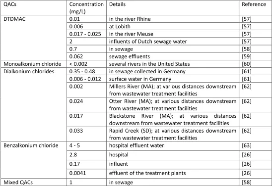
[26,52]. The measured QACs concentrations are in the ranges of μg/L (Table 1.1) and were
toxic to microalgae (Table 1.2). Although, the precise mechanisms are unclear [53], surfactant
toxicity on algae appeared to work on outside of the cell by interfering with the phospholipid
bilayer and altering the organization, stability and permeability of the membrane [54] and
more importantly, on inside of the cell by corrupting thylakoid organization and chlorophyll
synthesis with consequential impairment of photosynthetic capacity [55], leading to the cell
death [56].
1.3 Research Objectives
Use of Biolog microplate for functional or metabolic characterization is prevalent in
bacteria, yeast and fungi, however, it has not been well utilized in microalgal species and
especially in toxicity studies. Because of their ecological importance and sensitivity,
microalgae were selected as model organism and a single and community of microalgae were
employed with a functional or metabolic characterization technique to assess the toxicity of
a class of micropollutants, quaternary ammonium compounds (QACs) and the effects of
various water sources with complex compositions. The range of concentrations for QACs
present in various effluents and aquatic environment (Table 1.1) and the toxicity of QACs on
using water samples with complex compositions collected from natural water, wastewater
effluents and industrial wastewater. Often, it is difficult to differentiate the effects of waters
from different water bodies using a standard single algae growth inhibition test. The need for
improvement on EC50 as a measure of toxicity is better assessed using a community level
physiological profiling (CLPP) technique. In addition, the toxicity effects under mixotrophic and
heterotrophic growth conditions also will be evaluated, given the fact that the aquatic
environment contains a mixture of organic and inorganic carbon sources. The specific
objectives, which constitute the respective chapters of this thesis, are:
I. Assess the effects of quaternary ammonium compounds (QACs) on Scenedesmus obliquus
using Biolog plates under light and dark conditions (Chapter 2)
II. Develop microalgal bioassay based on the community level physiological profiling (CLPP)
(Chapter 3)
III. Assess water samples with complex compositions using microalgal bioassay based on the
Table 1.1 Concentrations of QACs in influent and effluent of wastewater, effluent of hospital, surface, river, sea water were found from different
literatures.
QACs Concentration
(mg/L)
Details Reference
DTDMAC 0.01 in the river Rhine [57]
0.006 at Lobith [57]
0.017 - 0.025 in the river Meuse [57]
2 influents of Dutch sewage water [57]
0.7 in sewage [58]
0.062 sewage effluents [59]
Monoalkonium chloride < 0.002 several rivers in the United States [60]
Dialkonium chlorides 0.35 - 0.48 in sewage collected in Germany [61]
0.006 - 0.012 surface water in Germany [61]
0.002 Millers River (MA); at various distances downstream
from wastewater treatment facilities
[62]
0.024 Otter River (MA); at various distances downstream
from wastewater treatment facilities
[62]
0.017 Blackstone River (MA); at various distances
downstream from wastewater treatment facilities
[62]
0.033 Rapid Creek (SD); at various distances downstream
from wastewater treatment facilities
[62]
Benzalkonium chloride 4 - 5 hospital effluent water [63]
2.8 hospital [26]
0.17 influent [26]
0.0041 effluent of the treatment plants [26]
0.05 - 0.1 in municipal sewage [63]
n.d. - 0.075 surface, river, sea water [47]
0.005 - 0.02 Main river in Germany [64]
0.05 river [65]
Table 1.2 Effective concentrations of 50% inhibition (EC50) and 10% inhibition (EC10) were obtained from other studies for microalgal species
under different quaternary ammonium compounds (QACs) and exposure duration. QACs are ordered in terms of hydrophobic chain length
(C12 top to C18 bottom).
QACs Molecular structure Duration
(hours)
EC50
(mg/L)
EC10
(mg/L)
Algal species Reference
DTAB
N+ Br
-96 1.50 - Chlorella
pyrenoidosa
[67]
96 0.55 - Scenedesmus
quadricauda
[67]
96 0.19 - Chlorella vulgaris
(FACHB-6)
[68]
DTAC
N+ Cl
-96 1.36 - Chlorella
pyrenoidosa
[67]
96 0.50 - Scenedesmus
quadricauda
[67]
EDDAB N+
Br
-96 0.20 - Chlorella vulgaris
(FACHB-6)
[68]
BDDAC
N+
Cl
-96 0.20 - Chlorella vulgaris
(FACHB-6)
[68]
TTAB N+
Br
-96 0.38 - Chlorella
pyrenoidosa
[67]
96 0.32 - Scenedesmus
quadricauda
[67]
96 0.18 - Chlorella vulgaris
(FACHB-6)
BDTAC
N+ Cl
-96 0.17 - Chlorella vulgaris
(FACHB-6)
[68]
CTAB N+
Br
-96 0.17 - Chlorella
pyrenoidosa
[67]
96 0.36 - Scenedesmus
quadricauda
[67]
144 0.32 0.034 Chlorella vulgaris [69]
96 0.16 - Chlorella vulgaris
(FACHB-6)
[68]
96 0.09 - Pseudokirchneriella
subcapitata
[70]
96 0.03 - Microcystis
aeruginosa
[70]
CTAC N+
Cl
-96 0.17 - Chlorella
pyrenoidosa
[67]
96 0.22 - Scenedesmus
quadricauda
[67]
96 0.15 0.022 Chlorella vulgaris [71]
96 0.14 - Chlorella vulgaris
(FACHB-6)
[68]
240 2.80 0.41 Dunaliella bardawil
(UTEX 200)
[72]
72 0.78 - Pseudokirchneriella
subcapitata
[73]
96 0.20 - Chlorella vulgaris
(FACHB-6)
[74]
EHDAB N+
Br
(FACHB-6) BDHAC
N+ Cl
-96 0.16 - Chlorella vulgaris
(FACHB-6)
[68]
CPB
N+ Br
-96 0.13 - Chlorella vulgaris
(FACHB-6)
[68]
STAB N+
Br
-96 0.10 - Chlorella
pyrenoidosa
[67]
96 0.15 - Scenedesmus
quadricauda
[67]
96 0.11 - Chlorella vulgaris
(FACHB-6)
[68]
STAC N+
Cl
-96 0.19 - Chlorella
pyrenoidosa
[67]
96 0.58 - Scenedesmus
quadricauda
[67]
DDAB
N+ Br
-72 0.021 - Pseudokirchneriella
subcapitata
[26]
DTDMAC
N+ Cl
-4 0.51 0.04 Natural community [57]
96 0.06 - Pseudokirchneriella
subcapitata
[70]
aeruginosa
96 0.07 - Navicula
pelliculosa
[70]
BAC
N+
Cl
-R
72 0.041 - Pseudokirchneriella
subcapitata
1.4 Literature Cited
[1] Dillon Consulting Ltd., A federal approach to contaminated sites, (1999).
[2] A.K. Chatterji, Introduction to environmental biotechnology, 2nd revise, prentice-hall
of india pvt. Ltd., 2007.
[3] P. V McCormick, M.B. O’Dell, Quantifying periphyton responses to phosphorus in the
Florida Everglades: A synoptic-experimental approach, J. North Am. Benthol. Soc. 15 (1996)
450–468. doi:10.2307/1467798.
[4] P.J. den Besten, M. Munawar, Ecotoxicological testing of marine and freshwater
ecosystems: Emerging techniques, trends and strategies, CRC press, Florida, U.S., 2016.
[5] C. Kienle, R. Kase, I. Werner, Evaluation of bioassays and wastewater quality: in vitro
and in vivo bioassays for the performance review in the project “Strategy MicroPoll,”
Dübendorf, Switzerland, 2011.
[6] ISO 11348-3, Water quality - Determination of the inhibitory effect of water samples
on the light emission of Vibrio fischeri (Luminescent bacteria test) - Part 3: method using
freeze-dried bacteria, 2007.
[7] ISO 8692, Water quality - Fresh water algal growth inhibition test with unicellular
green algae, 2012.
[8] ISO 20079, Water quality - Determination of the toxic effect of water constituents
and waste water on duckweed (Lemna minor) - Duckweed growth inhibition test, 2005.
[9] ISO 20665, Water quality - Determination of chronic toxicity to Ceriodaphnia dubia,
2008.
[10] ISO 15088, Water quality - determination of the acute toxicity of waste water to
[11] G. V. Aguirre-Martínez, M.A. Owuor, C. Garrido-Pérez, M.J. Salamanca, T.A. Del Valls,
M.L. Martín-Díaz, Are standard tests sensitive enough to evaluate effects of human
pharmaceuticals in aquatic biota? Facing changes in research approaches when performing
risk assessment of drugs, Chemosphere. 120 (2015) 75–85.
doi:10.1016/j.chemosphere.2014.05.087.
[12] A. Ginebreda, I. Munoz, M.L. de Alda, R. Brix, J. Lopez-Doval, D. Barcelo,
Environmental risk assessment of pharmaceuticals in rivers: relationships between hazard
indexes and aquatic macroinvertebrate diversity indexes in the Llobregat River (NE Spain),
Environ. Int. 36 (2010) 153–162. doi:10.1016/j.envint.2009.10.003.
[13] J. Margot, C. Kienle, A. Magnet, M. Weil, L. Rossi, L.F. de Alencastro, C. Abegglen, D.
Thonney, N. Chevre, M. Scharer, D.A. Barry, Treatment of micropollutants in municipal
wastewater: ozone or powdered activated carbon?, Sci. Total Environ. 461–462 (2013) 480–
498. doi:10.1016/j.scitotenv.2013.05.034.
[14] P.V. McCormick, M.J. Chimney, D.R. Swift, Diel oxygen profiles and water column
community metabolism in the Florida Everglades, USA. Arch. Hydrobiol. 140 (1997) 117-129.
[15] D.C. Miller, R.J. Geider, H.L. MacIntyre, Microphytobenthos: The ecological role of the
''secret garden'' of unvegetated, shallow-water marine habitats .2. Role in sediment stability
and shallow-water food webs. Estuaries 19 (1996) 202-212.
[16] D. van Wijk, M. Gyimesi-van den Bos, I. Garttener-Arends, M. Geurts, J. Kamstra, P.
Thomas, Bioavailability and detoxification of cationics: I. Algal toxicity of alkyltrimethyl
ammonium salts in the presence of suspended sediment and humic acid, Chemosphere. 75
(2009) 303–309. doi:10.1016/j.chemosphere.2008.12.047.
[17] Y. Oda, S. Nakamura, I. Oki, T. Kato, H. Shinagawa, Evaluation of the new system
(umu-test) for the detection of environmental mutagens and carcinogens, Mutat. Res.
Mutagen. Relat. Subj. 147 (1985) 219–229.
doi:http://dx.doi.org/10.1016/0165-1161(85)90062-7.
Miyamae, E. Rojas, J.-C. Ryu, Y.F. Sasaki, Single cell gel/comet assay: Guidelines for in vitro
and in vivo genetic toxicology testing, Environ. Mol. Mutagen. 35 (2000) 206–221.
doi:10.1002/(SICI)1098-2280(2000)35:3<206::AID-EM8>3.0.CO;2-J.
[19] J. Kwasniewska, G. NaŁęcz-Jawecki, A. Skrzypczak, G.A. PŁaza, M. Matejczyk, An
assessment of the genotoxic effects of landfill leachates using bacterial and plant tests,
Ecotoxicol. Environ. Saf. 75 (2012) 55–62.
doi:http://dx.doi.org/10.1016/j.ecoenv.2011.08.020.
[20] K. Sanfilippo, B. Pinto, M.P. Colombini, U. Bartolucci, D. Reali, Determination of trace
endocrine disruptors in ultrapure water for laboratory use by the yeast estrogen screen (YES)
and chemical analysis (GC/MS), J. Chromatogr. B. 878 (2010) 1190–1194.
doi:http://dx.doi.org/10.1016/j.jchromb.2010.03.025.
[21] E. Illés, E. Szabó, E. Takács, L. Wojnárovits, A. Dombi, K. Gajda-Schrantz, Ketoprofen
removal by O3 and O3/UV processes: Kinetics, transformation products and ecotoxicity, Sci.
Total Environ. 472 (2014) 178–184. doi:http://dx.doi.org/10.1016/j.scitotenv.2013.10.119.
[22] M. Wieczerzak, J. Namieśnik, B. Kudłak, Bioassays as one of the Green Chemistry tools
for assessing environmental quality: A review, Environ. Int. 94 (2016) 341–361.
doi:http://dx.doi.org/10.1016/j.envint.2016.05.017.
[23] J.R. Cain, F.R. Trainor, A bioassay compromise, Phycologia. 12 (1973) 227–232.
doi:10.2216/i0031-8884-12-3-227.1.
[24] W. Wang, An algal assay technique for aquatic toxicants, Illinois state water survey,
champaign, report of investigation 101 (1982).
[25] J. Cairns, E.S. of America, S. (Society), Multispecies toxicity testing, Pergamon press,
(1985).
[26] N. Kreuzinger, M. Fuerhacker, S. Scharf, M. Uhl, O. Gans, B. Grillitsch, Methodological
approach towards the environmental significance of uncharacterized substances - quaternary
doi:http://dx.doi.org/10.1016/j.desal.2006.10.036.
[27] B.A. Wilson, V.H. Smith, F. DeNoyelles, C.K. Larive, Effects of three pharmaceutical
and personal care products on natural freshwater algal sssemblages, Environ. Sci. Technol. 37
(2003) 1713–1719. doi:10.1021/es0259741.
[28] N.E. O’Connor, Impacts of sewage outfalls on rocky shores: Incorporating scale, biotic
assemblage structure and variability into monitoring tools, Ecol. Indic. 29 (2013) 501–509.
[29] S. Sabater, Diatom communities as indicators of environmental stress in the
Guadiamar River, S-W. Spain, following a major mine tailings spill, J. Appl. Phycol. 12 (2000)
113–124. doi:Doi 10.1023/A:1008197411815.
[30] R.J. Stevenson, P. V McCormick, R. Frydenborg, Methods for evaluating wetland
condition: Using algae to assess environmental conditions in wetlands. #11, US environmental
protection agency, office of water (2002).
[31] J.L. Garland, Analysis and interpretation of community-level physiological profiles in
microbial ecology, FEMS Microbiol. Ecol. 24 (1997) 289–300.
[32] J.L. Garland, A.L. Mills, Classification and characterization of heterotrophic microbial
communities on the basis of patterns of community-level-sole-carbon-source utilization.,
Appl. Environ. Microbiol. 57 (1991) 2351–2359.
[33] J.K. Dobranic, J.C. Zak, A microtiter plate procedure for evaluating fungal functional
diversity, Mycologia. 91 (1999) 756–765. doi:Doi 10.2307/3761529.
[34] C.-B. Zhang, J. Wang, W.-L. Liu, S.-X. Zhu, H.-L. Ge, S.X. Chang, J. Chang, Y. Ge, Effects
of plant diversity on microbial biomass and community metabolic profiles in a full-scale
constructed wetland, Ecol. Eng. 36 (2010) 62–68.
doi:http://dx.doi.org/10.1016/j.ecoleng.2009.09.010.
[35] S.J. Grayston, S. Wang, C.D. Campbell, A.C. Edwards, Selective influence of plant
doi:http://dx.doi.org/10.1016/S0038-0717(97)00124-7.
[36] M.P. Maila, P. Randima, K. Dronen, T.E. Cloete, Soil microbial communities: Influence
of geographic location and hydrocarbon pollutants, Soil Biol. Biochem. 38 (2006) 303–310.
doi:10.1016/j.soilbio.2005.05.006.
[37] Y. Yang, C.D. Campbell, L. Clark, C.M. Cameron, E. Paterson, Microbial indicators of
heavy metal contamination in urban and rural soils, Chemosphere. 63 (2006) 1942–1952.
doi:http://dx.doi.org/10.1016/j.chemosphere.2005.10.009.
[38] K.P. Weber, M. Gehder, R.L. Legge, Assessment of changes in the microbial
community of constructed wetland mesocosms in response to acid mine drainage exposure,
Water Res. 42 (2008) 180–188. doi:10.1016/j.watres.2007.06.055.
[39] K. Uchii, K. Matsui, R. Yonekura, K. Tani, T. Kenzaka, M. Nasu, Z. Kawabata, Genetic
and Physiological Characterization of the Intestinal Bacterial Microbiota of Bluegill (Lepomis
macrochirus) with Three Different Feeding Habits, Microb. Ecol. 51 (2006) 277–284.
doi:10.1007/s00248-006-9018-z.
[40] J.S. Buyer, D.P. Roberts, P. Millner, E. Russek-Cohen, Analysis of fungal communities
by sole carbon source utilization profiles, J. Microbiol. Methods. 45 (2001) 53–60. doi:Doi
10.1016/S0167-7012(01)00221-4.
[41] A. Pérez-Piqueres, V. Edel-Hermann, C. Alabouvette, C. Steinberg, Response of soil
microbial communities to compost amendments, Soil Biol. Biochem. 38 (2006) 460–470.
[42] H. Schmitt, P. Van Beelen, J. Tolls, C.L. Van Leeuwen, Pollution-induced community
tolerance of soil microbial communities caused by the antibiotic sulfachloropyridazine,
Environ. Sci. Technol. 38 (2004) 1148–1153. doi:10.1021/es034685p.
[43] K.P. Weber, E.J. Petersen, S. Bissegger, I. Koch, J. Zhang, K.J. Reimer, L. Rehmann, R.M.
Slawson, R.L. Legge, D.M. O'Carroll, Effect of gold nanoparticles and ciprofloxacin on microbial
catabolism: a community-based approach, Environ. Toxicol. Chem. 33 (2014) 44–51.
[44] Z. Tian-Yuan, W. Yin-Hu, Z. Lin-Lan, W. Xiao-Xiong, H. Hong-Ying, Screening
heterotrophic microalgal strains by using the Biolog method for biofuel production from
organic wastewater, Algal Res. 6, Part B (2014) 175–179.
doi:http://dx.doi.org/10.1016/j.algal.2014.10.003.
[45] B.R. Bochner, M. Siri, R.H. Huang, S. Noble, X.-H. Lei, P.A. Clemons, B.K. Wagner, Assay
of the multiple energy-producing pathways of mammalian cells, PLoS One. 6 (2011) e18147.
doi:10.1371/journal.pone.0018147.
[46] R.P. Schwarzenbach, B.I. Escher, K. Fenner, T.B. Hofstetter, C.A. Johnson, U. von
Gunten, B. Wehrli, The challenge of micropollutants in aquatic systems, Science (80-. ). 313
(2006) 1072 LP-1077.
[47] E. Olkowska, M. Ruman, Ż. Polkowska, Occurrence of surface active agents in the
environment, J. Anal. Methods Chem. 2014 (2014) 15 pages. doi:doi:10.1155/2014/769708.
[48] Y. Gao, Q. Shen, W. Ling, L. Ren, Uptake of polycyclic aromatic hydrocarbons by
Trifolium pretense L. from water in the presence of a nonionic surfactant, Chemosphere. 72
(2008) 636–643. doi:http://dx.doi.org/10.1016/j.chemosphere.2008.02.032.
[49] M.T. Garcı ́a, I. Ribosa, T. Guindulain, J. Sánchez-Leal, J. Vives-Rego, Fate and effect of
monoalkyl quaternary ammonium surfactants in the aquatic environment, Environ. Pollut.
111 (2001) 169–175. doi:http://dx.doi.org/10.1016/S0269-7491(99)00322-X.
[50] M.T. García, E. Campos, J. Sanchez-Leal, I. Ribosa, Effect of the alkyl chain length on
the anaerobic biodegradability and toxicity of quaternary ammonium based surfactants,
Chemosphere. 38 (1999) 3473–3483. doi:10.1016/s0045-6535(98)00576-1.
[51] M.A. Patrauchan, P.J. Oriel, Degradation of benzyldimethylalkylammonium chloride
by Aeromonas hydrophila sp. K., J. Appl. Microbiol. 94 (2003) 266–272.
doi:10.1046/j.1365-2672.2003.01829.x.
[52] E. Martínez-Carballo, A. Sitka, C. González-Barreiro, N. Kreuzinger, M. Fürhacker, S.
chromatography with mass spectrometry. Part I. Application to surface, waste and indirect
discharge water samples in Austria, Environ. Pollut. 145 (2007) 489–496.
doi:http://dx.doi.org/10.1016/j.envpol.2006.04.033.
[53] S. Vonlanthen, M.T. Brown, A. Turner, Toxicity of the amphoteric surfactant,
cocamidopropyl betaine, to the marine macroalga, Ulva lactuca, Ecotoxicology. 20 (2011)
202–207. doi:10.1007/s10646-010-0571-3.
[54] T. Cserháti, E. Forgács, G. Oros, Biological activity and environmental impact of
anionic surfactants, Environ. Int. 28 (2002) 337–348.
doi:https://doi.org/10.1016/S0160-4120(02)00032-6.
[55] A. Popova, R. Kemp, Effects of surfactants on the ultrastructural organization of the
phytoplankton, Chlamydomonas reinhardtii and Anabaena cylindrica, Fundam. Appl. Limnol.
/ Arch. F??r Hydrobiol. 169 (n.d.) 131–136.
[56] J.R.L. Walker, S. Evans, Effect of quaternary ammonium compounds on some aquatic
plants, Mar. Pollut. Bull. 9 (1978) 136–137.
doi:http://dx.doi.org/10.1016/0025-326X(78)90589-1.
[57] D.M. Tubbing, W. Admiraal, Inhibition of bacterial and phytoplanktonic metabolic
activity in the lower River Rhine by ditallowdimethylammonium chloride., Appl. Environ.
Microbiol. 57 (1991) 3616–3622.
[58] R.S. Boethling, Environmental fate and toxicity in wastewater treatment of
quaternary ammonium surfactants, Water Res. 18 (1984) 1061–1076.
doi:http://dx.doi.org/10.1016/0043-1354(84)90220-3.
[59] D.J. Versteeg, T.C.J. Feijtel, C.E. Cowan, T.E. Ward, R.A. Rapaport, An environmental
risk assessment for DTDMAC in The Netherlands, Chemosphere. 24 (1992) 641–662.
doi:http://dx.doi.org/10.1016/0045-6535(92)90219-H.
[60] V.T. Wee, J.M. Kennedy, Determination of trace levels of quaternary ammonium
Chem. 54 (1982) 1631–1633. doi:10.1021/ac00246a037.
[61] E. Schneider, K. Levsen, P. Dähling, F.W. Röllgen, Analysis of surfactants by newer
mass spectrometric techniques, Fresenius’ Zeitschrift Für Anal. Chemie. 316 (1983) 277–285.
doi:10.1007/BF00468920.
[62] M.A. Lewis, V.T. Wee, Aquatic safety assessment for cationic surfactants, Environ.
Toxicol. Chem. 2 (1983) 105–118. doi:10.1002/etc.5620020112.
[63] K. Kümmerer, A. Eitel, U. Braun, P. Hubner, F. Daschner, G. Mascart, M. Milandri, F.
Reinthaler, J. Verhoef, Analysis of benzalkonium chloride in the effluent from European
hospitals by solid-phase extraction and high-performance liquid chromatography with
post-column ion-pairing and fluorescence detection, J. Chromatogr. A. 774 (1997) 281–286.
doi:https://doi.org/10.1016/S0021-9673(97)00242-2.
[64] L. Huber, The behaviour of cationic activity surfactants from laundry softeners in
water and wastewater, Muenchener Beitraege Zur Abwasser - Fischerei - Und Flussbiologie.
31 (1979) 203–215.
[65] A. Utsunomiya, S. Naitou, I. Tomita, Studies on sodium linear alkylbenzenesulfonate
(LAS). III Distribution and fate of LAS and quaternary ammonium surfactant in the aquatic
environment, Eisei Kagaku. 35 (1989) 152–161. doi:10.1248/jhs1956.35.152.
[66] H.F. Sun, A. Takata, N. Hata, I. Kasahara, S. Taguchi, Transportation and fate of
cationic surfactant in river water, J. Environ. Monit. 5 (2003) 891–895. doi:10.1039/B308988F.
[67] G. Jing, Z. Zhou, J. Zhuo, Quantitative structure–activity relationship (QSAR) study of
toxicity of quaternary ammonium compounds on Chlorella pyrenoidosa and Scenedesmus
quadricauda, Chemosphere. 86 (2012) 76–82.
doi:https://doi.org/10.1016/j.chemosphere.2011.09.021.
[68] M. Zhu, F. Ge, R. Zhu, X. Wang, X. Zheng, A DFT-based QSAR study of the toxicity of
quaternary ammonium compounds on Chlorella vulgaris, Chemosphere. 80 (2010) 46–52.
[69] Z. Liang, F. Ge, H. Zeng, Y. Xu, F. Peng, M. Wong, Influence of cetyltrimethyl
ammonium bromide on nutrient uptake and cell responses of Chlorella vulgaris, Aquat.
Toxicol. 138 (2013) 81–87. doi:http://dx.doi.org/10.1016/j.aquatox.2013.04.010.
[70] M.A. Lewis, B.G. Hamm, Environmental modification of the photosynthetic response
of lake plankton to surfactants and significance to a laboratory-field comparison, Water Res.
20 (1986) 1575–1582. doi:http://dx.doi.org/10.1016/0043-1354(86)90123-5.
[71] F. Ge, Y. Xu, R. Zhu, F. Yu, M. Zhu, M. Wong, Joint action of binary mixtures of
cetyltrimethyl ammonium chloride and aromatic hydrocarbons on Chlorella vulgaris,
Ecotoxicol. Environ. Saf. 73 (2010) 1689–1695.
doi:http://dx.doi.org/10.1016/j.ecoenv.2010.06.003.
[72] X.-Y. Qv, J.-G. Jiang, Toxicity evaluation of two typical surfactants to Dunaliella
bardawil, an environmentally tolerant alga, Environ. Toxicol. Chem. 32 (2013) 426–433.
doi:10.1002/etc.2073.
[73] M.J. Rosen, F. Li, S.W. Morrall, D.J. Versteeg, The Relationship between the Interfacial
Properties of Surfactants and Their Toxicity to Aquatic Organisms, Environ. Sci. Technol. 35
(2001) 954–959. doi:10.1021/es0015141.
[74] X. Yin, G. Fei, W. Na, Z. Runliang, T. Nengguo, Selective algicidal activity of surfactant
and its mechanism, J. Hazardous, Toxic, Radioact. Waste. 15 (2011) 21–25.
Chapter 2. Assessment of the effects of quaternary ammonium
compounds (QACs) on
Scenedesmus obliquus
using Biolog plates
under light and dark conditions
[A revised version of this chapter has been prepared for submission to Journal of Aquatic
Toxicology]
Abstract
Toxicity effects of two quaternary ammonium compounds (QACs), dodecyl trimethyl
ammonium chloride (DTAC) and didecyl dimethyl ammonium bromide (DDAB), were assessed
using a freshwater microalgal strain, Scenedesmus obliquus. The toxicity experiments were
conducted under three different conditions; autotrophic, mixotrophic and heterotrophic.
Mixotrophic and heterotrophic conditions were carried out by utilizing YT Biolog plates. Data
were analyzed using effective concentration at 50% (EC50) for all growth conditions and the
effects of DTAC and DDAB were further analyzed for the differences in the growth inhibitions
for different individual organic carbons, as well in the generated profiles of effective
concentrations (EC50) for mixotrophic and heterotrophic conditions. In all the growth regimes,
the significant toxicity effects of DTAC and DDAB were conclusive with EC50 values of 0.48 ±
0.03 mg of DTAC/L and 1.18 ± 0.08 mg of DDAB/L for autotrophic conditions, 2.11 ± 0.06 mg
of DTAC/L and 1.35 ± 0.02 mg of DDAB/L for mixotrophic, and 1.46 ± 0.04 mg of DTAC/L and
0.52 ± 0.02 mg of DDAB/L for heterotrophic conditions on fifth day of treatment (120 hours).
If compared, DTAC inhibited the growth of Scenedesmus obliquus most in autotrophic
followed by heterotrophic and mixotrophic conditions. However, for DDAB, the order was
different as heterotrophic being the most sensitive followed by autotrophic and mixotrophic
conditions. In addition, the toxicity effects were significantly different (P < 0.001) depending
on the organic carbon that was utilized by Scenedesmus obliquus. Moreover, in the presence
of organic carbon, the light made Scenedesmus obliquus less sensitive to DTAC and DDAB.
Finally, the differences in the toxicity effects of DTAC and DDAB under mixotrophic and
heterotrophic conditions were further tested by forming four different profiles of effective
Keywords: Autotrophic, mixotrophic and heterotrophic microalgal bioassay, Scenedesmus
obliquus, YT Biolog plates, quaternary ammonium compounds (QACs)
2.1 Introduction
The presence of micropollutants in aquatic systems is receiving attention of scientific
community as their effects on the aquatic environment are very challenging to assess [1].
Among the diverse groups of micropollutants, quaternary ammonium compounds (QACs)
deserve special attention due to their many applications as surfactants, emulsifiers, asphalt,
fabric softeners, disinfectants, pesticides, corrosion inhibitors and personal care products
used in agricultural, domestic, healthcare and industrial applications [2, 3, 4]. QACs present a
low degree of selectivity in their biocidal action against different types of microorganisms; i.e.
bacteria, viruses, fungi, protozoa, and algae [5]. In addition, QACs promote bacterial strains to
be resistant to clinically important antimicrobial agents, a potential risk for human health [6].
Typically, surface water is mainly exposed to QACs-containing residual products by the
discharge of sewage effluents [7, 8] and they are found in the various aquatic environments
at µg/L to mg/L levels [9, 10]. This could pose negative effects on aquatic ecosystems [7] as
toxicity tests using standard test organisms revealed that concentration ranges found in
different environments are toxic to a wide variety of aquatic organisms, including algae, fish,
mollusks, barnacles, rotifers, starfish and shrimp [11].
Among standard test specimens, microalgae are being used increasingly for bioassay
in QACs' toxicity studies [12, 13, 14]. As a primary producer and also as a food source for
organisms of higher trophic levels, well-being of microalgae is crucial for the maintenance of
a healthy aquatic ecosystem. More importantly, their relatively higher sensitivity over
daphnids, rotifers, protozoans and fish make them popular in various toxicity studies [9, 15].
Microalgal toxicity studies in the past on QACs were conducted primarily on the
growth inhibition tests under autotrophic conditions following the standard guidelines [16, 17]
and the toxicity effects were evaluated by means of effective concentration (EC) [12, 13, 14].
Although, it seems to be a relatively simple protocol, a significant drawback is that equal EC
Moreover, toxicity effects of QACs under mixotrophic and heterotrophic growth conditions
were not evaluated, given the fact that the aquatic environment contains a mixture of organic
and inorganic carbon sources.
Therefore, in this study, microalgae toxicity experiments were conducted not only
under autotrophic growth condition but also, mixotrophic and heterotrophic conditions by
the use of Biolog plates. The use of Biolog plates allowed to quantify EC50’s for 57 different
carbon sources. The effects of two quaternary ammonium compounds (QACs), dodecyl
trimethyl ammonium chloride (DTAC) and didecyl dimethyl ammonium bromide (DDAB), were
evaluated in various ways using a freshwater algae strain, Scenedesmus obliquus. This species
was selected as it is a standard test species used in several occasions for the algal toxicity
experiments [18, 19, 20, 21].
2.2 Materials and Methods
2.2.1 Chemicals
Dodecyl trimethyl ammonium chloride (DTAC; purity ≥ 99%; CAS No. 112-00-5) and
didecyl dimethyl ammonium bromide (DDAB; 98% purity; CAS No. 2390-68-3) were purchased
from Sigma-Aldrich (MO, USA). These chemicals were selected among the other quaternary
ammonium compounds (QACs) due to their distinct molecular structures, DTAC with one and
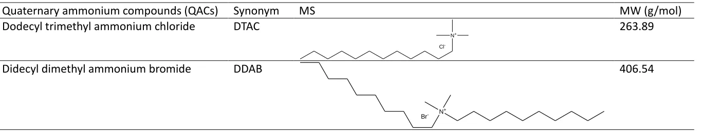
DDAB with two long hydrocarbon chains (Table 2.1). The solutions were prepared using
Table 2.1 Quaternary ammonium compounds (QACs), synonyms, molecular structures (MS) and molecular weights (MW) are shown. Molecular
structures were drawn by ChemDraw® Ultra (version 8.0).
Quaternary ammonium compounds (QACs) Synonym MS MW (g/mol)
Dodecyl trimethyl ammonium chloride DTAC
N+ Cl
-263.89
Didecyl dimethyl ammonium bromide DDAB
N+
Br
2.2.2 Microalgal strain and maintenance
An axenic culture of freshwater green algae, Scenedesmus obliquus (CPCC 5), was
obtained from the Canadian Phycological Culture Centre. The culture was maintained in a
modified HS (high salt) minimal medium [22] with 0.8 g of NaHCO3/L [23] at 25°C on shaking
(150 rpm) with a light intensity of 140 µE m-2 s-1 (16 hrs. Light: 8 hrs. dark) using an incubator
(Infors HT Multitron, Basel, Switzerland). The algal growth was determined routinely
measuring optical density (OD) at 600 nm with a 200 pro infinite series microplate reader
(Tecan, Männedorf, Switzerland). Cells in the exponential growth phase were used to
calculate the specific growth rate (0.82 ± 0.08 d-1, n = 3) and to use for the toxicity experiment.
The cell density (cells mL-1) was measured using a hemocytometer (Hausser Scientific, PA, USA)
and the correlation of optical density versus cell count was generated (Appendix A). To
maintain axenic condition, the culture was checked regularly for the bacterial contamination
by streaking onto nutrient agar plate and no contamination was found throughout the course
of experiments.
2.2.3 Toxicity tests on microalgae
The toxicity test was conducted under three different conditions; autotrophic,
mixotrophic and heterotrophic; autotrophic condition, according to the standard protocol of
Organisation for Economic Cooperation and Development [16] with modifications on the cell
density and use of 96 microplates and mixotrophic and heterotrophic conditions using Biolog
plates.
2.2.3.1 Autotrophic toxicity experiments
Inhibition efficiency tests in growth (%) were conducted using DTAC and DDAB
concentrations 0, 400, 800, 1200, 1600, 2000 and 2400 µg/L. The tests were performed in 96
microplates with a final volume of 150 µL and incubated as described above. Scenedesmus
obliquus in exponential phase was transferred to wells containing different concentrations of
DTAC and DDAB. Wells without DTAC and DDAB served as a control. A cell density of 1 × 10⁶
was calculated using the difference in fluorescence readings (excitation at 470 nm and
emission at 650 nm; [24]) using a M1000 pro infinite series microplate reader (Tecan,
Männedorf, Switzerland) on the fifth day of treatment.
2.2.3.2 Mixotrophic and heterotrophic toxicity experiments
The toxicity tests were performed using Biolog plates. Prefilled 96-well microtiter
plates (YT microplate; Biolog, CA, USA) were used under different treatment conditions. The
YT Biolog plates were selected as they have both sections of wells with or without included
tetrazolium dye (Biolog YT microplate; available from Biolog, CA, USA) and only 57 bottom
wells without tetrazolium dye were used for the analysis as tetrazolium dye can be light
sensitive (Biolog redox dye mixes; available from Biolog, CA, USA). Organic carbons and
sources in the Biolog plates were described in Table 2.2. The same cell density of 1 × 10⁶
cells/mL was exposed to final concentrations of 0, 400, 800, 1200, 1600, 2000 and 2400 µg/L
of DTAC and DDAB for mixotrophic and without two higher concentrations for heterotrophic
toxicity test. The Biolog plates contained multichannel pipetted 100 µL of mixed Scenedesmus
obliquus in 50 µL of modified HS media with a compound, either DTAC or DDAB. The Biolog
plates were kept in dark at 25°C for heterotrophic condition and incubated as above for
mixotrophic condition. Measurements of growths were determined using a M1000 pro
infinite series microplate reader (Tecan, Männedorf, Switzerland) measuring fluorescence
(excitation at 470 nm and emission at 650 nm; [24]). The fluorescence measurement reflecting
the growth was successfully used in this study for their known sensitivity to the quaternary
ammonium compound in microalgae [25], even at a lower pigment concentration per cell
under dark than light conditions [26]. Toxicity on the fifth day of treatment was used for
further analysis as it showed the prominent differences in growth from the control in
mixotrophic conditions and allowed the growth of the heterotrophic ones for the toxicity
effects to be effective. Moreover, the growth was in actively growing conditions on fifth day
Table 2.2 Organic carbons and sources in YT Biolog plates in 57 bottom wells are presented. Total represents the number of organic carbons in
each group of sources. One well is for negative control included water.
Carbon source Chemical Total
Carbohydrate D-Cellobiose, Gentiobiose, Maltose, Maltotriose, D-Melezitose, D-Melibiose, Palatinose, D-Raffinose,
Stachyose, Sucrose, D-Trehalose, Turanose, α-D-Glucose, D-Galactose, D-Psicose, Rhamnose, L-Sorbose, α-Methyl-Glucoside, β- Methyl-Glucoside, Amygdalin, Arbutin, Salicin, Maltitol, Mannitol, Sorbitol, Adonitol, Arabitol, Xylitol, i-Erythritol, Glycerol, L-Arabinose, Arabinose, D-Ribose, D-Xylose
34
Carboxylic acid Fumaric Acid, L-Malic Acid, Bromo-Succinic Acid, γ-Amino-Butyric Acid, α-Glutaric Acid, 2-
Keto-D-Gluconic Acid, Keto-D-Gluconic Acid
7
Amino acid L-Glutamic Acid 1
Fatty acid Tween 80 1
Polymer Dextrin, Inulin 2
Ester Succinic Acid Mono-Methyl Ester 1
Combination Succinic Acid Mono-Methyl Ester plus D-Xylose, N-Acetyl-L-Glutamic Acid plus D-Xylose, Quinic Acid
plus Xylose, Glucuronic Acid plus Xylose, Dextrin plus Xylose, α-Lactose plus Xylose, Melibiose plus Xylose, Galactose plus Xylose, m-Inositol plus Xylose, 1,2-Propanediol plus D-Xylose, Acetoin plus D-Xylose
![Figure 1.1 Steps for addressing a contaminated site (redrawn from, [1]).](https://thumb-us.123doks.com/thumbv2/123dok_us/1965208.1259167/18.595.73.522.298.537/figure-steps-addressing-contaminated-site-redrawn.webp)