_____________________________________________________________________________________________________
ISSN: 2231-0614
SCIENCEDOMAIN international
www.sciencedomain.org
A Subset of Genes Can Distinguish between
Bortezomib Responsive Versus Bortezomib
Resistant Myeloma
Yossi Cohen
1*, Odit Gutwein
1, Osnat Garach- Jehoshua
2, Adina Bar-Haim
3and Abraham Kornberg
11
Institute of Hematology, Assaf Harofeh Medical Center, Affiliated to Sackler Faculty of Medicine, Tel Aviv University, Israel. 2
Hematology Laboratory, Assaf Harofeh Medical Center, Affiliated to Sackler Faculty of Medicine, Tel Aviv University, Israel. 3
Department ofChemistry, Assaf Harofeh Medical Center, Affiliated to Sackler Faculty of Medicine, Tel Aviv University, Israel.
Authors’ contributions
This work was carried out in collaboration between all authors. Author YC designed the experiments, made the analyses and wrote the paper. All other authors assisted in patient recruitment and logistic support. All authors read and approved the final manuscript.
Article Information
DOI: 10.9734/BJMMR/2015/13421
Editor(s):
(1)Alex Xiucheng, Department of Biochemistry and Molecular Biology, University of Florida, USA.
Reviewers:
(1)Anonymous, Bulgaria. (2)Anonymous, Japan. (3)Anonymous, Belgium. Complete Peer review History:http://www.sciencedomain.org/review-history.php?iid=717&id=12&aid=6791
Received 16th August 2014 Accepted 20th September 2014 Published 5th November 2014
ABSTRACT
Despite the widespread use of proteasome inhibitors in the treatment of multiple myeloma, the mechanisms of the anti-myeloma activity and the molecular pathways that execute the tumor cell killing are still unknown. In the present work we compared gene expression profile changes in response to bortezomib treatment of cultured bone marrow samples from patients with bortezomib-sensitive versus bortezomib-resistant myeloma. The results showed a pronounced induction of>70 genes including>30 heat shock protein transcripts in both patient groups and therefore debate the anti-tumor action, attributed to the unfolded protein response. In contrast, a subset of 7 genes (MMP12, IL7R, MGST1, C3, CYP27A1, MIR148A and CXXC4) changed only in the samples from
the bortezomib-sensitive cases and therefore these tumor-associated genes might serve as predictors of the treatment efficacy, as well as for making of further insights onto the mechanism of action of proteasome inhibitors. In summary, we identified a subset of 7 genes which distinguished in our small series betweensensitive versus resistant tumor cells to bortezomib, which requires further assessment in a larger cohort of patients.
Keywords: Multiple myeloma; gene expression profile; MMP12; heat shock proteins; IL7R.
1. INTRODUCTION
Although the novel drug bortezomib (velcade) has become one of the most used agents for treatment of multiple myeloma (MM), the particular mechanism which executes its cytotoxic effect remains elusive due to the diversity of molecular changes induced by proteasome inhibition. One of the earliest hypotheses concerning the mechanism of tumor cell killing induced by bortezomib was derived from the role of the proteasome in degradation of IκB, the inhibitor ofNFκB [1-3]. However, because of the proved markedly less active reduction of the MM cells’ proliferation on the influence of IκB inhibitors in comparison with bortezomib [4,5], this explanation seemed incomplete. Bortezomib also caused 20-to-60-fold induction of the proapoptotic gene NOXA in various cancer cells [6] whereas in other models it impaired tumor growth via inhibition of HIF-1α
and repression of HIF-1 transcriptional activity with attenuation of the release of vascular endothelial growth factor (VEGF) [7]. Other studies suggested that the general accumulation of misfolded proteins in the endoplasmic reticulum (ER) is the major mechanism responsible for the antitumural activity of bortezomib [8,9]. The latter insult initiates the UPR signaling, which in turn stimulates splicing of inactive XBP1 [10] whereas spliced XBP1/XBP1s regulate(s) genes, which are responsible for the ER-associated degradation (ERAD) (e.g. EDEM), ending-up within the proteasome. XBP1s also induces genes that are responsible for protein folding such as p58IPK and a variety of ER chaperones [11]. Although UPR activation can regenerate protein homeostasis anditis also essential for plasma cell differentiation and survival by induction of various ER chaperones and folding enzymes [12-15], under prolonged and uncompensated ER stress the UPR promotes cellular apoptosis, known as terminal UPR [15-17]. The latter occurrence is mediated via the pro-apoptotic transcription factor CHOP (also known as GADD153 and DDIT3), which is induced via PERK and ATF6 pathways. CHOP causes down-regulation of
BCL2, thereby leading to caspase-dependent apoptosis [18,19]. In HNSCC cells, bortezomib induced apoptosis through induction of ER stress along with the generation of reactive oxygen species (ROS) that led to caspase activation whereas inhibition of NFκB was not sufficient to initiate apoptosis [20]. Consistent with the ER stress concept of bortezomib anti myeloma activity, it was found a correlation between the levels of immunoglobulin chain production and the sensitivity to proteasome inhibitors in sub-clones of both human IgG-secreting myeloma cell line JK-6L and murine myeloma cell line Ag8, transfected with expression plasmid, encoding the µ heavy (H) chain [21]. Moreover, pro-apoptotic factors of the ER stress response were induced to a greater extent in sub-clones producing high levels of µH-chains than in those producing no µH-chains. Conversely, MM cells became bortezomib-resistant through inhibition of unfolded protein accumulation by acquired mutations of the PSMB5 gene which prevented the catastrophic ER stress [22].
The present work deals with the cytotoxic mechanisms offered in the context of authentic MM cells from patients whose clinical response to bortezomib regimens was followed for years.
2. METHODS
2.1 Tissue Culture
CO2incubator. Cultures were fed two times per
week by replacing 70% of the medium with fresh supplement. The bortezomib used was VELCADE® for injection (Janssen-Cilag Ltd). For RNA extraction most of the medium was removed (leaving 200-300 µl) and after a vigorous pippetation the released cells were collected and fixed with liquid nitrogen in ~70 µl fractions.
2.2 RNA Extraction
Frozen samples were lysed by adding 300µllysis buffer to tubes. RNA was isolated by MagNA Pure Compact RNA Isolation procedure using MagNA Pure Compact instrument (Roche Diagnostics, Ltd, Israel). Integrity of RNA was examined by Agilent2100Bioanalyzer.
2.3 Gene Expression Profile (GEP)
Biotin-labeled cRNA was generated from 200 ng total RNA, hybridized onto GeneChip Human Gene1.0 ST Array (Affymetrix, Santa Clara, CA, USA) and the data were processed with the Affymetrix GeneChip Scanner 3000 and Affymetrix Expression Console. Normalization was done by the RMA method and fold change results were calculated relative to the fresh BM sample of each case. The microarray data were deposited on the public gene expression ominibus (GEO) accession number GSE51940 http://www.ncbi.nlm.nih.gov/geo/query/acc.cgi?a cc=GSE51940
2.4 Patients
The MM cases studied were selected according to their clinical fitness to the study design including extreme BM infiltration with tumor cells (> 90%), which eliminated the need for cell separation procedures known to bias the authentic GEP records [23-24] and long-term follow-up. The study was approved by the local institute review boards. We analysed BM samples from an overall group of five MM patients, two of which were newly diagnosed (cases A, B) and responded to velcade/dexamethasone (Vel/Dex) induction, another patient (case C) responded with nearly complete response to retreatment with Vel/Dex combination after long term remission of 30 months following initial treatment with bortezomib, dexamethasone and melphalan (VMP) as opposed to the other two patients who failed velcade regimens, one with newly
diagnosed MM (case D) showing primary resistance to Vel/Dex combination as reflected by increase in his paraprotein levels and persistence of > 95% plasma cells in repeated BM examination after 6 injections of velcade (days 1, 4, 8, 11, 29, 32, 36, 39); the second refractory case (case E) was heavily pre-treated and initially responded to Vel/Dex with VGPR (very good partial response) followed by long-term remission after consolidation stem cell transplantation (SCT) with melphalan 200 mg/m2. However, after almost 3 years the patient progressed and received second line treatment with lenalidomide/dexamethasone (Len/Dex) but was refractory to this regimen, then he partially responded to retreatment with Vel/Dex and continued with 2 salvage cycles of VD-PACE (velcade combined with dexamethasone, platinol, adriamicin, etoposide) and underwent second SCT while in VGPR (case D). Once again, after 6 months the patient progressed but now he became completely resistant to bortezomib with no response to Vel/Dex and to VD-PACE as evident by the development of chest wall plasmacytomas, pancytopenia, hypercalcemia, renal failure, sharp increase in his urine paraprotein levels as well as > 95% plasma cells in the BM aspirate and biopsy under treatment. At this stage his BM sample was examined in our present study and the patient entered a clinical trial with carfilzomib, pomalidomide and dexamethasone after which he achieved a PR (partial response), with drop in his paraprotein levels to almost 1/3 of his pre-treatment levels and improvement in all of his clinical and laboratory tests including repeated BM biopsy. The five patient’s characteristics are summarized in Table 1. Velcade was added to the cultured BM samples at a concentration of 2 µg/ml, which was found in our preliminary assessment to kill cultured primary MM cells within 24 hours (data not shown). In one of the cases (case C), velcade was also used at a concentration of 0.2
µg/ml, which showed almost the same GEP changes as the higher concentration. After 6-8 hours the treated and control cells were released from the bottom wells and they were then fixed with liquid nitrogen.
RESULTS
Table 1. Patient characteristics and response to treatment
Age Sex
Paraprotein CRAB*, FISH
ISS Velcade regimen
Response to velcade regimen
Salvage regimen (if given)/response Case
A
85♂ Non-secreting C, R, A, B t(4;14)
III Vel/Dex VGPR* after 16 injections of velcade
-
Case B
60♀ IgAk 3.0 g/dl C, R, A, B t(4;14)
III VCD CR* after 16 injections of velcade
-
Case C
75♀ IgAk 3.5 g/dl A, B N/A
III Vel/Dex VGPR* after 8 injections of velcade retreatment
NR** to lenalidomide
Case D
55♂ IgAk 3.1g/dl A, B; del 13q
II Vel/Dex NR** after 8 injections of velcade retreatment
R+D (PR) → SCT (VGPR)
Case E
63♂ IgAλ 3 g/dl, 16.6 g/day C,R,B; del P53
III Vel/dex NR** after 6 injections of velcade retreatment
Pomalidomide + Carfilzomib (PR)
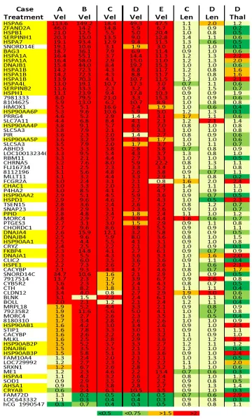
examined independently of their clinical response to bortezomib regimens (Fig. 1). The pronounced induction of HSP transcripts in the samples from our bortezomib resistant cases raises debates concerning the anti-malignancy role attributed to UPR activation [8-10, 17-19, 25-29] and excludes the possibility that the clinical inactivity of the drug in those patients resulted from inaccessible binding site or un blcokable proteasome in the tumor cells otherwise no induction of HSP transcripts could be elicited in vitro. Accordingly, the models of impaired drug binding used to explain the mechanism of bortezomib resistance on the basis of acquired resistance in mutant sub-clones [30-34] might be irrelevant to primary tumor cells as already noticed [35]. In addition, neither NOXA nor HIF1A, GADD45A, GADD45B, GADD45G, TNFRSF10B, FAS, FASLG, DAP3, CASP8, CASP7 or CASP1, which were reported to promote apoptosis in bortezomib treated cells [36,37], were induced markedly in any of the BM samples examined in our series and no BCL2 repression could be recognized. Furthermore, although the pro-apoptotic signaling molecule CHOP (DDIT3), which is considered to be induced and activated by ER stress [18,19], was induced to somewhat greater extent in our sensitive versus resistant to bortezomib cases, the differences in expression were too smallto explain the clinical differences seen. In contrast, a distinct subset of 7 genes (e.g., MMP12, IL7R, MGST1, C3, CYP27A1, MIR148A and CXXC4) was modulated exclusively in the BM samples from our bortezomib-responsive cases (Fig. 2), though the predictive value of this observation requires further assessment in a larger cohort of samples.
Within the limitations of our results, the first issue to be considered is the differences in the drug exposure in vitro versus in vivo. In the current study, bortezomib was added to cultures in
concentration much higher (~20 folds) than the usual peak plasma levels (89-120 ng/ml). However, bortezomib is rapidly and widely distributed to tissues and the mean 24-hour total radioactivity levels (TR) of 14C-bortezomib was found to be 43.5, 30.5 and 27.8 folds higher in the BM versus plasma of Sprague-Dawley rats after the first, third and fourth dose of the drug in a biweekly schedule, respectively [38]. Likewise, the area under the concentration-time curve (AUC; µg-eq. h/g) of TR between 0 and 72 hours post 14C-bortezomib injection (AUC0-72 h) was
31.7 and 24.1 folds higher in the BM versus plasma after the first and the fourth dose, respectively. Therefore, it seems reasonable that the bortezomib concentration, applied in vitro in the presented paper, is also reachable in the BM. In addition, in case C which was also examined with velcade concentration of 0.2 µg/ml the changes in GEP were almost the same as with 2
µg/ml.
4. DISCUSSION
Fig. 2.Genes modulated exclusively in bortezomib-responsive cases
or as yet unrecognized proteasome-related pro-apoptotic pathway. On the other hand, the excellent clinical response to bortezomib regimens despite the marked induction of numerous HSP transcripts in the BM samples from our bortezomib-responsive cases raises debates, connected with the role, attributed to HSPs on bortezomib resistance and concerning the expectations from HSP inhibitors to overcome drug resistance [45-47], though direct
ERAD inhibitors might synergized with bortezomib [48]. The second important finding was the subset of 7 genes which distinguished responsive/sensitive versus resistant to bortezomib myeloma. In considering the known roles of these genes, it seems reasonable that the induction of some of which, like MMP12 and C3was originated from contaminated macrophages which sensed the initial injury to adjacent tumor cells. For instance, MMP12 (macrophage metalloelastase) is a matrix metallopeptidase, which predominantly expressed by mature tissue macrophages and is implicated in pathological processes [49]. In contrast, IL7R was implicated in leukemogenesis [50]. Currently, we expand the study group in order to test the applicability of the findings in prediction of the clinical response to proteasome inhibitors on the basis of the in vitro results.
CONSENT
Not applicable.
ETHICAL APPROVAL
All authors hereby declare that all experiments have been examined and approved by the appropriate ethics committee and have therefore been performed in accordance with the Ethical standards laid down in the 1964 declaration of Helsinki.
ACKNOWLEDGMENTS
The authors thank Relly Forer (PhD), Inna Vulin (PhD) and Dina Volodarsky (MsC) from Dyn diagnostics Ltd for professional microarray and Bioinformatic service, Izhar Hardan (MD) from Meir hospital for support and advices and to DovZipory (PhD) from the Weizmann Institute of Science for the training and advices.
COMPETING INTERESTS
Authors have declared that no competing interests exist.
REFERENCES
1. Alkalay I, Yaron A, Hatzubai A, et al. In vivo stimulation of I kappa B phosphorylation is not sufficient to activate NF-kappa B. Mol Cell Biol. 1995;15:1294-1301.
2. Alkalay I, Yaron A, Hatzubai A, et al. Stimulation-dependent I kappa B alpha phosphorylation marks the NF-kappa B inhibitor for degradation via the ubiquitin-proteasome pathway. Proc Natl Acad Sci U S A. 1995;92:10599-10603.
3. Kanarek N, London N, Schueler-Furman O, et al. Ubiquitination and degradation of the inhibitors of NF-kappa B. Cold Spring Harb Perspect Biol. 2010;2:a000166. 4. Mitsiades N, Mitsiades CS, Poulaki V, et
al. Biologic sequelae of nuclear factor-kappa B blockade in multiple myeloma: Therapeutic applications blood. 2002;99:4079-4086.
6. Fernández Y, Verhaegen M, Miller TP, et al. Differential regulation of noxa in
normal melanocytes and melanoma cells by proteasome inhibition: Therapeutic
implications. Cancer Res. 2005;65:6294-6304.
7. Befani CD, Vlachostergios PJ, Hatzidaki E, et al. Bortezomib represses HIF-1α protein expression and nuclear accumulation by inhibiting both PI3K/Akt/TOR and MAPK pathways in prostate cancer cells. J Mol Med (Berl). 2012;90:45-54.
8. Nawrocki ST, Carew JS, Pino MS, et al. Bortezomib sensitizes pancreatic cancer cells to endoplasmic reticulum stress-mediated apoptosis. Cancer Res. 2005;65:11658–11666.
9. Nawrocki ST, Carew JS, Dunner K Jr, et al. Bortezomib inhibits PKR-like endoplasmic reticulum (ER) kinase and induces apoptosis via ER stress in human pancreatic cancer cells. Cancer Res. 2005;65:11510–11519.
10. Yoshida H, Matsui T, Yamamoto A, et al. XBP1 mRNA is induced by ATF6 and spliced by IRE1 in response to ER stress to produce a highly active transcription factor. Cell. 2001;107:881-891.
11. Lee AH, Iwakoshi NN, Glimcher LH. XBP-1 regulates a subset of endoplasmic reticulum resident chaperone genes in the unfolded protein response. Mol Cell Biol. 2003;23:7448-7459.
12. Iwakoshi NN, Lee AH, Glimcher LH. The X-box binding protein-1 transcription factor is required for plasma cell differentiation and the unfolded protein response. Immunol Rev. 2003;194:29-38.
13. Reimold AM, Iwakoshi NN, Manis J, et al. Plasma cell differentiation requires the transcription factor XBP-1. Nature. 2001;412:300-307.
14. Iwakoshi NN, Lee AH, Vallabhajosyula P, et al. Plasma cell differentiation and the unfolded protein response intersect at the transcription factor XBP-1. Nat Immunol. 2003;4:321-329.
15. Gass JN, Gifford NM, Brewer JW. Activation of an unfolded protein response during differentiation of antibody-secreting B cells. J BiolChem. 2002;277:49047– 49054.
16. Kim R, Emi M, Tanabe K, et al. Role of the unfolded protein response in cell death. Apoptosis. 2006;11:5–13.
17. Sano R, Reed JC. ER stress-induced cell death mechanisms. Biochim Biophys Acta. 2013;1833:3460-3470.
18. McCullough KD, Martindale JL, Klotz LO, et al. Gadd153 sensitizes cells to endoplasmic reticulum stress by down-regulating Bcl2 and perturbing the cellular redox state. Mol Cell Biol. 2001;21:1249-1259.
19. Harding HP, Zhang Y, Bertolotti A, et al. Perk is essential for translational regulation and cell survival during the unfolded protein response. Mol Cell. 2000;5:897-904.
20. Fribley A, Zeng Q, Wang CY. Proteasome inhibitor PS-341 induces apoptosis through induction of endoplasmic reticulum stress-reactive oxygen species in head and neck squamous cell carcinoma cells. Mol Cell Biol. 2004;24:9695–9704.
21. Meister S, Schubert U, Neubert K, et al. Extensive immunoglobulin production sensitizes myeloma cells for proteasome inhibition. Cancer Res. 2007;67:1783-1792.
22. Ri M, Iida S, Nakashima T, et al. Bortezomib-resistant myeloma cell lines: a role for mutated PSMB5 in preventing the accumulation of unfolded proteins and fatal ER stress. Leukemia. 2010;24:1506-1512. 23. Cohen Y, Garach-Jehoshua O, Bar-Chaim
A, at al. Niche-modulated and niche-modulating genes in bone marrow cells. Blood Cancer J. 2012;14:2:e97.
24. Cohen Y, Gutwein O, Garach-Jehoshua O, et al. The proliferation arrest of primary tumor cells out-of-niche is associated with widespread downregulation of mitotic and transcriptional genes. Hematology. 2014;19:286-92.
25. Wang Q, Shinkre BA, Lee JG, et al. The ERAD inhibitor eeyarestatin I is a bifunctional compound with a membrane-binding domain and a p97/VCP inhibitory group. PLoS One. 2010;12:e15479. 26. Obeng EA, Carlson LM, Gutman DM, et al.
Proteasome inhibitors induce a terminal unfolded protein response in multiple myeloma cells. Blood. 2006;107:4907–4916.
degradation are coupled events. FASEB J. 2000;14:769-778.
28. Werner ED, Brodsky JL, McCracken AA. Proteasome-dependent endoplasmic reticulum-associated protein degradation: an unconventional route to a familiar fate. Proc Natl Acad Sci. USA. 1996;93:13797-13801.
29. O'Hare T, Wiens GD, Whitcomb EA. Cutting edge: Proteasome involvement in the degradation of unassembled Ig light chains. J Immunol. 1999;163:11-14. 30. Wacker SA, Houghtaling BR, Elemento O,
et al. Using transcriptome sequencing to
identify mechanisms of drug action and resistance. Nat Chem Biol.
2012;8:235-237.
31. Stessman HA, Baughn LB, Sarver A, et al. Profiling bortezomib resistance identifies secondary therapies in a mouse myeloma model. Cancer Ther. 2013;12:1140-1150. 32. Shuqing Lü, Jianmin Wang. The resistance
mechanisms of proteasome inhibitor bortezomib. Biomarker Research. 2013;7771:1-13.
33. Suzuki E, Demo S, Deu E, et al. Molecular mechanisms of bortezomib resistant adenocarcinoma cells. PLoS One. 2011;6:e27996.
34. Franke NE, Niewerth D, Assaraf YG, et al. Impaired bortezomib binding to mutant β5 subunit of the proteasome is the underlying basis for bortezomib resistance in leukemia cells.
Leukemia. 2012;26:757-768.
35. Leung-Hagesteijn C, Erdmann N, Cheung G, et al. Xbp1s-negative tumor B cells and pre-plasmablasts mediate therapeutic proteasome inhibitor resistance in multiple myeloma. Cancer Cell. 2013;24:289-304. 36. Mujtaba T, Dou QP. Advances in the
understanding of mechanisms and therapeutic use of bortezomib. Discov Med. 2011;12:471-480.
37. Mitsiades N, Mitsiades CS, Poulaki V, et al. Molecular sequelae of proteasome inhibition in human multiple myeloma cells. Proc Natl Acad Sci. USA. 2002;99:14374-14379.
38. Hemeryck A, Geerts R, Monbaliu J, et al. Tissue distribution and depletion kinetics of bortezomib and bortezomib-related radioactivity in male rats after single and repeated intravenous injection of
14 C-bortezomib. Cancer Chemother Pharmacol. 2007;60:777-787.
39. Mulligan G, Mitsiades C, Bryant B, et al. Gene expression profiling and correlation with outcome in clinical trials of the proteasome inhibitor bortezomib. Blood. 2007;109:3177-3188.
40. Davenport EL, Moore HE, Dunlop AS, et al. Heat shock protein inhibition is associated with activation of the unfolded protein response pathway in myeloma plasma cells. Blood. 2007;110:2641-2649. 41. Zong WX, Li C, Hatzivassiliou G, et al. Bax
and Bak can localize to the endoplasmic reticulum to initiate apoptosis. J Cell Biol. 2003;162:59–69.
42. Selimovic D, Porzig BB, El-Khattouti A, et al. Bortezomib/proteasome inhibitor triggers both apoptosis and autophagy-dependent pathways in melanoma cells. Cell Signal. 2013;25:308-318.
43. Yu C, Rahmani M, Dent P. The hierarchical relationship between MAPK signaling and ROS generation in human leukemia cells undergoing apoptosis in response to the proteasome inhibitor Bortezomib. Exp Cell Res. 2004;295:555-566.
44. Landowski TH, Megli CJ, Nullmeyer KD, et al. Mitochondrial-mediated disregulation of Ca2+ is a critical determinant of Velcade (PS-341/bortezomib) cytotoxicity in myeloma cell lines. Cancer Res. 2005;65:3828–3836.
45. Hu J, Dang N, Menu E, De Bruyne E, et al. Activation of ATF4 mediates unwanted Mcl-1 accumulation by proteasome inhibition. Blood. 2012;119:826-37. 46. Khong T, Spencer A. Targeting HSP 90
induces apoptosis and inhibits critical survival and proliferation pathways in multiple myeloma. Mol Cancer Ther. 2011;10:1909-1917.
47. Roué G, Pérez-Galán P, Mozos A, et al. The Hsp90 inhibitor IPI-504 overcomes bortezomib resistance in mantle cell lymphoma in vitro and in vivo by down-regulation of the prosurvival ER chaperone BiP/Grp78. Blood. 2011;117:1270-1279. 48. Auner HW, Moody AM, Ward TH, et al.
49. Lee JT, Pamir N, Liu NC, et al. Macrophage Metalloelastase (MMP12) regulates adipose tissue expansion, Insulin Sensitivity, and Expression of Inducible nitric oxide synthase. Endocrinology. 2014;155:3409-20.
50. Tasian SK, Loh ML. Understanding the biology of CRLF2-overexpressing acute lymphoblastic leukemia. Crit Rev Oncog. 2011;16:13-24.
© 2015 Cohen et al.; This is an Open Access article distributed under the terms of the Creative Commons Attribution License (http://creativecommons.org/licenses/by/4.0), which permits unrestricted use, distribution, and reproduction in any medium, provided the original work is properly cited.
Peer-review history: