___________________________________________________________________________________________
1(4): 486-500, 2011
SCIENCEDOMAIN international
www.sciencedomain.org
Cell-Based Therapies in Musculoskeletal
Injuries: The Evolving Role of Bone
Marrow-Derived Mesenchymal Stem Cells
P. S. Pastides
1and W. S. Khan
1*1
University College London Institute of Orthopaedics and Musculoskeletal Sciences, Royal National Orthopaedic Hospital, Stanmore, Middlesex, HA7 4LP, United Kingdom.
Received 11th July 2011 Accepted 8th September 2011 Online Ready 28th September 2011
ABSTRACT
Aims: There is considerable interest in the potential of bone marrow-derived mesenchymal stem cells (BM-MSCs) in the management of musculoskeletal injuries. This review aims to summarise the information in the literature on the evolving role of these cells in the management of these complex heterogenous injuries.
Study design: Review Article.
Place and Duration of Study: University College London Institute of Orthopaedics and Musculoskeletal Sciences, Royal National Orthopaedic Hospital, Stanmore, Middlesex, HA7 4LP, United Kingdom.
Methodology: We reviewed the literature to identify studies on the use of BM-MSCs for the management of musculoskeletal injuries.
Results: There is an increasing and encouraging body of evidence to suggest that BM-MSCs have a significant role in the management of musculoskeletal injuries involving muscles, tendons, ligaments, bone, cartilage, menisci and nervous tissue.
Conclusion: Several characteristics of BM-MSCs make them ideal candidates in managing musculoskeletal injuries. Bone marrow is easy to obtain requiring minimal donor site morbidity, invasiveness and anaesthetic. Their autologous nature eliminates the issue of immunoreactions and ethical problems. The majority of studies in the literature however use small animal models, and further work in larger animals and ultimately ethically approved clinical trials should be explored before any significant clinical relevance can be assessed.
Keywords: Musculoskeletal; bone marrow; mesenchymal stem cells; scaffold; injection; cell culture; differentiation; integration;
1. INTRODUCTION
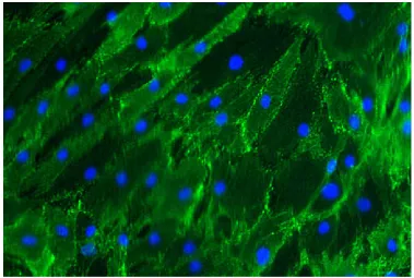
Tissue engineering is an exciting strategy being explored to deal with damaged or lost tissue. Tissue engineering can include the use of cells, scaffolds and growth factors in any combination (Mahapatra and Khan 2011; Nannaparaju et al., 2011). Stem cells are a self-renewing, slow-cycling cell population that exhibit high clonogenity, low cellular proliferation and the ability to undergo multilineage differentiation. Stem cells can be derived from a number of sources and are able to undergo chondrogenic, osteogenic and adipogenic differentiation (Pittenger et al., 1999). These cells are identified by a number of cell surface markers that they express on their cell surface including CD105 as shown in Figure 1. These cells are often used with scaffolds that provide a three-dimensional structural template. Scaffolds are natural (e.g. collagen and alginate) or synthetic (e.g. polyglycolic acid and polyacrylonitrile polyvinyl chloride) materials used for cell attachment and proliferation. The ideal scaffold is biocompatible and meets the biological needs of growing tissue.
Figure 1: Cell surface epitope characterisation of mesenchymal stem cells using cell surface staining with a primary antibody that recognises CD105 and a fluorescent secondary antibody that shows up green. The nuclei are counterstained with a blue dye. The figure shows strong staining for CD105, a mesenchymal stem cell marker.
et al., 1999; Short et al., 2003). Protocols for the culture (Freidenstein et al., 1970) and, chondrogenic, osteogenic and adipogenic differentiation of bne marrow-derived mesenchymal stem cells (BM-MSCs) have been described (Johnstone et al, 1998; Pittenger et al, 1999; Sekiya et al, 2002; Thanabalasundaram et al., in press). BM-MSCs have been associated with the repair and regeneration of musculoskeletal injuries.
Musculoskeletal injury can involve muscle, tendon, ligament, bone, meniscus and cartilage. The high incidence of such injuries highlights the need for novel, more effective treatments. Currently a lot of research is being carried out into this area. The use of BM-MSCs is one such option (Tucker et al., in press) and the aim of this review is to critique and clarify their findings.
2. METHODOLOGY
A thorough literature review was conducted and articles relating to the use of BM-MSCs for the treatment of musculoskeletal injuries were identified. The searched primarily focused on the use of the cells to treat common conditions affecting muscles, tendons, ligaments, bones, meniscii, cartilage and their role in osteoarthiritis.
3. RESULTS AND DISCUSSION
3.1 Muscle
Increased musculoskeletal activities can exert abnormal biomechanical environments onto muscle which can lead to injuries such as muscular strain and tears which can sufficiently hinder the patient’s mobility. The incidence of such injuries in the professional sporting setting is considerable and troublesome for both sporting professionals and their teams (Elliott et al., 2011).
Following muscular injury, repair and regeneration is triggered by local multipotent stem cells. BM-MSCs act as a reserve for muscle precursors and aid in its repair by migrating physiologically into the regenerating muscle. Ferrari et al. (1998) however showed that when BM-MSCs were administered systemically in immunodeficient mice, they had minimal impact on muscular regeneration. However, in cases of extended damage, they maintained the population of more differentiated, muscle-forming precursors (Ferrari et al., 1998).
3.2 Tendon
Tendons act to both transmit muscular forces but also as a store of elastic energy (Fukashiro et al., 1995; Oragui et al., 2011). Thus repetitive cyclical loading, such as that exhibited in the Achilles tendon may result in overuse injury (Kvist 1994), and consequently tendon rupture.
One study (Chong et al., 2007) looking at rabbit Achilles tendon using a fibrin gel into which BM-MSCs were seeded, demonstrated an increased modulus of elasticity by 32% and an increased proportion of collagen type I compared to the fibrin gel alone group at 3 weeks. However, after 6 and 12 weeks no difference was observed between groups. In a second study looking at rabbit Achilles tendon injuries, Ouyang et al. (2003) used 3 groups: group 1 received BM-MSCs seeded onto a poly-lactide-co-glycolide (PLGA) scaffold; group 2, a PLGA scaffold alone and group 3 received no laceration to act as a control. Group 1 exhibited greater tissue formation and remodelling compared to group 2. Both groups 1 and 2 had similar histological appearance to native tendon by 12 weeks. The tensile stiffness and modulus in group 1 was 87% and 62.6% respectively of a normal tendon whereas in group 2 they were 56.4% and 52.9% respectively suggestive of a potential benefit for these types of injuries.
In another study, incised rabbit patella tendons where treated with a collagen gel-BM-MSC composite (treatment group) or simply a collagen gel (control group) (Awad et al., 1999). Histologically, only 40% of the treatment group expressed more physiological, well integrated collagen types, whilst the remainders showed comparable collagen integration types to the control group. However, there was a demonstrable biomechanical advantage in the treatment group; tendon stiffness, modulus of elasticity, maximum stress and strain energy increased by 15%, 7%, 16% and 32% respectively as compared to the control group. This may suggest an advantageous biomechanical effect which is not apparent histologically and may simply be due to an increased cross sectional area provided by the cells.
Damage at the tendon bone interface in rabbits was also explored (Ouyang et al., 2004) using fibrin glue with and without BM-MSCs. Both groups showed evidence of perpendicular collagen type fibre formation. However, the control group had type I and III fibres only while the treatment group had a higher proportion of fibres and consisted of collagen type I, II and III. This demonstrates the ability of BM-MSCs to promote fibrocartilage-like tissue formation and thus aids in the healing process.
The results from these studies show improved biomechanical properties and healing rates of tendons when BM-MSCs are inserted.
3.3 Ligament
Ligaments stabilize joints and control their movements. During sporting activities, they are subjected to higher stress and strain rates as the joints are subjected to more forceful and exaggerated movements. Additionally, they help coordinate the complex movements required during sporting activities via a proprioceptive input into the nervous system (Frank, 2004).
The cruciate ligaments are probably the most famous and one of the commonest ligaments that can be damaged. Anterior cruciate ligament (ACL) tears can be caused by torsional forces created at the knee whilst the foot is firmly planted on the ground or upon landing or by direct blows to the outside of the knee. Damage to ligaments can also disrupt the physiological loads and movements of joints which may predispose to osteoarthritis (OA) (Muthuri et al., 2011).
ligament (Gobbi et al., 2009) or reconstruction using extra- or intra-articular tendon grafts therefore causing donor site morbidity (Streich et al., 2010). BM-MSCs potentially enhance the efficacy of some of the current treatments (Lim et al., 2004) or may act as a treatment in their own right (Al-Rashid and Khan, 2011). Kanaya et al. (2007) injected BM-MSCs into partial torn rat anterior cruciate ligaments and found that this appeared to accelerate their healing and concluded that they may be a viable treatment option. The ultimate failure load of the femur-ACL-tibia complex of the BM-MSC treated group was significantly higher than the control group. The transected area of the ACLs in the BM-MSC group demonstrated no retraction and a better histological grading score. The BM-MSCs survived the intra-articular environment and enhanced the healing of partial tears resulting in more biomechanically and histologically viable results.
This potential benefit is supported by other studies (Oe et al., 2011; Wei et al., 2011). In fact, Wei et al concluded that not only do BM-MSCs promote ligamentous healing, but expression of TGFβ1 and VEGF(165) within these cells significantly promoted angiogenesis of the reconstructed ligament at 3, 6, 12 weeks, with the best mechanical properties being achieved at 24 weeks.
Bilateral ACL reconstructions using hamstring tendon autografts were carried out on rabbits to investigate whether the use of BM-MSCs would reduce the incidence of early tendon autograft pull out following ligament reconstruction (Lim et al., 2004). The treatment limb autograft was coated with autologous BM-MSCs within a fibrin glue carrier. The control limb autograft received only fibrin glue. At 8 weeks post surgery the control had a distinct fibrous interface with the bone, containing collagen type I and III with occasional fibres resembling Sharpey’s fibres bridging the gap. Conversely the treatment group had matured zones of cartilage rich in collagen type II resembling normal entheses. The overall mean failure load and stiffness where 66% and 51% respectively, which was statistically significantly higher in the BM-MSC group. Despite this benefit, 44% of BM-MSC limbs failed by pullout highlighting that although the resulting biomechanical properties are apparent, more work is needed to evaluate this benefit.
BM-MSCs appear to be the optimum cell sources for ligament repair and produce better results biomechanically, histologically and physiologically.
3.4 Bone
The management of bone defects is challenging. Although bone defects commonly occur after musculoskeletal injury, they can also be caused by surgery or disease (Khan et al., 2009). Non-union is a significant problem affecting up to 10% of fractures (Axelrad et al., 2007). Any approach to deal with bone defects need to address three fundamental features for bone repair: osteoconduction, osteoinduction and osteogenesis. The current ‘gold standard’ autologous cancellous bone grafting is limited by tissue availability and donor site morbidity. Allogenic bone grafting has the potential for disease transmission. The use of growth factors including BMPs has shown promising results (Friedlaender et al., 2001) but their role is limited in the absence of an osteoconductive and osteogenic component.
Petite et al. (2000) investigated the results of using a coral scaffold with and without BM-MSCs, and with fresh bone marrow (FBM) in sheep. The BM-MSC seeded scaffold allowed bone deposition at the same rate as scaffold degradation. It was the only composite that resembled physiological bone and allowed bony union to occur after 16 weeks. Although the coral scaffold was osteoconductive resulting in bone deposition in the medullary centre, scaffold degradation outweighed bone deposition in coral and FBM scaffolds.
A different study evaluating the effect of BM-MSCs in healing rates of femoral fractures in rats, a porous hydroxyapatite-tricalcium phosphate ceramic cylinder was used. In one leg the rat received the cylinder alone and in the contralateral leg received the cylinder embedded with BM-MSCs. By 12 weeks union was complete in the BM-MSC group with bony ingrowth into the pores of the scaffold displaying increased strength (215%), stiffness (245%) and torsional energy absorbed (212%) compared to the scaffold side (Bruder et al., 1998b).
In a similarly designed study by Bruder et al. (1998a), a ceramic cylinder composed of hydroxyapatite and β-tricalcium phosphate was used to investigate fracture healing in a canine model. The dogs received a cylinder with or without BM-MSCs. In the dogs with cell-free cylinders union occurred between the implant and the bone cortex in ten of twelve dogs by 16 weeks however no callus was visible and most of the pores were filled with fibrous tissue. In the BM-MSC group, bone was distributed evenly throughout the implant and integrated well with the host bone. By 8 weeks solid union had occurred in all 12 bone implant interfaces with a continuous bridge of mineralized bone surrounding the defect. An osseous callus formed around the implant and the adjacent host bone in 84% of specimens. The implant pores were filled with woven or lamellar bone in direct contact with the ceramic (Bruder et al., 1998a). This finding is supported by another study using mice models and fracture of the tibia (Granero-Molto et al., 2009).
The preferred treatment for large bone defects is currently autologous bone grafts. However, the supply of suitable bone is limited and its collection is painful, with a risk of infection, nerve damage, and a loss of function (Calori GM et al 2011). Hence BM-MSC embedded scaffolds offer a safer, less destructive alternative with good results (Khaled et al., 2011).
3.5 Meniscus
The meniscus is a vital part of the joint. It acts to prevent the deterioration and degeneration of articular cartilage and the onset and development of osteoarthritis. For this reason, research into meniscus repair has been the recipient of particular interest from the orthopedic and bioengineering communities. They also function to absorb shock during dynamic loading and have a major role in the tribological properties of the knee joint (Makris et al., 2011).
Meniscal tears often occur in conjunction with ACL injuries (Noyes et al., 1980). Although lesions in the peripheral vascular region of the meniscus heal well but lesions in the central avascular area fail to do so (Klompmaker et al., 1996). The results with allograft are variable. Isolating meniscal cells from the resected region is another option that has been explored but the quantity and quality of these cells limits applications (Ha et al., 2011). Prosthetic replacement has received promising results from early animal studies but further studies are needed.
At 12 weeks, the cell-free scaffold group had many small round cells within the fibrin glue that were synthesising ECM in 25% of the specimens. The BM-MSC group at that stage contained an abundance of round cells within the scaffold which were surrounded completely by ECM in 75% specimens. In addition, cartilage-like tissue could also be seen (Izuta et al., 2001).
Walsh et al. (1999) investigated the effect of cell-seeded collagen type I scaffolds in rabbits. The rabbits underwent bilateral partial menisectomy and then either no treatment, autogenous periosteal graft, type I collagen sponge or a type I collagen sponge with BM-MSCs. Time-dependant osteoarthritic changes developed in all groups. These changes were greater in the autogenous periosteal graft group and fewer in the collagen scaffold group, especially when used with BM-MSCs. The addition of BM-MSCs to the scaffold enhanced fibrocartilage regeneration with evidence of mature bundles of collagen and proteoglycan.
In another study, rabbits with meniscal resection of the pars intermedia received either a hyaluronon/gelatine scaffold with or without MSCs, or received no treatment. The BM-MSC knees had high cellularity of chondrocyte-like morphology and extensive ECM resembling that of normal meniscus. There was nearly complete filling of the defects with good integration of the scaffold. The amount of fibrocartilage seen was significantly greater than in the cell-free scaffold. The cell-free scaffold group had a partially filled defect with good repair tissue integration but t extracellular matrix produced did not contain any type II collagen. The defect in the control was, in the majority, unfilled (Angele et al., 2008).
The effect of BM-MSCs when applied within a blood clot by sutures to a defected middle third of meniscus was investigated using a goat model. The goats were divided into four groups; Group 1 received only sutures, group 2 sutures and a blood clot, group 3 sutures, a blood clot and BM-MSCs and group 4 received nothing. Group 1 had 4 healed and 4 partially healed knees (3 at 75% and 1 at 50%) with no failed repairs. In group 2, 5 defects were healed and 2 partially healed (by 50%) and 1 failed repair. The repaired sections were more organised with less cellularity compared to group 1. Group 3 had 3 healed, 1 partially healed (25%) and 4 failed repairs. The repair site had reduced cellularity, increased matrix and increased orientation of the matrix. Finally, Group 4 had 7 knees with no healing and 1 partial healing (25%). This study therefore proved that BM-MSCs were detrimental to meniscal healing (Port et al., 1996).
Most of these studies show the positive effects of BM-MSCs on mensical healing. These cells have an ability to withstand the avascular meniscal conditions and appear well-suited to defects within this area. In vitro studies have demonstrated that the matrix forming phenotype of human meniscus cells can be enhanced by expansion in growth factors and altering the oxygen tension (Adesida et al., 2006; Adesida et al., 2007) and further work on these aspects is ongoing.
3.6 Cartilage
Articular cartilage is particularly suitable for tissue engineering applications as it is avascular, aneural and alymphatic (Khan and Hardingham, 2009). Articular cartilage shows a limited capacity for repair following injury. Cartilage injuries that extend to the subchondral bone show some signs of repair due to the release of BM-MSCs from the subchondral bone, and this principle is employed in microfractures (Punwar and Khan, 2011). Current treatments such as arthroscopic management, autologous osteochondral transfer and autologous chondrocyte implantation (ACI) have all shown positive results. However a systematic review comparing autologous chondrocyte implantation, osteochondral autograft transfer, matrix-induced autologous chondrocyte implantation and microfracture failed to identify a single technique consistently showing superior results compared with the others. It did however find that outcomes for microfracture tended to be worse in larger lesions (Magnussen et al., 2008).
Im et al. investigated the ability of BM-MSCs to treat cartilage defects (Im et al., 2001). They suspended BM-MSCs in Ham F-12 medium before injecting into full thickness cartilage defects in the patellar groove of rabbits. The control group received cell free medium. At 14 weeks the BM-MSC group contained reparative tissue resembling articular cartilage with a fully repaired subchondral bone layer. However, the reparative tissue of the control defects was thin, irregular and undifferentiated with less matrix collagen type II. Histological grading scores indicated the treatment group performed significantly better than the control (14.8 vs 8.9). Thus, the findings suggest the use of BM-MSCs in this way enhances cartilage repair but does not guarantee cartilaginous healing.
Wakitani et al. (1994) used BM-MSCs or periosteum derived mesenchymal stem cells (PD-MSCs) from rabbits and seeded them into a type 1 collagen gel. They were then implanted into large (3x6mm), full thickness osteochondral defects located in the weight bearing surface of the medial femoral condyle. The contralateral knee served as a control with its defect either left empty or filled with collagen free gel. The BM-MSC group produced reparative tissue more similar to hyaline cartilage with better integration by week 4. However by week 24, the quality of this tissue progressively declined as the thickness of the articular cartilage portion reduced below that of normal cartilage. The control group showed markedly inferior repair throughout the assessment. PD-MSCs yielded results very similar to BM-MSCs but in addition exhibited a progressive increase in surface irregularity.
Hybrid designs for scaffolds have also been used. Shao et al. (2006) seeded BM-MSCs into a Polycaprolactone (PCL) scaffold for the cartilage portion and a tricalcium phosphate-reinforced PCL scaffold for the bone portion. With fibrin glue, the seeded scaffolds were implanted into large osteochondral defects in the load bearing medial femoral condyle of rabbits. Controls received a cell free scaffold. After 6 months, repair tissue from the BM-MSC group was well integrated with host bone in all specimens. Most samples were hyaline like in nature with identifiable collagen type II and glycosaminoglycan, and an almost normal physiological stiffness. Defects in the control group were incompletely filled with fibrous repair tissue with little resemblance to cartilage or bone. However, some repair tissues in the BM-MSC group also experienced fissures and cracks at the integration site and microscopically demonstrated a lack of typical zonal arrangement (Shao et al., 2006).
similar to the previous scaffold studies. Although in support of Wakitani et al. (1994), it did not, however, report the trend in thinning of the reparative cartilage at 12 weeks. Unlike Shao et al there were signs of zonal organisation of the newly formed cartilage. Additionally, the PLGA scaffold was able to prevent leakage of the injected cells away from the defect, as reported by Im et al. (2001).
It is clear that BM-MSCs do have a role to play in the treatment of osteochondral defects, however it is an area of research that must be investigated further. The use of scaffolds, growth factors and altered culture conditions for in vitro expansion are synergistic factors that also require further evaluation (Khan et al., 2007; Khan et al., 2008).
3.7 Spinal Cord and Nerve Tissue
Spinal cord injuries have a significant socioeconomic impact on patients and on society as a whole. Even though endogenous stem cells are present in the spinal cord, recovery from spinal cord injuries is unpredictable. Sanchez-Ramos et al. (2000) have shown that BM-MSCs can successfully differentiate down the neuronal lineage in vitro. In a spinal cord injury animal model, Akiyama et al. (2002) have shown that BM-MSC implantation results in the formation of neural and myelin-producing cells, axonal regeneration and functional recovery. There have only been limited studies in humans. Park et al. (2005) showed that intra-lesional injections of bone marrow mononuclear cell fraction in five patients with acute spinal cord injury resulted in improvement in sensory and motor functions. Sykov´a et al. (2006) found similar results in 20 patients who were given intravenous injections in the acute post-injury period.
Nerve tissue is currently routinely replaced using autografts that results in variable regeneration and is associated with donor site morbidity. Current tissue engineering strategies are exploring nerve guidance conduits, and culture-expanded Schwann cells with polylactic acid and polylactic-co-glycolic acid scaffolds for regeneration through the conduit (Hadlock et al., 1998; Hadlock et al., 1999). There are currently no randomised controlled trials and better quality studies are needed before any definitive conclusions can be drawn on the role of stem cells in spinal cord and nerve tissue repair (Khan et al., 2009).
4. CONCLUSION
Unlike other sources of stem cells, bone marrow is easy to obtain requiring minimal donor site morbidity, invasiveness and anaesthetic and these properties make BM-MSCs an appropriate choice for musculoskeletal injuries (Kennard et al., 2011; Malik and Khan, 2011). For example, to obtain ACL fibroblasts an arthroscopy is required in an already injured knee, whereas BM-MSCs can be aspirated from the iliac crest. BM-MSCs can be isolated with relative ease due to their superior ability to bind to tissue culture plastic relative to other bone marrow cells (Petite et al., 2000). Once isolated, BM-MSCs can be easily proliferated in vitro
without losing their capacity for differentiation. Due to their multi-lineage potential, repair of complex injuries is possible. For example, when injected into a knee joint with injuries to the ACL, medial meniscus and cartilage of the femoral condyles, BM-MSCs mobilize to affected areas and contribute to regeneration (Agung et al., 2006).
cord MSCs (EUC-MSC) (Lovati et al., 2011). In addition, their autologous nature eliminates the issue of immunoreactions and ethical problems.
Any successful future therapy is likely to involve the use of scaffolds has been shown to be efficacious. They provide architectural support and prevent leakage of cells from the defect.
The limitation of the evidence to date is that most of the studies involve small numbers of animal models with inflicted injuries; this does mean that they findings may not be translated to trauma patients or human subjects. Further work in larger animal and ultimately ethically approved clinical trials should be explored before any clinical relevance can be assessed.
Key Points:
(i) Damage to musculoskeletal tissues can be treated with BM-MSCs.
(ii) BM-MSCs can be administered directly into the tissue defects via injection however there is a growing body of evidence to support the use of scaffolds. When scaffolds are used, there is still inconclusive evidence to support one medium over another.
(iii) As a cell source for tissue engineering, BM-MSCs are autologous in nature and hence eliminate the issue of immunoreactions and ethical problems. They can be harvested aspiration and have a high in vitro proliferation rate whilst maintaining their differentiating capacity.
(iv) The vast majority of these studies are at the small animal stage and therefore further work using larger animal models, and ideally humans is required.
COMPETING INTERESTS
Author has declared that no competing interests exist.
REFERENCES
Adesida, A.B., Brady, L.M., Khan, W.S., Hardingham, T.E. (2006). The matrix forming phenotype of human meniscus cells is enhanced by expansion in the presence of fibroblast growth factor 2 and hypoxia. Arthritis Res Ther., 8(3), R61.
Adesida, A.B., Brady, L.M., Khan, W.S., Millward-Sadler, J.S., Salter, D.M., Hardingham, T.E. (2007). Human meniscus cells express HIF-1alpha and increased SOX9 in response to low oxygen tension in cell aggregate culture and deposit collagen type I and II in a proteoglycan-deficient matrix. Arthritis Res Ther., 9(4), R69.
Agung, M., Ochi, M., Yanada, S., Adachi, N., Izuta, Y., Yamasaki, T., Toda, K (2006). Mobilization of bone marrow-derived mesenchymal stem cells into the injures tissues after intraarticular injection and their contribution to tissue regeneration. Knee Surg Sports Traumatol Arthrosc, 14, 1307-1314.
Akiyama, Y., Radtke, C., Honmou, O. (2002). Remyelination of the spinal cord following intravenous delivery of bone marrow cells. Glia, 39, 229–236
Al-Rashid, M., Khan, W.S. (2011). Stem cells and ligament repair. In: Berhardt LV (ed) Advances in Medicine and Biology, Nova Science Publishers, New York.
Aroen, A., Loken, S., Heir, S., Alvik, E., Ekeland, A., Granlund, O., Engebretsen, L. (2004). Articular cartilage lesions in 993 consecutive knee arthroscopies. American Journal of Sports Medicine, 32, 211-215.
Awad, H., Butler, D., Boivin, G., Smith, F., Malaviya, P., Huibregtse, B., Caplan, A. (1999). Autologous mesenchymal stem cell-mediated repair of tendon. Tissue Engineering, 5(3), 267-277.
Axelrad, T.W, Kakar, S., Einhorn, T.A. (2007). New technologies for the enhancement of skeletal repair. Injury, 38, S49–62.
Bruder, S.P., Kraus, K.H., Goldberg, V., Kadiyala, S. (1998a). The Effect of Implants Loaded with Autologous Mesenchymal Stem Cells on the Healing of Canine Segmental Bone Defects. Journal of Bone and Joint Surgery, 80(7), 985-996.
Bruder, S.P., Kurth, A.A., Shea, M., Hayes, W.C., Jaiswal, N., Kadiyala, S. (1998b). Bone regeneration by implantation of purified, culture-expanded human mesenchymal stem cells. Journal of Orthopaedic Research, 16(2), 155-162.
Calori, G.M., Mazza, E., Colombo, M., Ripamonti, C. (2011). The use of bone-graft substitutes in large bone defects: Any specific needs? Injury 2011 (Ahead of print) Chimutengwende-Gordon, M., Khan, W.S. (2011). Advances in the use of stem cells and
tissue engineering applications in bone repair. Curr Stem Cell Res Ther. (in press) Chong, A., Ang, A., Goh, J., Hui, J., Lim, A., Lee, E., Lim, B. (2007). Bone marrow-derived
mesenchymal stem cells influenced early tendon-healing in a rabbit Achilles tendon model. The Journal of Bone and Joint Surgery, 89, 74-81.
Elliott, M.C, Zarins, B, Powell, J.W., Kenyon, C.D. (2011) Hamstring muscle strains in professional football players: a 10-year review. American Journal of Sports Medicine, 39(4), 843-850.
Ferrari, G., Cusella-De Angelis, G., Coletta, M., Paolucci, E., Stornaiuolo, A., Cossu, G., Mavilio, F. (1998). Muscle regeneration by bone marrow-derived myogenic progenitors. Science, 279, 1528-1530.
Frank, C. (2004). Ligament structure, physiology and function. J Musculoskel Neuron Interact., 4(2), 199-201.
Friedlaender, G.E., Perry, C.R., Cole, J.D. (2001). Osteogenic protein-1 (bone morphogenetic protein-7) in the treatment of tibial non-unions. Journal of Bone and Joint Surgery,83-A, S151–158.
Freidenstein, A.J, Chailakhjan, R.K., Lalykina, K.S. (1970). The development of fibroblast colonies in monolayer cultures of guinea pig bone marrow and spleen cells. Cell Tissue Kinet, 4, 393-403.
Fukashiro, S., Komi, P., Jarvinen, M., Miyashita, M. (1995), In vivo achilles tendon loading' during jumping in humans. European Journal of Applied Physiology and Occupational Physiology, 71(5), 453-458.
Gidado, S., Khan, W.S., Marsh, D.R. (2009). The Effect of Changes in Oxygen Tension During Fracture Repair on Mesenchymal Stem Cell and Bone Activities. Current Research Journal of Biological Sciences, 1(1), 7-10.
Godwin, E.E., Young, N.J., Dudhia, J., Beamish, I.C., Smith, R.K.W. (2011). Implantation of bone marrow-derived mesenchymal stem cells demonstrates improved outcome in horses with overstrain injury of the superficial digital flexor tendon, DOI: 10.1111/j.2042-3306.2011.00363.
Granero-Molto, F., Weis, J.A., Miga, M., Landis, B., Myers, T., O'Rear, L., Longobardi, L., Duco Jansen, E., Mortlock, D., Spagnoli, A. (2009), Regenerative effects of transplanted mesenchymal stem cells in fracture healing. Stem Cells, 27, 1887-1898. Ha, J.K., Sung, J.H., Shim, J.C., Seo, J.G., Kim, J.G. (2011) Medial meniscus allograft
Hadlock, T., Elisseeff, J., Langer, R., Vacanti, J., Cheney, M. (1998). A tissue-engineered conduit for peripheral nerve repair. Archives of Otolaryngology- Head and Neck Surgery, 124, 1081-1086.
Hadlock, T., Sundback, C., Koka, R., Hunter, D., Cheney, M., Vacanti, J. (1999). A novel, biodegradable polymer conduit delivers neurotrophins and promotes nerve regeneration. Laryngoscope, 109, 1412-1416
Im, G., Kim, D., Shin, J., Hyun, C., Cho, W. (2001). Repair of cartilage defect in the rabbit with cultured mesenchymal stem cells from bone marrow. Journal of Bone and Joint Surgery, 83b, 289-294.
Izuta, Y., Ochi, M., Adachi, N., Deie, M., Yamasaki, T., Shinomiya, R. (2001). Meniscal repair using bone marrow-derived mesenchymal stem cells: experimental study using green fluorescent protein transgenic mice. The Knee, 12, 217-223.
Johnstone. B., Hering. T.M., Caplan, A.I., Goldberg, V.M., Yoo, J.U. (1998). In vitro chondrogenesis of bone marrow-derived mesenchymal progenitor cells. Experiments Cell Research, 238, 265-72.
Kanaya, A., Deie, M., Adachi, N., Nishimori, M., Yanada, S., Ochi, M. (2007). Intra-articular injection of mesenchymal stromal cells in partially torn anterior cruciate ligaments in a rat model. Arthroscopy, 23(6), 610-617.
Kennard, L., Tailor, H.D., Thanabalasundaram, G., Khan, W.S. (2011). Advances and Developments in the Use of Human Mesenchymal Stem Cells– A Few Considerations. Open Orthopaedic Journal., 5, 245-9.
Khaled, E.G., Saleh, M., Hindocha, S., Griffin, M., Khan, W.S. (2011). Tissue Engineering for Bone Production- Stem Cells, Gene Therapy and Scaffolds. Open Orthopaedic Journal, 5, 288-94.
Khan, W.S., Adesida, A.B., Hardingham, T.E. (2007). Hypoxic Conditions Increase HIF2a and Enhance Chondrogenesis in Stem Cells from the Infrapatellar Fat Pad of Osteoarthritic Patients. Arthritis Research Therapy, 9(3), R55.
Khan, W.S., Tew, S.R., Adesida, A.B., Hardingham, T.E . (2008). Human infrapatellar fat pad derived stem cells express the pericyte marker 3G5 and show enhanced chondrogenesis after expansion in fibroblast growth factor-2. Arthritis Research Therapy, 10(4), R74.
Khan, W.S., Hardingham, T.E. (2009). Stem Cell Applications and Cartilage Tissue Engineering Approaches Applicable in Hand Surgery, In: Beckingsworth RH (ed), Hand Surgery: Preoperative Expectations, Techniques and Results, Nova Science Publishers, New York.
Khan,W.S., Malik, A.A., Hardingham, T.E. (2009). Stem cell applications and tissue engineering approaches in surgical practice. Journal of Perioperative Practice, 19(4), 130-5.
Khan, W.S., Adesida, A.B., Tew, S.R., Lowe, E.T., Hardingham, T.E. (2010). Bone Marrow Derived Mesenchymal Stem Cells Express the Pericyte Marker 3G5 in Culture and Show Enhanced Chondrogenesis in Hypoxic Conditions. Journal of Orthopaedic Research, 28(6), 834-40.
Khan, W.S., Longo, U.G. (2011). The role of platelet rich plasma injections in Achilles tendonoses. In: Berhardt LV (ed.). Advances in Medicine and Biology, 37, Nova Science Publishers, New York.
Klompmaker, J., Veth, R.P., Jansen, H.W., Nielsen, H.K., de Groot, J.H., Pennings, A.J., Kuijer, R. (1996). Meniscal repair by fibrocartilage in the dog: characterization of the repair tissue and the role of vascularity. Biomaterials, 17, 1685-1691.
Kujala, U., Kaprio, J., Sarno, S. (1994), Osteoarthritis of weight bearing joints of lower limbs in former elite male athletes. BMJ, 308(6923), 231-234.
Kvist, M. (1994), Achilles tendon injuries in athletes. Sports Med, 18(3), 173-201.
Laboute, E., Savalli, L., Puig, P., Trouve, P., Sabot, G., Monnier, G., Dubroca, B. (2010). Analysis of return to competition and repeat rupture for 298 anterior cruciate ligament reconstructions with patellar or hamstring tendon autograft in sportspeople. Annals Physical Rehabilitation Medicine, 53(10), 598-614
Lim, J., Hui, J., Li, L., Thambyah, A., Goh, J., Lee, E. (2004). Enhancement of tendon graft osteointegration using mesenchymal stem cells in a rabbit model of anterior cruciate ligament reconstruction. The Journal of Arthroscopic and Related Surgery, 20(9), 899-910.
Lovati, A., Corradetti, B., Lange, C., Recordati, C., Bonacina, E., Bizzaro, D., Cremonesi, F. (2011). Comparison of equine bone marrow-, umbilical cord matrix and amniotic fluid-derived progenitor cells. Veterinary Research Communications, 35(2), 103-121. Mafi, P., Hindocha, S., Mafi, R., Griffin, M., Khan, W.S. (2011). Sources of Adult
Mesenchymal Stem Cells Applicable for Musculoskeletal Applications- A Systematic Review of the Literature. Open Orthopaedic Journal, 5, 238-44.
Magnussen, R.A., Dunn, W.R., Carey, J.L., Spindler, K.P. (2008). Treatment of focal articular cartilage defects in the knee: a systematic review. Clinical Orthopaedic Related Research, 466(4), 952-962.
Mahapatra, A., Khan, W.S. (2011). Editorial: Tissue Engineering in Orthopaedics and Musculoskeletal Sciences. Open Orthopaedic Journal, 5, 234-7.
Makris, E.A., Hadidi, P., Athanasiou, K.A. (2011). The knee meniscus: Structure-function, pathophysiology, current repair techniques, and prospects for regeneration. Biomaterials, 32(30), 7411-7431.
Malik, A., Khan, W.S. (2011). Editorial: Stem cell applications and tissue engineering approaches in orthopaedic surgery and musculoskeletal medicine. Current Stem Cell Research Therapeutics. (In press)
Muthuri, S.G., McWilliams, D.F., Doherty, M., Zhang, W. (2011). History of knee injuries and knee osteoarthritis: a meta-analysis of observational studies. Osteoarthritis Cartilage [Epub ahead of print]
Nannaparaju, M., Oragui, E., Khan, W.S. (2011). The role of stem cells, scaffolds and bioreactors in musculoskeletal tissue engineering. In: Xiao Y (ed) Mesenchymal Stem Cells, Nova Science Publishers, New York.
Noyes, F., Basset, R., Grood, E., Butler, D. (1980), Arthroscopy in acute traumatic hemarthrosis of the knee. Incidence of anterior cruciate tears and other injuries. The Journal of Bone and Joint Surgery, 2, 687-695.
Oe, K., Kushida, T., Okamoto, N., Umeda, M., Nakamura, T., Ikehara, S., Iida, H. (2011). New strategies for anterior cruciate ligament partial rupture using bone marrow transplantation in rats. Stem Cells and Development, 20(4), 671-679.
Oragui, E., Sachinis, N., Hope, N., Khan, W.S. (2011). Short Communication: Tendon Regeneration and Repair, and the Role of Mesenchymal Stem Cells. In: Xiao Y (ed) Mesenchymal Stem Cells, Nova Science Publishers, New York.
Ouyang, H. W., Goh, J. C., Thambyah, A., Teoh, S. H., Lee, E. H. (2003). Knitted poly-lactide-co-glycolide scaffold loaded with bone marrow stromal cells in repair and regeneration of rabbit Achilles tendon. Tissue Engineering, 9(3), 431-439.
Park, H.C., Shim, Y.S., Ha, Y. (2005). Treatment of complete spinal cord injury patients by autologous bone marrow cell transplantation and administration of granulocyte-macrophage colony stimulating factor. Tissue Engineering, 11, 913–922.
Petite, H., Viateau, V., Bensaid, W., Meunier, A., Pollack, C., Bourguignon, M., Oudina, K., Sedel, L., Guillemin, G. (2000). Tissue-engineered bone regeneration. Nature Biotechnology, 18, 959-963.
Pittenger, M., Mackay, A., Beck, S., Jaiswal, R., Douglas, R., Mosca, J., Moorman, M., Simonetti, D., Craig, S., Marshak, D. (1999). Multilineage Potential of Adult Human Mesenchymal Stem Cells. Science, 284, 143-147.
Port, J., Jackson, D.W., Lee, T.Q., Simon, T.M. (1996). Meniscal Repair Supplemented With Exogenous Fibrin Clot and Autogenous Cultured Marrow Cells in the Goat Model. The American Journal of Sports Medicine, 24(4), 547-555.
Punwar, S., Khan, W.S. (2011). Mesenchymal Stem Cells and Articular Cartilage Repair: Clinical Studies and Future Direction. Open Orthopaedic Journal, 5, 295-300.
Redman, S., Oldfield, S., Archer, C. (2005). Current strategies for articular cartilage repair. European Cells and Materials, 9, 23-32.
Sanchez-Ramos, J., Song, S., Cardozo-Pelaez, F., et al. (2000). Adult bone marrow stromal cells differentiate into neural cells in vitro. Experimental Neurology, 164, 247–256. Schnabel, L.V., Lynch, M.E., van der Meulen, M.C., Yeager, A.E., Kornatowski, M.A., Nixon,
A.J. (2009). Mesenchymal stem cells and insulin-like growth factor-I gene-enhanced mesenchymal stem cells improve structural aspects of healing in equine flexor digitorum superficialis tendons. Journal of Orthopaedic Research, 27(10), 1392-1398. Sekiya, I., Vuoristo, J.T., Larson, B.L., Prockop, D.J. (2002). In vitro cartilage formation by
human adult stem cells from bone marrow stroma defines the sequence of cellular and molecular events during chondrogenesis. Proc Natl Acad Sci., U S A., 99, 4397-4402. Shao, X., Goh, J., Hutmacher, D., Lee, E., Zigang, G. (2006). Repair of large osteochondral
defects using hybrid scaffolds and bone marrow-derived mesenchymal stem cells in a rabbit model. Tissue Engineering, 12(6), 1539-1551.
Sherman, M., Warren, R., Marshall, J., Savatsky, G. (1988). A clinical and radiographical analysis of 127 anterior cruciate insufficient knees. Clinical Orthopaedics and Related Research, 227, 229-237.
Short, B., Brouard, N., Occhioduro-Scott, T., Ramakrishnand, A., Simmons, P.J. (2003). Mesenchymal stem cells. Archives Medical Research, 34, 565-71.
Streich, N.A., Zimmermann, D., Bode, G., Schmitt, H. (2010). Reconstructive versus non-reconstructive treatment of anterior cruciate ligament insufficiency. A retrospective matched-pair long-term follow-up. International Orthopaedics, 35(4), 607-13
Sykov´a, E., Homola, A., Mazanec, R. (2006). Autologous bone marrow transplantation in patients with subacute and chronic spinal cord injury. Cell Transplant, 15, 675–687 Thanabalasundaram, G., Arumalla, N., Tailor, H.D., Khan, W.S. (2011). Regulation of
differentiation of mesenchymal stem cells into musculoskeletal cells. Current Stem Cell Research and Therapy. (in press)
Tucker, B.A., Karamsadkar, S.S., Khan, W.S., Pastides, P. (2011). The Role of Bone Marrow Derived Mesenchymal Stem Cells in Sports Injuries. Journal of Stem Cells (in press).
Uematsu, K., Hattori, K., Ishimoto, Y., Yamauchi, J., Habata, T., Takakura, Y., Ohgushi, H., Fukuchi, T., Sato, M. (2005). Cartilage regeneration using mesenchymal stem cells and a three-dimensional poly-lactic-glycolic acid (PLGA) scaffold, Biomaterials, 26, 4273-4279.
Wei, X., Mao, Z., Hou, Y., Lin, L., Xue, T., Chen, L., Wang, H., Yu, C. (2011). Local administration of TGFβ-1/VEGF165 gene-transduced bone mesenchymal stem cells for Achilles allograft replacement of the anterior cruciate ligament in rabbits. Biochemical and Biophysical Research Communication, 406(2), 204-210.