4(30): 4868-4878, 2014 SCIENCEDOMAINinternational
www.sciencedomain.org
Hosting the Unwanted: Stethoscope
Contamination Threat
Gabriele Messina
1*, Emma Ceriale
2, Sandra Burgassi
1, Carmela Russo
2,
Nicola Nante
1, Lorenzo Mariani
3, Lucilla Taddei
4, Daniele Lenzi
5and Pietro Manzi
5 1Department of Molecular and Developmental Medicine, University of Siena, Laboratory of Environmental Hygiene, Italy. 2University of Siena, Post Graduate School in Public Health, Italy. 3Rugani Clinic, Hospital Direction, Siena, Italy. 4Alta Val D’Elsa Hospital, Hospital Direction, Local Health Unit 7, Siena, Italy. 5Teaching Hospital “Le Scotte”, Hospital Direction, Siena, Italy.Authors’ contributions This work was carried out in collaboration between all authors. Author GM conceived and designed the study, acquired data, carried out the data analysis, drafted the article, did critical revision of the manuscript for important intellectual content. Author EC carried out the data analysis, collaborated in drafting the article. Author SB conceived and designed the study, collaborated in drafting acquired the article. Author CR acquired data, collaborated in drafting the article. Author NN conceived and designed the study, did critical revision of the manuscript for important intellectual content. Author LM conceived and designed the study, collaborated in drafting the article. Author LT conceived and designed the study, collaborated in drafting the article. Author DL conceived and designed the study, did critical revision of the manuscript for important intellectual content. Author PM conceived and designed the study, did critical revision of the manuscript for important intellectual content. All authors read and approved the final manuscript.
Received 9thMay 2014
Accepted 3rdJune 2014
Published 1stJuly 2014
ABSTRACT
Aims: Stethoscopes represent a vehicle of bacteria and other microorganisms and may play a role in the spread of health-care associated infections (HAIs). We aimed to evaluate the contamination levels of stethoscopes before and after use of a disinfecting technique (DT).
Study Design:Matched cross-over study.
Place and Duration of Study:The study was conducted in July 2012 and involved three hospitals in Siena Province (Italy). Two were public hospitals with about 750 and 140 beds, and the other was private with 40 beds.
Methodology: We evaluated: i) contamination on 74 shared and non shared stethoscopes; ii) bacterial load before and after use of a DT. Total bacterial count (TBC) at 36ºC and 22ºC, Staphylococcusspp., molds,Enterococcusspp., Pseudomonasspp., Escherichia coliand total coliforms bacteria were evaluated. Mann Whitney and Wilcoxon tests were used for comparisons (p<0.05).
Results:Before DT, 49 stethoscopes were positive for TBC at 36ºC, 48 for TBC at 22ºC, 40 for Staphylococcus spp., 18 for methicillin-resistant Staphylococcus aureus, 33 for coliforms (9 forEscherichia coli), 5 forEnterococcusspp. and 2 for molds. After cleaning, the percentage reduction in CFUs was close at 100% in most comparisons. Shared stethoscopes proved to be less contaminated than non shared ones (p<0.05).
Conclusion: Our results suggest that stethoscopes may be potential vehicles of HAIs. The DT was effective in reducing bacterial contamination.
Keywords: Stethoscope; health care-associated infections; hospital, medical devices; hygiene.
ABBREVIATIONS
HAI: Health-care associated infections; DT: Disinfecting technique; TBC: Total bacterial count; CFU: Colony forming unit; MRSA: Methicillin-resistant Staphylococcus Aureus.
1. INTRODUCTION
Health care-associated infections (HAIs), also referred to as "nosocomial" or "hospital" infections, are contracted in hospitals or other health care facilities without being present or incubating at the time of admission. They can affect patients in any type of care setting and can also appear after discharge. HAIs are the most frequent adverse event of health care [1]. Hospital infections may be caused by any agent, including bacteria, fungi and viruses, as well as other less common types of pathogens. They represent a significant cause of morbidity and mortality and may increase health care costs [2,3]. Most involve the urinary tract, bloodstream, surgical sites and respiratory tract. They are also a considerable problem for certain categories of patients, such as those with immune deficiency or suppression, intensive care patients, chemotherapy patients, recipients of organ transplants, diabetics and so forth [4,5].
Considering all these aspects, the education and sensitization of young health care providers to use of disinfecting techniques remain important. The aim of this study was to evaluate contamination levels of stethoscopes before and after use of a disinfecting technique (DT).
2. MATERIALS AND METHODS
2.1 Settings
A matched cross-over study involving three hospitals in Siena Province (Italy) was conducted in July 2012. Two were public hospitals with about 750 and 140 beds, and the other was private with 40 beds. To represent the heterogeneity of hospital departments and staff, the following hospital units were selected: intensive care, operating theatres, emergency units and medical units such as cardiology. These units provided different scenarios. Intensive care units have doctors/nurses who follow strict protocols and hygiene is a high priority. Patients may be unconscious and are generally critical, some with immunodeficiency or infections. Operating theatres are designed and operated to have a low contamination load. Emergency units have a very high volume of patients and many doctors/nurses participate in daily activity, making hygiene heterogeneous. Medical units are places where patients have contact with doctors and visitors.
Before the study began, meetings were held between the hospital management and the principal researcher. This is was necessary to explain the project, establish the necessary contacts, and avoid any bias in conducting the study. It was considered important to avoid bias caused by doctors/nurses knowing when the investigation would be run, as this might prompt changes in hygiene. It was also decided that stethoscope sampling would be on the same day in each hospital, to prevent news of the study circulating and modifying hygienic behaviour.
2.2 Study Population
74 stethoscopes were analyzed, including shared (47) and personal (27) ones.
2.3 Disinfecting Technique
2.4 Data Collection
The experimental protocol required a first sample (swab) H(0) from one half of each stethoscope membrane before cleaning it with the product, and a second sample H(1) from the other half of the stethoscope membrane after cleaning. Samples were obtained by swabbing the stethoscope surface with sterile cotton pads for approximately 5 seconds per sample. Cleaning the stethoscope diaphragm with the product took approximately 20-25 seconds. All samples were obtained by the principal investigator who was escorted by a doctor of the hospital management. All doctors/nurses encountered during the visit to the units were informed by the principal researcher/hospital management doctor of the study and were asked if there was any problem about taking stethoscope samples. There were no objections. A new pack of product was used for every stethoscope. The following information was also recorded at the time of sampling: hospital ID, department ID, doctor/nurse ID. Records were indexed with a unique ID. The same ID was assigned to the pack of sanitizing product. All the information was recorded and stored in a database for future analysis.
2.5 Laboratory Analysis
Analysis was carried out in the Hygiene and Environmental Laboratory of the University of Siena, where the swabs were placed in 1ml of phosphate buffered saline, shaken in a vortex mixer and the liquid sown (0.1ml/plate) in Petri dishes containing: plate count agar (PCA) for total microbial load of mesophilic and psychrophilic microorganisms incubating at 36ºC and 22ºC, respectively; mannitol salt agar for Staphylococcusspp.,Pseudomonascetrimide for Pseudomonas spp., Slanetz & Bartley medium for Enterococcus spp., Brilliance E. coli/coliform spp. chromogenic medium for Escherichia coli and coliform bacteria, Acinetobacterbase forAcinetobacterspp, and Brilliance methicillin-resistantStaphylococcus aureus (MRSA) MRSA2 medium for methicillin-resistant Staphylococcus aureus incubating at 36ºC.Clostridium difficileagar base was supplemented withClostridium difficileselective supplement and 7% defibrinated horse blood forClostridium difficile spp, with incubation for 48 hours at 36ºC in an anaerobiosis jar. Anaerobiosis was obtained using a gas generating kit.
All the sowings were made by the same technician of the Department of Physiopathology, Experimental Medicine and Public Health involved in the study. The Petri dishes were read by the principal researcher and the technician. The results were expressed as colony-forming units per swab (CFU/ 0.1ml). The plates were read 24 and 48 hours after sowing. All bacteria/mould counts were added to the previous database for further use.
2.6 Statistical Analysis
Data cleaning of the database was performed. Descriptive analysis (mean, standard deviation, median, interquartile range, minimum, maximum) of the data for all types of microbes/molds was performed at H(0) and H(1). To reveal differences in bacterial contamination before and after use of the product the Wilcoxon signed-rank test was used, while the Mann-Whitney test was used to detect difference between personal and shared stethoscopes and differences among three hospitals.
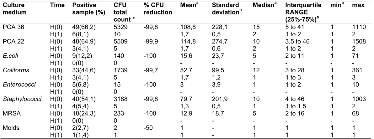
Table 1. Descriptive statistics of stethoscope variables at H(0) and H(1): Number and percentage of positive samples, overall CFU count, percentage reduction in CFUs between H(0) and H(1), mean, standard deviation, median, interquartile
range, minima and maxima
Culture
medium Time Positivesample (%) CFUtotal count *
% CFU
reduction Mean
a Standard
deviationa Median
a Interquartile
RANGE (25%-75%)a
mina max
PCA 36 H(0) 49(66,2) 5329 -99,8 108,8 228,1 15 5 to 41 1 1110
H(1) 6(8,1) 10 1,7 0,5 2 1 to 2 1 2
PCA 22 H(0) 48(64,9) 5509 -99,9 114,8 274,7 10 3.5 to 46 1 1508
H(1) 3(4,1) 5 1,7 0,6 2 1 to 2 1 2
E.coli H(0) 9(12,2) 140 -100 15,6 23,7 5 2 to 11 1 71
H(1) 0(0) 0 - - -
-Coliforms H(0) 33(44,6) 1739 -99,7 52,7 99,5 12 3 to 28 1 361
H(1) 3(4,1) 5 1,7 1,2 1 1 to 3 1 3
Enterococci H(0) 5(6,8) 15 -100 3 3,9 1 1 to 2 1 10
H(1) 0(0) 0 - - -
-Staphylococci H(0) 40(54,1) 3188 -99,8 79,7 201,9 10 4 to 46 1 1003
H(1) 4(5,4) 5 1,3 0,5 1 1 to 1.5 1 2
MRSA H(0) 18(24,3) 233 -100 12,9 18,7 5 2 to 16 1 68
H(1) 0(0) 0 - - -
-Molds H(0) 2(2,7) 2 -50 1 - 1 1 1 1
H(1) 1(1,4) 1 1 - 1 1 1 1
Pseudomonas spp., Acinetobacter spp. and Clostridium difficile were not detected on the stethoscopes. * summing all CFUs on the stethoscopes,
3. RESULTS AND DISCUSSION
The Table 1 shows the variables at H(0) and H(1) with mean, standard deviation, median, interquartile range, minima, maxima, overall CFU count, percentage reduction in CFUs between H(0) and (H(1), number of positive samples and their percentages for all stethoscopes. No samples contained Pseudomonas, Acinetobacter spp. or Clostridium difficileat H(0) or H(1). The CFUs’ reduction is close at 100% in most comparisons. In cases the number of CFUs in H(1) did not correspond to 0, statistical tests were carried to highlight differences between pre- and post- cleaning. Significant differences were detected in the comparison for TBC at 36ºC and 22ºC, Coliforms, Staphylococcus spp. (p<0.001), No differences emerged in the molds comparison (p<0.563). Only 2/74 stethoscopes were contaminated with molds before the DT, and one after cleaning. Two stethoscope diaphragms also carried a visible film of undefined material (solidified gel or other dirt) which was only partly removed by the disinfecting technique (Fig. 1). These stethoscopes, at H(1), did not show microbial contamination.
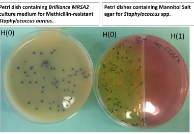
The comparison between physicians’/nurses’ stethoscopes and shared ones showed significant differences in some cases. We recorded increased contamination of private stethoscopes by E. coli (P=0.0360), Enterococcus spp. (P=0.0024), Staphylococcus spp. (P=0.0164) and MRSA (P=0.0060). No significant statistical difference was found between shared and private stethoscopes for TBC at 36ºC (P=0.2496), TBC at 22ºC (P=0.2235) and coli form (P=0.3583) contamination. The (Fig. 2) shows an example of cultures for MRSA from swabs taken before and after cleaning. No statistical differences were also found in contamination among the three hospitals (P>0.05).
Stethoscopes are a universal tool of the medical profession and a potential source of nosocomial infections. They are used in direct contact with numerous patients every day and are often not routinely cleaned [24]. Several studies, as well as our own, have investigated microbial contamination of stethoscopes and, consequently, their role in the transmission of health-care-associated infections. Some of these infections are very dangerous, for example in the European Union, methicillin-resistant Staphylococcus aureus (MRSA) is frequently isolated in hospitals of Italy, Spain, Greece, Portugal and Great Britain [25].
In line with our results, the bacteria most commonly isolated are gram-positive cocci, especially Staphylococcus spp. [5,19,20,24,26]. Other bacteria frequently isolated are Enterobacteriaceae: Enterobacter spp., coliforms spp, Citrobacter spp., Klebsiella spp. and Serratia spp. being microorganisms considered by several studies [5,18,19,26-28]. We too found coli forms and E. coli in our samples; indeed, before cleaning, 40 out of 74 were positive forStaphylococcus spp., including 18 for MRSA, 33 for coliforms (with 3 E. coli),5 forEnterococcusspp. and 2 for molds.
removed some of the print on membranes of cheaper stethoscopes. This was probably due to the moist and adhesive nature of the product and the wiping action of the swab.
Left:
A) first sample H(0): use of cotton swab, no use of DT
B) second sample H(1): use of DT, before of cotton swab
Figure 1
Right:
C) first sample H(0): use of cotton swab, no use of DT
D) second sample H(1): use of DT, use of cotton swab
A B C D
The disinfecting technique eliminated bacterial load, but did not always remove dirt from stethoscopes (see part B)
Stethoscopes pre and post use of Disinfecting Technique
Fig. 1. Stethoscopes pre and post use of disinfecting technique
The cleaning product contains about 30% ethyl alcohol and has elastic consistency that attaches to and removes dirt. Both features disinfect. Our study demonstrates that after cleaning the percentage reduction in CFUs in all samples was 99.8% for TBC at 36ºC and 99.9% for TBC at 22ºC. These values are larger than the percentage reductions obtained in the surveys mentioned above. This disinfecting technique also determined a 99.7% reduction in coliforms, 99.8% in Staphylococcus spp., and 100% in E. coli, Enterococcus spp. and MRSA at H(1). Molds showed a different pattern, decreasing from two to one positive sample. This apparent ineffectiveness of the technique for molds could be due to a relative initial absence of molds on the stethoscopes. To test the disinfecting technique for molds, greater initial mould contamination is necessary. The tested product does not wet the article to be cleaned, dispensing with a drying step.
stethoscopes after every use, 32% cleaned them many times per day but not after every use, 11% cleaned them weekly and 3.8% never cleaned them [17].
Petri dishes containing Mannitol Salt
agar for
Staphylococcus
spp.
Petri dish containing
Brilliance MRSA2
culture medium for Methicillin-resistant
Staphylococcus aureus
.
H(1)
H(0)
H(0)
Figure 2
Example of cultures of swabs from stethoscopes taken
before and after cleaning
Fig. 2. Example of cultures of swabs from stethoscopes taken pre and post cleaning
We also compared microbial contamination of stethoscopes of physicians/nurses and shared stethoscopes and found some significant statistical differences. We recorded greater contamination on non-shared stethoscopes for: E. coli, Enterococcus spp, Staphylococcus spp. and MRSA. Since health care professionals presumably use their own stethoscopes more often than shared ones, the former are more likely to be contaminated. Other reasons for greater contamination of personal stethoscopes could be that shared stethoscopes are subject to established hygiene practices. In fact, we found that in intensive care units, standard protocols require the disinfection of stethoscopes which are placed at every bedside. Other departments may also follow this procedure. On the contrary, a study by Whittington et al. conducted in an intensive care unit showed that the diaphragms of bedside stethoscopes had greater bacterial contamination than personal ones, though the latter are more frequently contaminated by pathogenic bacteria [22]. This could sustain our hypothesis that personal stethoscopes are used with greater frequency than shared ones and are therefore more often colonized by pathogenic bacteria.
technique to keep microbe concentrations low with repeated use and to test how long the effect lasts.
Our results indicate that educational programmes on disinfection procedures for doctors are important, especially for young staff. They help make health personnel more aware of these aspects, often overlooked or considered marginal. Habits acquired early in professional life are more likely to last.
It would be also useful to calculate the attributable risk of nosocomial infection caused by stethoscopes. This information, linked to hospital expenses, would infer savings in health care costs.
4. CONCLUSION
The results of the present study suggest that the disinfecting technique was effective in reducing stethoscope microbiological load, however its efficacy should not be a reason to neglect standard hygiene and cleanliness practices. Stethoscopes may also be disinfected by simple traditional methods, such as swabbing with sodium hypochlorite which normally eliminates bacteria.
CONSENT
The study did not involve patients, so the consent has not been applied. However we were authorized by the medical administrations of Siena teaching hospital “Le Scotte”, the Rugani Clinic and Alta Val D’Elsa Hospital in Siena Province Italy.
ETHICAL APPROVAL
Not applicable.
FUNDING SOURCES
The study was conducted under a Master Research and Service Agreement (D.R. 341/2012, signed 6thMarch 2012, Exhibit 4 signed 9thJuly 2012) between the University of Siena and
Joker AG/SA which financed the research.
COMPETING INTERESTS
Authors have declared that no competing interests exist.
REFERENCES
1. Bates DW, Larizgoitia I, Prasopa-Plaizier N, Jha AK. And Research Priority Setting Working Group of the WHO World Alliance for Patient Safety. Global priorities for patient safety research.BMJ. 2009;338:b1775.
2. Mitchell A, Dealwis N, Collins J, Chew K, Taylor R, Schwab U, Narayanan M. Stethoscope or 'Staphoscope'? Infection by auscultation. J Hosp Infect. 2010;76(3):278-279.
4. Jones JS, Hoerle D, Riekse R. Stethoscopes: A potential vector of infection? Ann Emerg Med. 1995. 26(3):296-299.
5. Mangi RJ, Andriole VT. Contaminated stethoscopes: A potential source of nosocomial infections.Yale J Biol Med. 1972;45(6):600-604.
6. Messina G, Ceriale E, Burgassi S, Russo C, Defranceschi C, Mariani L, Taddei L, Lenzi D, Manzi P. Impact of a disinfecting technique on microbial contamination of computer keyboards and telephone handsets. Journal of Hospital Administration. 2013;2(4):1-6.
7. Messina G, Ceriale E, Lenzi D, Burgassi S, Azzolini E, Manzi P. Environmental contaminants in hospital settings and progress in disinfecting techniques.Biomed Res Int. 2013;2013:429780.
8. Messina G, Quercioli C, Burgassi S, Nistico F, Lupoli A, Nante N. How many bacteria live on the keyboard of your computer? Am J Infect Control. 2011;39(7):616-618. 9. Tang PH, Worster A, Srigley JA, Main CL. Examination of staphylococcal stethoscope
contamination in the emergency department (pilot) study (EXSSCITED pilot study). CJEM. 2011;13(4):239-244.
10. Lecat P, Cropp E, McCord G, Haller NA. Ethanol-based cleanser versus isopropyl alcohol to decontaminate stethoscopes.Am J Infect Control. 2009;37(3):241-243. 11. Manzi P, Liberatore S, Morgante A. Asepsis and disinfection in infection control.
Designing for Health.2010(119):37-40.
12. Alleyne SA, Hussain AM, Clokie M, Jenkins DR. Stethoscopes: Potential vectors of Clostridium difficile.J Hosp Infect. 2009;73(2):187-189.
13. Bandi S, Conway A. Question 2. Does regular cleaning of stethoscopes result in a reduction in nosocomial infections? Arch Dis Child. 2012;97(2):175-177.
14. Breathnach AS, Jenkins DR, Pedler SJ. Stethoscopes as possible vectors of infection by staphylococci.BMJ. 1992;305(6868):1573-1574.
15. Marinella MA, Pierson C, Chenoweth C. The stethoscope. A potential source of nosocomial infection? Arch Intern Med. 1997;157(7):786-790.
16. Messina G, Burgassi S, Russo C, Ceriale E, and Mariani L. Indagine sulla contaminazione microbica di stetoscopi, telefoni e tastiere di computer presso una casa di cura.Mondo Sanitario. 2012;19(10):1-5.
17. Muniz J, Sethi RK, Zaghi J, Ziniel SI, and Sandora TJ. Predictors of stethoscope disinfection among pediatric health care providers.Am J Infect Control; 2012.
18. Nunez S, Moreno A, Green K, and Villar J. The stethoscope in the Emergency Department: A vector of infection? Epidemiol Infect. 2000;124(2):233-237.
19. Sengupta S, Sirkar A, Shivananda PG. Stethoscopes and nosocomial infection.Indian J Pediatr. 2000;67(3):197-199.
20. Genne D, de Torrente A, Humair L, Siegrist HH. Level of stethoscope contamination in the hospital environment.Schweiz Med Wochenschr. 1996;126(51-52):2237-2240. 21. Sood P, Mishra B, Mandal A. Potential infection hazards of stethoscopes. J Indian
Med Assoc. 2000;98(7):368-370.
22. Whittington AM, Whitlow G, Hewson D, Thomas C, Brett SJ. Bacterial contamination of stethoscopes on the intensive care unit.Anaesthesia. 2009;64(6):620-624.
23. CONFARMA.Study of disinfectant efficacy by the microbe carrier test under simulated conditions of use; 2010.
24. Smith MA, Mathewson JJ, Ulert IA, Scerpella EG, Ericsson CD. Contaminated stethoscopes revisited.Arch Intern Med. 1996;156(1):82-84.
25. European Centre for Disease Prevention and Control. EARSS Annual Reports; 2010. 26. Youngster I, Berkovitch M, Heyman E, Lazarovitch Z, Goldman M. The stethoscope as
27. Uneke CJ, Ogbonna A, Oyibo PG, Onu CM. Bacterial contamination of stethoscopes used by health workers: public health implications.J Infect Dev Ctries. 2010;4(7):436-441.
28. Zuliani Maluf ME, Maldonado AF, Bercial ME, Pedroso SA. Stethoscope: A friend or an enemy? Sao Paulo Med J. 2002;120(1):13-15.
29. Saloojee H, Steenhoff A. The health professional's role in preventing nosocomial infections.Postgrad Med J. 2001;77(903):16-19.
30. Bernard L, Kereveur A, Durand D, Gonot J, Goldstein F, Mainardi JL, Acar J, Carlet J. Bacterial contamination of hospital physicians' stethoscopes. Infect Control Hosp Epidemiol. 1999;20(9):626-628.
31. Rutala WA, Weber DJ. And The Healthcare Infection Control Practices Advisory Committee (HICPAC). Guideline for Disinfection and Sterilization in Healthcare Facilities; 2008.
© 2014 Messina et al.; This is an Open Access article distributed under the terms of the Creative Commons Attribution License (http://creativecommons.org/licenses/by/3.0), which permits unrestricted use, distribution, and reproduction in any medium, provided the original work is properly cited.
Peer-review history:
The peer review history for this paper can be accessed here: